This article contains AI generated content.
· UpdatedAchondroplasia Genes and Biomarkers – 4 Genes And 6 Biomarkers To Track
Introduction
Living with achondroplasia — or supporting someone who does — often means navigating a healthcare system that reacts to complications as they arise rather than anticipating them at a molecular level. Most appointments address what went wrong: foramen magnum narrowing, sleep apnea episodes, spinal stenosis progression. The conversation rarely makes it down to the signaling pathways and measurable markers that determine how the body is actually coping. That gap between diagnosis and deep understanding is where a lot of useful information gets lost.
Generic health advice fails people with achondroplasia in a specific way. Growth charts built for typical populations are meaningless here. Risk assessments developed for the general public don't weight the right things. Nutritional and exercise recommendations designed for standard skeletal architecture miss the specific vulnerabilities — and specific strengths — that come with a permanently overactive FGFR3 receptor. What's needed is a more precise framework.
This article takes that more precise approach. It focuses on four genes and gene pathways that drive achondroplasia and shape its complications, and then covers six biomarkers that give a real-time read on how the body is managing the downstream consequences. For each gene and each biomarker, there are practical plans — both with and without supplements — grounded in what the evidence actually supports.
Better information leads to better decisions. Not cures, not miracle protocols — just sharper conversations with clinicians, earlier detection of complications, and a clearer understanding of which interventions are actually moving the right levers. The genetics section comes first, since achondroplasia is fundamentally a gene-driven condition with molecular pathways that can now be targeted therapeutically. The biomarker section follows as a practical complement. Complementary approaches with real clinical evidence round out the picture.
Summary
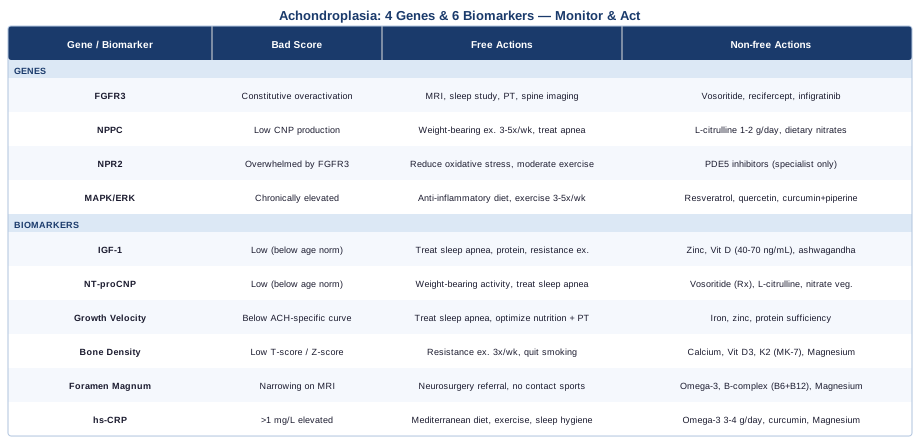
This article decodes achondroplasia at the molecular level — starting with the FGFR3 gain-of-function mutation that drives the condition, then tracing its effects through the CNP/NPR2 signaling axis and the MAPK/ERK cascade that turns the receptor's overactivation into impaired bone growth. For each of these four genetic layers, you'll find a plan: what to do without supplements, and what targeted nutritional or pharmacological tools may support the pathway. The biomarker section covers six measurable signals — from IGF-1 and NT-proCNP to bone density and inflammatory markers — with cost ranges, testing guidance, and concrete action plans for suboptimal results. Beyond genetics and biomarkers, the article includes a synthesis of the molecular research revolution that produced the first FDA-approved precision therapy for achondroplasia, and five complementary modalities with meaningful clinical evidence. If you've been managing this condition reactively, this is what a proactive framework looks like.
Understanding the Genetic Roots of Achondroplasia
Achondroplasia is caused by one of the most precisely characterized mutations in human genetics: a single nucleotide change in the FGFR3 gene that locks the receptor into a permanently active state. But understanding the full molecular picture requires going beyond that single mutation. Several genes and pathways interact with FGFR3 signaling, and understanding them is what opens the door to targeted interventions — both the medical treatments now available and the lifestyle and nutritional strategies that can support the system around them.
Gene 1: FGFR3 — The Core Driver
FGFR3 (Fibroblast Growth Factor Receptor 3) is located on chromosome 4p16.3 and encodes a transmembrane receptor that, under normal circumstances, acts as a biological brake on bone growth. Its role is to limit chondrocyte (cartilage cell) proliferation in the growth plate so that skeletal development stays proportionate. In achondroplasia, a single point mutation — the G380R substitution (glycine to arginine at position 380) in roughly 98% of cases — makes the receptor constitutively active. The brake is permanently engaged. Chondrocyte proliferation is suppressed throughout fetal development and childhood, producing the characteristic pattern of shortened long bones, proximal limb reduction, midface hypoplasia, macrocephaly, and lumbar lordosis.
This mutation is autosomal dominant: one copy is sufficient to cause the full condition. In approximately 80% of cases, it arises de novo — a new mutation, not inherited. The rate of new mutation at this specific nucleotide position is among the highest known in the human genome. Achondroplasia occurs in roughly 1 in 25,000 births and is the most common skeletal dysplasia (GeneReviews: Achondroplasia, NCBI).
FGFR3 Is Confirmed — The Plan Without Supplements
The FGFR3 mutation cannot be altered by lifestyle or supplementation; gene-editing approaches remain experimental. What can be addressed is the downstream signaling the mutation drives. Vosoritide (brand name Voxzogo), an analog of C-type natriuretic peptide (CNP), was FDA-approved in 2021 for children aged 5 and older. It is administered once daily by subcutaneous injection and works by counteracting FGFR3 overactivation downstream. A pivotal randomized trial demonstrated significant improvements in annualized growth velocity compared to placebo (Savarirayan et al., NEJM 2020).
Beyond vosoritide, comprehensive FGFR3-related monitoring is non-negotiable. Every child with achondroplasia should receive: MRI of the foramen magnum in infancy to assess cervicomedullary compression risk; annual or biannual polysomnography (sleep study) through childhood; regular audiological and ophthalmological assessment; and spine imaging in adulthood to monitor for stenosis. Physical therapy adapted to achondroplasia biomechanics helps maximize functional capacity. Surgical options — foramen magnum decompression, spinal decompression, limb lengthening — are indicated based on clinical assessment and shared decision-making with a skeletal dysplasia specialist.
Emerging therapies include recifercept (an FGFR3 decoy receptor), infigratinib (an FGFR inhibitor), and TransCon CNP. These are in various stages of late-stage clinical trials. Discuss eligibility for trials with a skeletal dysplasia center.
FGFR3 Is Confirmed — The Plan With Supplements
No supplement directly reverses FGFR3 overactivation. The following nutrients support the systems most stressed by FGFR3-driven pathology and should be considered as foundational support alongside medical management:
Vitamin D3 (1,000–4,000 IU/day, adjusted to serum level of 40–70 ng/mL): Supports chondrocyte function, bone mineralization, and immune regulation. Achondroplasia patients often have reduced outdoor activity; deficiency is common. Test quarterly until stable, then twice yearly.
Vitamin K2 (MK-7) (90–200 mcg/day): Activates osteocalcin and directs calcium to bone rather than soft tissue. Pair with vitamin D for synergy. Long-term daily use; no cycling required.
Magnesium glycinate (200–400 mg/day): Supports bone metabolism, sleep architecture (critical given sleep apnea prevalence), and muscle function. Side effect: loose stool at high doses — titrate down if needed.
Zinc (15–25 mg/day with food): Involved in growth plate biology and IGF-1 signaling. If used long-term, pair with 1–2 mg copper to avoid copper depletion. Test serum zinc before supplementing.
Omega-3 fatty acids (EPA+DHA 2–4 g/day): Reduce systemic inflammation, which may exacerbate joint and spinal pain. Take with meals. No cycling needed; monitor for blood-thinning effects if on anticoagulants.
Gene 2: NPPC — The CNP Signaling Gene
NPPC encodes C-type natriuretic peptide (CNP), a small paracrine signaling molecule produced predominantly in bone and cartilage tissue. CNP functions as the body's endogenous counterbalance to FGFR3 overactivation. When CNP binds its receptor (NPR2) on chondrocytes, it triggers production of cyclic GMP (cGMP), which activates protein kinase G and subsequently suppresses the MAPK/ERK cascade — the same cascade that overactive FGFR3 chronically drives. In simple terms, CNP is the natural antidote to the problem that FGFR3 creates.
Individuals with lower NPPC expression or reduced endogenous CNP production have less buffering capacity against FGFR3 overactivation. The therapeutic logic of vosoritide and other CNP-pathway drugs is to pharmacologically amplify exactly this signal. A rare gain-of-function mutation in NPPC causes a skeletal overgrowth syndrome — the mirror image of achondroplasia — confirming the direct regulatory relationship.
Supporting NPPC Expression — Without Supplements
Endogenous CNP production in cartilage appears to be stimulated by intermittent mechanical loading on chondrocytes. This is one mechanistic reason why appropriate weight-bearing physical activity matters in achondroplasia — not primarily for cardiovascular benefit, but for signaling. Aquatic exercise is often recommended for joint comfort, but some weight-bearing activity (walking, adapted resistance work) is likely important for growth plate signaling through the CNP axis.
Sleep quality is the second major lever. CNP production has circadian variation, and sleep-disordered breathing — which disrupts normal endocrine and paracrine signaling — should be treated aggressively with CPAP or surgical intervention when indicated. Untreated sleep apnea suppresses multiple anabolic signals simultaneously.
Frequency: Weight-bearing activity 3–5 times per week at moderate intensity, adapted to physical capacity and current orthopedic status. Sleep: target 7–9 hours with active apnea management.
Supporting NPPC Expression — With Supplements
Vosoritide (prescription) is pharmacologically what NPPC biology points toward — a CNP analog that does what the body's own NPPC gene produces, at a higher effective concentration. For those not on vosoritide, no over-the-counter supplement directly mimics CNP.
Dietary nitrates from leafy greens (spinach, arugula, beets) support the broader nitric oxide / cGMP signaling system, which shares overlapping architecture with the CNP/NPR2/cGMP axis. The mechanistic crossover is real but should not be overstated. Daily dietary inclusion of nitrate-rich vegetables — aim for 2–3 servings — has no downside and supports vascular and signaling health broadly.
L-citrulline (1–2 g/day): Supports endogenous nitric oxide production and cGMP activity through the arginine pathway. The evidence base is cardiovascular, not achondroplasia-specific. Generally well tolerated; monitor blood pressure.
Gene 3: NPR2 — The Receptor That Amplifies the CNP Signal
NPR2 (Natriuretic Peptide Receptor B) encodes the surface receptor that CNP binds to on chondrocytes. When CNP engages NPR2, the receptor's intrinsic guanylyl cyclase domain activates and generates cGMP — the second messenger that drives the anti-FGFR3 signaling cascade. NPR2 is a molecular rheostat for skeletal growth: loss-of-function mutations cause acromesomelic dysplasia with limb shortening, while gain-of-function mutations cause overgrowth syndromes. The dose-response relationship is well-established and clinically actionable.
In achondroplasia, NPR2 itself is structurally intact — but its activity is overwhelmed by the constitutively signaling FGFR3. The therapeutic approach is therefore to flood the system with more CNP input (vosoritide), or to amplify the cGMP signal further downstream by preventing its breakdown.
Phosphodiesterase 5 (PDE5) inhibitors — drugs that prevent cGMP degradation — have shown modest rescue effects in mouse models of achondroplasia by extending the half-life of the cGMP generated through NPR2. Sildenafil and riociguat are the principal candidates being studied. This research is in early clinical stages.
Supporting NPR2 Signaling — Plans With and Without Supplements
Without supplements: The primary strategy is maximizing endogenous CNP to ensure NPR2 is receiving sufficient ligand (see NPPC section). No lifestyle intervention is known to directly upregulate NPR2 receptor expression or density. Avoiding factors that chronically suppress cGMP signaling — particularly oxidative stress from poor diet, sedentary behavior, and smoking — is the practical goal.
With supplements: No over-the-counter supplement is known to directly activate NPR2 equivalent to CNP. PDE5 inhibitors work downstream by preventing cGMP degradation — these are prescription medications and not appropriate for self-use, particularly in children. For adults: discuss emerging data on PDE5 inhibitors and achondroplasia outcomes with a skeletal dysplasia specialist. Monitoring: if NT-proCNP testing is available, assess every 6–12 months to track NPR2 pathway activity indirectly.
Gene 4: The MAPK/ERK Pathway — Where the Damage Is Done
The MAPK/ERK signaling cascade is the primary effector pathway through which constitutively active FGFR3 suppresses chondrocyte proliferation. This pathway includes: RAS proteins (encoded by KRAS, NRAS, HRAS), RAF kinases (encoded by BRAF), MEK1 and MEK2 (encoded by MAP2K1 and MAP2K2), and ERK1/2 (encoded by MAPK3 and MAPK1).
In achondroplasia, overactive FGFR3 continuously feeds RAS-RAF-MEK-ERK signaling, which then inhibits chondrocyte division and differentiation at the growth plate. This is the final common pathway where the upstream genetic defect is translated into impaired skeletal growth.
MEK inhibition has been explored as a therapeutic angle. Meclozine — an antihistamine used for motion sickness — was found incidentally to inhibit MEK signaling and, in mouse models of achondroplasia, to promote endochondral bone growth. Trametinib, a dedicated MEK inhibitor used in oncology, is under investigation in skeletal dysplasias. Neither is current standard of care for achondroplasia.
Supporting MAPK/ERK Balance — Without Supplements
Chronic inflammation is one driver of aberrant baseline MAPK/ERK activation across tissues. An anti-inflammatory dietary pattern — Mediterranean-style, high in olive oil, fatty fish, vegetables, legumes, and nuts, low in ultra-processed food and refined sugar — reduces the systemic inflammatory pressure on multiple signaling pathways simultaneously. This does not counteract the FGFR3-driven MAPK overactivation in the growth plate, but it reduces the inflammatory load on surrounding tissues and joints.
Regular moderate exercise (3–5 times/week) additionally modulates MAPK/ERK signaling through physiological rather than pathological activation — the difference between acute beneficial signaling and chronic suppressive signaling.
Supporting MAPK/ERK Balance — With Supplements
Resveratrol (100–500 mg/day): Has demonstrated MAPK/ERK modulating properties in multiple preclinical and early clinical contexts. No direct achondroplasia data exists; use as a general anti-inflammatory support. Cycle: 5 days on, 2 days off for long-term use. Generally well tolerated; avoid high doses with anticoagulants.
Quercetin (500–1,000 mg/day): Inhibits MAPK pathway activity in preclinical models. Often paired with resveratrol for broader coverage. Cycle similarly; take with food to improve absorption.
Curcumin with piperine (500–1,000 mg/day): Broad modulator of inflammatory and MAPK signaling; clinical evidence is primarily in cancer and inflammatory conditions. Take with black pepper extract (piperine) for bioavailability. Avoid high doses in pregnancy. Cycle: 8 weeks on, 2 weeks off.
These supplements address the systemic signaling environment — they are not targeted FGFR3 pathway therapies and should be framed accordingly.
Moving from the genetic layer to measurable markers in the body, the following section covers six biomarkers that provide practical, trackable data on how the condition is being managed over time.
6 Biomarkers to Monitor in Achondroplasia
Because the genetic cause in achondroplasia is known and fixed, biomarker monitoring serves a specific purpose: tracking complications before they become symptomatic, assessing treatment response, and optimizing the metabolic and nutritional conditions in which the body operates. These are the six most clinically meaningful markers to follow.
Biomarker 1: IGF-1 (Insulin-Like Growth Factor 1)
Why it matters: IGF-1 is the principal mediator of growth hormone action on bone and cartilage. Chondrocytes express IGF-1 receptors, and IGF-1 normally stimulates their proliferation and differentiation. In achondroplasia, overactive FGFR3 blunts the growth plate's response to IGF-1 — but IGF-1 levels themselves may be normal, reduced, or suboptimal depending on sleep quality, nutrition, and stress load. Identifying an independent IGF-1 deficit — on top of FGFR3 overactivation — creates an additional therapeutic lever. It also informs whether growth hormone therapy may be beneficial in a particular individual.
How to Measure It
Standard fasting serum test. Reference ranges are age- and sex-specific. Cost: approximately $50–150 depending on insurance and laboratory. Order through a pediatric endocrinologist or general practitioner. Measure every 6–12 months in actively growing children; annually in adults. Pair with a growth hormone stimulation test if IGF-1 is consistently low and GH deficiency is suspected.
If the Score Is Low — Without Supplements
Sleep is the highest-yield intervention. IGF-1 secretion is tightly coupled to slow-wave sleep stages. Untreated sleep apnea — extremely common in achondroplasia — dramatically suppresses nocturnal GH release and downstream IGF-1 production. Aggressive CPAP adherence or surgical resolution of airway obstruction can produce meaningful IGF-1 recovery within weeks.
Protein intake (1.6–2.2 g/kg/day) and adequate total calorie sufficiency are required foundations for GH/IGF-1 axis function. Chronic undernutrition, which is underrecognized in children with feeding difficulties, suppresses IGF-1 regardless of GH status.
Resistance exercise — adapted to capacity — stimulates GH secretion. Even twice-weekly sessions of moderate-intensity resistance work in adults produce measurable GH/IGF-1 improvements.
If the Score Is Low — With Supplements
Zinc (15–25 mg/day): Zinc deficiency directly impairs GH receptor sensitivity and IGF-1 production. Test serum zinc — deficiency is common in children with restricted diets. Pair with 1–2 mg copper if using long-term.
Vitamin D (maintain 40–70 ng/mL): Vitamin D receptors are present on GH-secreting pituitary cells; deficiency correlates with reduced IGF-1 in multiple cohort studies.
Ashwagandha (KSM-66 extract) (300–600 mg/day): Shown to support IGF-1 and reduce cortisol in adults. Cortisol chronically suppresses GH release, so this is a relevant secondary mechanism. Use in adults only; cycle 8 weeks on, 2 weeks off. Side effects: mild GI upset in some; avoid in pregnancy.
Growth hormone therapy (prescription) may be considered in confirmed GH deficiency — its role in achondroplasia without documented GH deficiency is debated. Discuss specifically with a pediatric endocrinologist who has experience in skeletal dysplasias.
Biomarker 2: NT-proCNP (N-Terminal Pro-C-Type Natriuretic Peptide)
Why it matters: NT-proCNP is a stable breakdown product of the CNP precursor molecule. Because CNP itself has a very short half-life in circulation, NT-proCNP serves as a practical surrogate marker for CNP production and pathway activity. Since CNP is the body's endogenous counterbalance to FGFR3 overactivation, tracking NT-proCNP gives an indirect window onto how robustly the compensatory system is functioning. In children on vosoritide, NT-proCNP is used to confirm that the CNP-pathway drug is biologically active at the expected level.
How to Measure It
Serum or plasma test. Not part of standard panels — requires a specialty or academic laboratory. Cost: approximately $100–250. Reference ranges are strongly age-dependent (NT-proCNP is physiologically higher in children). Measure at baseline before initiating any CNP-pathway therapy, and every 6 months during treatment. Some major children's hospitals order this routinely for patients on vosoritide.
If the Score Is Low — Without Supplements
Weight-bearing physical activity is the primary lifestyle lever for supporting endogenous CNP production in cartilage. In the absence of vosoritide, a consistently low NT-proCNP in an eligible child may strengthen the clinical argument for initiating targeted therapy — document and present this to the treating skeletal dysplasia specialist.
Treat sleep apnea: disrupted sleep reduces the circadian expression of multiple cartilage-active signaling molecules including CNP. Optimizing sleep architecture is a low-cost first step.
If the Score Is Low — With Supplements
Vosoritide (prescription) is the pharmacological intervention that directly targets this pathway. For non-prescription support, dietary nitrate-rich vegetables (spinach, beets, arugula) and L-citrulline (1–2 g/day) support the broader cGMP system with which CNP signaling overlaps. Evidence specifically for NT-proCNP improvement is absent — frame these as general cGMP-supportive strategies.
Biomarker 3: Growth Velocity
Why it matters: Growth velocity — the annualized rate of height gain — is the most sensitive clinical signal of whether any intervention is working. Standard population growth charts are not appropriate for achondroplasia. Achondroplasia-specific reference charts (such as those developed by Hoover-Fong and colleagues) must be used to interpret whether a child's growth trajectory is within expected range for the condition, or whether velocity is further suppressed. This biomarker is the primary endpoint in all major clinical trials of new achondroplasia therapies.
How to Measure It
Accurate stadiometer measurement at every clinic visit — ideally every 3–6 months during active growth. Cost: effectively zero. Calculate annualized velocity: [(height at visit 2 minus height at visit 1) divided by months between visits] multiplied by 12. Ensure consistent measurement technique and timing to minimize variability. Plot against achondroplasia-specific reference curves.
If the Score Is Lower Than Expected — Without Supplements
Investigate systematically: Is sleep apnea undertreated? Is caloric intake adequate for the growth phase? Is there an intercurrent illness or orthopedic complication that has suppressed activity and appetite? Are there psychosocial factors affecting nutrition? Address each in sequence. Ensure engagement with a physical therapist familiar with skeletal dysplasias. If not on vosoritide and the child is eligible, this is a primary clinical conversation to have.
If the Score Is Lower Than Expected — With Supplements
Growth velocity cannot be meaningfully elevated above the ceiling imposed by FGFR3 overactivation through supplementation. However, nutritional deficiencies create a floor below that ceiling — correct them first. Ensure iron status is adequate (deficiency impairs GH response), zinc is replete, and total protein and calorie intake supports the growth phase. Pediatric nutritional consultation is appropriate for children with consistently suboptimal velocity.
Biomarker 4: Bone Mineral Density (DXA Scan)
Why it matters: Adults with achondroplasia present a complex bone health picture. The bones are structurally different — shorter with altered cortical geometry — and standard DXA interpretation using typical reference populations can be misleading. Some studies suggest increased volumetric bone density in achondroplasia, yet altered bone geometry creates specific mechanical vulnerabilities, particularly at the spine. Spinal stenosis and the spinal loading pattern from exaggerated lumbar lordosis place unusual cumulative stress on vertebral structures. Monitoring bone mineral density helps identify osteoporosis in adulthood and guides decisions about fracture risk, especially as patients move into their 40s and beyond.
How to Measure It
DXA scan of the lumbar spine and femoral neck. Cost: approximately $150–350, typically covered by insurance for adults with risk factors. In achondroplasia, request size-adjusted analysis and discuss with the radiologist the need for appropriate reference population selection. Repeat every 1–2 years in adults with suboptimal results or on treatment; every 3 years if stable and normal.
If the Score Is Low — Without Supplements
Weight-bearing resistance exercise (3 sessions per week) is the most evidence-supported non-pharmacological intervention for bone density improvement. Aquatic exercise provides cardiovascular benefit but is less effective for osteogenic stimulus than weight-bearing activity. Reduce alcohol intake; eliminate smoking. Review any medications with bone-depleting effects (long-term corticosteroids, anticonvulsants). Ensure adequate sleep — bone remodeling is preferentially active during sleep.
If the Score Is Low — With Supplements
Calcium (1,000–1,200 mg/day total from food and supplements): Food sources preferred; supplement in 500 mg doses maximum to limit absorption ceiling. Vitamin D3 (2,000–4,000 IU/day targeting 40–70 ng/mL serum): Essential co-factor for calcium absorption. Vitamin K2 (MK-7) (90–200 mcg/day): Activates osteocalcin and reduces vascular calcium deposition. Magnesium (300–400 mg/day): Required for vitamin D metabolism and bone matrix quality.
For documented osteoporosis, prescription bisphosphonates or other bone-protective agents may be appropriate — discuss with an endocrinologist. All supplements: daily, long-term, no cycling required. Monitor DXA annually while optimizing, then extend to 2-year intervals when stable.
Biomarker 5: Foramen Magnum and Spinal Canal Dimensions
Why it matters: The foramen magnum is the opening at the base of the skull through which the brainstem passes. In achondroplasia, abnormal FGFR3-driven bone formation at the skull base frequently narrows this opening. Cervicomedullary compression is one of the most dangerous potential complications — it can cause central sleep apnea, sudden infant death, hypotonia, and, without intervention, significant neurological harm. Critically, it can be present without obvious symptoms in infants. Lumbar spinal canal narrowing is separately responsible for the claudication and leg pain that affect many adults.
How to Measure It
MRI (preferred: no radiation, best soft tissue detail) or CT of the brain and cervical spine for foramen magnum assessment. MRI of the lumbar spine for stenosis evaluation in adults. Cost: $500–$2,000+ depending on region and insurance coverage. Current guidelines from pediatric skeletal dysplasia groups recommend MRI of the foramen magnum in all infants with achondroplasia during the first year, repeated if symptoms emerge. Foramen magnum cross-sectional area below established thresholds warrants urgent neurological review.
If Dimensions Are Concerning — Without Supplements
This is primarily a surgical domain. Cervicomedullary decompression — removal of the posterior rim of the foramen magnum and upper cervical laminae — is indicated when compression is symptomatic or associated with central apnea, hypotonia, or hyperreflexia. Timing decisions require a neurosurgeon experienced in achondroplasia. Lumbar stenosis may be managed conservatively with physical therapy and posture-focused rehabilitation in mild cases; surgical decompression is appropriate for significant functional compromise. Avoid contact sports and high-fall-risk activities when foramen magnum narrowing is significant.
If Dimensions Are Concerning — With Supplements
No supplement alters bony dimensions. However, supporting neurological health during monitoring or perioperatively is reasonable. Omega-3 fatty acids (EPA+DHA 2–4 g/day) support neurological membrane integrity. B12 and B6 (from a complete B-complex): Support myelin and nerve function, relevant in the context of possible cord compression. Magnesium glycinate (300–400 mg/day): Modest neuroprotective properties in the context of compression-related nerve stress. These are supportive measures only; they do not replace surgical intervention when indicated.
Biomarker 6: High-Sensitivity CRP and Inflammatory Markers
Why it matters: Chronic low-grade inflammation accelerates joint degeneration, spinal pain, and musculoskeletal decline — concerns that are heightened in achondroplasia due to the altered biomechanics of a skeleton built to different proportions. Exaggerated lumbar lordosis, genu varum (bowing of the legs), and altered gait patterns create cumulative mechanical stress that, over decades, translates into pain and joint damage. Tracking hs-CRP and optionally IL-6 gives a practical baseline and lets both the patient and clinician assess whether dietary and lifestyle changes are producing measurable anti-inflammatory effects. This is a biomarker where lifestyle genuinely moves the needle.
How to Measure It
Hs-CRP: standard blood test, cost $20–50. Target: below 1 mg/L (an optimal threshold advocated in longevity-focused frameworks); clinically acceptable below 3 mg/L. IL-6: less commonly ordered, cost $50–100; useful when hs-CRP is borderline elevated. Measure as part of an annual metabolic panel; reassess every 3–6 months when actively intervening.
If the Score Is Elevated — Without Supplements
Mediterranean diet: the most consistently anti-inflammatory dietary pattern in human trial data. High in extra virgin olive oil, fatty fish (2–3 times/week), vegetables, legumes, nuts, and whole grains; low in ultra-processed food, refined sugar, and seed oils. Regular moderate-intensity exercise (3–5 times/week) reduces CRP independent of weight loss. Adequate sleep (7–9 hours per night): sleep deprivation is a potent driver of systemic inflammation, and this is especially relevant in achondroplasia given sleep apnea prevalence — aggressively treat any sleep-disordered breathing.
If the Score Is Elevated — With Supplements
Omega-3 fatty acids (EPA+DHA 3–4 g/day): The most robustly evidence-supported supplement for reducing hs-CRP, demonstrated in multiple meta-analyses. Take with food; monitor for blood-thinning effects if on anticoagulants.
Curcumin with piperine (500–1,000 mg/day): Reduces CRP and IL-6 in meta-analyses of inflammatory conditions. Cycle: 8 weeks on, 2 weeks off. Avoid high doses in pregnancy or with anticoagulants.
Magnesium (300–400 mg/day): Inverse association between magnesium status and CRP is well-documented. Daily; no cycling required.
Vitamin D (maintain 40–70 ng/mL serum): Vitamin D insufficiency is consistently associated with elevated inflammatory markers. Retest every 3 months until stable.
The Research Paradigm Shift That Changed Achondroplasia
For most of the 20th century, achondroplasia was managed purely through complication surveillance and repair. There was no therapy targeting the underlying mechanism. That changed with the identification of FGFR3 as the causal gene in 1994, and the decades of basic and translational research that followed. Here are the ten most impactful findings from that body of work — the ideas that collectively produced the first precision-medicine treatment for achondroplasia and are now shaping what comes next.
1. The Mutation Is Gain-of-Function, Not Loss-of-Function
Most genetic diseases involve a broken gene. Achondroplasia involves a gene that works too well in the wrong direction. The FGFR3 receptor's job is to limit bone growth; the mutation makes it do that job permanently and excessively. This distinction matters because it means loss-of-function gene therapy approaches won't work — the goal is to counteract overactivation, not to restore a missing function.
2. The Growth Plate Is the Target — and the Challenge
All clinically meaningful therapy must reach the growth plate cartilage. Growth plate cartilage is largely avascular (has no direct blood supply), which creates a genuine drug delivery challenge. Understanding this shaped the development of CNP analogs like vosoritide — molecules that can be delivered systemically but accumulate in cartilaginous tissue.
3. CNP Is the Body's Own Antidote
Identifying that CNP naturally counteracts FGFR3 downstream signaling through the NPR2/cGMP/PKG axis was the research breakthrough that enabled targeted therapy. It shifted the field from gene correction to pathway modulation — a fundamentally more achievable therapeutic goal in the near term.
4. Vosoritide Proved Pathway Modulation Works
The randomized controlled trial leading to FDA approval of vosoritide in 2021 demonstrated that a CNP analog could produce meaningful, sustained improvements in growth velocity — a first in the history of achondroplasia pharmacology. It validated the entire molecular framework built over the preceding two decades.
5. Growth Velocity Is More Informative Than Final Height
Clinical research has moved away from adult height as the primary endpoint. Growth velocity measured over the active growth period is more sensitive to intervention, more clinically actionable, and more reflective of skeletal biology. Maximizing velocity during critical windows may also reduce the severity of spinal and craniofacial complications.
6. Sleep Apnea Is the Most Underdiagnosed Systemic Complication
Research consistently documents that both obstructive and central sleep apnea are more prevalent in achondroplasia than clinically recognized, and that treating them improves neurodevelopmental trajectory, IGF-1 levels, cardiovascular health, and quality of life. It is the most actionable systemic complication in the current evidence base.
7. FGFR3 Is Expressed Beyond the Skeleton
FGFR3 is expressed in the brain, inner ear, and other tissues. Hearing loss, hydrocephalus, and some cognitive differences associated with achondroplasia are not coincidental — they reflect the broader expression pattern of a constitutively active receptor that extends far beyond the growth plate.
8. Multiple Pathways Can Be Targeted Simultaneously
The pipeline now includes FGFR3 decoy receptors (recifercept), FGFR inhibitors (infigratinib), MEK inhibitors (meclozine, trametinib), PDE5 inhibitors (sildenafil), and CNP analogs (TransCon CNP). These target different nodes of the same pathological network, opening the door to combination approaches and earlier intervention.
9. Biomarkers Enable Monitoring, Not Just Diagnosis
NT-proCNP and growth velocity together are emerging as practical monitoring biomarkers for treatment response — making clinical decisions less subjective and more data-driven. This is the beginning of biomarker-guided therapy in achondroplasia.
10. Quality of Life Is a Primary Scientific Endpoint
Recent clinical trials mandate patient-reported outcomes measuring pain, mobility, psychological wellbeing, and social participation alongside height data. The research community has formally acknowledged that height is one metric in a larger picture of health and function — and the clinical conversation should follow.
Complementary Approaches with Clinical Evidence
None of the following modalities addresses the FGFR3 mutation or substitutes for medical management. What they offer is evidence-supported support for the pain, musculoskeletal burden, respiratory function, and psychological wellbeing that accompany achondroplasia across a lifetime. Five modalities with meaningful human clinical evidence are included here.
Yoga
Yoga combines physical postures, controlled breathing, and mindfulness into a practice that has accumulated a strong evidence base for chronic musculoskeletal pain and spinal health. For achondroplasia, where exaggerated lumbar lordosis, hip flexor tightness, chronic low back pain, and joint hypermobility are common, yoga provides a low-impact tool for building core strength, improving posture, and developing body awareness — all adaptable to different physical dimensions and limitations.
A systematic review and meta-analysis found that yoga produced statistically significant reductions in chronic low back pain intensity and disability compared to control conditions, with effects maintained at 6- and 12-month follow-up. While no trial has enrolled achondroplasia patients specifically, the biomechanical profile studied maps directly onto the challenges this population faces.
Work with an instructor who has experience adapting postures for different body types. Prioritize gentle core stabilization (not crunches — deep transversus abdominis engagement), hip flexor release, and thoracic mobility. Avoid unsupported inversions if cervicomedullary status is uncertain. Start with yin yoga or restorative yoga as gentler entry points. Practice 2–3 times per week; sessions of 30–45 minutes are sufficient to produce benefit.
Tai Chi
Tai chi is a slow, deliberate movement practice that has been extensively studied for its effects on balance, proprioception, and fall prevention. These outcomes are directly relevant in achondroplasia, where an altered center of gravity, shortened limbs, and genu varum increase fall risk throughout adulthood. Tai chi additionally offers benefits for joint pain, stress, and cardiovascular function — all co-occurring concerns in this population.
A meta-analysis found that tai chi significantly reduced fall incidence and improved postural stability in adults with musculoskeletal conditions compared to controls. For achondroplasia, where altered gait mechanics are a daily reality and proprioceptive training is rarely prescribed, tai chi offers a structured and widely available approach to balance rehabilitation.
Begin with a beginner-level class or structured video program. Sessions of 20–40 minutes, 2–3 times per week, produce balance benefits within 8–12 weeks of consistent practice. Inform the instructor of any joint or spinal limitations. Chair-based tai chi programs are available for those with significant lower-limb or lumbar restrictions and provide comparable balance training benefits.
Massage Therapy
Therapeutic massage directly addresses the chronic muscle tension and myofascial tightness that accumulate around the achondroplasia skeletal framework. The paraspinal muscles, hip flexors, and thoracic musculature carry disproportionate tension due to the exaggerated lumbar lordosis and the mechanical demands placed on them by altered posture and gait. Deep tissue and myofascial release techniques can reduce pain, improve range of motion, and support participation in physical rehabilitation.
A Cochrane review found low-to-moderate quality evidence that massage therapy reduces chronic musculoskeletal pain intensity and improves physical function compared to inactive controls, with effects observable at both short- and medium-term follow-up. Massage should be part of a multimodal approach — not a standalone pain management strategy.
Work with a registered massage therapist who is comfortable adapting to different body proportions and who understands the significance of spinal complications in achondroplasia. Focus sessions on the lumbar-thoracic junction, hip flexors, and posterior chain. Frequency: every 2–4 weeks for maintenance; weekly during pain flares. Always communicate any neurological symptoms — tingling, weakness, or altered sensation — before and during sessions so pressure near areas of spinal compromise can be appropriately modified.
Breathing-Based Therapies
Achondroplasia is associated with two distinct respiratory challenges: restrictive chest wall mechanics in some individuals (due to altered thoracic cage geometry) and sleep-disordered breathing (both obstructive and central), which is prevalent and often underdiagnosed. Breathing-based therapies — including diaphragmatic breathing training, inspiratory muscle training (IMT), and slow-breathing protocols — address respiratory mechanics and autonomic regulation, and are practical complements to medical management of sleep apnea.
A randomized controlled trial found that inspiratory muscle training significantly improved respiratory muscle strength and quality of life in patients with restrictive respiratory conditions. Separately, slow-breathing protocols (5–6 breath cycles per minute) have been shown to activate parasympathetic tone, reduce cortisol, and lower systemic inflammatory markers across multiple randomized trials.
Begin with diaphragmatic breathing practice: 10 minutes daily, lying or sitting, focusing on belly expansion. Progress to slow-breathing protocol: inhale for 5 counts, exhale for 5 counts, 10 minutes per session. For inspiratory muscle training, use a resistance-based breathing device (such as Threshold IMT) for 5–10 minutes/day at 30–50% of maximal inspiratory pressure. Always maintain active treatment for obstructive sleep apnea alongside — breathing exercises and CPAP/surgery address different aspects of the respiratory picture.
Mindfulness Meditation / MBSR
Mindfulness-Based Stress Reduction (MBSR) is a structured 8-week program combining sitting meditation, body scan, and gentle movement that has been robustly tested for chronic pain, anxiety, and psychological wellbeing. The psychological burden of living with a visible physical difference — including experiences of social stigma, chronic pain-related anxiety, and the cumulative weight of medical decision-making — is clinically significant in achondroplasia populations, particularly during adolescence and major life transitions.
A meta-analysis of 39 randomized controlled studies found that MBSR and mindfulness meditation produced significant reductions in anxiety, depression, and pain in people with chronic health conditions (Goyal et al., JAMA Internal Medicine, 2014). Effects on anxiety were moderate in magnitude and sustained at follow-up — making this one of the better-supported psychological interventions available.
Enroll in an 8-week MBSR program — available through hospital wellness centers, academic medical centers, and online platforms. Daily practice of 20–45 minutes during the program. After completion, maintain 10–20 minutes of daily meditation. Insight Timer and the Mindfulness-Based Stress Reduction Workbook by Bob Stahl and Elisha Goldstein provide accessible entry points. Consider referral to a health psychologist with chronic condition experience for more targeted support around achondroplasia-specific stressors.
Conclusion
Achondroplasia is not a condition with a lifestyle fix or a supplement protocol that reverses the underlying biology. The FGFR3 G380R mutation is permanent. But what has changed dramatically is the ability to understand, measure, and target the pathways that mutation drives — and a precision medicine framework built around that understanding is now genuinely available.
The four genes covered here — FGFR3, NPPC, NPR2, and the MAPK/ERK cascade — are not abstract science. They map directly onto the therapies currently in use (vosoritide) and those entering clinical practice (recifercept, TransCon CNP, MEK inhibitors). The six biomarkers are practical tools: track IGF-1, NT-proCNP, growth velocity, bone density, structural dimensions, and inflammatory markers, and you have a real dashboard rather than a list of things to worry about.
The next smart step is to bring this framework to a skeletal dysplasia specialist — not as a replacement for their clinical judgment, but as a foundation for a more informed and productive conversation. Ask which biomarkers are worth monitoring given your specific situation, where you sit in the vosoritide eligibility window if you haven't yet discussed it, and what clinical trials might be relevant. Better information, consistently applied, is what separates reactive management from proactive care.
Musculoskeletal: Bone Conditions Spine Conditions
Neurological: Spinal Cord Conditions
Respiratory: Sleep & Breathing Disorders
Autoimmune: Inflammatory Conditions