This article contains AI generated content.
· UpdatedACL Calcification Genes And Biomarkers – 6 Genes And 7 Biomarkers To Track
Introduction
Discovering that your ACL has calcified is one of those findings that raises more questions than it answers. The ligament responsible for stabilizing your knee is accumulating calcium deposits — and the standard response is typically physical therapy, anti-inflammatories, and a monitoring approach that rarely explains why it happened in the first place. Most people walk out of their appointment with a diagnosis but no real mechanistic understanding of what their body is doing.
The frustrating reality is that ACL calcification is rarely arbitrary. It reflects a specific failure in the systems your body uses to keep calcium in bone and out of soft tissue. Those systems depend on proteins that require vitamin K2 to function, on minerals that compete directly with calcium at the crystal-formation level, and on inflammatory signals that — when chronically elevated — can reprogram your ligament's own cells to start behaving like bone-forming cells. Generic interventions that ignore these mechanisms are treating the result, not the process.
A more precise approach starts with measurement. Blood biomarkers can reveal whether your calcification-inhibiting proteins are active, whether your calcium metabolism is running correctly, and whether underlying inflammation is quietly sustaining the problem. Separately, your genetic profile can explain structural predispositions — certain gene variants alter how well your body transports calcification inhibitors, how efficiently it activates them, or how strongly it responds to osteogenic signals in soft tissue. Neither tool alone gives you the full picture; together they point toward root causes with a specificity that symptoms and imaging cannot.
Better data does not promise a cure. But it meaningfully changes the conversation you can have with a clinician, and it allows for interventions targeted to your actual biology rather than population averages. This article covers a biomarker-first strategy — seven specific blood markers, why each one matters, and what to do when results are poor — followed by a genetics-based approach for deeper context, a summary of the book that most completely reframes soft-tissue calcification in practical terms, and three evidence-supported complementary therapies worth considering alongside the rest.
Summary
This article approaches ACL calcification from the angles most clinicians skip: what your blood is telling you right now, and what your genes may be predisposing you toward. The biomarker section identifies seven measurable signals — including undercarboxylated MGP (ucMGP), RBC magnesium, serum phosphate, and hsCRP — that reflect whether your body's calcification-control systems are functioning or failing. Each biomarker section includes a specific plan for improving a poor result, both with and without supplements, including frequencies, dosing, and realistic side effects. The genetics section covers six key genes — among them ANKH, GGCX, ENPP1, and VDR — that can structurally compromise your calcification defenses, and explains practical compensatory strategies for each. Beyond the two main strategies, you will find a detailed summary of Vitamin K2 and the Calcium Paradox by Kate Rheaume-Bleue — arguably the most practical book ever written on why calcium ends up in the wrong places — plus three complementary therapies with documented human evidence for calcification-related conditions. The goal is to give you the right questions and the right tests before your next clinical appointment.
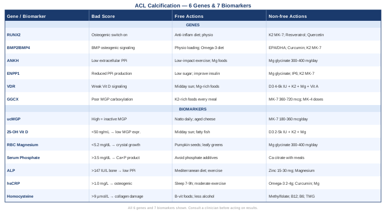
7 Key Biomarkers to Track for ACL Calcification
Why Biomarkers Reveal What Imaging Cannot
An MRI or X-ray shows you the consequence — calcium already deposited where it should not be. Biomarkers show you the process that created that consequence, and whether it is still running. The body maintains sophisticated defenses against soft-tissue calcification. When those defenses are intact, ectopic calcification does not occur. When they are compromised — by deficiencies, dysregulation, or inflammation — calcium follows the path of least resistance into ligaments, tendons, and cartilage.
The seven markers below correspond to the most critical checkpoints in that system. Most can be ordered through a primary care physician or functional medicine lab. Where costs are given, they reflect approximate US pricing without insurance.
Biomarker 1: Undercarboxylated Matrix Gla Protein (ucMGP)
Matrix Gla Protein (MGP) is the most potent known inhibitor of soft-tissue calcification. It functions by physically binding calcium ions and crystal nucleation sites in connective tissue, blocking the initial seeding that allows calcium deposits to grow. The critical detail: MGP is only active in its carboxylated form. Carboxylation requires vitamin K2 as a cofactor. When vitamin K2 is insufficient, MGP circulates in its undercarboxylated, inactive form — ucMGP — and the protective mechanism fails. Research consistently links elevated circulating ucMGP with soft-tissue and vascular calcification. Schurgers and colleagues have published extensively on this relationship, establishing ucMGP as both a mechanistic biomarker and a practical target for intervention (see ucMGP and calcification research on PubMed).
How to Measure It
ucMGP requires a specialized assay not included in standard blood panels. In Europe, VitaK BV (Netherlands) is the primary reference laboratory. In the United States, some functional medicine labs offer it through specialty order, and it is occasionally available through integrative medicine clinics. Cost range: $80–$200. You are looking for low ucMGP — meaning most circulating MGP is carboxylated and active. High ucMGP signals functional vitamin K2 insufficiency and reduced soft-tissue protection.
If the Score Is Bad: The Plan Without Supplements
The most powerful dietary correction is natto — Japanese fermented soybeans — which contains roughly 100 times more MK-7 per gram than any other food. Even one tablespoon daily provides a clinically meaningful MK-7 dose. Other K2 sources: aged hard cheeses (Gouda, Edam), goose and duck liver, pasture-raised egg yolks, and dark poultry meat. Reduce or eliminate processed foods with phosphate additives, which compete with the absorption and utilization of fat-soluble nutrients including K2.
If the Score Is Bad: The Plan With Supplements or Equipment
MK-7 (menaquinone-7) supplementation: 180–360 mcg per day, taken with a fat-containing meal. MK-7's half-life is approximately 72 hours, making once-daily dosing effective for maintaining consistent tissue levels. No cycling is needed for most healthy adults — K2 is non-toxic at these doses. Important exception: individuals on warfarin or vitamin K antagonists must consult their physician before starting K2, as it interacts directly with anticoagulation. Pair with vitamin D3 (see biomarker 2) — D3 and K2 are synergistic in calcium direction. Retest ucMGP after 3–6 months to verify response.
Biomarker 2: 25-OH Vitamin D
Vitamin D is commonly framed as a bone mineral — but its role in connective tissue health extends well beyond that. The vitamin D receptor (VDR) is expressed in ligament fibroblasts, synovial tissue, and immune cells throughout the joint environment. Low vitamin D drives two problems simultaneously: it increases the production of pro-inflammatory cytokines (which, as will be discussed under hsCRP, directly promote osteogenic reprogramming of soft tissue), and it reduces MGP expression in connective tissue cells. In practical terms, vitamin D deficiency removes both an anti-inflammatory and a direct calcification-inhibiting influence from the ACL environment.
The literature on vitamin D and musculoskeletal soft-tissue health is increasingly strong. Multiple studies associate lower 25-OH vitamin D with greater rates of tendon and ligament pathology and poorer healing outcomes (see vitamin D and tendon/ligament research on PubMed).
How to Measure It
A standard 25-hydroxyvitamin D blood test — widely available from any lab. Cost: $30–$80. Optimal functional range: 50–80 ng/mL (125–200 nmol/L). The conventional lower limit of "normal" (30 ng/mL) is now viewed by many researchers as insufficient for anti-inflammatory and connective tissue protection purposes. Retesting every 3–6 months is appropriate while optimizing levels.
If the Score Is Bad: The Plan Without Supplements
Midday sun exposure for 15–30 minutes on a large skin surface area (arms, chest, back) several times per week is the most direct natural source. Skin tone, latitude, and season significantly affect synthesis efficiency. Dietary sources contribute modestly: fatty fish (salmon, mackerel, sardines), beef liver, and pasture-raised egg yolks. Reducing indoor sedentary time during daylight hours is a meaningful structural change.
If the Score Is Bad: The Plan With Supplements or Equipment
Vitamin D3: 2,000–5,000 IU per day with a fat-containing meal. The critical co-administration: K2 (MK-7, 180–360 mcg/day) — vitamin D increases intestinal calcium absorption, and without K2 to direct that calcium into bone, the risk of soft-tissue deposition increases. Magnesium (300–400 mg/day) is also essential — it is required for the enzymatic conversion of vitamin D to its active hormonal form. Monitor levels at 3-month intervals and adjust dose to achieve 50–80 ng/mL. Safe for long-term daily use at these doses in most healthy adults; contraindicated in granulomatous diseases (sarcoidosis) without specialist oversight.
Biomarker 3: RBC Magnesium
Magnesium acts as a direct biochemical competitor to calcium in soft tissue. At the cellular and extracellular level, magnesium inhibits hydroxyapatite crystal nucleation — it binds to the same sites on the extracellular matrix where calcium phosphate crystals would otherwise begin to form. When magnesium is abundant, crystal formation is structurally impeded. When it is depleted, calcium proceeds largely unopposed.
Standard serum magnesium is an unreliable indicator of true magnesium status. The body prioritizes serum magnesium tightly — it pulls from intracellular stores to maintain serum levels, meaning serum can look normal while tissue deficiency is significant. RBC (red blood cell) magnesium reflects intracellular stores and is the clinically meaningful test. Epidemiological data consistently shows that magnesium insufficiency is widespread in populations eating processed diets, and this has direct implications for ectopic calcification risk (see magnesium and ectopic calcification research).
How to Measure It
Order specifically as RBC magnesium — not serum magnesium. Available through most specialty labs including SpectraCell and Quest Diagnostics. Cost: $50–$100. Optimal range: 5.2–6.5 mg/dL (approximately 2.1–2.7 mmol/L in some lab reporting formats). A result below 5.0 mg/dL represents a clear deficiency signal even if serum magnesium appears normal.
If the Score Is Bad: The Plan Without Supplements
Prioritize magnesium-dense foods daily: pumpkin seeds (highest per-gram concentration), dark leafy greens (spinach, Swiss chard), almonds, black beans, avocado, and quinoa. Eliminate the primary depleters: alcohol consumption, high refined-sugar intake, excess caffeine, and medications that increase renal magnesium wasting (diuretics, proton pump inhibitors). Chronic psychological stress activates a neuroendocrine cascade that directly increases urinary magnesium excretion — stress reduction is not peripheral to this biomarker.
If the Score Is Bad: The Plan With Supplements or Equipment
Magnesium glycinate: 300–400 mg elemental magnesium per day, taken in the evening — glycinate is well-absorbed and has a mild calming effect that suits evening use. Magnesium malate is preferred by some for daytime use given its role in energy metabolism. Avoid magnesium oxide — absorption is under 10% and it primarily acts as a laxative. The dose-limiting side effect of magnesium supplementation is loose stools, which varies by form and individual tolerance. No cycling required for most healthy adults. Retest RBC magnesium at 3 months; continued long-term supplementation is safe and often necessary given modern dietary patterns.
Biomarker 4: Serum Inorganic Phosphate
Phosphate's role in ectopic calcification is straightforward: when phosphate rises, the calcium-phosphate product (Ca × P) in the bloodstream increases, creating thermodynamic conditions where calcium phosphate crystals spontaneously precipitate in soft tissue. This mechanism is dramatically evident in chronic kidney disease, where severe hyperphosphatemia drives calcification of arteries, joints, and soft tissue throughout the body. What is less appreciated is that even modestly elevated phosphate — still within the "normal" range — can tip the balance toward calcification in susceptible individuals.
The modern Western diet is saturated with inorganic phosphate additives — present in processed meats, cola beverages, fast food, packaged cheese products, and shelf-stable baked goods. These additive phosphates are more bioavailable than the naturally occurring phosphate in whole foods, making them disproportionately impactful on serum phosphate levels.
How to Measure It
Serum phosphorus is part of a standard basic or comprehensive metabolic panel. Cost: included in a standard panel, $20–$60 standalone. Optimal functional range: 2.5–3.5 mg/dL. Many labs report the upper normal limit as 4.5 mg/dL, but functional and integrative medicine practitioners targeting ectopic calcification risk typically aim for the lower half of the reference range. Values consistently above 3.7 mg/dL in the context of soft-tissue calcification are worth addressing.
If the Score Is Bad: The Plan Without Supplements
Scan ingredient labels and eliminate products containing sodium phosphate, dicalcium phosphate, phosphoric acid, sodium hexametaphosphate, and similar additives. These appear in canned meats, fast food, cola drinks, processed cheeses, and many packaged snacks. Replace with whole-food protein sources: fresh fish, eggs, legumes, and unprocessed meat — these contain naturally bound phosphate that is significantly less bioavailable. Plant-based phosphate (bound to phytate) is particularly poorly absorbed.
If the Score Is Bad: The Plan With Supplements or Equipment
Pharmacological phosphate binders are reserved for clinical hyperphosphatemia under nephrology management. For dietary-range elevations, calcium citrate taken with meals binds some gastrointestinal phosphate before absorption; consult a physician before using this approach, as it also increases calcium intake. The primary intervention here is dietary — no supplement compensates for an additive-phosphate-rich diet. Focus is elimination, not supplementation.
Biomarker 5: Alkaline Phosphatase (ALP)
Tissue-nonspecific alkaline phosphatase (TNAP), encoded by the ALPL gene, sits at a critical junction in calcification regulation. Its primary relevant function: degrading inorganic pyrophosphate (PPi), which is the body's main endogenous inhibitor of mineralization. Where PPi is abundant, calcium crystals cannot form. Where PPi is depleted — because TNAP is overactive — calcification proceeds more easily.
Elevated ALP (bone fraction specifically) in the context of soft-tissue calcification is a meaningful signal: it suggests that the PPi brake on mineralization is being excessively removed. Context matters — ALP is also elevated in liver disease, bone turnover states, and certain medications — so interpretation requires fractionation when the cause is unclear (see TNAP, pyrophosphate and calcification research).
How to Measure It
ALP is part of a comprehensive metabolic panel. Cost: included in a standard panel. Normal adult range varies by lab, approximately 44–147 IU/L. If consistently elevated, request a fractionated ALP to distinguish bone-derived from liver-derived ALP. A bone ALP above 40 IU/L in adults not in active skeletal growth warrants further investigation in the context of soft-tissue calcification.
If the Score Is Bad: The Plan Without Supplements
If liver-driven: prioritize Mediterranean-style dietary patterns, reduce alcohol intake, improve insulin sensitivity through exercise and refined-carbohydrate reduction. If bone-driven: address vitamin D, K2, and phosphate status first (as above). Weight management reduces ALP across both tissue sources. Identify and address any medication-driven elevation (some anticonvulsants and corticosteroids raise ALP).
If the Score Is Bad: The Plan With Supplements or Equipment
Zinc (15–30 mg zinc bisglycinate or picolinate per day, with meals) supports appropriate ALP enzyme kinetics — zinc is a cofactor and dysregulation of ALP is partly zinc-dependent. Magnesium supports the broader mineralization regulation context. Avoid high-dose phosphate supplements or phosphate-rich protein powders. The primary strategy is addressing root causes (metabolic liver, bone mineral balance) rather than suppressing ALP directly.
Biomarker 6: High-Sensitivity CRP (hsCRP)
The connection between inflammation and calcification is not merely associative — it is mechanistic. Inflammatory cytokines, particularly TNF-α, IL-1β, and IL-6, have been demonstrated in human and cell-model research to stimulate osteogenic reprogramming of connective tissue fibroblasts: they activate RUNX2 and BMP signaling in ligament cells, effectively telling those cells to begin depositing mineral. Sustained low-grade inflammation is therefore not just a pain driver — it is a calcification driver at the molecular level.
High-sensitivity CRP (hsCRP) is the most accessible and widely validated marker of systemic low-grade inflammation. Peter Attia has consistently emphasized hsCRP as a central longevity and cardiovascular marker. Its relevance to connective tissue calcification is increasingly supported by research linking chronic inflammatory states to elevated rates of soft-tissue mineralization.
How to Measure It
Specify high-sensitivity CRP explicitly — standard CRP does not detect low-grade inflammation. Cost: $30–$80. Optimal target: below 0.5 mg/L. Below 1.0 mg/L is considered low-risk for most practitioners. Above 3.0 mg/L signals significant chronic inflammation; above 10 mg/L usually indicates active infection or autoimmune flare and should prompt immediate investigation.
If the Score Is Bad: The Plan Without Supplements
Sleep quality is among the most powerful levers for hsCRP — even one week of poor sleep measurably elevates inflammatory cytokines. Prioritize 7–9 hours of consistent, dark, cool-environment sleep. Regular moderate-intensity exercise (not excessive high-intensity training, which acutely spikes CRP) reduces baseline inflammation over time. A diet low in refined carbohydrates, trans fats, and seed oils, and high in polyphenols and omega-3-rich foods, reduces systemic inflammatory tone. Gum disease (periodontitis) is a frequently overlooked source of persistently elevated hsCRP — dental evaluation is worthwhile if values remain high despite other interventions.
If the Score Is Bad: The Plan With Supplements or Equipment
Omega-3 fatty acids (EPA + DHA): 2–4 grams per day from high-quality fish oil or algal oil, taken with meals to minimize GI side effects. Well-documented for reducing hsCRP and inflammatory cytokines in multiple randomized trials. No standard cycling; safe for continuous long-term use. Liposomal or phospholipid-complexed curcumin: 500–1,000 mg per day — the enhanced-absorption forms matter significantly, as standard curcumin is poorly bioavailable. Evidence for hsCRP reduction is consistent across multiple studies. Magnesium reduces IL-6 and CRP independently. If hsCRP remains above 3.0 mg/L despite these interventions, investigate gut dysbiosis, dental pathology, or occult metabolic syndrome before escalating supplements.
Biomarker 7: Homocysteine
Homocysteine is an intermediate amino acid in the methionine metabolism cycle. Its elevation in the context of ACL calcification is relevant through two distinct pathways. First, high homocysteine directly impairs collagen cross-linking by inhibiting lysyl oxidase — the enzyme that gives collagen its tensile strength. Weakened collagen architecture in the ACL creates microinjury, triggering an inflammatory repair cycle that, if sustained, feeds the calcification process. Second, elevated homocysteine promotes oxidative stress and endothelial dysfunction — both of which amplify the inflammatory signaling that drives osteogenic reprogramming of fibroblasts.
Thomas Dayspring and other lipidologists who work at the vascular level have long emphasized homocysteine as a marker that the conventional reference range treats far too permissively. The same argument applies in connective tissue biology.
How to Measure It
Plasma homocysteine — a straightforward venous blood test. Cost: $50–$100. Optimal target: below 8–9 µmol/L. Some practitioners working from cardiovascular and connective tissue risk frameworks target below 7 µmol/L. Conventional lab "normal" extends to 15 µmol/L in many references — this threshold is considered inadequate in functional medicine contexts.
If the Score Is Bad: The Plan Without Supplements
Prioritize B-vitamin-dense whole foods: dark leafy greens (folate), eggs (choline and B12), lean meat and fish (B6 and B12), and legumes. Reduce or eliminate alcohol, which interferes with B-vitamin absorption and methionine metabolism. Adequate dietary protein (not just animal protein; plant sources work well for methionine cycling) supports the remethylation pathway. Regular moderate exercise reduces homocysteine by approximately 10–15% independent of diet.
If the Score Is Bad: The Plan With Supplements or Equipment
Methylfolate (5-MTHF, not folic acid): 400–800 mcg per day — the active form bypasses the MTHFR enzyme, which a significant portion of the population has genetic variants in. Methylcobalamin (B12): 500–1,000 mcg per day sublingually or in capsule form — the methylated form is preferable for direct remethylation support. Pyridoxal-5-phosphate (B6): 25–50 mg per day — the active coenzyme form, used in the transsulfuration pathway. TMG (trimethylglycine, betaine): 500–1,000 mg per day — operates through the alternative betaine-homocysteine methyltransferase (BHMT) pathway and is particularly effective when the folate pathway is compromised. Cycling is not required for B vitamins at these doses; do not exceed 100 mg/day of B6 long-term due to peripheral neuropathy risk at higher doses. Retest plasma homocysteine at 3 months.
Building on these seven measurable signals gives you a biochemical map of your individual calcification risk profile. Understanding the genetic layer beneath that map adds a further dimension — explaining why certain deficiencies persist and why certain pathways are harder to regulate.
What Your Genes Say About ACL Calcification Risk
Reading the Genetic Layer
Genetics do not determine destiny in calcification any more than in most complex physiological processes. But they set the baseline difficulty. A person with ANKH loss-of-function variants has structurally lower extracellular pyrophosphate — the main crystal inhibitor — and will require more deliberate compensatory effort than someone without that variant. Knowing this prevents years of frustration with interventions that work for the population average but underperform for specific genetic profiles.
Consumer genetic testing (23andMe, AncestryDNA) combined with third-party analysis tools (Genetic Genie, FoundMyFitness by Rhonda Patrick) can surface many of the variants discussed below. Clinical genetic panels for mineralization or connective tissue disorders are available through rheumatology or medical genetics consultations for more definitive analysis.
Gene 1: RUNX2
RUNX2 (Runt-related transcription factor 2) is the master transcriptional switch for osteoblast differentiation. In physiological conditions, it is silenced in soft tissues like ligaments. In ectopic calcification, RUNX2 becomes pathologically activated in fibroblasts and tenocytes — reprogramming them to behave as bone-forming cells. Variants that increase RUNX2 expression or reduce the transcriptional suppressors that keep it off in soft tissue can predispose the ACL environment to osteogenic conversion.
The human evidence for RUNX2 polymorphisms in soft-tissue calcification is primarily from studies of calcific aortic valve disease and heterotopic ossification; direct ACL-specific human data is limited, but the mechanistic pathway is well-established and widely referenced in the ectopic calcification literature (see RUNX2 and soft tissue calcification on PubMed).
If the Gene Is Bad: The Plan Without Supplements
The inflammatory signals that activate RUNX2 in non-osseous tissue — primarily TNF-α and TGF-β1 — are substantially reduced by the same lifestyle interventions that lower hsCRP: sleep quality, anti-inflammatory diet, and moderate exercise. Appropriate mechanical loading through physiotherapy is also important — mechanobiological signals from physiological tension keep ACL fibroblasts in a functional rather than osteogenic state. Prolonged immobilization removes these signals and may upregulate osteogenic pathways.
If the Score Is Bad: The Plan With Supplements or Equipment
Vitamin K2 (MK-7, 180–360 mcg/day) activates MGP, which directly antagonizes RUNX2-driven mineralization at the extracellular level. Trans-resveratrol has demonstrated inhibitory effects on RUNX2 transcription in human cell studies: 200–500 mg per day, taken with a fat-containing meal. Quercetin inhibits BMP-RUNX2 signaling: 500–1,000 mg per day with meals. These supplements carry a reasonable mechanistic argument but limited direct human clinical trial evidence for ACL calcification specifically. A reasonable protocol: cycle on for 3 months, take 4 weeks off, and track symptoms and inflammatory markers.
Gene 2: BMP2 and BMP4
Bone morphogenetic proteins 2 and 4 (BMP2/BMP4) are among the most powerful known inducers of osteogenic differentiation. They are essential in bone formation and repair, but their aberrant activation in soft tissue is a primary driver of heterotopic ossification and ligament calcification. Variants that increase BMP2/4 signaling, or that reduce the natural BMP antagonists — noggin, chordin, gremlin — tip the balance toward soft-tissue mineralization.
The clinical relevance of this pathway is illustrated at the extreme by fibrodysplasia ossificans progressiva, a rare but instructive condition caused by an activating mutation in the BMP receptor ACVR1, which causes progressive ossification of connective tissue throughout the body. ACL calcification does not involve such severe dysregulation, but the same pathway operates on a continuum (see BMP2 and heterotopic ossification research).
If the Gene Is Bad: The Plan Without Supplements
Mechanical loading within the physiological range — guided physiotherapy, not aggressive loading — has documented suppressive effects on BMP-mediated osteogenesis in tendons and ligaments. Optimize dietary omega-3 to omega-6 ratio by reducing seed oil consumption and increasing fatty fish intake; pro-inflammatory fatty acids sensitize BMP signaling in injured tissue. Avoid excessive dietary calcium without corresponding K2 — excess calcium can accelerate BMP-driven mineralization in a permissive genetic background.
If the Score Is Bad: The Plan With Supplements or Equipment
Omega-3 fatty acids (EPA/DHA, 2–4 g/day): reduce the inflammatory amplification of BMP signaling. Liposomal curcumin (500–1,000 mg/day): shows inhibitory effects on BMP-driven osteogenic signaling in cell models, with limited but promising human data for calcification-prone conditions. K2 (MK-7, 180–360 mcg/day): the downstream MGP activation counters the calcification output of BMP signaling even when upstream signaling is elevated. Cycle curcumin and resveratrol (3 months on, 4 weeks off); omega-3 and K2 can be taken continuously.
Gene 3: ANKH
ANKH encodes the ANK protein — a multi-pass transmembrane channel that transports inorganic pyrophosphate (PPi) from the intracellular environment to the extracellular space. Extracellular PPi is the body's primary local inhibitor of hydroxyapatite crystal formation: wherever PPi is abundant, calcium crystals cannot nucleate and grow. ANKH is therefore a gatekeeper for calcification protection in cartilage and ligament tissue.
Loss-of-function variants in ANKH are the most firmly established genetic contributors to familial calcium pyrophosphate deposition (CPPD) disease — a condition that frequently involves calcification of knee joint cartilage and the knee's supporting ligaments, including the ACL and posterior cruciate ligament. The human genetic evidence here is among the strongest in the ectopic calcification field (see ANKH and CPPD on PubMed).
If the Gene Is Bad: The Plan Without Supplements
Since ANKH dysfunction depletes extracellular PPi, the compensatory logic targets PPi from other sources and direct crystal-inhibition alternatives. ENPP1 (see the next gene) is the primary enzymatic producer of extracellular PPi — supporting its function matters. Regular low-impact exercise (swimming, cycling) maintains joint fluid circulation and helps distribute PPi and other calcification inhibitors throughout the joint space. Maintain adequate magnesium to support direct crystal inhibition independent of the PPi pathway.
If the Score Is Bad: The Plan With Supplements or Equipment
Magnesium glycinate or malate (300–400 mg/day): magnesium inhibits hydroxyapatite crystal formation independently of PPi by competing with calcium at mineralization sites. This is the most directly compensatory supplement for reduced ANKH function. No supplement currently restores ANKH channel function directly. For symptomatic CPPD confirmed or suspected in association with ANKH variants, rheumatological management (colchicine 0.6 mg twice daily is a standard prophylactic option) should be discussed with a specialist. This is not a self-management condition — ANKH-associated CPPD warrants medical oversight.
Gene 4: ENPP1
ENPP1 (ectonucleotide pyrophosphatase/phosphodiesterase 1) is the enzyme responsible for generating most of the extracellular pyrophosphate that protects against calcification. It does this by cleaving extracellular ATP into AMP and PPi. Together with ANKH (which transports intracellular PPi out), ENPP1 forms the two-component system that maintains adequate PPi in the extracellular space around connective tissue.
The ENPP1 K121Q polymorphism is a relatively common variant that reduces enzyme efficiency. It has been linked to increased calcification tendency and metabolic dysregulation in multiple human cohort studies, and severe loss-of-function variants in ENPP1 cause generalized arterial calcification of infancy — demonstrating the critical role of ENPP1 in preventing ectopic mineralization throughout the body (see ENPP1 K121Q and calcification on PubMed).
If the Gene Is Bad: The Plan Without Supplements
Since ENPP1 function is impaired, reduce the metabolic conditions that maximize demand on its already-reduced PPi output. High sugar intake and alcohol consumption increase cellular ATP turnover without proportional ENPP1 compensation. Insulin resistance is associated with ENPP1 dysfunction — improving insulin sensitivity through dietary adjustment (lower refined carbohydrate, higher fiber) and exercise has both direct and indirect benefits for the ENPP1-PPi pathway.
If the Score Is Bad: The Plan With Supplements or Equipment
Magnesium glycinate (300–400 mg/day): compensates for reduced PPi by directly inhibiting crystal nucleation. Inositol hexaphosphate (IP6) — found in whole grains and legumes, or as a supplement (1,000–2,000 mg/day, taken away from meals to avoid chelation of dietary minerals) — acts as a structural analog to pyrophosphate, inhibiting crystal growth. Evidence for IP6 is primarily preclinical and early-stage human, but mechanistically sound and low-risk. Cycle 3 months on, 4 weeks off. K2 (MK-7, 180–360 mcg/day) remains essential to ensure MGP-level protection is intact even when the PPi layer is compromised.
Gene 5: VDR (Vitamin D Receptor)
The VDR gene encodes the vitamin D receptor through which all genomic actions of calcitriol (the active form of vitamin D) are mediated. Four common VDR single nucleotide polymorphisms — FokI, BsmI, TaqI, and ApaI — affect receptor efficiency. Certain allele combinations are associated with measurably reduced vitamin D signaling even at adequate serum 25-OH vitamin D concentrations. For those with poor VDR function, achieving the anti-inflammatory and calcification-inhibitory effects of vitamin D requires higher circulating levels.
Ali Torkamani of Scripps Research, a leading figure in genomic medicine and precision health, has emphasized in his educational work that genetic variants in metabolic receptors and enzymes often require dietary, supplemental, and lifestyle compensation rather than replacement — the VDR case is a clear example.
If the Gene Is Bad: The Plan Without Supplements
Aim for the higher end of the vitamin D functional range (60–80 ng/mL) rather than just "normal." Maximize sun exposure quality: midday UVB, large skin area, 15–30 minutes several times per week, avoiding sunscreen during that brief period. Combine vitamin D-rich foods consistently with magnesium-rich foods — magnesium enables vitamin D activation and VDR function simultaneously. Maintain consistent sleep and exercise, both of which improve sensitivity to hormonal signaling including vitamin D.
If the Score Is Bad: The Plan With Supplements or Equipment
Higher-dose vitamin D3 may be required for individuals with poor VDR function: 4,000–6,000 IU/day, with the target of achieving serum levels of 60–80 ng/mL. This higher target compensates for receptor inefficiency. Always co-administer the complete triad: K2 (MK-7, 180–360 mcg/day), magnesium glycinate (300–400 mg/day), and vitamin A (from food or supplements — 2,000–5,000 IU retinol, not beta-carotene) — vitamins A, D, and K2 are synergistic nuclear hormone receptor ligands. Monitor serum 25-OH vitamin D and calcium every 3–6 months, particularly at higher doses.
Gene 6: GGCX (Gamma-Glutamyl Carboxylase)
GGCX encodes the enzyme that carboxylates vitamin K-dependent proteins — including MGP and osteocalcin. This enzyme, using vitamin K2 as a cofactor, adds the carboxyl groups that activate these proteins. A critical and underappreciated implication: even if vitamin K2 intake is adequate, reduced GGCX enzyme activity means that MGP remains undercarboxylated and inactive. GGCX variants can therefore produce a functional vitamin K2 deficiency in the absence of any dietary deficiency.
Several GGCX polymorphisms have been associated with altered carboxylation efficiency in human studies, and GGCX mutations cause a severe clinical syndrome (pseudoxanthoma elasticum variant) characterized by widespread ectopic calcification of elastic tissue. More common GGCX variants with milder effects remain under-researched for soft-tissue calcification but represent a plausible and undertested contributor.
If the Gene Is Bad: The Plan Without Supplements
If GGCX efficiency is genetically reduced, the compensatory strategy is substrate saturation: dramatically increase the available vitamin K2 so that even operating at reduced enzymatic efficiency, sufficient MGP gets carboxylated to protect soft tissue. This means K2-rich foods at every meal — natto, aged hard cheeses, pasture-raised animal products. Reduce competing factors: warfarin and similar medications directly block the GGCX cycle; statins may reduce K2 availability in peripheral tissue. If either of these medications are being taken, this genetic finding warrants specific clinical discussion.
If the Score Is Bad: The Plan With Supplements or Equipment
For those with confirmed GGCX variants who show persistently elevated ucMGP despite standard K2 supplementation, higher-dose MK-7 (360–720 mcg/day) saturates the reduced-efficiency GGCX enzyme with sufficient substrate to drive adequate carboxylation. This is beyond the standard therapeutic dose and should be discussed with a physician. MK-4 at pharmacological doses (1,500–15,000 mcg/day, doses used in Japanese clinical trials for osteoporosis) reaches tissue distributions that MK-7 does not fully cover and may offer additional benefit. Monitor ucMGP every 3–6 months to calibrate the response.
The Book That Changes How You Think About Calcium
Vitamin K2 and the Calcium Paradox by Kate Rheaume-Bleue — a Canadian naturopathic physician — is the most practically useful book written on the subject of ectopic calcification. It synthesizes decades of biochemical and clinical research into a framework that most physicians were never taught, and its core argument applies directly to soft-tissue calcification in the ACL and surrounding connective tissue.
1. The Core Paradox
The book's central insight is this: millions of people are simultaneously losing calcium from their bones and depositing it in soft tissue. This is not a contradiction — it is the direct consequence of calcium management without vitamin K2. Without K2 to direct calcium to bone and block it from soft tissue, calcium supplementation or even adequate dietary calcium does not build bone effectively. It circulates and deposits wherever the crystal-formation threshold is lowest — arteries, cartilage, ligaments.
2. Matrix Gla Protein as the Hero
Rheaume-Bleue dedicates substantial attention to MGP, explaining clearly how it was originally characterized in shark cartilage — tissue that never calcifies under normal conditions — by biologist Paul Price. MGP is the biological reason connective tissue in cartilaginous species remains flexible. When K2 is adequate, MGP is activated, and this protection extends to human connective tissue. When K2 is insufficient, MGP is inactive and that protection disappears.
3. Why MK-7 Outperforms MK-4 for Soft Tissue
Not all K2 is equivalent in its reach. MK-4 (menaquinone-4), the form produced by animals from K1, is rapidly metabolized and cleared from the blood within hours. MK-7 (menaquinone-7), produced by bacteria during fermentation, remains in circulation for approximately 72 hours. This prolonged half-life means MK-7 has time to reach peripheral soft tissues — arteries, ligaments, tendons — and activate MGP throughout the body. For soft-tissue calcification protection, MK-7 is the relevant form.
4. The Rotterdam Study
The book is anchored partly in the Rotterdam Heart Study, one of the largest long-term nutritional cohort studies ever conducted. Among its findings: higher dietary menaquinone (K2) intake was associated with a 57% reduction in aortic calcification risk and a 26% reduction in all-cause mortality. This was not a study of supplements — it was a prospective observation of dietary K2 intake in thousands of people over years. And the protective association was specific to K2, not K1.
5. Calcium Supplements Without K2 Are a Problem
Rheaume-Bleue builds a compelling case that the billions of calcium supplement doses taken annually without concurrent K2 may be creating more calcification burden than they resolve. Multiple large randomized trials — including the CAIFOS and WHI studies — have shown that calcium supplements increase cardiovascular calcification events. The problem is not calcium; it is the absence of the directional protein. K2 turns calcium supplementation from a potential liability into an appropriate tool.
6. Natto Is Uniquely Positioned
A tablespoon of natto delivers more MK-7 than any other food by an enormous margin — roughly 100 times more than aged cheese, which is the next best option. The book makes a practical case for acquiring a taste for it or incorporating it into recipes where its flavor and texture can be masked (mixed into miso soups, combined with sauces). For those who cannot manage the taste, high-purity MK-7 derived from Bacillus subtilis natto fermentation is the supplement equivalent.
7. K2 Deficiency Is the Default, Not the Exception
Using ucMGP data from European population studies, the book argues that circulating undercarboxylated MGP — indicating insufficient K2 to activate the available protein — is the statistical norm in populations eating a modern processed diet. This is not a detectable clinical condition in most healthcare settings. No one will diagnose you with K2 deficiency at a routine physical. But the biochemical consequence — impaired calcification protection — operates silently.
8. K1 and K2 Are Not Interchangeable
One of the most important misunderstandings the book dispels: eating vitamin K in leafy greens (K1) does not protect soft tissue the way K2 does. K1 is primarily hepatic — it goes to the liver to activate clotting factors. K2 goes to bone, arteries, and connective tissue. They are chemically related but functionally distinct in their tissue distribution. You cannot meet the soft-tissue K2 requirement through spinach.
9. The D3 and K2 Interaction
The book explains the physiological relationship between D3 and K2 in terms that matter clinically. Vitamin D3 increases calcium absorption from the gastrointestinal tract. This is beneficial when K2 is adequate to direct that calcium into bone and inhibit it from soft tissue. Without K2, vitamin D3-driven calcium absorption increases calcium availability without increasing calcium placement accuracy. Taking D3 without K2 — as the majority of people who supplement with D3 do — amplifies the calcium paradox.
10. Highest-Risk Populations
The book identifies specific groups with the greatest unmet K2 need: individuals on long-term warfarin therapy (which directly blocks the K2 carboxylation cycle), those on statins (which may reduce peripheral K2 synthesis), people with fat malabsorption conditions (K2 is fat-soluble; conditions like celiac disease, Crohn's, or cystic fibrosis significantly impair absorption), post-menopausal women taking calcium supplements without K2, and anyone whose diet is low in fermented foods and pasture-raised animal products. ACL calcification patients on any of these lists warrant particularly close attention to K2 status.
Complementary Approaches Worth Considering
Low-Level Laser Therapy (Photobiomodulation)
Low-level laser therapy (LLLT) — also called photobiomodulation — uses specific wavelengths of red and near-infrared light to penetrate tissue and drive measurable cellular effects: increased mitochondrial ATP production, reduced inflammatory cytokine expression, and modulation of fibroblast behavior. For calcific tendinopathies — the closest well-studied analog to ACL calcification — LLLT has accumulated meaningful human clinical evidence that distinguishes it from most alternative physical therapies.
A systematic review and meta-analysis examining LLLT for calcific shoulder tendinopathy found statistically significant reductions in pain and improvements in shoulder function compared to sham treatment. The same inflammatory pathways — TNF-α, IL-6, and NF-κB signaling — that LLLT downregulates in calcific shoulder tendinopathy are directly implicated in ACL ectopic calcification. While ACL-specific randomized trials are sparse (given its relative rarity), the mechanistic overlap is strong and the evidence from adjacent musculoskeletal calcification conditions is worth incorporating (see LLLT and calcific tendinopathy research).
For realistic application: use a device in the 630–850 nm wavelength range with 25–100 mW output. Apply directly over the knee at medial and lateral aspects for 60–120 seconds per treatment point, 3–5 times per week. A standard course is 8–12 weeks. Home devices from manufacturers such as Joovv or Mito Red Light offer accessible options; clinical-grade devices provide higher irradiance. LLLT has an excellent safety profile at therapeutic doses — avoid use directly over suspected malignant tissue, and inform your physiotherapist that you are incorporating it.
Tai Chi for Joint Mobility and Inflammatory Reduction
Tai chi is a slow-movement, weight-shifting practice that combines joint mobility, proprioceptive training, and controlled breathing. Its relevance to ACL calcification is grounded in three mechanisms: it maintains knee joint range of motion through gentle, non-provocative movement; it consistently reduces systemic inflammatory markers including CRP and IL-6 across randomized trials; and it improves proprioception — consistently impaired when intra-ligamentous pathology is present.
A randomized controlled trial published in Arthritis Care & Research found that tai chi significantly reduced pain and improved physical function in patients with knee osteoarthritis, with associated reductions in systemic inflammatory biomarkers. While knee osteoarthritis and ACL calcification are distinct conditions, the overlapping mechanisms — inflammatory pain, proprioceptive disruption, and joint mobility limitation — make this evidence directly applicable. Tai chi is one of the few exercise modalities with consistent RCT evidence across multiple knee pathologies (see tai chi and knee pathology RCTs).
For practical application: begin with Yang-style tai chi, the most accessible form, through a beginner class or structured video program. Three to five sessions of 30–45 minutes per week. The first 4–6 weeks are a learning curve — do not assess benefits before 8 weeks. Inform your instructor of the ACL pathology and avoid any posture that provokes sharp knee pain. Sustained practice over 12+ weeks consistently demonstrates measurable improvements in both pain scores and inflammatory markers in knee-pathology studies.
Breathing-Based Therapies
The connection between breathing patterns and calcification operates through two documented mechanisms. First, chronic dysfunctional or excessive breathing (habitual over-breathing or mouth breathing) disrupts CO2/O2 balance and can affect calcium ion excitability and handling in soft tissue. Second, and more directly supported by human research: breathing practices that activate the parasympathetic nervous system reliably suppress cortisol secretion and pro-inflammatory cytokine production — including the IL-6 and TNF-α that drive osteogenic reprogramming of connective tissue fibroblasts.
A study in Frontiers in Human Neuroscience demonstrated that slow, controlled breathing (around 6 cycles per minute) significantly increased vagal tone and reduced markers of systemic inflammation in participants with elevated baseline inflammatory states. The parasympathetic-inflammation link is well-established — the vagus nerve directly modulates the inflammatory reflex via the spleen and macrophage populations throughout the body (see slow breathing and inflammation on PubMed). Evidence for direct calcification benefit is preliminary, but given the low risk and strong anti-inflammatory rationale, the application is straightforward to incorporate.
For practical implementation: practice diaphragmatic slow breathing for 10–15 minutes daily. Inhale for 4–5 counts, exhale for 6–8 counts (the longer exhale is the key — it activates the parasympathetic response). Consistency over duration — a daily 12-minute practice outperforms occasional 45-minute sessions. Buteyko breathing and CO2-tolerance training are more structured approaches for those interested in going deeper; a qualified Buteyko practitioner can assess your baseline and guide progression. Avoid Wim Hof-style hyperventilation phases without cardiovascular assessment.
Conclusion
ACL calcification is not a random misfortune. It is the result of specific deficiencies — of MGP carboxylation, of pyrophosphate, of magnesium, of appropriate calcium direction — sometimes compounded by genetic variants that make these systems structurally harder to maintain. The seven biomarkers covered in this article give you a measurable window into each of those failure points. The six genes add a layer of explanation for why some individuals face this problem despite doing many things right.
The clearest next step is a targeted lab panel: ucMGP, 25-OH vitamin D, RBC magnesium, serum phosphate, ALP, hsCRP, and homocysteine. These seven tests are either part of routine panels or orderable through functional medicine labs. Bring the results to a rheumatologist or integrative medicine physician familiar with calcification pathology — ideally one willing to act on functional ranges rather than only conventional cutoffs. If accessible, add a genetic panel or upload existing consumer DNA data to a third-party analysis tool and review the six genes discussed.
No intervention described here replaces clinical oversight, and none guarantees reversal. What they offer is something more durable: a clearer picture of what is actually happening in your body, and a specific basis for intervening on the right targets rather than the most obvious ones.
Musculoskeletal: Joint Conditions Tendon & Ligament Conditions Sports Injuries
Autoimmune: Inflammatory Conditions