This article contains AI generated content.
· UpdatedActinomycosis - 4 Genes And 6 Biomarkers To Track
Introduction
Actinomycosis is not the kind of condition most people have heard of, yet for those living through it, the frustration is anything but abstract. It is a chronic bacterial infection caused by Actinomyces species — bacteria that are part of the normal flora of the mouth, gut, and female reproductive tract in virtually every healthy adult. Most of the time they cause no harm. But when tissue barriers are disrupted — by dental trauma, surgery, a poorly positioned IUD, or immune suppression — they can spread slowly through soft tissue, forming dense fibrous lesions that mimic cancer, tuberculosis, or Crohn's disease. The average time between first symptoms and correct diagnosis is somewhere between six months and two years.
What makes this gap so wide is not just the rarity of the diagnosis. It is the fact that two people with the same bacterial exposure can have completely different outcomes. One person clears the infection with a short course of antibiotics; another faces relapse after relapse, tissue destruction, and prolonged suffering. That asymmetry almost certainly reflects individual differences in immune genetics, nutritional status, and systemic inflammatory tone — factors that standard clinical care rarely measures with any depth. Generic advice to "complete the antibiotic course and rest" is accurate but leaves out the question of why the body failed to contain these bacteria in the first place.
This article takes a more targeted approach. The first main section walks through six biomarkers worth tracking throughout the course of actinomycosis — markers that reveal how severely your immune system is activated, whether your body has the nutritional reserves to sustain a prolonged defense, and how effectively treatment is actually working over time. A second section then examines four immune-related genes — TLR2, IL-6, TNF-α, and DEFB1 — that shape innate bacterial recognition and clearance, with practical protocols for each.
Neither section is a substitute for medical care. But more precise biological information turns passive waiting into active, informed participation. When you can track inflammation, immune competence, and tissue stress with real numbers, every conversation with your physician becomes more focused and productive. And that is where cautious, evidence-grounded hope lives: not in shortcuts, but in better information leading to smarter decisions.
Summary
Actinomycosis progresses slowly, mimics serious diseases, and recurs in people whose immune defenses are chronically under-resourced. This article builds a practical monitoring framework directly around the biology of the infection itself, giving you measurable targets rather than vague recommendations.
What you will find here: Six biomarkers — including CRP, procalcitonin, albumin, and fibrinogen — with precise measurement guides, cost ranges, and actionable recovery plans for when results are abnormal. Four immune genes — TLR2, IL-6, TNF-α, and DEFB1 — that influence bacterial recognition and chronic inflammation, with lifestyle and supplement protocols for poor variants. A summary of ten high-impact immune science findings from well-documented research that challenges the "just take antibiotics" mindset. And four evidence-assessed complementary modalities with realistic application guides for the specific context of chronic bacterial infection.
The biomarker section is the most clinically actionable starting point: you can measure these markers today, track them across treatment, and use the data to have more productive conversations with your doctor. The genetics section adds a layer of personalization — explaining why some people's immune systems struggle more than others to contain Actinomyces once it has breached tissue barriers. Together, these two lenses offer a significantly more complete picture than standard infectious disease monitoring alone.
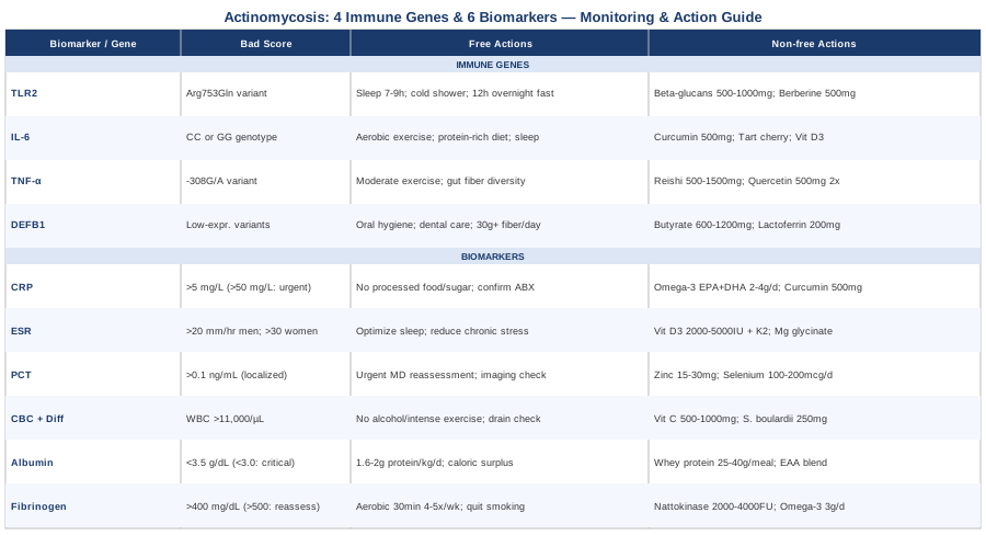
6 Biomarkers to Track Through Actinomycosis
Actinomycosis unfolds on a slow timeline — often requiring six to twelve months of antibiotic therapy even in straightforward cases. That extended arc makes biomarker tracking unusually valuable, not as a one-time snapshot but as a running series of data points that reveal whether the infection is retreating, stable, or silently progressing. The six markers below span acute-phase inflammation, bacterial burden signaling, immune cell activity, and nutritional resilience. No single test tells the full story; together, they give a substantially richer picture of what is actually happening inside the tissue.
Biomarker 1: C-Reactive Protein (CRP)
Why it matters
CRP is a protein produced by the liver in direct response to interleukin-6 signaling — the same cytokine pathway activated by bacterial invasion. During active actinomycosis it is typically elevated, often substantially, and it responds to successful antibiotic treatment faster than most other systemic markers. That responsiveness makes serial CRP measurement one of the most practical tools for gauging whether therapy is penetrating the infection. Actinomyces colonies are surrounded by dense fibrotic stroma that limits drug diffusion; a CRP that fails to decline after three to four weeks of appropriate treatment is a concrete signal that source control may be inadequate, that surgical drainage might be needed, or that the prescribed antibiotic regimen is insufficient for the infection depth.
Research on bacterial soft tissue infections consistently demonstrates CRP as a reliable early indicator of treatment response. Studies published in Clinical Microbiology and Infection have highlighted CRP's sensitivity to reductions in bacterial load, making it particularly useful in conditions like actinomycosis where imaging changes often lag weeks behind clinical reality. A single elevated value is less informative than a trend; the trajectory across monthly measurements is where the clinical signal lives.
How to measure it
High-sensitivity CRP (hs-CRP) is the preferred version, as it detects lower-level inflammation useful for baseline monitoring and trend detection. Cost ranges from $10–$40 at commercial labs in the United States. Results are typically available within 24–48 hours and can be ordered through any primary care physician as part of a general inflammatory panel.
Target during actinomycosis treatment: CRP trending steadily downward toward below 5 mg/L. Values persistently above 50 mg/L in the context of known infection warrant prompt clinical reassessment. Monthly testing is a reasonable minimum frequency during active treatment.
If the score is high: the plan without supplements
The most direct intervention when CRP remains elevated is ensuring adequate antibiotic penetration and source control. The standard approach for invasive actinomycosis — per NCBI StatPearls clinical reference on actinomycosis — involves high-dose intravenous penicillin G in the acute phase, transitioning to oral amoxicillin for months, with surgical drainage of any abscess collections. Confirm with your physician that route, dose, and duration match current infectious disease guidelines for your specific presentation. Beyond antibiotics, eliminating dietary ultra-processed foods and added sugars — both of which independently drive systemic NF-κB activation and hepatic CRP output — reduces background inflammatory noise and ensures your CRP readings more faithfully reflect the infection itself.
If the score is high: the plan with supplements or equipment
Omega-3 fatty acids (EPA and DHA combined) at 2–4 grams per day from a high-quality fish oil have well-documented CRP-reducing effects confirmed across multiple meta-analyses on inflammatory conditions. Take with the largest meal of the day for best absorption. Frequency: daily throughout the treatment period; no cycling required. Monitor for blood-thinning effects if taking anticoagulants.
Curcumin, in a phosphatidylcholine-complexed or liposomal form for meaningful bioavailability, at 500–1000 mg per day has shown consistent CRP-lowering effects in clinical trials. It does not replace antibiotic therapy but can support inflammatory balance during the prolonged treatment course. Avoid high-dose curcumin if taking immunosuppressive medications or if there is any hepatobiliary issue.
Biomarker 2: Erythrocyte Sedimentation Rate (ESR)
Why it matters
ESR measures how quickly red blood cells settle in a test tube — a rate driven by changes in blood protein composition, particularly the fibrinogen and immunoglobulin elevations characteristic of inflammatory states. In actinomycosis, ESR is almost universally elevated during the active phase and tends to normalize slowly, making it a useful complement to CRP rather than a replacement. Where CRP captures changes over days to weeks, a persistently elevated ESR over months reflects the chronic, low-grade inflammatory activity that characterizes actinomycosis even as acute bacterial load is declining.
One particularly practical use of ESR in actinomycosis management is in distinguishing true relapse from post-treatment fibrotic scar tissue, which can appear similar on imaging. When clinical symptoms are ambiguous, a rising ESR alongside rising CRP makes active infection significantly more likely than quiescent fibrosis. This distinction directly influences whether antibiotic therapy should be extended or whether a biopsy is warranted.
How to measure it
ESR is a standard laboratory test costing $10–$30. Normal values differ by age and sex: generally below 20 mm/hour for men under 50 and below 30 mm/hour for women under 50, with slightly higher accepted upper limits in older adults. Monthly measurement during treatment establishes a trend; a downward trajectory is the target even if absolute values remain elevated for the first several months of therapy.
If the score is high: the plan without supplements
Persistently elevated ESR should prompt a conversation about treatment duration adequacy. Current infectious disease guidelines support extended oral amoxicillin or penicillin courses — often six to twelve months — precisely because actinomycosis is slow-moving and fibrotic tissue impairs drug delivery. A still-elevated ESR at week eight is not automatically a treatment failure; it is expected given the infection's biology. What matters is whether the trend is consistently downward. Non-pharmacologically, optimizing sleep (sustained poor sleep elevates ESR through cortisol-driven inflammatory gene expression) and reducing chronic psychosocial stress are measurably effective ways to ensure ESR readings are not inflated by factors unrelated to the infection itself.
If the score is high: the plan with supplements or equipment
Vitamin D3 deficiency is associated with higher ESR through its role in macrophage differentiation and inflammatory cytokine suppression. Testing serum 25-OH vitamin D and supplementing to the 40–60 ng/mL range — typically 2000–5000 IU/day depending on baseline — has mechanistic plausibility and low risk. Always co-supplement with vitamin K2 (100–200 mcg/day, as MK-7 form) when using higher vitamin D doses to protect calcium metabolism. Magnesium glycinate at 200–400 mg at night supports vitamin D metabolism and independently reduces inflammatory markers in several clinical studies. These are complementary to antibiotic therapy, not substitutes for it.
Biomarker 3: Procalcitonin (PCT)
Why it matters
Procalcitonin is currently one of the most specific blood biomarkers for bacterial infection. It rises in response to bacterial toxins and pro-inflammatory cytokines released during active infection and remains low during viral illness and most non-infectious inflammatory states — a specificity that CRP does not match. In actinomycosis, PCT's behavior is nuanced: because this is typically a chronic, localized infection rather than a systemic septic process, PCT levels may not be as dramatically elevated as in acute bacteremia or sepsis. Nevertheless, a rising PCT in a patient being treated for known actinomycosis is a clinically significant warning: it suggests systemic spread, secondary bacterial co-infection, or failure of source control.
PCT is also increasingly used to guide antibiotic duration decisions. Research demonstrating that PCT-guided antibiotic stewardship protocols can reduce unnecessary antibiotic exposure without worsening outcomes has been published across multiple settings. For a condition where patients face many months of antibiotics, PCT provides an objective anchor for de-escalation decisions.
How to measure it
PCT is a blood test available at most hospital-based and larger commercial laboratories. Cost: $30–$80 in the US. A value below 0.1 ng/mL is generally normal. Values between 0.1–0.5 ng/mL suggest possible localized bacterial infection; values above 0.5 ng/mL indicate more active bacterial inflammatory activity. In known actinomycosis, even mild elevations are worth monitoring serially every four to six weeks rather than interpreting in isolation.
If the score is high: the plan without supplements
A rising PCT during actinomycosis treatment is a medical red flag requiring prompt physician reassessment — it is not something addressable through lifestyle modification. The clinical priorities are: confirming antibiotic adherence and adequacy, reviewing imaging for new or enlarging abscess collections requiring surgical drainage, and ruling out a secondary bacterial co-infection. PCT in active bacterial infection is a signal to the immune system's need for medical escalation, not nutritional adjustment. Do not delay clinical contact if PCT is trending upward.
If the score is high: the plan with supplements or equipment
Supporting immune clearance capacity during active bacterial infection involves ensuring micronutrients critical for neutrophil and macrophage function are not depleted. Zinc (15–30 mg elemental zinc per day, not exceeding 40 mg to avoid copper depletion) is essential for neutrophil oxidative burst activity. Selenium (100–200 mcg per day as selenomethionine) supports glutathione peroxidase enzyme activity relevant to immune cell function. Both should be taken alongside — not instead of — appropriate antibiotic therapy. Duration: throughout the acute treatment phase, tapering off after normalization of inflammatory markers. Higher doses of zinc in particular can impair copper balance over time and are not justified for indefinite use.
Biomarker 4: Complete Blood Count (CBC) with Differential
Why it matters
The complete blood count with differential remains one of the most informative and cost-effective tests for monitoring any bacterial infection. In actinomycosis, the characteristic finding is leukocytosis — elevated total white blood cell count — driven primarily by neutrophilia, confirming active bacterial immune stimulation. As treatment succeeds and bacterial load declines, WBC and neutrophil counts should trend toward normalization. A persistent neutrophilia after three to four weeks of appropriate therapy is a concrete signal that bacterial source control is insufficient — either the antibiotic is not reaching the infection site, an undrained abscess is maintaining the reservoir, or the infection is more extensive than appreciated on imaging.
One diagnostically useful nuance: as actinomycosis transitions from acute to chronic, the differential often shifts toward relative monocyte elevation. This reflects the transition from acute bacterial clearance toward granuloma formation — the histopathological signature of actinomycosis — and provides a window into which phase of the immune response is currently dominant. Tracking this shift alongside absolute WBC count adds interpretive depth to routine monitoring.
How to measure it
CBC with differential is one of the most affordable blood tests available, costing $10–$30. Normal total WBC range: 4,500–11,000 cells/µL. Neutrophils should represent 50–70% of total WBC during health; values above this in the context of known infection indicate ongoing bacterial immune stimulation. Monthly testing throughout treatment is reasonable minimum monitoring.
If the score is high: the plan without supplements
Sustained leukocytosis signals that bacterial burden remains too high for the immune system to contain passively — the primary interventions remain medical. Review imaging for collections requiring surgical drainage. Confirm antibiotic appropriateness (penicillin, amoxicillin, and clindamycin are the standard options depending on presentation and allergy status) and that the duration aligns with infectious disease guidelines for the specific site of infection. Eliminate background variables that inflate WBC independently: chronic sleep deprivation, heavy exercise during acute infection, and alcohol consumption all drive WBC upward and should be controlled to ensure test results reflect infection status rather than confounders.
If the score is high: the plan with supplements or equipment
Vitamin C at 500–1000 mg per day supports neutrophil migration and bactericidal activity — neutrophils concentrate vitamin C at levels far above plasma, reflecting its functional importance in immune cell killing. A 2017 review in Nutrients supported the role of adequate vitamin C in neutrophil function and overall immune defense. Saccharomyces boulardii probiotic supplementation during antibiotic therapy (250–500 mg per day, taken at least two hours from the antibiotic dose) helps maintain gut-associated immune cell populations that otherwise deplete during prolonged antibiotic use. Both can be taken daily throughout the treatment course at these doses without significant side effects.
Biomarker 5: Serum Albumin
Why it matters
Serum albumin is the primary protein in blood plasma, synthesized exclusively by the liver. During significant bacterial infection, inflammatory cytokines — particularly IL-6 and TNF-α — directly suppress hepatic albumin synthesis while shifting liver production toward acute-phase proteins like CRP and fibrinogen. The result: any active, prolonged infection drives albumin downward over weeks to months. In actinomycosis, which frequently requires six to twelve months of treatment, albumin becomes a critical indicator of both inflammatory status and the body's nutritional reserves. Low albumin during prolonged antibiotic therapy means tissue repair, immune protein synthesis, and antibiotic binding capacity are all being compromised — many antibiotics are heavily protein-bound, and low albumin affects their effective free concentration in tissue.
Peter Attia has highlighted serum albumin as one of the most underappreciated markers of physiological resilience — it reflects both hepatic function and the integrated state of protein metabolism under inflammatory stress. For a metabolically demanding condition like actinomycosis, this framing is directly applicable.
How to measure it
Serum albumin is included in any comprehensive metabolic panel (CMP), costing $15–$40 at most labs. Target range: 3.5–5.0 g/dL. Levels below 3.5 g/dL in the context of active infection indicate significant nutritional-inflammatory strain and are associated with impaired wound healing and immune competence. Below 3.0 g/dL represents a clinical priority. Monthly monitoring during treatment is justified.
If the score is low: the plan without supplements
Increasing dietary protein intake is the most impactful primary step. Aim for 1.6–2.0 grams of high-quality complete protein per kilogram of body weight per day — a level consistently shown to support albumin synthesis under inflammatory stress. Prioritize eggs, fish, poultry, and full-fat dairy. Reducing any caloric deficit is equally important: calorie restriction during active infection impairs hepatic albumin synthesis regardless of protein intake. Physical rest rather than high-intensity exercise is appropriate during acute infection phases, since intense training acutely suppresses albumin as part of its inflammatory signaling.
If the score is low: the plan with supplements or equipment
Whey protein isolate (25–40 grams added to a meal) is among the most efficient dietary interventions for driving hepatic albumin synthesis due to its high leucine content. Leucine at 2–3 grams per dose is a direct activator of hepatic mTOR signaling relevant to albumin production. For patients whose antibiotic-related GI effects limit high oral protein tolerance, essential amino acid (EAA) blends — providing the full albumin-stimulating amino acid pattern in smaller volume — are a practical alternative. A registered dietitian with experience in infection-related malnutrition can be particularly valuable for patients with persistently low albumin during extended treatment.
Biomarker 6: Fibrinogen
Why it matters
Fibrinogen is a coagulation protein and acute-phase reactant that rises during bacterial infection as part of the same hepatic shift that suppresses albumin. In actinomycosis specifically, elevated fibrinogen carries additional significance: fibrin and fibrinogen deposition are key components of the dense fibrotic stroma that surrounds Actinomyces colonies — the histopathological feature that defines this infection. Persistently elevated fibrinogen in a patient under antibiotic therapy may reflect ongoing fibrotic tissue activity even as bacterial load is declining. This can explain why some patients continue to have symptoms, imaging abnormalities, and elevated inflammatory markers long after bacterial clearance has occurred — the fibrotic response has taken on its own momentum.
Beyond infection monitoring, elevated fibrinogen is also an independent cardiovascular risk factor that researchers including Thomas Dayspring have emphasized in the context of metabolic health assessment. For patients managing a prolonged illness that limits activity and disrupts diet, tracking fibrinogen alongside lipid panels adds meaningful risk context.
How to measure it
Fibrinogen is ordered as a standalone test or as part of a coagulation panel. Cost: $20–$50. Normal range: 200–400 mg/dL. Values persistently above 500 mg/dL during actinomycosis treatment suggest ongoing fibrotic or inflammatory activity requiring clinical reassessment rather than treatment de-escalation.
If the score is high: the plan without supplements
Moderate aerobic exercise at 30 minutes per session, four to five days per week, has the strongest non-pharmacological evidence base for reducing fibrinogen — though during acute infection this must be calibrated carefully to avoid depleting immune resources. As the infection stabilizes under treatment, gradual reintroduction of daily walking progressing to light aerobic activity is the evidence-aligned approach. Anti-inflammatory dietary patterns (Mediterranean-style eating with abundant vegetables, fatty fish, olive oil, and minimal processed foods) consistently reduce fibrinogen in clinical trials independently of other markers. Smoking cessation produces a significant and rapid reduction in fibrinogen, with measurable effects within weeks.
If the score is high: the plan with supplements or equipment
Nattokinase (2000–4000 FU/day, taken between meals) is a fibrinolytic enzyme derived from fermented soybeans that has shown fibrinogen-reducing effects in small clinical trials. It must not be taken alongside anticoagulant medications without physician oversight and is contraindicated in the peri-surgical period. Omega-3 fatty acids at therapeutic doses (3–4 grams EPA/DHA combined) modestly reduce fibrinogen in addition to their CRP effects, making them a highly efficient dual-action supplement during actinomycosis treatment. Cycle nattokinase in three-month intervals with a four-week break and reassess fibrinogen at the end of each cycle; side effects at recommended doses are minimal in otherwise healthy adults.
With these six biomarkers mapped and monitored, the picture shifts from reactive to proactive — treatment decisions become data-driven rather than symptom-driven. The next dimension worth understanding is why some individuals are biologically predisposed to more serious or prolonged actinomycosis in the first place.
Immune Genetics and Actinomycosis Susceptibility
The gap between bacterial exposure and clinical bacterial infection is largely immunological, and immunological response strength is substantially heritable. Research specifically investigating the genetics of actinomycosis susceptibility is limited — this is a rare condition, and large genetic association studies have not been conducted. However, the immune architecture governing the recognition and clearance of Actinomyces species is the same one governing innate immunity to all gram-positive bacteria, and that architecture is well-characterized. The four genes below represent key decision points in the cascade that either contains or fails to contain a Actinomyces breach.
TLR2: The Bacterial Recognition Gate
What the gene does
TLR2 encodes Toll-Like Receptor 2, a pattern recognition receptor expressed on innate immune cells that detects bacterial cell wall components — specifically lipoteichoic acid and peptidoglycan, the structural molecules characteristic of gram-positive bacteria including all Actinomyces species. When TLR2 binds these structures, it activates NF-κB signaling, triggers proinflammatory cytokine release, and recruits neutrophils to the infection site. The Arg753Gln polymorphism (rs5743708) is a reduced-function variant associated with blunted TLR2 signaling — meaning immune cells carrying this variant are slower to recognize gram-positive bacterial invasion and mount a proportionate response.
Multiple studies have linked reduced-function TLR2 variants to increased susceptibility and severity in gram-positive bacterial infections, including staphylococcal skin infections and oral bacterial disease. Given that Actinomyces are gram-positive organisms whose primary portals of entry are oral and mucosal surfaces where TLR2 is the first-line recognition receptor, this genetic link is mechanistically direct. The variant is most common in European populations, estimated at around 5–10% carrier frequency.
If the gene is suboptimal: the plan without supplements
Several behavioral interventions are known to upregulate TLR2 expression and functional priming. Adequate sleep (7–9 hours per night with consistent timing) is the most impactful: sleep deprivation selectively suppresses TLR-mediated innate immune activation. Time-restricted eating patterns of 10–12 hours of feeding with a 12–14-hour overnight fast support circadian immune gene expression, including TLR pathway cycling. Brief cold water exposure — 2–3 minutes of cold shower or immersion three to four times per week — activates catecholamine release that transiently upregulates innate immune surveillance. These interventions carry no cost and minimal risk, and their immune-priming mechanisms are well-supported.
If the gene is suboptimal: the plan with supplements or equipment
Beta-glucans derived from oat bran or medicinal mushrooms (particularly Lentinus edodes and Grifola frondosa) are TLR2 and TLR4 partial agonists that can upregulate receptor expression and prime innate immune responsiveness. Standard dose: 500–1000 mg/day of purified beta-1,3/1,6-glucan, taken in the morning. Evidence from immune priming studies in immunocompromised and at-risk populations is promising, though large trials specifically in TLR2 variant carriers are lacking. Berberine at 500 mg two to three times per day has shown TLR pathway modulating effects in experimental models, with some human data supporting immune innate activation. Cycle berberine: eight weeks on, two to four weeks off. Side effects at this dose are primarily GI (mild diarrhea or cramping in sensitive individuals); do not combine with other drugs metabolized by CYP3A4 without pharmacist review.
IL-6 Gene Variants: The Inflammatory Amplifier
What the gene does
IL6 encodes interleukin-6, the primary cytokine driving the acute-phase response — the signal that tells the liver to produce CRP, serum amyloid A, and fibrinogen while suppressing albumin synthesis. The IL6 gene contains a well-studied functional promoter polymorphism, -174G/C (rs1800795), that influences how much IL-6 is released in response to bacterial stimulation. The CC genotype is associated with lower IL-6 production, potentially blunting the acute-phase response and reducing macrophage activation speed. The GG genotype may produce an exaggerated inflammatory response — which in actinomycosis translates to more intense fibrotic tissue formation, since IL-6 is one of the cytokines driving the fibrotic stromal reaction that walls off bacterial colonies.
This is not a simple good/bad gene. In the context of actinomycosis, low IL-6 producers may have a slower, less efficient initial immune response that allows Actinomyces to establish deeper tissue involvement before detection. High IL-6 producers may contain the bacteria more effectively but at the cost of excessive fibrotic tissue damage — which can explain persistent pain, dysfunction, and imaging abnormalities even after bacterial clearance.
If the gene is suboptimal: the plan without supplements
For low IL-6 producers: maintaining adequate protein intake, performing regular moderate aerobic exercise (which acutely stimulates beneficial muscle-derived IL-6 distinct from the inflammatory form), and ensuring sufficient dietary iron and folate — both required for immune cell proliferation — support appropriate macrophage activation. For high IL-6 producers: anti-inflammatory dietary patterns, particularly emphasizing polyphenol-rich vegetables and fatty fish, reduce resting IL-6 expression. Optimizing sleep quality (IL-6 is highly sensitive to sleep disruption) is essential in both cases.
If the gene is suboptimal: the plan with supplements or equipment
For high IL-6 producers: curcumin in phosphatidylcholine-complexed form (500–1000 mg/day) specifically downregulates IL-6 transcription through NF-κB inhibition in multiple clinical trials on inflammatory conditions. Tart cherry extract (480 mg standardized to anthocyanins, twice daily) reduces IL-6 in exercise and post-surgical inflammatory studies with good safety profile. Both can be taken continuously during the treatment period. For low IL-6 producers: vitamin D3 supplementation to the 40–60 ng/mL serum range supports appropriate immune cell differentiation and cytokine responsiveness across multiple immune cell types, including macrophages and dendritic cells — making it the most widely applicable immune-support intervention regardless of IL-6 variant.
TNF-α (TNFA): Granuloma Formation and Bacterial Containment
What the gene does
Tumor necrosis factor alpha is a cytokine essential for macrophage activation, bactericidal killing, and — critically for actinomycosis — granuloma formation. The granuloma is the body's structural solution to bacteria it cannot kill outright: immune cells wall off the colony, attempting to contain rather than eliminate it. Actinomyces colonies are the prototypical example of this in action. The TNFA gene promoter variant -308G/A (rs1800629) influences TNF-α expression levels. The A allele (high producers) is associated with more effective granuloma formation but also with more aggressive tissue damage during the inflammatory response.
The clinical significance of TNF-α in actinomycosis is directly demonstrated by what happens when it is pharmacologically blocked: patients receiving TNF-α inhibitor drugs for autoimmune conditions — biologics like infliximab or adalimumab — face dramatically elevated risk of actinomycosis and similar granulomatous infections, because without TNF-α signaling, granuloma integrity collapses and bacterial containment fails. This biological fact underscores the centrality of this pathway to actinomycosis defense.
If the gene is suboptimal: the plan without supplements
For low TNF-α producers: early and aggressive antibiotic treatment is particularly important, since inadequate granuloma formation allows the infection to spread more widely before containment. Avoiding immunosuppressive medications unless clinically critical, supporting gut microbiome health through dietary fiber diversity, and maintaining regular moderate exercise (which acutely stimulates TNF-α release from macrophages) all support appropriate inflammatory activation. For high TNF-α producers prone to excessive tissue inflammation: anti-inflammatory dietary patterns and stress management are priority interventions, as chronic psychological stress amplifies TNF-α expression through glucocorticoid resistance mechanisms.
If the gene is suboptimal: the plan with supplements or equipment
For low TNF-α producers: reishi mushroom (Ganoderma lucidum) and turkey tail (Trametes versicolor) extracts contain beta-glucans and triterpenes that directly support macrophage TNF-α production. Standardized extracts at 500–1500 mg/day are reasonable starting doses with favorable safety profiles. For high TNF-α producers: quercetin at 500 mg twice daily specifically inhibits TNF-α transcription through AP-1 pathway modulation and has been studied in inflammatory disease models. Pterostilbene at 250 mg/day (a more bioavailable analog of resveratrol) has similar mechanisms. Cycle quercetin: six to eight weeks on, two to four weeks off. These supplements should not be taken as a reason to delay or reduce antibiotic therapy.
DEFB1 (Defensin Beta 1): Mucosal Frontline Defense
What the gene does
DEFB1 encodes beta-defensin 1, an antimicrobial peptide secreted by epithelial cells throughout the body — including the oral mucosa, gastrointestinal lining, and female reproductive tract: precisely the sites where actinomycosis originates. Beta-defensin 1 directly disrupts bacterial cell membranes, killing bacteria before the adaptive immune system has even been recruited. It is one of the earliest chemical barriers the body deploys against bacterial colonization. Functional variants in the DEFB1 5' untranslated region (particularly rs11362 and rs1800972) affect expression levels of this peptide in mucosal tissue.
Research has associated lower DEFB1 expression with increased susceptibility to oral bacterial infections, including periodontal disease caused by Actinomyces and related gram-positive species. Since Actinomyces israelii is the most common cause of cervicofacial actinomycosis — arising from oral mucosal breach — DEFB1 expression in oral epithelial cells represents a plausible susceptibility gateway.
If the gene is suboptimal: the plan without supplements
Good oral hygiene is directly protective: reducing the total bacterial load competing against diminished beta-defensin expression at the oral mucosa lowers the probability of tissue invasion following any dental procedure or trauma. Regular dental care, particularly prophylactic cleaning before any planned dental surgery, is especially important for individuals who may have this susceptibility. Gut microbiome diversity supports DEFB1 expression in intestinal epithelial cells through short-chain fatty acid (SCFA) production — specifically butyrate, which is a known transcriptional activator of defensin gene expression. Dietary diversity and high fiber intake (30+ grams per day from varied plant sources) is the most accessible way to support this pathway.
If the gene is suboptimal: the plan with supplements or equipment
Butyrate supplementation (sodium butyrate or tributyrin at 600–1200 mg per day with meals) directly upregulates defensin gene expression in intestinal epithelial cells — a well-replicated finding in gut mucosal biology. Lactoferrin at 200–400 mg/day is a glycoprotein with direct antimicrobial activity against gram-positive bacteria and may partially compensate for reduced beta-defensin expression in oral and GI mucosa. It is available as an oral supplement with good tolerability. Colostrum supplements containing bioactive immunoglobulins and lactoferrin offer passive mucosal immune support during high-risk periods such as post-dental procedures, post-surgical recovery, or during antibiotic therapy. Duration for all: throughout any identified high-risk period or as continuous support during extended actinomycosis treatment.
What Immune Science Tells Us About Recovering from Chronic Bacterial Infection
Among the most accessible and research-dense treatments of immune function currently reaching the general public, the Huberman Lab podcast — specifically the "How to Enhance Your Immune System to Fight Colds, Flu, and Other Pathogens" episode and related immune-focused episodes — stands out for drawing on peer-reviewed neuroscience, immunology, and behavioral science to produce actionable protocols. While the podcast does not address actinomycosis specifically, its framework for understanding innate immune regulation, and the behavioral levers that modulate it, applies directly to the chronic bacterial infection context.
1. Sleep Is the Single Most Impactful Immune Variable
Huberman synthesizes research showing that during slow-wave sleep, T-cell adhesion to lymphoid tissues is enhanced, cytokine regulation is optimized, and immunological memory is consolidated. A single night of poor sleep (under six hours) can reduce natural killer cell activity by over 70% according to University of California sleep research. For actinomycosis patients managing a months-long treatment timeline, consistent 7–9 hours of sleep at regular times is not background hygiene — it is primary treatment support.
2. Morning Sunlight Calibrates the Immune Circadian Clock
Light exposure within 30–60 minutes of waking — ideally outdoors, 10–20 minutes on a clear day — triggers the morning cortisol peak that primes innate immune readiness. Cortisol in its acute pulsatile form is immunologically activating and anti-infective; it is only in the chronically elevated state (from sustained psychosocial stress) that it becomes immunosuppressive. This behavioral tool costs nothing and requires no equipment.
3. Cold Exposure Activates Innate Immune Priming
Brief cold water immersion (2–4 minutes cold shower or cold water to the neck, three to four times weekly) triggers a catecholamine surge that activates natural killer cells and primes macrophage function. Research from Radboud University by Kox and colleagues demonstrated that individuals trained in this approach showed measurably blunted inflammatory cytokine responses to bacterial endotoxin challenge compared to controls — a directly relevant finding for managing excessive inflammation in actinomycosis.
4. Nasal Breathing Maintains Antimicrobial Mucosal Defense
Nitric oxide produced in the nasal sinuses during nasal breathing has direct antimicrobial properties and modulates bronchial immune function. Chronic mouth breathing bypasses this mechanism. For patients with cervicofacial actinomycosis, restoring nasal breathing where anatomically possible supports the mucosal immune layer most relevant to this infection's primary entry point.
5. Exercise Has a U-Shaped Relationship with Immune Function
Moderate aerobic activity (30–45 minutes at 60–70% maximum heart rate, five days per week) increases natural killer cell circulation, beneficial muscle-derived IL-6 signaling, and lymphocyte trafficking. Elite endurance athletes in heavy training phases show a documented "open window" of immune depression. For actinomycosis patients, this means prioritizing daily walking and moderate movement over high-intensity training until inflammatory markers have normalized.
6. Chronic Loneliness Upregulates Pro-Inflammatory Gene Expression
Research from UCLA and Carnegie Mellon has documented that chronic social isolation upregulates NF-κB pathway activity at the leukocyte gene expression level — measurable in blood cells. This is the same NF-κB pathway that drives excess cytokine production in actinomycosis tissue. The implication is that social connection is not a soft wellbeing suggestion; it has a direct inflammatory gene expression consequence.
7. Zinc, Vitamin D, and Vitamin C Are the Most Consistently Immune-Implicated Micronutrients
Zinc deficiency impairs thymic T-cell production and neutrophil bactericidal activity. Vitamin D deficiency impairs macrophage differentiation and beta-defensin expression (directly overlapping with the DEFB1 genetic section above). Vitamin C deficiency reduces neutrophil migration speed and killing capacity. All three are commonly low in populations consuming highly processed diets — making dietary audit and targeted supplementation directly relevant for chronic infection patients.
8. Chronic Cortisol Is Immunosuppressive; Acute Cortisol Is Priming
The distinction between the cortisol of healthy stress response (acute, pulsatile, morning-peaking) and the cortisol of chronic psychological overload (flat, persistently elevated) is immunologically critical. Chronic cortisol downregulates IL-2, reduces NK cell activity, and impairs adaptive immune memory. Stress management in this context is immune management, not psychological comfort.
9. Antibiotic-Induced Microbiome Disruption Compromises Gut-Associated Immune Tissue
Approximately 70% of immune cells are resident in gut-associated lymphoid tissue, and the gut microbiome directly regulates systemic immune tone through short-chain fatty acid production and defensin stimulation. Long-course antibiotics — unavoidable in actinomycosis — substantially disrupt this ecosystem. Targeted probiotic use during and after treatment (specifically Lactobacillus rhamnosus GG and Saccharomyces boulardii) is one of the most directly applicable evidence-based adjuncts for this patient group.
10. Deliberate Parasympathetic Rest Is Required for Immune Recovery
The immune repair processes — cell regeneration, lymphocyte education, antibody maturation — occur preferentially during parasympathetic states. Techniques that reliably shift autonomic balance toward parasympathetic dominance include the physiological sigh (two short inhales through the nose followed by one long exhale through the mouth, repeated three to five times), yoga nidra, and non-sleep deep rest (NSDR) sessions of 10–30 minutes. Huberman recommends deliberate daily rest as a non-negotiable component of recovery from any prolonged immune challenge.
Complementary Approaches That May Support Recovery
Standard antibiotic therapy remains the cornerstone of actinomycosis treatment. The following complementary modalities are best understood as adjuncts: ways to reduce inflammatory burden, restore microbiome integrity, and support immune function during what is often a prolonged treatment course. It is important to acknowledge that direct human clinical evidence for most of these interventions in actinomycosis specifically is limited, given how rare the condition is. The evidence cited below relates to closely analogous contexts — chronic bacterial infection, surgical wound healing, antibiotic-associated immune disruption — and should be interpreted accordingly.
Microbiome-Directed Therapies
Actinomyces species are commensals whose transition to pathogen reflects a disruption of the microbial balance in oral, gut, or vaginal ecosystems. Microbiome-directed therapy addresses this context in two ways: restoring protective commensal communities displaced by antibiotics, and reducing the ecological conditions that favor Actinomyces overgrowth. Because actinomycosis treatment typically requires months of antibiotic exposure, antibiotic-associated microbiome disruption is a secondary concern that directly impairs gut-based immune competence during recovery.
A Cochrane systematic review on probiotics for antibiotic-associated diarrhea confirmed that Lactobacillus rhamnosus GG and Saccharomyces boulardii reduce antibiotic-associated microbiome disruption with moderate strength of evidence across multiple trial populations. For systemic immune restoration after actinomycosis, a diet incorporating 30+ different plant species per week — the target associated with microbiome diversity in the American Gut Project data — supports microbial ecosystem recovery through selective fermentation.
During actinomycosis antibiotic therapy: take L. rhamnosus GG (10 billion CFU/day) and/or S. boulardii (5–10 billion CFU/day), separated from each antibiotic dose by at least two hours. Continue for a minimum of four weeks after completing the antibiotic course. Dietary application: introduce fermented foods (plain yogurt, kefir, kimchi) once gastrointestinal tolerance is confirmed. Probiotic supplementation and dietary fiber diversity are complementary rather than interchangeable — prioritize both.
Low-Level Laser Therapy (Photobiomodulation)
Low-level laser therapy (LLLT), also called photobiomodulation, uses specific wavelengths of red and near-infrared light (typically 630–1000 nm) to stimulate mitochondrial cytochrome c oxidase, increasing cellular ATP production and reducing local inflammatory cytokine levels. In the context of actinomycosis — particularly cervicofacial or oral presentations where surgical drainage of abscesses is common — LLLT has been explored as an adjunct to support tissue healing, reduce local inflammation, and accelerate wound repair after surgical intervention.
A randomized controlled study in Lasers in Medical Science examining LLLT applied to soft tissue wounds following oral surgical procedures demonstrated significantly reduced inflammation, pain intensity, and healing time compared to sham treatment. For oral actinomycosis patients who frequently require repeated dental surgical procedures, this evidence base is directly transferable. Standard protocols use 4–8 J/cm² delivered over multiple sessions (three to five per week in the initial postoperative period), declining in frequency as healing progresses.
Photobiomodulation for cervicofacial actinomycosis is best accessed through a licensed physiotherapist or dental specialist with LLLT equipment. Home red-light panels at 660 nm and 850 nm wavelengths can provide superficial anti-inflammatory support and are increasingly accessible at $100–$400 for consumer devices. Sessions of 10–20 minutes per targeted area, following device-specific distance guidelines, are reasonable. This is an adjunctive wound-support tool; it does not replace surgical drainage or antibiotic therapy and its systemic immune effects from home devices, while plausible, are less rigorously characterized.
Mindfulness Meditation and MBSR
Mindfulness-based stress reduction (MBSR) is an eight-week structured program combining body scan meditation, sitting meditation, and mindful movement. Its relevance to actinomycosis is primarily through the immune-modulating effects of stress reduction: as detailed in the immune science section, chronic psychological stress sustains cortisol elevation, NF-κB activation, and inflammatory cytokine production — all of which impair the immune response required to contain and resolve a chronic bacterial infection. The prolonged nature of actinomycosis treatment itself generates significant illness-related stress, creating a feedback loop that may extend and intensify the inflammatory burden.
Research by Kabat-Zinn and colleagues published in Psychosomatic Medicine demonstrated that MBSR participants showed measurably higher antibody titers to influenza vaccine compared to controls — a validated proxy for improved adaptive immune competence. A 2016 meta-analysis in Annals of the New York Academy of Sciences confirmed that mindfulness interventions produce consistent reductions in CRP and IL-6 across inflammatory conditions. These effects are modest in magnitude but clinically relevant for patients managing conditions where inflammation both drives pathology and responds to behavioral input.
Accessible MBSR programs are available online, including through the University of Massachusetts Medical School where the method originated. Starting with 10 minutes per day of guided body scan practice is sufficient to produce measurable changes in stress physiology within four weeks. For actinomycosis patients navigating a treatment timeline of many months, this practice also directly helps manage the diagnostic uncertainty and anxiety that commonly accompany rare, slowly resolving disease — a benefit that is practical regardless of the magnitude of its inflammatory effects.
Breathing-Based Therapies
Breathing-based therapies — including slow diaphragmatic breathing at four to six breaths per minute, the physiological sigh protocol, and cyclic controlled hyperventilation followed by breath retention — modulate the autonomic nervous system and through it, immune function. For actinomycosis patients, the specific relevance is twofold: activating parasympathetic recovery states suppressed during chronic illness, and — in the case of controlled hyperventilation protocols — transiently activating the sympathetic catecholamine surge that primes innate immune cell activity.
A study led by Matthijs Kox at Radboud University (published in PNAS 2014) demonstrated that individuals trained in combined meditation, exposure, and breathing techniques showed significantly blunted inflammatory cytokine responses and reduced flu-like symptoms when injected with bacterial endotoxin, compared to untrained controls — a finding directly relevant to managing the inflammatory balance in chronic bacterial infection. While this study was conducted in healthy volunteers, its mechanism — catecholamine-mediated innate immune modulation — is transferable to clinical contexts with appropriate caution.
For practical application during actinomycosis: the physiological sigh protocol (two short inhales through the nose followed by one long exhale through the mouth, repeated three to five times) provides the fastest available reduction in sympathetic overdrive during acute pain or anxiety episodes. For immune priming, a controlled breathing protocol of 30 deep diaphragmatic cycles followed by comfortable breath retention can be practiced three to four times per week in the morning. Caution: avoid breath-holding near water and do not practice during fever or acute infection flare. Evidence for breathing protocols in clinical infection populations specifically is still developing; begin conservatively and monitor your response.
Conclusion
Actinomycosis is a condition where the gap between receiving the correct diagnosis and understanding why your body allowed it to develop in the first place is often wide — and that gap has real consequences for recovery quality and the likelihood of relapse. Tracking inflammatory biomarkers like CRP, procalcitonin, albumin, and fibrinogen across treatment turns passive antibiotic compliance into an active biological monitoring practice. Understanding your immune genetic profile — whether your TLR2, IL-6, TNF-α, or DEFB1 variants are working for or against you — adds a layer of personalized context that generic infectious disease protocols simply cannot provide.
None of this displaces the high-dose, long-duration antibiotic therapy that remains the foundation of actinomycosis treatment. But better biological information reliably leads to better clinical conversations: earlier identification of inadequate source control, more precise nutritional and supplementation support, and a clearer understanding of why your immune system may need more time or more targeted help than average.
The most practical next step is to request a comprehensive inflammatory panel — CRP, ESR, CBC with differential, procalcitonin, albumin, and fibrinogen — at your next medical appointment and to use those numbers as a baseline for tracking treatment response over monthly intervals. If genetic testing is accessible to you through a functional medicine clinician or consumer panel, reviewing TLR2 and IL-6 variants adds the personalization layer. Then build the behavioral foundations: sleep, protein-adequate nutrition, moderate movement, and deliberate stress management. These are not secondary to medication — they are the biological infrastructure that determines whether your immune system can do what antibiotics prepare the ground for but cannot complete alone.
Infectious Ear, Nose & Throat Skin
Autoimmune: Inflammatory Conditions
Infectious: Bacterial Infections