This article contains AI generated content.
· UpdatedAcute Hemorrhagic Edema of Infancy — 5 Genes And 6 Biomarkers To Track
Introduction
When a parent first notices large, coin-shaped bruise-like lesions spreading across their infant's cheeks, ears, and limbs seemingly overnight, the instinctive reaction is panic. Acute hemorrhagic edema of infancy (AHEI) is a rare small-vessel vasculitis that typically strikes children between four months and two years of age, and its presentation is alarming in ways that seem disproportionate to its usual outcome. Most cases resolve on their own within one to three weeks. But most parents are handed that fact as a conclusion rather than an explanation, and it leaves a trail of unanswered questions in its wake.
The questions that linger are legitimate ones. Why did this happen to this child and not another? Could it recur after a future infection or vaccination? How would a parent or clinician know if a case is evolving toward a more serious complication? Is there a biological predisposition that could be identified early — and if so, what can be done with that information? These are not anxious overcorrections. They are the right things to be asking about a condition that is poorly understood even by many pediatric specialists.
AHEI has a limited dedicated research base, in part because of its rarity and its tendency to resolve before extensive workup is completed. But that does not mean the underlying biology is a blank page. The condition shares immunological mechanisms with well-studied vasculitides like IgA vasculitis (formerly Henoch-Schönlein Purpura), and research on related conditions provides meaningful clues about which genes may shape susceptibility and which biomarkers can track disease activity and detect the early signs of complications.
This article looks at both of those angles in depth. The first section focuses on the six most clinically useful biomarkers for monitoring AHEI — what each one reveals, how it is measured, what an abnormal result may signal, and what steps can be taken in response. The genetics section follows as a complementary lens, covering five gene variants that may influence immune complex processing and vascular inflammation in predisposed children. Neither section offers simple answers, but both offer better questions — and that is where clearer decisions begin.
Summary
Acute hemorrhagic edema of infancy (AHEI) is a self-limiting vasculitis that looks far worse than it usually is — but its biology is more complex than the typical "wait and watch" advice suggests. This article goes deeper: it identifies the six most useful biomarkers for monitoring AHEI from diagnosis through recovery, and the five gene variants most likely to shape immune vulnerability in affected children. You will find specific guidance on what each biomarker reveals, how to measure it, what abnormal values mean, and what actions — medical and non-medical — may help. The genetics section explains which variants affect how immune complexes are processed and cleared, and what parents and clinicians can do when those variants are present. Beyond these core strategies, the article also covers what one of the most evidence-referenced podcasts on human biology says about immune system regulation, and explores three complementary approaches — including the autoimmune protocol and microbiome-directed therapies — that have human clinical support relevant to immune-mediated inflammatory conditions. The goal throughout is precision over reassurance: information specific enough to actually change what you track, what you ask, and what you do next.
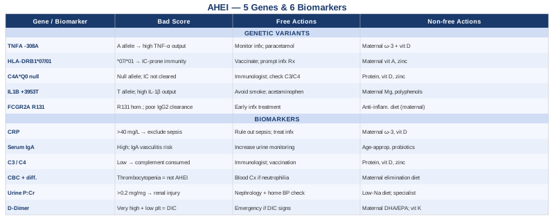
6 Biomarkers Worth Tracking in Acute Hemorrhagic Edema of Infancy
AHEI is diagnosed primarily on clinical grounds — the characteristic lesions, the age of onset, the absence of systemic organ involvement — but laboratory biomarkers play a critical role in ruling out more dangerous mimics, monitoring severity, and detecting the early signs of renal or coagulation complications. The six markers below are the most informative for this condition, selected for both their diagnostic relevance and their practical accessibility in a pediatric clinical setting.
Biomarker 1: C-Reactive Protein (CRP)
Why it matters. CRP is synthesized by the liver in response to interleukin-6 (IL-6) and other pro-inflammatory cytokines. In AHEI, levels are typically mildly to moderately elevated, reflecting the underlying inflammatory vasculitis. CRP does not diagnose AHEI, but its trajectory matters considerably: a CRP that remains stubbornly high or surges after initial improvement suggests either a persistent infectious trigger, a secondary bacterial infection, or a misdiagnosis — particularly meningococcemia or other serious infections that can mimic AHEI's purpuric presentation.
How to measure it. High-sensitivity CRP (hs-CRP) is measured from a standard blood draw and costs between $15 and $40 in most clinical settings. Standard CRP is adequate for acute monitoring in infants; hs-CRP adds value in follow-up when lower-level inflammation is being assessed. In the context of AHEI, most clinicians measure it at presentation and repeat it every 48–72 hours during the acute phase, or sooner if the child deteriorates.
If the score is bad — the plan without supplements. If CRP is substantially elevated (above 40–50 mg/L in an infant), the priority is ruling out sepsis, bacterial meningitis, and other serious infections before attributing the presentation to AHEI. Antibiotic therapy may be initiated while the clinical picture clarifies. Once an infectious trigger is identified (upper respiratory infections, otitis media, and urinary tract infections are the most common preceding events in AHEI), addressing that underlying cause is the most direct way to bring CRP down. Rest, hydration, and removal of any suspected pharmacological trigger (if a recent medication was introduced) are the first non-pharmacological steps.
If the score is bad — the plan with supplements or equipment. In an infant, supplementation decisions rest entirely with the treating physician. If the child is breastfed, the mother's diet has indirect relevance: a diet rich in omega-3 fatty acids (from fatty fish, walnuts, or flaxseed) reduces the production of pro-inflammatory eicosanoids and may modestly lower inflammatory burden. Vitamin D sufficiency in nursing mothers is also relevant, as deficiency is linked to dysregulated innate immunity. These are supportive measures, not treatments for active disease.
Biomarker 2: Serum IgA
Why it matters. IgA is the immunoglobulin class most associated with mucosal immunity and post-infectious immune responses. Its role in AHEI is still debated, but elevated serum IgA and IgA deposits in vessel walls have been found in a meaningful subset of AHEI cases — similar to what is seen in IgA vasculitis (Henoch-Schönlein Purpura), the condition most frequently confused with AHEI. The distinction matters because IgA vasculitis carries a higher risk of renal involvement. Serum IgA levels help contextualize the likely immunological mechanism and can influence decisions about renal monitoring frequency.
How to measure it. Serum immunoglobulin quantification (IgA, IgG, IgM) is ordered as a standard panel. Cost typically ranges from $30 to $80. In infants, interpreting IgA levels requires age-adjusted reference ranges, as normal values at 4–12 months differ significantly from adult norms. Skin biopsy with direct immunofluorescence, while more invasive, is the definitive test to determine whether IgA deposition is occurring in the vessel walls — this is not routinely done but becomes relevant if the diagnosis is uncertain or if renal complications emerge.
If the score is bad — the plan without supplements. An elevated serum IgA at diagnosis signals a heightened need for urinary monitoring (see Biomarker 5). It does not require immediate treatment beyond standard management of the underlying trigger. If a mucosal infection preceded the episode, treating that infection thoroughly is important. Avoiding re-exposure to the likely trigger (a specific antibiotic, a food antigen if relevant) during recovery is reasonable.
If the score is bad — the plan with supplements or equipment. IgA dysregulation has links to gut microbiome composition — a well-established connection in IgA vasculitis research. Probiotic supplementation appropriate for infant age (under physician guidance) may support mucosal immune regulation over time. For nursing mothers, a diet that supports gut barrier integrity — emphasizing diverse plant fibers, fermented foods, and adequate zinc — is a reasonable complementary measure. There are no supplements that directly reduce pathological IgA in an acute episode.
Biomarker 3: Complement C3 and C4
Why it matters. The complement system is a key mediator of immune complex clearance. In conditions where immune complexes deposit in small vessel walls — as happens in AHEI — complement proteins are consumed, sometimes causing measurably low C3 and C4 levels. Normal complement levels in a child with purpura argue against lupus-related vasculitis or hypocomplementemic urticarial vasculitis. Significantly low C3 or C4 values in AHEI warrant closer attention and may suggest a genetic predisposition to complement deficiency — a factor that can increase susceptibility to recurrent episodes.
How to measure it. C3 and C4 are measured from a standard serum sample, usually as part of a complement panel. Cost ranges from $40 to $120 depending on whether additional complement markers (CH50, C1q) are included. Testing is most informative during the acute phase when consumption would be at its peak. Low values should be rechecked during recovery to confirm normalization.
If the score is bad — the plan without supplements. Persistently low complement after the acute phase has resolved should prompt evaluation by a pediatric immunologist for primary complement deficiency. Children with inherited complement deficiencies have elevated lifetime risks for encapsulated bacterial infections (meningococcal, pneumococcal, and Haemophilus influenzae), and this has direct implications for vaccination schedules. If the child has a first-degree relative with recurrent vasculitis, low complement, or autoimmune disease, genetic testing for complement pathway variants becomes a more pressing conversation.
If the score is bad — the plan with supplements or equipment. Complement function depends heavily on adequate zinc, vitamin D, and protein intake. In infants, these are primarily addressed through breast milk quality (if breastfeeding) or formula selection. Pediatric immunologists sometimes use targeted nutritional support in children with partial complement deficiencies, though this is individualized and not a standard protocol.
Biomarker 4: Complete Blood Count (CBC) with Differential
Why it matters. The CBC provides a rapid window into systemic immune activity. In AHEI, leukocytosis is common, reflecting the infectious or inflammatory trigger. Eosinophilia may be present, suggesting a hypersensitivity component. Thrombocytopenia — a low platelet count — is not typical of AHEI, and its presence should redirect the diagnostic workup toward immune thrombocytopenic purpura (ITP), disseminated intravascular coagulation (DIC), or another diagnosis. A normal or elevated platelet count, alongside leukocytosis, is a reassuring pattern consistent with AHEI rather than a coagulation disorder.
How to measure it. CBC with differential is one of the most affordable and widely available blood tests, costing $15–$40. It is typically ordered at presentation and repeated if the clinical picture changes. In an infant presenting with purpura, the platelet count is the single most important number: a platelet count above 150,000/μL strongly argues against thrombocytopenic purpura as the cause of the bleeding lesions.
If the score is bad — the plan without supplements. Marked leukocytosis (above 15,000–20,000/μL) with fever warrants exclusion of serious bacterial infection. If the white count is driven by neutrophilia, blood cultures and a clinical reassessment should precede any decision to discharge. Persistent eosinophilia after the acute episode raises the question of parasitic infection, food allergy, or atopic predisposition — each of which has different implications for future prevention.
If the score is bad — the plan with supplements or equipment. If eosinophilia persists beyond the acute episode, an allergist or pediatric gastroenterologist may evaluate for food protein sensitivity. In breastfed infants, a maternal elimination trial (typically dairy and soy first) is a low-risk first step under dietitian guidance. This does not treat active AHEI but may reduce the immune activation burden that predisposed to the episode.
Biomarker 5: Urinary Protein-to-Creatinine Ratio
Why it matters. Renal involvement in AHEI is uncommon but not absent. The urinary protein-to-creatinine ratio (UP:UC) is the most sensitive outpatient marker of early glomerular injury. In the absence of significant proteinuria or hematuria, renal involvement can be confidently excluded. However, because AHEI and IgA vasculitis share immunological overlaps, and because renal complications in IgA vasculitis can emerge weeks after the skin lesions resolve, urinary monitoring should continue for four to six weeks after apparent recovery — not just during the acute phase.
How to measure it. A spot urine sample (random, not 24-hour) for protein and creatinine is inexpensive ($15–$30) and can be done at any pediatric clinic. A ratio above 0.2 mg/mg in a child is generally considered elevated and warrants further evaluation. Urine dipstick for blood and protein provides a rapid initial screen, but the protein-to-creatinine ratio is more sensitive and precise. Microscopic urinalysis for red cell casts adds specificity if proteinuria is detected.
If the score is bad — the plan without supplements. A persistently elevated UP:UC ratio in a child recovering from AHEI should prompt nephrology referral. Most children will not develop significant renal disease, but the ones who do need early identification. Blood pressure monitoring at home (using a validated pediatric cuff) is a practical addition for any infant with confirmed proteinuria — hypertension is an early sign of glomerular compromise.
If the score is bad — the plan with supplements or equipment. At the infant stage, no supplementation protocol addresses renal inflammation directly outside of medical management. For nursing mothers, reducing dietary sodium and ensuring adequate hydration supports normal renal filtration in the infant. If the child has progressed to solid foods, a low-sodium, minimally processed diet is appropriate supportive care. Quercetin and omega-3 supplementation have anti-inflammatory renal benefits in adult research, but their use in infants is not established and should only occur under specialist guidance.
Biomarker 6: D-Dimer and Coagulation Panel
Why it matters. D-dimer is a fibrin degradation product elevated whenever significant clotting and clot breakdown are occurring simultaneously. In AHEI, a mildly elevated D-dimer may reflect the vascular inflammation and localized coagulation activation associated with purpuric lesions. A markedly elevated D-dimer, however — particularly when combined with falling platelets, falling fibrinogen, and prolonged clotting times — raises concern for disseminated intravascular coagulation (DIC), a medical emergency that requires immediate intervention. This is why the coagulation panel (PT, aPTT, fibrinogen, D-dimer) is valuable at AHEI presentation, even though DIC is rare in true AHEI.
How to measure it. D-dimer is measured from plasma and costs $30–$70. A full coagulation panel adds PT and aPTT at additional cost. These are typically ordered once at presentation in infants with extensive purpura, and repeated only if there is clinical deterioration. A normal coagulation panel at diagnosis is significantly reassuring and shifts the diagnostic probability toward AHEI rather than a coagulopathy-driven process.
If the score is bad — the plan without supplements. A high D-dimer in the context of clinical deterioration, thrombocytopenia, and coagulopathy requires emergency pediatric or hematology consultation. AHEI itself is not the diagnosis in this scenario, and pursuing a coagulation-focused workup takes priority. If the coagulation panel is modestly abnormal but the clinical picture is otherwise consistent with AHEI, repeat testing in 48–72 hours is reasonable.
If the score is bad — the plan with supplements or equipment. In infants who are recovering and have had documented coagulation activation, omega-3 fatty acids through breast milk (via maternal supplementation with 1–2g DHA/EPA daily, with physician awareness) have mild anti-thrombotic properties. Vitamin K adequacy is important for coagulation factor production and is typically ensured through standard neonatal vitamin K prophylaxis — confirming this was given is worthwhile if there is any question.
5 Genes That May Shape Vulnerability in Acute Hemorrhagic Edema of Infancy
Dedicated genetic studies on AHEI are essentially absent from the literature, a reflection of how rarely the condition is studied as its own entity rather than as a variant of IgA vasculitis or leukocytoclastic vasculitis broadly. What follows is built on the well-characterized genetic landscape of related small-vessel vasculitides, leukocytoclastic mechanisms, and complement biology — with explicit acknowledgment that this is extrapolated evidence, not direct AHEI-specific findings. The practical value lies in identifying which children may benefit from more vigilant monitoring and what biological pathways are most worth supporting.
Gene 1: TNFA — TNF-Alpha Promoter Variants
What the gene does. The TNFA gene encodes tumor necrosis factor-alpha, a master regulator of the inflammatory response. The -308 G>A polymorphism (rs1800629) is the most studied variant: the A allele is associated with higher TNF-alpha production in response to immune triggers. Higher circulating TNF-alpha amplifies vascular endothelial activation, increases capillary permeability, and drives leukocyte recruitment to vessel walls — each of which is central to the AHEI disease process.
Studies in pediatric IgA vasculitis and other leukocytoclastic conditions have found enrichment of the -308A allele compared to healthy controls, suggesting that this variant may predispose to more robust inflammatory responses after infectious or antigenic triggers. Given AHEI's immunological proximity to IgA vasculitis, this association is biologically plausible and clinically worth considering, particularly in children who experience recurrent or unusually severe episodes.
If the gene is bad — the plan without supplements. A child known to carry the TNFA -308A allele (through genetic testing) should be monitored more closely during and after any significant infection or vaccination. Parents should be educated to recognize early AHEI signs and instructed to seek evaluation promptly rather than observing at home. Avoiding unnecessary antibiotic exposure — particularly to medications (like penicillin-class antibiotics and NSAIDs) that are frequently documented as AHEI triggers — is a preventive priority. Fever management with paracetamol/acetaminophen rather than ibuprofen during infections is sensible, as NSAIDs can influence vascular prostaglandin balance.
If the score is bad — the plan with supplements or equipment. In a breastfeeding mother whose infant carries this variant, dietary strategies that reduce systemic TNF-alpha signaling have indirect relevance. Omega-3 supplementation (1–2g EPA/DHA daily) competitively inhibits arachidonic acid-derived pro-inflammatory eicosanoids and has been shown in multiple trials to modestly reduce circulating TNF-alpha. Vitamin D at levels sufficient to maintain maternal serum 25(OH)D above 40 ng/mL supports regulatory T-cell function, which counterbalances TNF-alpha-driven inflammation. Curcumin has NF-κB-inhibiting properties relevant to TNF-alpha signaling but is not appropriate for direct infant supplementation; its use in nursing mothers has limited evidence and should be discussed with a physician before starting.
Gene 2: HLA-DRB1 — Immune Recognition and Autoantigen Presentation
What the gene does. HLA-DRB1 encodes a component of the MHC class II molecule, which presents antigens to CD4+ T helper cells. Specific HLA-DRB1 alleles determine which antigens trigger an immune response and which are ignored. In IgA vasculitis, HLA-DRB1*07 and HLA-DRB1*01 have been consistently associated with disease susceptibility in multiple pediatric cohorts. The implication is that children carrying these alleles may be more likely to mount immune complex-forming responses to common microbial antigens — particularly those from streptococcal and respiratory viruses — leading to vasculitic episodes rather than straightforward recovery.
If the gene is bad — the plan without supplements. HLA typing in children who have had AHEI or who have a strong family history of vasculitis or IgA nephropathy is not standard practice, but it becomes relevant in recurrent or atypical cases. For children known to carry high-risk HLA-DRB1 alleles, preventing the triggering infections is the most actionable intervention: ensuring age-appropriate vaccinations are current, practicing good hand hygiene to reduce respiratory virus exposure, and treating otitis media and streptococcal pharyngitis promptly and completely reduces the frequency of the immune triggers that initiate cascades in susceptible children.
If the score is bad — the plan with supplements or equipment. HLA-DRB1 alleles cannot be modified, but the downstream consequences of their activity — heightened T-helper cell activation and immune complex formation — can be modulated. For nursing mothers, adequate vitamin A (primarily from whole food sources such as liver and orange vegetables) supports mucosal immunity and reduces the proinflammatory T-helper 1/17 bias associated with some HLA-DRB1 risk alleles. Zinc adequacy is also important for thymic T-cell development and regulatory T-cell function.
Gene 3: C4A — Complement Component 4A and Immune Complex Clearance
What the gene does. The C4A gene encodes complement component C4A, a key protein in the classical complement pathway that facilitates the opsonization and clearance of immune complexes. The C4A*Q0 null allele — a gene deletion leading to absent C4A protein — is strongly associated with immune complex-mediated diseases including lupus, IgA nephropathy, and leukocytoclastic vasculitis. When C4A is absent or insufficient, immune complexes are not cleared efficiently and tend to deposit in small vessel walls, triggering the leukocytoclastic inflammation that defines conditions like AHEI.
This is one of the most biologically coherent genetic connections to AHEI. Children with C4A null alleles have impaired immune complex clearance, and even common infections generate immune complexes that cannot be efficiently removed — increasing the risk of vessel wall deposition and the downstream vasculitic cascade.
If the gene is bad — the plan without supplements. C4A null allele status cannot be corrected, but its consequences can be managed. Preventing immune complex-forming infections through vaccination and early treatment is the primary lever. In children with known or suspected C4A deficiency, any febrile illness or purpuric eruption warrants lower-threshold evaluation — not reassurance at home. Complement levels (C3, C4) should be checked at baseline and during acute episodes to understand how severely complement is being consumed. A pediatric immunologist experienced in complement disorders should be involved in care for confirmed complement deficiency.
If the score is bad — the plan with supplements or equipment. No supplement replaces missing C4A protein. However, supporting the overall complement system through adequate protein intake (ensuring the infant receives appropriate total calories from breast milk or formula), vitamin D, and zinc helps maximize the function of the complement proteins that are present. In older children transitioning to solid foods, a diet varied in whole proteins and rich in micronutrients supports overall immune homeostasis.
Gene 4: IL1B — Interleukin-1 Beta and Vascular Inflammation
What the gene does. Interleukin-1 beta (IL-1β), encoded by IL1B, is one of the earliest and most potent mediators of the inflammatory cascade. The +3953 C>T polymorphism (rs1143634) and the -511 C>T promoter variant are associated with higher IL-1β production. Elevated IL-1β drives endothelial activation, increases vascular permeability, and promotes neutrophil trafficking to inflamed tissue — all of which contribute to the edema and hemorrhagic purpura that characterize AHEI. In pediatric vasculitic conditions, high-IL1B producers tend to have more pronounced inflammatory responses to otherwise manageable antigenic triggers.
If the gene is bad — the plan without supplements. IL-1β signaling amplification cannot be corrected at the genetic level, but fever and infection — which are the primary triggers for IL-1β release — can be managed proactively. Avoiding unnecessary inflammatory insults (tobacco smoke exposure, indoor pollutants, chronic sleep disruption in the infant) reduces the baseline activation of IL-1β pathways. When fever occurs, acetaminophen can reduce IL-1β-driven temperature elevation and may modestly dampen the inflammatory cascade; this should be used judiciously and as directed by a pediatrician.
If the score is bad — the plan with supplements or equipment. In nursing mothers, a diet emphasizing polyphenol-rich foods (berries, green tea, dark leafy vegetables, olive oil) has demonstrated IL-1β-modulating properties in human observational and intervention studies. Magnesium adequacy is worth noting: magnesium deficiency upregulates NLRP3 inflammasome activity, which is a primary driver of IL-1β processing and release. Maternal magnesium supplementation at 200–400mg/day (as glycinate or malate, which are better tolerated) is well-supported for general inflammatory regulation, with indirect benefits for the breastfed infant.
Gene 5: FCGR2A — Fc Gamma Receptor and Immune Complex Processing
What the gene does. FCGR2A encodes Fc gamma receptor IIa (FcγRIIa), expressed on phagocytes including neutrophils, monocytes, and macrophages. This receptor binds the Fc region of IgG antibodies to facilitate phagocytosis and clearance of immune complexes. The H131R polymorphism (rs1801274) affects binding affinity: the R131 allele binds IgG2 poorly, reducing the efficiency of immune complex removal from the bloodstream. Children carrying the R131/R131 homozygous genotype have a demonstrated impairment in immune complex clearance that has been linked to susceptibility to immune complex-mediated vasculitis.
In the context of AHEI, where immune complex deposition in dermal capillaries is the central pathological event, impaired FcγRIIa function creates a sustained immune complex burden that favors vessel wall deposition rather than systemic clearance. This is a biologically compelling connection, though direct AHEI-specific FCGR2A data does not yet exist.
If the gene is bad — the plan without supplements. FCGR2A genotyping is not standard care but may be considered by pediatric immunologists evaluating children with recurrent or atypical episodes. Practically, the management implication is the same as for C4A deficiency: minimize immune complex-forming triggers, treat infections early, and maintain a lower threshold for clinical evaluation during febrile illnesses. For children with confirmed immune complex clearance impairment, prophylactic antibiotic protocols may be considered during high-risk periods (such as streptococcal epidemics), in consultation with a pediatric infectious disease specialist.
If the score is bad — the plan with supplements or equipment. Receptor expression and affinity cannot be changed by supplementation, but the upstream burden of immune complexes that overwhelm an already-impaired system can be reduced. Preventing IgG-mediated immune complex formation means reducing chronic antigenic stimulation: ensuring appropriate management of recurrent infections, addressing food sensitivities if present (particularly IgG-mediated ones), and avoiding unnecessary antigenic exposures during recovery periods. For nursing mothers, an anti-inflammatory diet that reduces circulating immune complex load is a reasonable supportive strategy.
What Andrew Huberman's Immune System Episodes Reveal About Infant Vasculitis
The Huberman Lab podcast has produced several episodes on immune function, inflammation, and how early-life immune calibration shapes long-term health. While none directly address AHEI, the episodes with immunologist Dr. Mark Davis and physician-scientist Dr. Roger Seheult cover mechanisms that are directly relevant: how the innate immune system responds to novel antigens, how complement and antibody-mediated responses can overshoot, and what conditions increase the likelihood that an immune response damages host tissue rather than clearing the pathogen.
These episodes synthesize a substantial body of peer-reviewed immunology research and are worth engaging with if you want to build a working mental model of why AHEI happens — not just that it does. The ten most clinically actionable insights for families navigating AHEI are summarized below.
1. The Innate Immune Response Is the First Responder — and the Main Driver of AHEI
AHEI is not primarily a failure of adaptive immunity. The initial trigger — a virus, a bacterium, occasionally a vaccine or medication — activates the innate immune system, which then generates the cytokine environment that drives vascular inflammation. Understanding this explains why the condition resolves on its own: once the triggering antigen clears, the innate activation dissipates.
2. Fever Modulation Affects Inflammatory Amplitude
Huberman's episodes highlight that fever is not simply a symptom — it is a regulated immune tool. Suppressing fever completely may blunt the immune response; leaving it unchecked amplifies inflammatory cytokine production. Moderate temperature management (treating discomfort rather than aggressively suppressing every fever elevation) is consistent with immune system logic.
3. Sleep Is the Single Most Potent Immune Regulator
Multiple studies cited across Huberman's immune episodes demonstrate that even one night of disrupted sleep significantly impairs phagocytic function, antibody production, and regulatory T-cell activity. For infants with AHEI — whose sleep is often disrupted by discomfort — supporting sleep consolidation through environmental optimization (darkness, sound control, temperature regulation) is not trivial.
4. Vitamin D Acts as an Immune Thermostat
Vitamin D receptors are present on virtually every immune cell. Deficiency shifts the immune balance toward pro-inflammatory responses and away from regulatory tolerance. Huberman references multiple studies showing that vitamin D sufficiency correlates with reduced autoimmune and inflammatory disease incidence. In nursing mothers, maintaining 25(OH)D above 40 ng/mL ensures the infant receives adequate vitamin D through breast milk — though direct infant supplementation is typically recommended regardless of maternal status.
5. Omega-3 Fatty Acids Shift Resolution Biology, Not Just Inflammation
A nuanced point from the inflammation episodes: omega-3s do not simply reduce inflammation. They promote the production of specialized pro-resolving mediators (SPMs) — lipids that actively orchestrate the resolution phase of inflammation and restore vascular homeostasis. This is directly relevant to AHEI, where resolution of the inflammatory episode is the therapeutic goal.
6. The Gut Microbiome Calibrates Systemic Immune Tone
Huberman's microbiome episodes with Dr. Justin Sonnenburg explain how gut bacterial diversity sets the baseline tone of immune regulation. Low microbiome diversity in early infancy is associated with heightened immune reactivity. Breastfeeding, avoidance of unnecessary antibiotics, and exposure to diverse environments all support early microbiome diversity.
7. Stress Hormones Directly Impair Immune Complex Clearance
Cortisol, the primary stress hormone, suppresses phagocyte activity — including the macrophage-mediated clearance of immune complexes that is already impaired in some genetically predisposed children. In infants who are distressed (from pain, overstimulation, or sleep deprivation), cortisol-driven immune suppression can compound underlying vulnerabilities.
8. Repeated Low-Grade Infections Build Different Immunity Than Single Major Infections
Episodes on immune memory explain that the nature of prior antigen exposure shapes how the immune system responds to future triggers. Children raised in environments with higher microbial diversity tend to have better-calibrated innate immune responses — less prone to overshooting. This is part of the biological rationale behind the hygiene hypothesis and its relevance to immune-mediated conditions.
9. Cold Exposure Has Anti-Inflammatory Mechanisms — But Not in Infants
Huberman frequently discusses deliberate cold exposure as a tool for reducing systemic inflammation through norepinephrine release and Nrf2 pathway activation. This is relevant context for older family members or parents managing their own inflammatory health, but it has no application to infants and should not be pursued in children with active skin conditions.
10. Resolution of Inflammation Is an Active Process That Requires Adequate Nutrition
One of the most important conceptual shifts in recent immunology, highlighted across multiple episodes: inflammation does not simply stop. Resolution is actively driven by specific mediators that require precursors — primarily EPA, DHA, and arachidonic acid metabolites. Nutritional adequacy during and after an AHEI episode directly supports this resolution process.
Complementary Approaches Worth Knowing About
Given that AHEI is immune-mediated, self-limiting, and affects the youngest patients, complementary approaches must be chosen carefully — prioritizing those with meaningful biological rationale and human clinical support, not merely theoretical plausibility.
The Autoimmune Protocol — Sarah Ballantyne
The Autoimmune Protocol (AIP), developed and documented by Dr. Sarah Ballantyne in The Paleo Approach, is a dietary elimination and reintroduction protocol specifically designed to reduce gut permeability, dampen systemic immune activation, and support regulatory immune mechanisms. While AHEI is not autoimmune in the strict sense, it involves dysregulated immune complex processing and inflammatory vascular injury — mechanisms that AIP is designed to modulate.
The core of the AIP protocol eliminates grains, legumes, dairy, eggs, nightshades, nuts, seeds, and refined sugars for 30–90 days, then systematically reintroduces foods to identify individual immune triggers. A pilot study published in Inflammatory Bowel Diseases (2017) demonstrated significant improvements in inflammatory biomarkers and clinical disease activity in Crohn's disease and ulcerative colitis patients following the AIP diet over six weeks, providing a proof-of-concept for the protocol's immunomodulatory effects in clinical populations.
For AHEI specifically, the AIP is not applicable to the infant directly — it is a nursing mother's tool. A breastfeeding mother following a simplified elimination protocol (focusing on dairy, gluten, and high-lectin foods as the primary targets) may reduce the quantity of pro-inflammatory dietary antigens transmitted through breast milk. This is speculative for AHEI specifically but grounded in established mechanisms of dietary antigen transmission. Any elimination protocol in a nursing mother should be implemented with dietitian support to prevent nutritional deficiencies.
Microbiome-Directed Therapies
The infant gut microbiome is one of the most powerful modulators of early immune calibration. Microbial colonization in the first months of life directly shapes the balance between pro-inflammatory and regulatory immune populations. Dysbiosis — low microbial diversity or absence of key beneficial species — shifts the immune system toward heightened reactivity, which may lower the threshold for conditions like AHEI after an infectious trigger.
Clinical evidence for microbiome intervention in vasculitic conditions is modest but growing. A 2021 study in Frontiers in Immunology found altered gut microbiome composition in children with IgA vasculitis compared to controls, with depletion of Faecalibacterium prausnitzii and other butyrate-producing species that support regulatory T-cell function. While AHEI-specific microbiome data does not exist, the shared immunological features are relevant.
Practically, the most accessible microbiome-directed intervention for infants is breastfeeding continuation (which provides prebiotic oligosaccharides that feed Bifidobacterium species), avoidance of unnecessary antibiotic courses, and age-appropriate probiotic supplementation with strains like Lactobacillus rhamnosus GG and Bifidobacterium longum infantis under pediatrician guidance. These are long-term immune calibration tools, not acute AHEI treatments, and their benefit would be measured in reduced recurrence risk rather than shortened acute episodes.
Mindfulness Meditation and MBSR — For Parents and Caregivers
This recommendation is directed at parents, not the infant. Watching a child develop alarming purpuric lesions is acutely stressful, and parental stress is not a trivial clinical variable. Elevated maternal cortisol affects breast milk composition, infant cortisol levels, and — through the nervous system modulation of immune function — can compound the inflammatory environment in a vulnerable infant. There is a well-characterized bidirectional relationship between caregiver psychological state and infant immune and inflammatory outcomes.
Mindfulness-based stress reduction (MBSR) has robust evidence for reducing parental distress in families managing pediatric illness. A 2016 systematic review in Pediatrics found significant reductions in parental anxiety and depression scores following MBSR interventions, with secondary improvements in parental responsiveness and child outcomes across multiple pediatric conditions. The practical format for parents in acute situations does not require a formal eight-week MBSR course: five minutes of focused breathing, body scan practice before sleep, and brief periods of non-reactive attention during the infant's symptoms are entry points that fit the reality of parenting through an acute medical episode.
The indirect benefits for the infant are real: a calmer caregiver provides better sleep environment curation, clearer symptom observation, and more measured decision-making in conversations with medical teams. These are not trivial contributions to a child's recovery trajectory.
Conclusion
Acute hemorrhagic edema of infancy is, in most cases, a self-resolving condition — but that fact carries more weight when you understand the biology behind it rather than simply accepting it on faith. The six biomarkers covered here give families and clinicians a structured monitoring framework: a way to distinguish a typical AHEI trajectory from one that needs closer attention, and to detect the rare complications before they escalate. The five genetic variants provide context for why some children seem predisposed to more significant immune responses after ordinary infections, and what monitoring and lifestyle choices follow from that understanding.
The next smart step depends on where you are in this process. If your child is currently in an acute episode, the priority is ensuring the biomarker panel has been completed — particularly the platelet count, complement levels, and urinalysis — and that follow-up urine monitoring is planned through six weeks after resolution. If you are in a post-episode period and trying to understand recurrence risk, a conversation with a pediatric immunologist about complement function and genetic predisposition is worth requesting. And if you are a nursing mother wanting to reduce the immune activation burden reaching your infant, the dietary and lifestyle levers in this article give you concrete, evidence-grounded starting points. Better information, applied precisely, produces better decisions — and that is exactly what this condition requires.