This article contains AI generated content.
· UpdatedAlkaptonuria: 3 Genes and 6 Biomarkers to Track
Introduction
Living with alkaptonuria means navigating a condition that most doctors encounter once in a career, if at all. The darkening urine, the joint pain that arrives decades before most people expect it, the slow accumulation of pigment in cartilage and connective tissue — these are not vague symptoms that fit a broad diagnostic category. Alkaptonuria is precise in its mechanism, and managing it well requires precision in return.
Generic health advice built around inflammation, diet, or lifestyle will only go so far. Alkaptonuria is driven by a specific enzymatic failure: the body cannot break down homogentisic acid (HGA), a metabolite of tyrosine and phenylalanine. Without the enzyme homogentisate 1,2-dioxygenase — encoded by the HGD gene — HGA accumulates, oxidizes, and binds irreversibly to collagen. The damage that follows — to joints, heart valves, and kidneys — is not random. It is predictable, measurable, and increasingly addressable.
What has changed in the last decade is the quality of the tools available to track and intervene. Urinary biomarkers, cartilage degradation markers, oxidative stress indicators, and even genetic profiling now give a clearer picture of where a person stands and how fast things may be moving. This matters enormously, because the window for meaningful intervention is real but not unlimited.
This article covers the six most useful biomarkers for monitoring AKU progression and response to intervention, followed by a look at the three genes most relevant to understanding this condition — including one that is a therapeutic target rather than a disease cause. A summary of the landmark clinical research that has reshaped treatment thinking comes next, along with a section on complementary approaches with actual clinical grounding. Better information, used consistently over time, genuinely changes outcomes in this disease.
6 Biomarkers to Track in Alkaptonuria
Tracking alkaptonuria without measuring biomarkers is like navigating without a map. The condition progresses silently for years before symptoms become debilitating. The biomarkers below are the most practical, most evidence-backed tools available — ranging from simple urine tests to specialized cartilage markers that capture what standard panels miss entirely.
1. Urinary Homogentisic Acid (HGA)
Why it matters: Urinary HGA is the defining biomarker of alkaptonuria. It reflects the rate at which HGA is being produced and excreted, and it is directly proportional to the amount of HGA available to oxidize and damage tissues. In untreated adults, urinary HGA typically ranges from 1 to 8 grams per day. Every gram excreted daily is HGA that could have bound to collagen and contributed to ochronosis or joint destruction.
How to measure it: A 24-hour urine collection analyzed by high-performance liquid chromatography (HPLC) or gas chromatography-mass spectrometry (GC-MS) is the gold standard. Some centers offer random urine HGA normalized to creatinine, which is more convenient but slightly less precise. Cost typically ranges from $80 to $200 depending on the lab and country. Specialist metabolic centers offer this routinely; general labs may need to send samples to a reference laboratory.
If the score is bad — the plan without supplements: The most powerful non-pharmacological strategy is a controlled reduction in dietary tyrosine and phenylalanine intake. Both amino acids are precursors to HGA. This does not mean eliminating protein — it means shifting toward lower-phenylalanine proteins and reducing high-intake foods like red meat, poultry, and high-protein dairy. This approach modestly reduces urinary HGA (by roughly 10–20%) and should be guided by a metabolic dietitian to avoid protein deficiency. Hydration also matters: maintaining high urine output reduces the concentration of HGA available to oxidize in connective tissues.
If the score is bad — the plan with supplements or medication: Nitisinone (NTBC) is the only pharmaceutical capable of dramatically reducing HGA. By inhibiting HPD (the enzyme one step upstream of HGD), it blocks HGA production at the source. The SONIA 2 clinical trial demonstrated a greater than 90% reduction in urinary HGA with 10 mg daily nitisinone. This is covered further in the Strategy 3 section. For those not on nitisinone, ascorbic acid (vitamin C) at doses of 500–1000 mg daily has been studied as an inhibitor of HGA oxidation and polymerization — it does not reduce HGA production but may slow its conversion into the damaging ochronotic pigment. No significant toxicity at these doses. Monitor urinary HGA every 3–6 months when stable, monthly during treatment changes.
2. Serum Homogentisic Acid
Why it matters: While urinary HGA reflects total daily excretion, serum HGA gives a snapshot of circulating HGA load at a given moment. The two values are correlated but not identical. Serum HGA is particularly relevant because circulating HGA is what reaches joint cartilage, cardiac valves, and other connective tissues. In healthy individuals, serum HGA is essentially undetectable. In untreated AKU patients, it can reach 400–1000 µmol/L.
How to measure it: Serum HGA requires specialized metabolic labs. It is not widely available through routine clinical labs but is offered by metabolic disease centers and academic hospitals with mass spectrometry capability. Cost is roughly $100–$250. Samples must be processed quickly to prevent ex vivo HGA oxidation, which can falsely lower measured values if not properly handled.
If the score is bad — the plan without supplements: The dietary interventions that reduce urinary HGA also lower serum HGA. Additionally, avoiding prolonged fasting helps reduce the catabolic breakdown of body proteins that temporarily spikes amino acid availability and HGA production. Gentle regular movement (walking 30 minutes daily) supports metabolic efficiency without stressing the joints.
If the score is bad — the plan with supplements or equipment: Nitisinone remains the most effective intervention. Ascorbic acid supplementation (500–1000 mg daily) continues to have a supportive role. Some researchers have explored whether NAC (N-acetylcysteine) as a thiol antioxidant might reduce HGA-mediated oxidative damage, but clinical evidence in AKU specifically is limited and it should be considered only with medical guidance.
3. High-Sensitivity C-Reactive Protein (hs-CRP)
Why it matters: Chronic HGA accumulation triggers a persistent low-grade inflammatory state, particularly in joint tissues where ochronotic pigment deposits activate immune responses. Elevated hs-CRP reflects this systemic inflammation. In AKU, tracking hs-CRP helps gauge whether inflammation is accelerating — which correlates with faster joint deterioration — and provides a non-specific but practical indicator of disease activity. Levels above 1 mg/L suggest elevated risk; levels above 3 mg/L indicate high systemic inflammation. Peter Attia has consistently emphasized hs-CRP as one of the most actionable and underused routine markers in preventive medicine.
How to measure it: hs-CRP is available through any standard blood panel. It costs $10–$40 and can be ordered by any physician. It requires no special sample handling. Test at least twice per year; more frequently during symptomatic flares or treatment changes.
If the score is bad — the plan without supplements: Anti-inflammatory dietary changes have meaningful impact: reducing ultra-processed foods, refined carbohydrates, and omega-6-heavy vegetable oils while increasing oily fish, olive oil, and non-starchy vegetables. Sleep quality is a major driver — poor sleep consistently elevates CRP. Aim for 7–9 hours in a cool, dark environment. Moderate-intensity aerobic exercise (20–30 minutes, 4–5 days per week) also reliably lowers CRP, though joint status in AKU determines which modalities are appropriate (swimming and cycling are generally safer than high-impact activities).
If the score is bad — the plan with supplements or equipment: Omega-3 fatty acids (2–4 g EPA+DHA daily) have robust evidence for lowering CRP. Cycle with a break every 3–4 months if taking high doses. Curcumin with piperine (500–1000 mg daily with food) has moderate evidence as an anti-inflammatory adjunct. Magnesium glycinate (200–400 mg at night) supports sleep quality, which indirectly reduces CRP. No significant cycling needed for these. Sauna use (3–4 sessions per week at 80°C for 15–20 minutes) has been associated with reduced inflammatory markers in observational studies, though direct AKU evidence is absent.
4. CTX-II (C-Terminal Telopeptide of Type II Collagen)
Why it matters: CTX-II is released into urine when type II collagen — the dominant collagen of joint cartilage — is being degraded. In alkaptonuria, ochronotic pigment causes progressive cartilage destruction starting in the spine and large joints. CTX-II is a direct readout of how fast that destruction is occurring. It is one of the most condition-relevant biomarkers in AKU because it captures the biological consequence of HGA accumulation in joint tissue, not just the upstream HGA burden. Elevated CTX-II means cartilage is actively breaking down, even when pain has not yet fully declared itself.
How to measure it: Urinary CTX-II is measured by ELISA and normalized to urinary creatinine. Specialist labs and academic rheumatology centers offer this test. Cost ranges from $80 to $200. A second-morning urine sample (to avoid the overnight fasting effect) is the preferred collection method. Annual monitoring is standard; every 6 months if actively managing joint disease.
If the score is bad — the plan without supplements: Weight management is critical — each kilogram of excess body weight adds approximately 3–4 kg of load across the knee joint and more across lumbar discs. A 5% reduction in body weight measurably lowers cartilage stress markers. Low-impact movement (swimming, cycling, aquatic therapy) maintains cartilage nutrition through synovial fluid circulation without mechanical overload. Avoiding repetitive joint loading activities (jumping, heavy axial loading) is particularly important given that AKU joint cartilage is structurally compromised earlier than in the general population.
If the score is bad — the plan with supplements or equipment: Collagen peptides (10–15 g daily with vitamin C) have shown in randomized trials to support cartilage matrix synthesis, though studies in AKU specifically are lacking. Undenatured type II collagen (UC-II, 40 mg daily) works via oral tolerance mechanisms to reduce cartilage-directed immune activity. Glucosamine sulfate (1500 mg daily) and chondroitin sulfate (1200 mg daily) have moderate evidence for slowing cartilage loss in osteoarthritis and represent a reasonable adjunct. These supplements have good safety profiles; no specific cycling is required. Water immersion physical therapy devices improve range of motion and reduce joint load effectively for AKU patients with significant joint involvement.
5. Kidney Function Panel (eGFR and Urinary Creatinine)
Why it matters: Kidney stones are a recognized complication of alkaptonuria, with some studies reporting prevalence of around 4–5% in AKU patients. HGA itself may contribute to renal tubular damage and stone formation. Monitoring eGFR (estimated glomerular filtration rate) provides a trend line for kidney health over time. A declining eGFR — even within the normal range — is meaningful in the context of a known metabolic condition affecting renal tissue. Urinary creatinine provides context for normalizing other urinary biomarkers.
How to measure it: eGFR is derived from serum creatinine and is available on any comprehensive metabolic panel. Cost is minimal ($15–$50 as part of a panel). Monitor annually at minimum; twice per year if there is a history of kidney stones or if on nitisinone (which raises plasma tyrosine and phenylalanine, potentially affecting renal handling of these amino acids).
If the score is bad — the plan without supplements: Adequate hydration is the most impactful free intervention — targeting 2–3 liters of fluid daily prevents HGA concentration in the urinary tract. Reducing dietary oxalate (spinach, nuts, chocolate in large quantities) is prudent if oxalate stones have been identified. Alkalizing urine through dietary shifts (increased fruits and vegetables, reduced processed foods) may reduce stone risk, though the specific benefit in HGA-related kidney stones needs further study.
If the score is bad — the plan with supplements or equipment: Potassium citrate (under medical supervision) is the standard pharmacological approach for stone prevention and mild renal protection. Magnesium supplementation (300–400 mg daily) reduces urinary oxalate and is well-tolerated. Vitamin B6 (50 mg daily) reduces endogenous oxalate production. If eGFR is declining, consultation with a nephrologist familiar with rare metabolic diseases is essential before making medication changes, as nitisinone itself requires monitoring of kidney markers due to elevated upstream metabolites.
6. Plasma Vitamin C (Ascorbic Acid)
Why it matters: Vitamin C occupies a specific and underappreciated role in AKU. Ascorbic acid is a potent inhibitor of HGA auto-oxidation — the process by which HGA converts into the reactive quinone species that then polymerizes and binds to collagen to form ochronotic pigment. Higher circulating vitamin C may slow the rate at which accumulated HGA causes tissue damage, even without reducing HGA production itself. Many AKU patients have suboptimal plasma vitamin C levels, partly because oxidative stress from the condition itself depletes ascorbate reserves. Optimal plasma levels are generally considered above 50 µmol/L; deficiency begins below 23 µmol/L.
How to measure it: Plasma vitamin C can be measured by specialized labs using HPLC. It is not routinely included in standard panels. Cost ranges from $30 to $80. Samples must be processed promptly as ascorbate degrades quickly. Some academic medical centers include this in metabolic disease monitoring panels. Testing once or twice yearly is reasonable for AKU management.
If the score is bad — the plan without supplements: Increasing fresh fruit and vegetable intake is the most effective dietary approach: bell peppers, citrus fruits, kiwi, and broccoli are particularly high in vitamin C. Smoking severely depletes ascorbate. Reducing high-sugar foods, which compete with vitamin C at the cellular transport level, also helps raise effective tissue levels. Cooking destroys vitamin C, so incorporating raw or lightly steamed vegetables into daily meals improves status meaningfully.
If the score is bad — the plan with supplements or equipment: Supplemental vitamin C at 500–1000 mg daily in divided doses is the standard approach for AKU, supported by mechanistic studies on HGA oxidation inhibition. Higher doses (above 2000 mg daily) may cause gastrointestinal discomfort and carry theoretical risk of oxalate kidney stone formation at very high doses — relevant given AKU's renal vulnerability. Liposomal vitamin C improves absorption at lower doses. No specific cycling is required. Consider testing plasma levels 8–12 weeks after starting supplementation to confirm adequacy.
The Genetic Picture: 3 Genes Worth Understanding
Alkaptonuria is one of the few diseases where the genetic cause is essentially singular and well-characterized. But understanding the genetics in more detail — including the genes that influence treatment response and disease severity — offers actionable insight that goes beyond simply knowing you carry HGD mutations.
Gene 1: HGD — The Causative Gene
What it does: The HGD gene encodes homogentisate 1,2-dioxygenase, the enzyme that normally cleaves the benzene ring of homogentisic acid in the tyrosine catabolism pathway. When both copies of HGD are dysfunctional (alkaptonuria is autosomal recessive), HGA cannot be degraded and accumulates in the body. More than 200 distinct pathogenic variants have been identified in HGD, ranging from missense mutations to frameshift deletions. The type of mutation correlates with residual enzyme activity, which in turn affects HGA burden and likely disease severity, though this relationship is still being fully characterized.
If the gene is bad — the plan without supplements: Since there is no way to restore HGD enzyme function through lifestyle or diet, the strategy is to reduce substrate load — specifically tyrosine and phenylalanine intake. A metabolic dietitian can design a tyrosine-restricted diet that keeps dietary intake roughly 20–30% below typical levels without compromising total protein nutrition. Low-phenylalanine medical foods developed for PKU management can be adapted for AKU but require professional guidance. This does not normalize HGA but reduces daily HGA burden in a meaningful way.
If the gene is bad — the plan with supplements or equipment: For HGD mutation carriers with confirmed AKU, nitisinone is the only targeted pharmacological intervention. Genetic testing via a clinical genetics panel that includes HGD sequencing (cost: $300–$800, covered by some insurers for rare metabolic disease workup) confirms mutation type and zygosity. Knowing the specific mutation can also guide genetic counseling for family members. Vitamin C supplementation (500–1000 mg daily) addresses HGA-mediated oxidative damage independent of genotype. See NIH GARD information on alkaptonuria for resources on genetic testing pathways.
Gene 2: HPD — The Therapeutic Target
What it does: The HPD gene encodes 4-hydroxyphenylpyruvate dioxygenase (4-HPPD), the enzyme immediately upstream of HGD in the tyrosine degradation pathway. HPD is not a disease gene in alkaptonuria — it functions normally in AKU patients. However, it is the target of nitisinone, the only approved pharmacological treatment for AKU. By inhibiting 4-HPPD, nitisinone blocks the production of HGA before it even forms, reducing urinary HGA by over 90%.
Understanding HPD matters for two reasons: first, variants in HPD could theoretically affect how efficiently nitisinone inhibits the enzyme, influencing drug response; second, because HPD inhibition raises upstream metabolites (tyrosine and phenylalanine in plasma), HPD function becomes central to monitoring side effect risk. Elevated plasma tyrosine from nitisinone use can cause keratopathy (eye irritation from tyrosine crystal deposits) and requires ophthalmologic monitoring.
If the gene is relevant — the plan without supplements: HPD is not a disease-causing gene, so there is no "bad HPD genotype" to address. However, understanding the HPD pathway means understanding what happens when nitisinone is used: plasma tyrosine rises substantially. A low-tyrosine, low-phenylalanine diet becomes important during nitisinone therapy to prevent keratopathy. This dietary management is the primary non-supplement intervention around HPD inhibition.
If the gene is relevant — the plan with supplements or equipment: Regular slit-lamp eye examinations (every 6–12 months) are essential for anyone on nitisinone to catch tyrosine-related keratopathy early. Some clinicians also monitor plasma tyrosine directly (target: below 700 µmol/L on treatment). Ophthalmology-grade lubricating eye drops help manage mild corneal changes. Diet remains the primary tool for controlling plasma tyrosine on therapy.
Gene 3: NFE2L2 (NRF2) — The Oxidative Stress Modifier
What it does: NFE2L2, which encodes the transcription factor NRF2, is the master regulator of the cellular antioxidant response. When cells encounter oxidative stress, NRF2 migrates to the nucleus and activates hundreds of cytoprotective genes including those encoding superoxide dismutase, glutathione peroxidase, and heme oxygenase. In alkaptonuria, the auto-oxidation of HGA generates reactive oxygen species continuously. The rate at which this oxidative burden is cleared depends substantially on baseline NRF2 pathway activity, which varies by genotype.
Individuals with common NFE2L2 variants associated with lower NRF2 activity may accumulate oxidative damage faster, potentially accelerating ochronosis and joint destruction even at similar HGA levels. This gene does not cause AKU, but it is a meaningful modifier of how damaging accumulated HGA becomes. Human evidence for this relationship in AKU specifically is limited to mechanistic studies and biochemical inference; large clinical cohort data in AKU is lacking due to the disease's rarity.
If the gene is bad — the plan without supplements: Lifestyle factors that activate the NRF2 pathway through hormetic stress include regular moderate exercise (particularly resistance training and high-intensity intervals), intermittent fasting, and sauna exposure. These interventions upregulate endogenous antioxidant production without supplementation. Cold exposure (brief cold showers or cold immersion) is another NRF2 activator with emerging evidence. Reducing chronic background oxidative load — through sleep quality, reduced alcohol, and eliminating smoking — helps preserve NRF2 pathway responsiveness.
If the gene is bad — the plan with supplements or equipment: Sulforaphane (from broccoli sprouts or concentrated supplements, 10–30 mg daily) is the most potent dietary NRF2 activator with human clinical evidence. It can be cycled 5 days on, 2 days off to prevent pathway adaptation. Quercetin (500 mg twice daily with food) and resveratrol (150–500 mg daily) also support NRF2 activity, though evidence strength is lower. NAC (N-acetylcysteine, 600–1200 mg daily in divided doses) supports glutathione synthesis, a key downstream antioxidant. These are supportive adjuncts — not substitutes for reducing HGA burden — and carry low risk of side effects at these doses when taken with food.
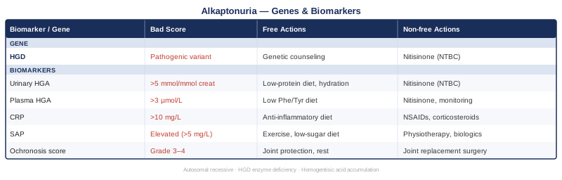
Quick Reference: Genes and Biomarkers at a Glance
What the SONIA 2 Trial Teaches Us About AKU — 10 Things Worth Knowing
For a disease as rare as alkaptonuria, the SONIA 2 randomized controlled trial (Ranganath et al., 2020, New England Journal of Medicine) is as close to a paradigm-shifting landmark as this field has seen. It challenged the long-standing clinical orthodoxy of watchful waiting and produced findings that reshape how every biomarker and gene discussed above should be interpreted. These are the ten things worth understanding.
1. Nitisinone Reduces Urinary HGA by More Than 90%
The trial's primary finding: 10 mg/day of nitisinone in adults with AKU reduced urinary HGA excretion from a mean of approximately 25 mmol/24h to under 1 mmol/24h. No dietary intervention, supplement, or lifestyle strategy comes close to this magnitude of reduction. For a biomarker that directly drives tissue damage, this is not an incremental improvement — it is a near-complete suppression of the root pathological driver.
2. But Clinical Benefit Was Harder to Demonstrate Statistically
This is the uncomfortable nuance that challenges simplistic enthusiasm. The trial's primary clinical endpoint — a composite ochronosis score (AKU Severity Score Index, or AKUSSI) — did not reach statistical significance for clinical improvement at the pre-specified level over 4 years. This does not mean nitisinone doesn't work clinically; it likely means the disease progresses slowly, the trial duration may have been too short for full clinical effect to materialize, and the measurement tools for ochronosis need refinement. Understanding this distinction is essential for any AKU patient making treatment decisions.
3. Early Treatment Is Almost Certainly the Key
The trial enrolled adults who already had established disease. The most compelling implication of the trial is not what it showed in these patients — it is what it suggests for younger patients treated before ochronosis accumulates. Reducing HGA by over 90% from early adulthood is mechanistically expected to prevent the downstream tissue damage that cannot be reversed. This is why diagnosis in childhood or early adulthood, with prompt HGA monitoring, is critical.
4. Plasma Tyrosine Rises on Nitisinone — and Must Be Managed
Because nitisinone inhibits HPD (the enzyme upstream of HGD), it causes accumulation of 4-hydroxyphenylpyruvate and eventually elevated plasma tyrosine. The SONIA 2 trial documented keratopathy (corneal tyrosine deposits) in a proportion of treated patients, confirming that ophthalmologic monitoring and dietary tyrosine restriction are essential accompaniments to nitisinone therapy. Treating one biochemical problem without managing its consequence is not complete care.
5. Ochronosis Is Largely Irreversible — Prevention Is the Goal
The pigment that accumulates in cartilage and connective tissue from HGA polymerization does not disappear when HGA is suppressed. SONIA 2 confirmed what mechanistic reasoning already suggested: once ochronotic deposits are established, they persist. The therapeutic ambition for nitisinone — and for aggressive HGA monitoring in general — is to prevent new accumulation, not to reverse existing damage. This fundamentally reframes biomarker tracking as a prevention tool rather than a recovery tool.
6. Urinary HGA Is Validated as the Primary Monitoring Biomarker
The SONIA 2 trial used 24-hour urinary HGA as its primary biochemical outcome, cementing its status as the gold-standard monitoring biomarker. This validation matters: it means the effort and cost of measuring urinary HGA accurately is supported by the highest level of clinical trial evidence available for this disease.
7. The AKU Severity Score Index Captures What Standard Measures Miss
AKUSSI is a composite score incorporating ochronosis burden across multiple tissues (ears, eyes, skin, joints), functional capacity, and reported symptoms. Standard rheumatology measures like joint pain scores or imaging alone miss the multi-system nature of AKU. The trial's use of AKUSSI highlights that AKU monitoring requires composite assessment, not single-organ focus.
8. AKU Has a Predictable Natural History — Which Makes Biomarker Tracking Powerful
AKU follows a relatively predictable trajectory: HGA accumulation is lifelong, ochronosis begins appearing in the third decade, joint involvement accelerates in the fourth and fifth decades, and cardiac and renal complications emerge later. This predictability means that biomarker trends over time have meaningful prognostic value. A rising CTX-II trend or falling eGFR is not noise — it reflects where in the natural history a person currently sits.
9. Treatment Decisions Should Not Wait for Symptoms
The SONIA 2 findings reinforce a principle that challenges traditional symptom-driven medicine: in alkaptonuria, waiting for pain or functional limitation to appear before intervening means watching irreversible damage accumulate. The biochemical case for early intervention — measured through urinary HGA — is strong, even when clinical symptoms are absent. This aligns with the broader preventive medicine philosophy championed by physicians like Peter Attia around biomarker-driven, early-stage intervention.
10. Nitisinone Availability Varies — But the Evidence Framework Stands Everywhere
Nitisinone is approved by the European Medicines Agency (EMA) and has been studied under compassionate use in many countries. Access varies by jurisdiction and healthcare system. But the framework the trial establishes — measure HGA, monitor ochronosis systematically, protect kidneys and eyes, manage plasma tyrosine with diet — is universally applicable regardless of whether nitisinone is accessible. The evidence does not just support a drug; it supports a complete monitoring and management approach.
Complementary Approaches With Clinical Grounding
Alkaptonuria's primary damage is biochemical and structural, but the pain, functional limitation, and psychological weight of managing a lifelong rare disease create real targets for complementary support. The following modalities have meaningful evidence relevant to AKU's clinical manifestations.
Low-Level Laser Therapy (Photobiomodulation) for Joint Pain
Low-level laser therapy (LLLT) — also called photobiomodulation — delivers specific wavelengths of light (typically 630–904 nm) to tissues at sub-thermal intensities, stimulating mitochondrial activity and reducing local inflammation. In alkaptonuria, where joint cartilage degradation leads to osteoarthritis-like pain in the spine and large joints, LLLT offers a non-pharmacological pain management tool that does not introduce further metabolic load.
A systematic review and meta-analysis published by Bjordal et al. (World Association for Laser Therapy) found LLLT to be significantly effective for chronic joint pain reduction compared to placebo, with benefits in both pain intensity and functional scores. Individual randomized trials in knee and spinal osteoarthritis — the joint presentations most common in advanced AKU — show consistent short-term pain reduction.
For AKU specifically, LLLT can be applied to symptomatic joints under guidance of a physiotherapist or rehabilitation physician trained in photobiomodulation. A typical protocol involves 8–12 sessions over 4–6 weeks, with wavelengths of 780–904 nm and dosing of 4–6 J/cm². Maintenance sessions monthly may help sustain benefit. Side effects are minimal when applied correctly. This modality does not address the underlying HGA burden but provides meaningful symptomatic support.
Yoga for Joint Mobility and Spinal Function
Yoga's relevance for alkaptonuria lies in its capacity to maintain joint range of motion, improve spinal flexibility, and strengthen the surrounding musculature — all of which decelerate the functional consequences of ochronotic joint destruction. As the AKU spine stiffens and large joints lose range, targeted mobility work becomes more, not less, important.
A randomized controlled trial by Ward et al. in Archives of Internal Medicine (Iyengar yoga for chronic low back pain) demonstrated clinically meaningful improvements in disability and pain over 24 weeks. While conducted in non-AKU populations, the spinal focus directly maps to the lumbar involvement that is among the earliest and most disabling aspects of AKU-related ochronosis.
For AKU patients, the appropriate yoga style is restorative or Iyengar (prop-supported), not vigorous vinyasa or hot yoga. Poses that decompress the lumbar spine — supported bridge, supine spinal twist, legs-up-the-wall — and maintain hip and shoulder range of motion are particularly relevant. A qualified yoga therapist who understands musculoskeletal limitations can modify practices safely. Joint loading must be carefully managed: weight-bearing poses on already-compromised cartilage should be avoided or adapted. Two to three sessions per week is a sustainable starting target.
Mindfulness-Based Stress Reduction (MBSR) for Chronic Pain and Psychological Burden
Living with a rare, progressive, and poorly understood condition carries a psychological burden that is frequently undertreated. Chronic pain from ochronotic joint disease is compounded by the cognitive and emotional weight of uncertainty. MBSR — the structured 8-week program developed by Jon Kabat-Zinn — addresses both the sensory and emotional dimensions of chronic pain through sustained attentional training.
A meta-analysis by Veehof et al. published in Pain demonstrated that mindfulness-based interventions produce significant reductions in pain intensity, depression, anxiety, and pain-related disability in chronic pain populations. The effect sizes are moderate but meaningful, and the benefits tend to persist at 6–12 month follow-up — a finding that distinguishes MBSR from purely symptomatic interventions.
For alkaptonuria patients, MBSR offers an especially relevant tool because it addresses the overlap between physical pain experience and the anxiety associated with monitoring a progressive rare disease. The standard MBSR protocol (8 weekly 2.5-hour sessions plus a one-day retreat) is available in-person and online in validated formats. Daily home practice of 30–45 minutes is part of the program. No physical contraindications exist. The main barrier is time commitment; starting with a guided app-based mindfulness program for 10–15 minutes daily is a realistic entry point before committing to the full MBSR curriculum.
Conclusion
Alkaptonuria is a rare condition, but it is not a mystery. Its mechanism is understood, its biomarkers are measurable, and its genetic basis is defined. What has historically been missing is a practical framework that translates that scientific understanding into consistent, trackable action — and that is what monitoring the six biomarkers above, understanding the three genes involved, and applying the evidence from the SONIA 2 trial provides.
The clearest next step is this: if urinary HGA has not been measured recently, arrange it. If hs-CRP and kidney function have not been tracked annually, add them to your next blood panel. If you do not know your HGD mutation status, genetic testing through a metabolic disease specialist can clarify your situation and open conversations about treatment eligibility. None of these steps require waiting — and in alkaptonuria, the evidence strongly suggests that time spent without measurement is time spent without the information needed to protect your tissues.
Musculoskeletal: Joint Conditions Spine Conditions
Autoimmune: Connective Tissue Conditions
Urological: Kidney Conditions