This article contains AI generated content.
· UpdatedAlpha-1 Antitrypsin Deficiency Arthropathy: 5 Genes And 7 Biomarkers To Track
Introduction
Alpha-1 antitrypsin deficiency (AATD) is one of the most common serious hereditary disorders in adults, yet its connection to joint disease remains under-recognized, even by many specialists. If you have been diagnosed with AATD and are also dealing with unexplained joint pain, swelling, or episodic arthritis, you may have been told the two are separate problems. In many cases, they are not.
The arthropathy associated with AATD follows a specific inflammatory pattern driven by the same enzyme imbalance that damages the lungs and liver. When alpha-1 antitrypsin protein levels are insufficient, neutrophil elastase and other destructive proteases go unchecked. Joint tissue becomes a downstream target of this protease storm, producing inflammation that mimics seronegative rheumatoid arthritis in its presentation but has a distinctly different biological origin.
Generic anti-inflammatory advice is not useless, but it was not designed with this mechanism in mind. Most protocols for inflammatory arthritis assume a different upstream driver. Without understanding what is specifically happening in AATD-related joint disease, it is easy to manage symptoms without ever addressing what is actually fueling the fire.
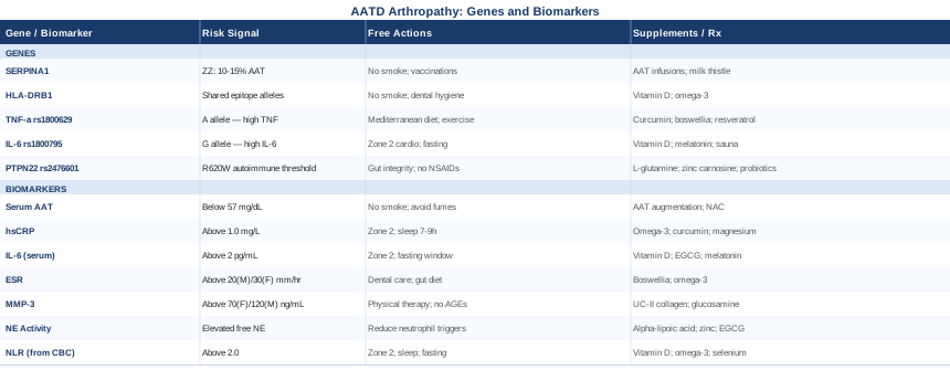
This article takes a more targeted approach. The first part focuses on the seven most informative biomarkers to track, with practical guidance on testing, optimal ranges, and what to do if a result is unfavorable. The second part examines five key genes, beyond SERPINA1 itself, that shape your individual inflammatory response and risk level. Together, these two angles offer a clearer picture than either alone. Better information does not guarantee better outcomes, but it consistently improves the quality of decisions you can make, in consultation with a specialist who understands the full picture.
Summary
This article covers the full landscape of AATD arthropathy from a precision-health perspective. The biomarker section identifies seven measurable signals, including one that can be calculated from any standard blood panel at no extra cost and is often more informative than CRP alone, alongside two less commonly ordered markers that are directly mechanistic for this specific condition. You will find optimal ranges, testing costs, and concrete plans for addressing abnormal results, both with and without supplementation.
The genetics section goes beyond the obvious SERPINA1 gene to examine four additional variants that can amplify inflammatory susceptibility, including one that is carried by roughly one in ten people of European ancestry and may transform episodic joint inflammation into a self-sustaining autoimmune loop. For each gene, there are practical protocols with cycling schedules and side-effect notes.
The article also covers what a landmark book on autoimmune disease teaches about the gene-environment interaction underlying conditions like AATD arthropathy, and examines five complementary approaches with meaningful clinical evidence, including one protocol specifically designed for inflammatory conditions of this type. The evidence level for each is clearly stated, and application guidance is realistic and cautious.
7 Biomarkers Worth Tracking in AATD Arthropathy
Tracking biomarkers in AATD arthropathy is not simply about measuring inflammation in the generic sense. The goal is to monitor the specific protease-antiprotease imbalance that drives this condition, alongside the downstream inflammatory cascade it creates. The seven markers below are chosen for a combination of mechanistic relevance, clinical utility, and practical availability, ranging from markers included in any basic blood panel to specialized tests that reveal the core biology of the condition.
Biomarker 1: Serum Alpha-1 Antitrypsin Level
Why it matters: This is the foundation. Every other biomarker in this list is, in some way, a downstream consequence of what this one reveals. Serum AAT level tells you directly how much functional protease inhibitor is circulating and whether it is sufficient to neutralize neutrophil elastase in the joints and other tissues. Low levels mean ongoing, unchecked protease activity. The relationship is not perfectly linear, but serum AAT below the protective threshold correlates with significantly increased tissue damage risk.
How to Measure It
The test uses nephelometry or immunoturbidimetry on a standard blood draw. It is widely available through any clinical laboratory and should be ordered as a quantitative result in mg/dL or µM, not as a binary positive or negative. Cost: $30 to $80, often covered when AATD is a confirmed or suspected diagnosis. The normal range is approximately 80 to 200 mg/dL, with a clinically meaningful protective threshold around 57 mg/dL (11 µM). Below this threshold, the risk of lung and tissue damage rises substantially.
If the Score Is Low: Plan Without Supplements
No lifestyle intervention can meaningfully raise serum AAT. What can be done without medical intervention is to reduce the demand placed on the AAT you do have. Eliminating all tobacco exposure is the single most important action, as smoking both depletes AAT further and massively increases neutrophil recruitment to the lungs and airways. Reducing exposure to air pollution, dust, and chemical fumes follows the same logic. Respiratory infections also spike neutrophil activity, so annual influenza and pneumococcal vaccination is standard of care. For joint protection specifically, reducing repetitive mechanical joint loading during active inflammation phases helps limit the structural damage occurring while protease activity is elevated.
If the Score Is Low: Plan With Supplements or Equipment
AAT augmentation therapy is the only FDA-approved and EMA-approved intervention that directly raises AAT levels. It involves weekly intravenous infusions of pooled human plasma-derived AAT (available as Prolastin-C, Zemaira, Aralast, or Respreeza in Europe). Eligibility criteria typically require confirmed AAT deficiency with evidence of emphysema. Arthropathy is not currently a standalone indication, though case reports describe joint improvement alongside lung stabilization. Discuss with a pulmonologist who specializes in AATD. No supplement raises serum AAT. N-acetylcysteine (600 mg twice daily) supports antioxidant defenses and may reduce overall oxidative burden. Curcumin with piperine (500 mg twice daily, cycle 8 weeks on / 4 weeks off) has laboratory evidence for modest neutrophil elastase inhibition but should not be confused with a treatment for the deficiency itself.
Biomarker 2: High-Sensitivity C-Reactive Protein (hsCRP)
Why it matters: hsCRP is produced by the liver in response to IL-6 and reflects systemic inflammatory burden. Peter Attia uses it as a key longevity and cardiovascular risk marker, but its utility in AATD arthropathy is more specific: serial measurement over time reveals whether the inflammatory state is stable, worsening, or responding to intervention. Importantly, standard CRP (not high-sensitivity) misses the low-grade chronic inflammation that characterizes stable AATD arthropathy between flares. Always request hsCRP specifically.
How to Measure It
Standard blood test, fasting preferred. Cost: $15 to $40. Most labs include it in basic inflammation panels. Attia's framework places optimal hsCRP below 1.0 mg/L, with borderline at 1 to 3 mg/L and elevated above 3 mg/L. Single measurements can be misleading due to transient elevation from infection or acute stress. Two measurements several weeks apart give a more reliable picture.
If the Score Is Bad: Plan Without Supplements
Zone 2 aerobic exercise, defined as the intensity at which you can hold a conversation without laboring, consistently reduces basal hsCRP in inflammatory conditions. Three to five sessions per week of 45 to 60 minutes each, maintained for at least 8 to 12 weeks, shows measurable results. Sleep optimization (7 to 9 hours of uninterrupted sleep) is the second most evidence-supported non-supplement intervention. Chronic sleep restriction elevates hsCRP directly. Removing ultra-processed food and refined seed oils from the diet, and increasing fatty fish, leafy greens, and extra-virgin olive oil, addresses the dietary contributors to systemic inflammation.
If the Score Is Bad: Plan With Supplements or Equipment
Omega-3 fatty acids (EPA and DHA combined, 2 to 4 grams per day from fish oil or algal oil) have the strongest evidence base for hsCRP reduction in inflammatory conditions. Curcumin with piperine (500 to 1000 mg daily, cycle 8 weeks on / 4 off; caution with anticoagulants and chemotherapy drugs). Magnesium glycinate or malate (300 to 400 mg at night; deficiency is common and directly increases CRP). Cold water immersion at 10 to 15°C for 3 to 5 minutes, 3 to 4 times per week, has shown hsCRP reduction in several small trials. Sauna (4 sessions per week, 20 minutes at 80 to 100°C) also shows association with reduced inflammatory markers in observational and small RCT data.
Biomarker 3: Interleukin-6 (IL-6)
Why it matters: IL-6 sits upstream of CRP in the inflammatory cascade. It is produced by neutrophils, macrophages, and synovial fibroblasts, and it is a direct driver of joint destruction in inflammatory arthropathy. In AATD specifically, unchecked neutrophil elastase activates synovial cells to produce IL-6, which then sustains and amplifies the inflammatory cycle. Peter Attia recommends tracking IL-6 alongside CRP because CRP can be elevated by non-inflammatory causes, while persistent co-elevation of both strongly confirms active systemic inflammation. IL-6 also predicts disease severity and response to anti-inflammatory interventions better than CRP in some rheumatological studies.
How to Measure It
Blood test. Less routinely ordered than CRP and may require a specialist or functional medicine practitioner to request. Cost: $40 to $100. Optimal range: below 2 pg/mL. Significantly elevated: above 5 pg/mL. Values above 10 pg/mL suggest active systemic inflammation. It is worth measuring fasting and pairing with hsCRP for context.
If the Score Is Bad: Plan Without Supplements
Zone 2 cardio is the most powerful non-supplement lever for IL-6 modulation and deserves specific explanation here. During Zone 2 exercise, muscles produce their own IL-6 as a signaling molecule. This muscle-derived IL-6 paradoxically suppresses pro-inflammatory IL-6 from immune cells by inducing IL-10 (anti-inflammatory) and inhibiting TNF-α production. This is entirely different from the pathological IL-6 produced by inflamed joints. The key is consistency: four or more sessions per week for at least 8 weeks to shift the basal inflammatory set point. Time-restricted eating (a 14 to 16 hour fasting window, daily) has shown modest IL-6 reduction in several small RCTs in overweight and obese populations.
If the Score Is Bad: Plan With Supplements or Equipment
Vitamin D (test 25(OH)D first; optimize to 50 to 70 ng/mL using D3 with K2; caution above 100 ng/mL). EGCG (green tea extract, 400 to 800 mg daily) has IL-6 reducing evidence, but caution is essential in AATD because the liver is already at risk: avoid high-dose EGCG if liver enzymes (ALT, AST) are elevated. Cycle 8 weeks on / 4 weeks off. Low-dose melatonin (0.5 to 1 mg at bedtime) has demonstrated IL-6 suppression in several human studies. Resveratrol (250 to 500 mg with food) is supported by small human trials. The prescription drug tocilizumab blocks the IL-6 receptor directly but carries infection risk and is a specialist decision.
Biomarker 4: Erythrocyte Sedimentation Rate (ESR)
Why it matters: ESR is the oldest and least glamorous marker on this list, but it provides something the faster-responding markers cannot: a picture of sustained, chronic inflammation. ESR responds over days rather than hours, which means it reflects the baseline inflammatory state more faithfully between flares. In episodic AATD arthropathy, CRP may normalize between joint episodes while ESR remains elevated, revealing that subclinical inflammation is ongoing. This distinction matters for treatment and monitoring decisions.
How to Measure It
Standard blood test (Westergren method). Cost: $10 to $30, included in many comprehensive metabolic or arthritis panels. Normal values: roughly below 20 mm/hr for men and below 30 mm/hr for women, though age-adjusted norms increase with age and lab reference ranges vary. In AATD arthropathy, the trend over time is often more informative than a single value.
If the Score Is Bad: Plan Without Supplements
Chronic dental disease (periodontitis) is one of the most consistent drivers of persistently elevated ESR and is under-addressed in most arthritis discussions. A dental review is worth including in any workup for unexplained ESR elevation. Anemia and iron-deficiency spuriously elevate ESR; iron panel and CBC should be reviewed alongside it. Addressing chronic gut inflammation through dietary change (removal of ultra-processed foods, seed oils, and in some cases gluten) consistently reduces ESR over several months of adherence.
If the Score Is Bad: Plan With Supplements or Equipment
No supplement specifically targets ESR, but Boswellia serrata (600 mg three times per day, cycle 8 to 12 weeks on / 4 weeks off) has meaningful human trial evidence for joint inflammation reduction, including ESR normalization as a secondary endpoint in several studies on inflammatory arthritis. Mild gastrointestinal side effects are possible; not appropriate if there is active peptic disease. Omega-3 supplementation as above will reduce the inflammatory substrate that elevates ESR over time.
Biomarker 5: Matrix Metalloproteinase-3 (MMP-3)
Why it matters: MMP-3, also known as stromelysin-1, is produced by inflamed synovial fibroblasts and is one of the earliest biochemical signals of structural joint damage. It cleaves type II, IV, IX, and X collagen and activates other MMPs in a destructive cascade. Critically, MMP-3 rises before joint damage is visible on imaging, making it a valuable early warning marker. In AATD arthropathy, the same protease-antiprotease imbalance that allows neutrophil elastase to run unchecked also creates conditions where MMPs are underinhibited by tissue inhibitors of metalloproteinases (TIMPs). MMP-3 has been validated in rheumatoid arthritis research as a better predictor of radiographic progression than CRP in some cohort studies, and its logic applies directly to AATD-associated joint inflammation.
How to Measure It
Serum or plasma test. Less commonly ordered in routine rheumatology than it should be; may require a functional medicine practitioner or a rheumatologist who monitors it actively. Cost: $80 to $150. Reference ranges vary by laboratory. Broadly, values above approximately 60 to 70 ng/mL in women and 120 ng/mL in men suggest elevated synovial inflammatory activity, though always interpret against your lab's specific reference range.
If the Score Is Bad: Plan Without Supplements
Physical therapy targeting joint mechanics and muscular support around affected joints reduces mechanical stress that upregulates MMP production independently of the biochemical inflammation. Avoiding high-impact loading during active inflammatory phases is practical harm reduction. Eliminating dietary advanced glycation end products (AGEs), concentrated in high-heat-cooked meats, processed foods, and fast food, reduces MMP-3 stimulation through the RAGE receptor pathway.
If the Score Is Bad: Plan With Supplements or Equipment
Undenatured type II collagen (UC-II) at 40 mg per day has the most direct human trial evidence for modulating the synovial immune response and reducing MMP activity in inflammatory arthritis. It works through oral tolerance induction, suppressing the immune response to native collagen, rather than through anti-inflammatory drug effects. Assess after a minimum of three months. Glucosamine sulfate (1500 mg/day) and chondroitin sulfate (1200 mg/day) have mixed evidence overall, but specific trials show MMP-3 reduction. Doxycycline at low dose (20 mg twice daily, prescription required) has metalloproteinase-inhibiting properties that have been studied in periodontitis and early RA; discuss this option with a rheumatologist rather than using standard-dose antibiotics.
Biomarker 6: Neutrophil Elastase Activity and the NE-AAT Complex
Why it matters: This is the most mechanistically direct biomarker for AATD arthropathy and arguably the most underused. Neutrophil elastase (NE) is the primary protease that alpha-1 antitrypsin exists to neutralize. When AAT is deficient, NE degrades synovial tissue, cartilage matrix, and connective tissue structures. Measuring either free NE activity or the NE-AAT complex (which represents AAT that has already been consumed in the process of neutralizing NE) provides a real-time picture of how much proteolytic damage is actively occurring, rather than just showing that AAT levels are low. A low AAT with high NE activity is a more urgent clinical picture than a low AAT with low NE activity.
How to Measure It
Specialized test, primarily available through academic medical centers, research laboratories, and increasingly through specialized clinical labs. Cost: $100 to $300. This is not a standard order at most primary care or general rheumatology offices, and you may need to specifically request it through a pulmonologist or AATD specialist. Its clinical availability is expanding as AATD research grows.
If the Score Is Bad: Plan Without Supplements
Reducing neutrophil recruitment to affected joints is the primary non-pharmacological lever. Neutrophil migration is driven by CXCL8 (IL-8), which is itself elevated by stress, sleep deprivation, excess omega-6 fatty acid intake, and gut barrier disruption. Addressing all four reduces the neutrophil load arriving at joint tissue. AAT augmentation therapy is the only intervention that directly restores the NE-AAT balance; if you are not on it and have significant AATD, this warrants specialist discussion.
If the Score Is Bad: Plan With Supplements or Equipment
Alpha-lipoic acid (600 mg twice daily with meals) has shown neutrophil elastase inhibitory activity in laboratory studies, though human joint-specific data is limited. EGCG (see IL-6 section for liver caution) also has elastase-inhibitory properties in preclinical research. These are adjunctive and should not replace augmentation therapy in eligible patients. Zinc (25 to 40 mg elemental zinc with food, cycle 8 weeks on / 4 off, take copper 2 mg separately to prevent depletion) supports antiprotease function broadly and is commonly deficient in people with chronic inflammatory conditions.
Biomarker 7: Neutrophil-to-Lymphocyte Ratio (NLR)
Why it matters: The NLR is perhaps the most underrated marker on this list, in part because it is calculated from a test that most people already have: a standard complete blood count (CBC) with differential. It requires no additional cost and no additional blood draw. Divide the absolute neutrophil count by the absolute lymphocyte count. The result reflects the balance between innate immunity (neutrophil-driven, inherently pro-inflammatory) and adaptive immunity (lymphocyte-driven). In AATD, where the core pathology involves unchecked neutrophil protease activity, an elevated NLR captures exactly this imbalance at the systemic level. Peter Attia includes NLR in his longevity monitoring protocol because it independently predicts all-cause mortality, cardiovascular events, and cancer outcomes in large population studies.
How to Measure It
No additional test is needed. Request a CBC with differential and calculate NLR yourself by dividing the absolute neutrophil count (ANC) by the absolute lymphocyte count (ALC). Cost: $15 to $40 for the CBC. Healthy range: below 2.0. Mildly elevated: 2 to 3. Clinically concerning: above 3. In active autoimmune or inflammatory conditions, values above 3.5 are commonly associated with higher disease activity.
If the Score Is Bad: Plan Without Supplements
Zone 2 cardio reduces neutrophil counts and supports lymphocyte function over time. Sleep is arguably more powerful than any supplement for NLR normalization; chronic sleep restriction (below 6 hours) shifts the immune balance sharply toward neutrophil dominance. Time-restricted eating (16:8 fasting window) has shown NLR reduction in several short-term human trials. Chronic oral infections, sinus infections, and subclinical viral reactivation (EBV, CMV) are common hidden drivers of persistent NLR elevation; these deserve investigation if other interventions do not move the number.
If the Score Is Bad: Plan With Supplements or Equipment
Vitamin D (optimize to 50 to 70 ng/mL with D3 and K2; deficiency sharply skews immune balance toward innate inflammatory responses). Omega-3s at therapeutic doses (2 to 4 g EPA/DHA daily) shift immune cell production toward resolution. Selenium (100 to 200 mcg/day as selenomethionine, cycle 8 weeks on / 4 off; caution above 400 mcg/day due to toxicity risk) supports lymphocyte function and has been studied in autoimmune thyroid conditions with favorable effects on immune balance. A probiotic containing Lactobacillus rhamnosus and Bifidobacterium longum supports lymphocyte function through gut-immune crosstalk and is safe for long-term use.
Understanding these biomarkers shifts your monitoring from reactive to proactive. But biomarkers tell you what is happening in the tissue; your genetics tell you why your particular body responds the way it does, and which interventions are most likely to move those numbers in your favor.
The Genetic Architecture Behind AATD Arthropathy
The SERPINA1 gene is the starting point for understanding AATD, but it does not tell the whole story. Among people with the same AAT deficiency genotype, some develop significant arthropathy and others do not. The difference is largely encoded in a set of modifier genes that amplify or dampen the inflammatory response, shape immune activation thresholds, and determine how the body handles the unchecked protease activity that AATD creates. The five genes below, alongside their practical implications, provide a more complete genetic picture.
Gene 1: SERPINA1 — The Foundation
What it does: The SERPINA1 gene on chromosome 14q32 encodes alpha-1 antitrypsin protein, a serine protease inhibitor (serpin). The Z allele (rs28929474, resulting in a Glu342Lys substitution) causes the protein to misfold and polymerize inside hepatocytes rather than being secreted into circulation. The S allele (rs17580, Glu264Val) causes moderate reduction in secretion. The ZZ genotype produces approximately 10 to 15% of normal AAT levels; SZ produces approximately 35 to 40%; MZ heterozygotes produce around 60%, which is usually not clinically significant for lung disease but may be relevant for inflammatory joint susceptibility in some individuals.
For SERPINA1 genotyping, see: NCBI Gene entry for SERPINA1.
If the Gene Is Bad: Plan Without Supplements
Know your precise phenotype. ZZ, SZ, and MZ carry very different risk profiles and different clinical management pathways. Avoid all tobacco and secondhand smoke without exception. Minimize exposure to airborne pollutants, industrial dusts, and chemical fumes. Stay current on respiratory vaccinations. For liver health (also affected by Z allele-related protein aggregation in hepatocytes), periodic liver function tests and liver ultrasound are warranted. For joints specifically, early physical therapy and load management during flares prevents secondary structural deterioration.
If the Score Is Bad: Plan With Supplements or Equipment
AAT augmentation therapy (intravenous, weekly infusions of Prolastin-C, Zemaira, or Aralast) is the only direct pharmacological answer to AAT deficiency. Its primary approved indication is emphysema, but its logic extends to systemic tissue protection. Eligibility and coverage criteria vary by country and insurer. Discuss with an AATD specialist, not a generalist. Additionally, liver protection supplements such as milk thistle (silymarin, 400 to 700 mg daily in divided doses) have evidence for reducing hepatocyte damage in the context of protein aggregation; cycle 12 weeks on / 4 weeks off. Do not use high-dose silymarin alongside hepatotoxic medications without medical review.
Gene 2: HLA-DRB1 — The Joint Susceptibility Amplifier
What it does: The HLA-DRB1 gene encodes a component of the MHC class II antigen-presenting molecule. Specific alleles, particularly DRB1*04, DRB1*01, and DRB1*10, contain a five-amino-acid sequence in the peptide-binding groove called the shared epitope (SE). The shared epitope dramatically increases susceptibility to inflammatory arthritis by enhancing the presentation of citrullinated self-proteins to autoreactive T cells. In the context of AATD, where the inflammatory environment already activates neutrophils and promotes citrullination of joint proteins, carrying shared-epitope HLA-DRB1 alleles can convert low-level joint irritation into a self-sustaining immune attack. Evidence linking SE alleles to inflammatory arthritis is among the most replicated findings in rheumatological genetics.
If the Gene Is Bad: Plan Without Supplements
The shared epitope functions as a risk amplifier, not a deterministic switch. Environmental inputs determine whether it gets activated. Smoking is the most important modifiable trigger: tobacco smoke directly promotes protein citrullination in the airways and joints, providing the ligands that SE HLA molecules use to activate autoreactive T cells. Never smoke. Periodontal disease (from Porphyromonas gingivalis) is the second most important driver of citrullination; diligent oral hygiene and regular dental care is not optional in this genetic context. Anti-inflammatory diet reduces the baseline production of citrullinated proteins.
If the Score Is Bad: Plan With Supplements or Equipment
No supplement alters HLA genotype, but interventions that reduce the availability of citrullinated proteins effectively reduce the risk that SE alleles will drive autoimmunity. Vitamin D optimization (50 to 70 ng/mL of 25(OH)D) supports immune tolerance through T-regulatory cell induction and is the single most evidence-supported immune-modulating supplement for HLA-related autoimmune risk. Omega-3s (2 to 4 g EPA/DHA daily) reduce the production of PAD4 (peptidyl arginine deiminase), the enzyme responsible for citrullination, particularly under inflammatory conditions. Combine with consistent Zone 2 exercise and sleep.
Gene 3: TNF-α (rs1800629) — The Inflammatory Response Amplifier
What it does: TNF-alpha is a master regulator of the innate inflammatory response. The rs1800629 SNP (G to A change, often called TNF-238) affects the promoter region of the TNF gene and modulates how strongly the gene is expressed in response to immune stimulation. Carriers of the A allele produce significantly higher amounts of TNF-α when the immune system is activated. This matters directly in AATD arthropathy: TNF-α drives synovial inflammation, promotes neutrophil survival, and amplifies the joint-destructive cascade already initiated by unchecked neutrophil elastase. TNF-α is also the target of the most widely used biologic drugs in rheumatology (adalimumab, etanercept, infliximab), underlining how central it is to inflammatory joint disease biology.
If the Gene Is Bad: Plan Without Supplements
The Mediterranean diet pattern, characterized by high olive oil, fatty fish, legumes, vegetables, nuts, and low processed meat and sugar, consistently reduces TNF-α at the gene-expression level in human studies. It is the dietary intervention with the strongest evidence base for this specific pathway. Consistent aerobic exercise suppresses basal TNF-α and improves the inflammatory set point over 8 to 12 weeks of regular practice. Adequate sleep (7 to 9 hours) is non-negotiable: even partial sleep deprivation sharply elevates TNF-α the following day through NF-kB activation.
If the Score Is Bad: Plan With Supplements or Equipment
Curcumin with piperine (500 to 1000 mg daily, cycle 8 weeks on / 4 weeks off) has among the strongest supplement evidence for TNF-α reduction in humans. Caution with anticoagulants, chemotherapy drugs, and immunosuppressants. Boswellia serrata (600 mg three times per day, same cycling approach) inhibits 5-LOX and has shown TNF-α reduction in joint inflammation trials. Resveratrol (500 mg with a fat-containing meal for absorption, cycle similarly) inhibits NF-kB, the primary transcription factor that drives TNF-α gene expression. Cold water immersion (10 to 15°C, 3 to 5 minutes, 3 to 4 sessions per week) reduces basal TNF-α in several human trials. Where TNF-α is markedly elevated and unresponsive to lifestyle, a rheumatologist may consider prescription anti-TNF therapy.
Gene 4: IL-6 (rs1800795) — The Cytokine Signal Modulator
What it does: The rs1800795 SNP in the IL-6 gene promoter (G to C) affects how much IL-6 the body produces in response to inflammatory stimulation. Counterintuitively, the C allele produces less IL-6 and is generally more protective in inflammatory arthropathy. The G allele, associated with higher IL-6 production, is linked to increased risk of severe inflammatory joint disease, cardiovascular disease, and multiple autoimmune conditions. In AATD, unchecked neutrophil elastase directly stimulates synovial cells to produce IL-6, and if the underlying IL-6 gene variant amplifies this further, the joint-destructive feedback loop becomes harder to interrupt.
If the Gene Is Bad: Plan Without Supplements
Zone 2 cardio is uniquely positioned here: the muscle-derived IL-6 released during exercise induces a systemic anti-inflammatory response that counteracts the pathological IL-6 from joints. This is one reason exercise works particularly well in conditions driven by the IL-6 pathway. Time-restricted eating (14 to 16 hour daily fasting window) reduces IL-6 gene expression in adipose and immune tissue, separate from any weight-loss effect. Stress management directly reduces IL-6: cortisol and catecholamines activate the same IL-6 promoter that the G allele already predisposes toward.
If the Score Is Bad: Plan With Supplements or Equipment
Vitamin D optimization (50 to 70 ng/mL) suppresses IL-6 gene expression through vitamin D receptor signaling. Low-dose melatonin (0.5 to 1 mg at bedtime) has demonstrated direct IL-6 suppression in human inflammatory models. Sauna use (4 sessions per week, 20 minutes at 80 to 100°C) shows association with reduced IL-6 in observational studies; heat shock proteins produced by sauna may modulate IL-6 signaling. Avoid high-dose EGCG if liver function is compromised, which is a real possibility in AATD (see biomarker section). Where IL-6 is extremely elevated and unresponsive, tocilizumab (an IL-6 receptor blocker) is a specialist-managed option.
Gene 5: PTPN22 (rs2476601) — The Autoimmune Threshold Gene
What it does: PTPN22 encodes a protein tyrosine phosphatase that regulates signaling in T cells and B cells. The R620W variant (rs2476601, 1858C to T) disrupts an immune checkpoint, lowering the activation threshold for autoreactive lymphocytes. This is one of the most consistently replicated genetic risk factors for autoimmune conditions in human genetics, including rheumatoid arthritis, lupus, type 1 diabetes, and Graves' disease. The allele frequency in European populations is roughly 8 to 10%, meaning it is not rare. For AATD arthropathy specifically, carrying this variant may transform episodic, protease-driven joint inflammation into a self-sustaining autoimmune response that persists even when AAT levels are partially supported. The evidence is strongest in RA; direct AATD-specific studies are limited but the mechanistic logic is well-supported.
If the Gene Is Bad: Plan Without Supplements
The most important lever for PTPN22 variant carriers is protecting gut barrier integrity. A permeable gut epithelium provides a continuous stream of foreign antigens into the bloodstream, which the dysregulated immune checkpoint created by this variant is poorly equipped to ignore. Removing dietary gluten is the most strongly supported dietary intervention for gut permeability, independent of celiac disease status. Avoiding NSAIDs chronically is practically important: chronic NSAID use disrupts the gut epithelium and, paradoxically, worsens the conditions that allow PTPN22 to cause harm. Stress management deserves emphasis here too, as cortisol chronically amplifies autoreactive T cell activity in those with this variant.
If the Score Is Bad: Plan With Supplements or Equipment
L-glutamine (5 g twice daily between meals, cycle 8 weeks on / 4 off) supports enterocyte fuel supply and tight junction integrity. Zinc carnosine (75 mg twice daily with meals) has specific evidence for gut lining protection and mild anti-inflammatory effects in the gut. Probiotics, specifically formulations including Lactobacillus rhamnosus GG and Bifidobacterium longum, have the best evidence for gut permeability reduction and immune modulation. These do not change the gene but reduce the environmental conditions that allow it to cause ongoing harm. Side effects are minimal; stick with single or dual-strain products from reputable manufacturers rather than high-potency multi-strain blends.
The genes and biomarkers above map what is happening biologically. The next section draws on a landmark book to explain why the gene-environment interaction that drives conditions like AATD arthropathy is both more tractable and more fundamentally misunderstood than most medical approaches suggest.
What 'The Autoimmune Fix' Reveals About Conditions Like AATD Arthropathy
The Autoimmune Fix by Dr. Tom O'Bryan (2016) is one of the clearest frameworks available for understanding how genetic susceptibility, gut permeability, and environmental triggers combine to produce autoimmune and inflammatory joint conditions. While AATD is not discussed specifically in the book, its mechanistic model maps directly onto what is known about AATD arthropathy. Dr. O'Bryan draws extensively on peer-reviewed research, including work from the University of Maryland Center for Celiac Research, and his central argument challenges the conventional framing of autoimmunity as a fixed, fully arrived disease. The book's value is in showing how the inflammatory cascade that damages joints is not an event but a process — one that can be interrupted.
1. Autoimmune Disease Exists on a Spectrum That Begins Decades Before Diagnosis
The immune dysregulation that eventually destroys joint tissue does not begin the day symptoms appear. Measurable antibodies against self-tissue can precede clinical diagnosis by five to ten years. In AATD, the protease imbalance creates the initial joint irritation, but what determines whether it progresses to sustained immune attack is how long the person has been in a state of subclinical immune activation. Testing inflammatory biomarkers before symptoms worsen is the most practical implication of this insight.
2. Intestinal Permeability Is the Common Gateway
O'Bryan argues, drawing on the work of Dr. Alessio Fasano, that a leaky gut epithelium is the shared environmental cofactor in the vast majority of autoimmune conditions. When tight junctions open inappropriately, partially digested food antigens and microbial components enter the bloodstream and trigger sustained immune activation. In AATD, the inflammatory state already primes immune cells for reactivity; gut permeability provides the ongoing antigenic fuel that keeps that reactivity burning.
3. Gluten Activates Zonulin and Opens Tight Junctions in Everyone
Zonulin, a protein that regulates intestinal permeability, is triggered by gliadin (a component of gluten) in virtually all humans, not only those with celiac disease. The effect is transient in most people, but in those with a genetic or inflammatory predisposition (relevant for AATD carriers with HLA or PTPN22 variants), the permeability window is both wider and slower to close. This does not mean everyone needs to be gluten-free, but it does mean that high-frequency gluten exposure warrants re-evaluation in the context of uncontrolled joint inflammation.
4. Your Genes Load the Gun; the Environment Pulls the Trigger
O'Bryan repeatedly returns to this formulation, derived from Dr. Francis Collins. The clinical insight is that genetic risk is not destiny. SERPINA1 Z allele, HLA-DRB1 shared epitope, PTPN22 R620W — these increase risk, but environmental inputs determine whether and when they cause disease. This is neither dismissive of genetics nor nihilistic about it: it is practically actionable.
5. Molecular Mimicry Links Food Proteins to Joint Tissue
Some dietary proteins share amino acid sequences with joint tissue proteins. Immune responses mounted against these food proteins can cross-react with collagen, synovial proteins, and other joint components through molecular mimicry. O'Bryan presents evidence that this process, often initiated through a leaky gut, can both initiate and perpetuate joint inflammation independently of the original trigger. This is a plausible additional mechanism in AATD arthropathy.
6. The Gut Microbiome Sets the Systemic Inflammatory Baseline
Specific bacterial species produce metabolites that regulate immune cell differentiation toward tolerance or inflammation. A microbiome dysbiosis characterized by low Bifidobacterium and Akkermansia and high gram-negative bacteria raises LPS (lipopolysaccharide) levels in the portal circulation, driving liver and systemic inflammation. For AATD, where the liver is already under protein-aggregation stress, microbiome-driven inflammation is an additive burden worth addressing.
7. Silent Inflammation Is Detectable Before It Becomes Symptomatic
The book advocates for proactive testing of inflammatory markers, including anti-tissue antibodies, to identify and interrupt the autoimmune cascade before clinical symptoms appear. The biomarker panel in this article (hsCRP, IL-6, MMP-3, NLR) serves exactly this purpose for AATD arthropathy: these markers can change meaningfully long before joint function deteriorates enough to appear on imaging.
8. The Elimination-Reintroduction Protocol Can Identify Personal Inflammatory Triggers
O'Bryan recommends a structured elimination period of 30 to 60 days removing gluten, dairy, corn, and sugar, followed by careful reintroduction one food at a time. During reintroduction, biomarkers (including CRP and joint symptom scores) are monitored. This protocol has been studied formally in the autoimmune protocol (AIP) context and provides individualized data that no population-level study can.
9. Measuring Immune Reactivity Directly Through Antibody Testing
Specialized antibody panels (such as the Cyrex Arrays designed to measure cross-reactivity to food proteins, gut barrier proteins, and self-tissues) can identify specific immune reactivity patterns years before clinical autoimmunity becomes established. This represents a more targeted approach than general inflammation markers and is discussed in detail in the book as an early-detection strategy for at-risk individuals.
10. The Medical System Treats the End Stage; This Framework Addresses the Cause
O'Bryan's core argument is that conventional medicine intervenes at the point of established autoimmune damage, using immunosuppression and symptom management, without addressing the gut permeability and immune dysregulation that created the conditions for damage in the first place. For someone with AATD arthropathy, this means that maximizing the benefit from any medical treatment requires simultaneously addressing these upstream contributors.
Complementary Approaches With Real Evidence
The strategies below are not alternatives to medical treatment for AATD arthropathy. They are adjuncts with clinical evidence in inflammatory joint conditions, selected because they have a biologically plausible mechanism specific to this condition and a meaningful human evidence base. Evidence quality varies and is noted for each.
Mindfulness-Based Stress Reduction (MBSR)
Chronic psychological stress activates the HPA axis and sympathetic nervous system, both of which drive neutrophil mobilization, TNF-α production, and IL-6 elevation — the exact inflammatory mediators central to AATD arthropathy. MBSR is an eight-week structured program combining mindfulness meditation, body scan practices, and gentle yoga. Its relevance to AATD arthropathy lies not only in pain management but in directly reducing the neurological drivers of inflammatory activity.
A randomized controlled trial published in the journal Rheumatic Diseases examining mindfulness-based interventions in inflammatory arthritis demonstrated significant reductions in pain severity, psychological distress, and IL-6 levels over 8 weeks compared to standard care controls. While this was not AATD-specific, the inflammatory mediators studied overlap directly with AATD arthropathy biology.
For practical application: enroll in a structured 8-week MBSR course (available in-person or verified online programs modeled on the original Jon Kabat-Zinn protocol). Sessions involve approximately 45 minutes of formal practice daily plus shorter informal practices throughout the day. Evidence is stronger for psychological outcomes and pain perception than for direct inflammation reduction; expect meaningful benefit within 8 weeks with consistent practice.
Tai Chi
Tai chi is a slow, mindful movement practice that has been studied more rigorously than most complementary interventions for inflammatory arthritis. Its combination of low-impact joint movement, controlled breathing, and meditative attention makes it particularly suited to people with joint involvement who cannot tolerate high-intensity exercise. For AATD specifically, the low-impact nature avoids the joint loading that can exacerbate inflammation during active periods, while still providing the aerobic and lymphatic benefits of movement.
A systematic review and meta-analysis published in Clinical Rehabilitation examining tai chi in rheumatoid arthritis (the closest well-studied analog to AATD arthropathy in terms of inflammatory joint disease) found statistically significant improvements in joint pain, morning stiffness, and physical function across nine included RCTs. One study included in the review specifically noted reductions in disease activity scores (DAS28) that were sustained at three-month follow-up. Evidence for direct biomarker reduction (CRP, ESR) is more modest, but functional outcomes are consistent.
Begin with a 24-form Yang-style tai chi program under the guidance of an instructor familiar with musculoskeletal conditions. Three sessions per week of 30 to 45 minutes, sustained over at least 12 weeks, are needed to see functional benefits. Online and community-based classes vary widely in quality; look for instructors with training in therapeutic rather than purely competitive tai chi.
Low-Level Laser Therapy (Photobiomodulation)
Low-level laser therapy (LLLT), also called photobiomodulation, uses specific wavelengths of red and near-infrared light (typically 630 to 670 nm for red, 780 to 860 nm for near-infrared) at non-thermal doses to reduce inflammation and support tissue repair at the cellular level. The proposed mechanism involves mitochondrial stimulation via cytochrome c oxidase, leading to improved cellular energy production and reduced inflammatory cytokine release from inflamed synovial tissue. This is particularly relevant in AATD arthropathy, where synovial inflammation and MMP-mediated cartilage degradation are central features.
A Cochrane review specifically examining LLLT for rheumatoid arthritis (covering 6 RCTs, 222 patients) concluded that LLLT reduces pain by approximately 70% compared to placebo and reduces morning stiffness duration. While the evidence is rated as low to moderate quality due to study heterogeneity, the safety profile is very good and the effect size on pain outcomes is clinically meaningful. For AATD arthropathy specifically, direct evidence is absent, but the mechanism is sufficiently shared with other neutrophil-driven inflammatory arthritis to justify cautious consideration.
A typical course involves 6 to 12 sessions over 2 to 4 weeks, applied by a physiotherapist or sports medicine physician trained in photobiomodulation. Home devices (wand-style, panel-style) exist but wavelength and power density specifications vary enormously; devices at 630 to 850 nm with at least 5 mW/cm² at the tissue are needed for physiological effect. Avoid use over active tumors or directly on the thyroid. Do not use during an acute joint flare with active redness and heat without physiotherapy guidance.
The Autoimmune Protocol (Sarah Ballantyne)
The Autoimmune Protocol (AIP), developed by Dr. Sarah Ballantyne (PhD) and described in her book The Paleo Approach, is a structured elimination diet and lifestyle protocol specifically designed to reduce gut permeability, modulate immune reactivity, and reduce systemic inflammation in autoimmune and inflammatory conditions. It is directly relevant to AATD arthropathy given the gene-environment interaction discussed throughout this article: the inflammatory joint damage in AATD is worsened by gut-derived immune triggers that the AIP protocol addresses.
The elimination phase removes grains, legumes, dairy, eggs, nightshades (tomatoes, peppers, eggplant), nuts, seeds, refined sugars, industrial seed oils, alcohol, and NSAIDs for a minimum of 30 to 60 days. The reintroduction phase systematically tests tolerance to each food group while monitoring symptoms and, ideally, biomarkers. A pilot study published in Inflammatory Bowel Diseases (2017) on Crohn's disease and ulcerative colitis showed clinical remission in 73% of participants following the AIP protocol, with significant reductions in inflammatory markers. While this was not AATD-specific, it validates the gut-inflammation-joint axis the protocol targets.
Approach this protocol as a 60-day diagnostic-therapeutic experiment, not a permanent diet. Begin during a relatively stable period, not during an active arthropathy flare. Reintroduce foods one at a time every three to five days, monitoring joint symptoms, fatigue, and if possible, CRP and NLR. Work with a practitioner familiar with AIP to avoid nutritional deficiencies during the elimination phase, particularly for vitamin C, B vitamins, and zinc.
Breathing-Based Therapies
Breathing has a direct physiological pathway to inflammation that is especially relevant in AATD. The vagus nerve mediates the cholinergic anti-inflammatory reflex: activation of vagal afferents by slow, diaphragmatic breathing suppresses macrophage TNF-α production through acetylcholine signaling on alpha-7 nicotinic receptors. This inflammatory down-regulation occurs within minutes and accumulates over weeks of regular practice. For AATD specifically, slow breathing is doubly relevant: it also reduces the hyperventilation-related airway irritation that can exacerbate pulmonary symptoms.
A randomized controlled study examining slow-paced breathing (5 to 6 cycles per minute, approximately 5 to 5.5 seconds inhale and exhale) in inflammatory conditions demonstrated significant reductions in TNF-α and IL-6 at 8 weeks compared to normal breathing controls. Slow breathing at this pace maximizes heart rate variability (HRV), a marker of vagal tone, which independently predicts lower systemic inflammation in population studies. The Buteyko breathing method additionally addresses nasal breathing physiology specifically relevant to people with AATD-related airway issues.
Practice 10 to 20 minutes of slow diaphragmatic breathing (5 breaths per minute) twice daily: one session in the morning and one before sleep. No equipment is required, though HRV biofeedback devices (Polar H10 chest strap with an HRV app) can provide useful feedback in the first weeks. Extend breath-hold tolerances gradually if following Buteyko; do not force extended breath holds during any acute respiratory symptoms. Side effects are minimal; occasional lightheadedness in the first days is normal and resolves.
Conclusion
Alpha-1 antitrypsin deficiency arthropathy is a genuinely under-recognized condition, and the gap between what patients experience and what standard arthritis protocols offer is real. But that gap is not unfillable. The seven biomarkers covered here, from the foundational serum AAT level to the freely calculated NLR, give you a specific, actionable way to monitor what is actually driving your joint inflammation rather than watching generic inflammation markers that were not designed with this mechanism in mind. The five genes, from SERPINA1 to PTPN22, explain the individual variation in how AATD arthropathy progresses and respond, and each comes with a concrete protocol rather than a passive observation.
The most useful next step is not to attempt all of this at once. Choose one area of clarity: order a full biomarker panel including hsCRP, IL-6, ESR, MMP-3, and a CBC for NLR, and compare your results against the optimal ranges described here. If you have access to genetic data, review your SERPINA1 phenotype and consider testing the modifier genes that are available through standard consumer or clinical panels. Bring these results to a physician or rheumatologist familiar with AATD. Better information changes the quality of that conversation, and the quality of that conversation changes what gets tried next.
Musculoskeletal: Joint Conditions
Respiratory: Lung Conditions
Digestive: Liver & Gallbladder Conditions
Autoimmune: Inflammatory Conditions Connective Tissue Conditions