This article contains AI generated content.
· UpdatedAneurysmal Bone Cyst Genes And Biomarkers: 7 Genes And 6 Biomarkers To Track
Introduction
An aneurysmal bone cyst diagnosis carries a particular kind of confusion. The imaging looks alarming — cavernous, blood-filled spaces expanding inside bone, sometimes causing visible deformity or fracture risk — yet the pathologist calls it benign. That label is technically correct, but it does little to explain why an otherwise healthy child or young adult has a structurally destructive lesion in their femur, pelvis, or spine. And it does nothing to predict whether, after surgery, the cyst will stay gone.
The recurrence problem is real. Depending on surgical technique, cyst location, and patient age, recurrence rates after curettage range from 10% to nearly 60% in published series. The variables that drive that spread are rarely discussed in the typical post-surgical consultation. Yet since the early 2000s, researchers have pinpointed a specific chromosomal translocation — a rearrangement involving the USP6 gene on chromosome 17 — that drives roughly 65–70% of primary ABCs. That discovery opened the door to understanding why these cysts form, why they sometimes come back, and what molecular pathways might be partially modified to support better outcomes.
Generic bone health advice — take calcium, do weight-bearing exercise, avoid injury — is not wrong. But it misses the molecular specificity that matters for a condition like ABC. What drives bone destruction here is not poor nutrition or inactivity; it is a specific cascade of events: USP6 overexpression activating NF-κB signaling, NF-κB driving matrix metalloproteinase production, and both NF-κB and VEGF conspiring to create the blood-filled cavities that define the condition on imaging. Each step in that cascade is, to varying degrees, modifiable through targeted interventions.
This article maps seven genes most relevant to aneurysmal bone cyst biology and explains what each one does, what happens when it is disrupted, and what practical steps — dietary, lifestyle-based, and supplementary — may help modulate its downstream effects. It then covers six biomarkers worth tracking between imaging appointments. Complementary approaches and a focused synthesis of Peter Attia's bone health framework round out the picture, offering a more precise and actionable understanding than standard advice alone can provide.
Summary
In the genetics section below, you will find the seven genes at the center of ABC biology — USP6, CDH11, CNBP, OMD, VEGFA, RUNX2, and MMP9 — each explained clearly with its specific role in bone destruction and recovery. For each gene, you will find a concrete plan: what to do without supplements and what to consider adding, with dosages, cycling schedules, and side effect notes. This is not generic bone health advice; it maps directly to the molecular mechanisms that drive ABC.
The biomarker section gives you six trackable lab values: ALP, P1NP, CTX, hs-CRP, vitamin D, and serum VEGF. These translate the molecular biology into numbers you can monitor at regular intervals, with guidance on interpretation and specific plans for improving out-of-range results. For a condition where imaging is expensive and recurrence can be clinically silent, these markers provide a meaningful early-warning layer.
Beyond those two frameworks, the article summarizes the ten most actionable ideas from Peter Attia's bone health approach, reviews five complementary modalities with genuine human evidence, and closes with a clear next step. Whether you are newly diagnosed, post-surgical, or helping someone else navigate this condition, the goal here is simple: better information enables better decisions.
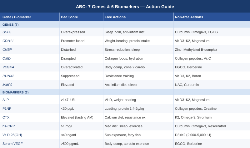
The 7 Genes Behind Aneurysmal Bone Cyst: What the Research Shows
Understanding the genetic basis of ABC is not just an academic exercise. The molecular events that drive these cysts — USP6 overexpression, NF-κB activation, MMP production, excessive angiogenesis — correspond to biological pathways that respond to targeted interventions. That is what makes genetics, for this condition, the more actionable starting framework.
A critical clarification before diving in: in ABC, the gene disruptions described below are somatic mutations. They occur in the tumor cells themselves, not in the patient's germline DNA. This means you cannot correct them with supplementation the way one might support an inherited MTHFR variant. What you can do is modulate the downstream biological environment those disrupted genes create — the inflammation, the matrix degradation, the excessive blood vessel formation — to support healing and potentially reduce the conditions that favor recurrence. Researchers like Ali Torkamani at Scripps Research have emphasized this kind of pathway-level thinking as the practical bridge between genetic findings and clinical action.
Gene 1: USP6 — The Master Switch
What it does. USP6 (Ubiquitin Specific Peptidase 6) encodes a deubiquitinase — an enzyme that removes ubiquitin tags from proteins, effectively preventing their degradation and prolonging their activity. In normal cells, USP6 is expressed at very low levels with tightly regulated activity. In ABC tissue, a chromosomal translocation places the USP6 coding sequence under the control of a highly active gene promoter, causing it to be expressed at massively elevated levels.
What its overexpression causes. Overactive USP6 activates two critical downstream pathways: NF-κB (nuclear factor kappa B), the master regulator of inflammation and bone-destroying osteoclast activity, and the matrix metalloproteinase (MMP) system, which degrades collagen and other structural proteins in bone matrix. The result is local bone destruction, cystic cavity formation, and a chronic inflammatory microenvironment that makes recurrence more likely after surgery. This mechanism has been documented across multiple studies published since Oliveira and colleagues first identified the USP6 translocation in 2004. PubMed research on USP6 in ABC
If USP6 downstream signaling is active: the plan without supplements
The most powerful non-supplemental modulator of NF-κB is sleep quality. Chronic sleep deprivation consistently elevates NF-κB activity across multiple tissue types; targeting 7–9 hours of uninterrupted sleep per night is one of the best-supported ways to reduce systemic inflammatory tone. An anti-inflammatory dietary pattern — Mediterranean-style, low in ultra-processed foods, refined sugar, and industrial seed oils — blunts NF-κB signaling through multiple mechanisms including reduced circulating lipopolysaccharide and lower saturated fat load. Stress management is not peripheral here: chronic cortisol exposure directly activates NF-κB, so structured recovery practices reduce the molecular burden at the root level.
If USP6 downstream signaling is active: the plan with supplements
Curcumin (curcuminoids): The best-documented natural NF-κB inhibitor. Dose: 500–1,500 mg/day of standardized curcuminoids with piperine (black pepper extract) to improve bioavailability. Frequency: daily with meals. Cycling: 8 weeks on, 2 weeks off to assess tolerance. Side effects: GI discomfort at high doses; potential interaction with anticoagulants — discuss with your physician if post-surgical.
Omega-3 fatty acids (EPA + DHA): Modulate NF-κB and COX-2 inflammatory signaling through multiple prostaglandin pathways. Dose: 2–4 g/day EPA+DHA combined from quality fish oil or algal oil. Frequency: daily with food. Side effects: fishy burps if taken without food; mild blood-thinning effect at higher doses — important to disclose to surgeons before any procedures.
EGCG (epigallocatechin gallate): Directly inhibits multiple MMPs including MMP-9, and downregulates NF-κB through IKK inhibition. Dose: 400–800 mg/day standardized EGCG from green tea extract. Frequency: daily, taken away from iron-containing meals (EGCG binds dietary iron). Cycling: 12 weeks on, 4 weeks off. Side effects: hepatotoxicity risk at doses above 800 mg/day has been documented in case reports; stay within the standard range and take with food.
Gene 2: CDH11 — The Disrupted Bone Builder
What it does. CDH11 (Cadherin 11, also called OB-cadherin) is a cell adhesion molecule that plays a central role in osteoblast differentiation and bone matrix formation. Under normal circumstances, it anchors osteoblasts in place and provides the mechanical and chemical signals needed for new bone synthesis.
What its disruption causes. In roughly 70% of USP6-positive ABCs, the translocation t(16;17)(q22;p13) fuses the active CDH11 promoter region to the USP6 coding sequence. This simultaneously places USP6 under CDH11's strong promoter — driving overexpression — and disrupts CDH11's own function. The result is impaired osteoblast behavior at precisely the location where bone formation is needed most. This helps explain why ABCs often persist: the bone-building signal is disrupted while the bone-destroying signal is amplified. PubMed studies on CDH11 and USP6 in ABC
If CDH11 function is compromised: the plan without supplements
Weight-bearing mechanical load is the most potent non-pharmacological activator of osteoblast activity. During active lesion phases, this must be done under orthopedic guidance — walking is usually appropriate; high-impact activities are contraindicated until bone is structurally stabilized. Protein intake is a second pillar: osteoblasts require adequate amino acids to produce collagen, and most people in the 12–25 age group affected by ABC consume less than optimal protein. Targeting 1.4–2.0 g/kg/day of dietary protein from quality sources reduces the burden on supplementation. Food-first sources of calcium and phosphorus — dairy, legumes, leafy greens, canned fish with bones — round out the dietary foundation.
If CDH11 function is compromised: the plan with supplements
Vitamin D3 + K2: Vitamin D3 activates the vitamin D receptor in osteoblasts, directly upregulating differentiation and RUNX2 expression (covered in Gene 6). K2 as MK-7 ensures calcium is directed into bone matrix rather than soft tissue. D3 dose: 2,000–5,000 IU/day guided by blood levels; target 40–60 ng/mL 25(OH)D. K2 as MK-7: 100–200 mcg/day. Frequency: daily with a fat-containing meal (both are fat-soluble). Cycling: continuous — monitor 25(OH)D every 12 weeks until stable. Side effects: D3 toxicity is rare but real above 100 ng/mL; never supplement without periodic monitoring. NIH Office of Dietary Supplements: Vitamin D
Magnesium glycinate or malate: Required for over 300 enzymatic reactions including osteoblast function; most people consume significantly less than optimal from diet alone. Dose: 200–400 mg/day of elemental magnesium in glycinate or malate form (better tolerated than oxide). Frequency: daily, often better tolerated in the evening. Side effects: loose stools at high doses — start low and titrate up over two weeks. NIH Office of Dietary Supplements: Magnesium
Gene 3: CNBP — The RNA Regulator Often Overlooked
What it does. CNBP (Cellular Nucleic Acid Binding Protein, formerly ZNF9) is an RNA-binding protein that regulates the translation of multiple target messenger RNAs. It acts as a modulator of cell stress responses and influences how mesenchymal cells respond to metabolic and inflammatory signals.
What its disruption causes. CNBP is the second most common fusion partner with USP6 in ABC, accounting for approximately 10–15% of USP6-rearranged cases. Its disruption alters normal translational regulation in bone-resident mesenchymal cells, and the CNBP promoter region drives USP6 overexpression in a similar but distinct fashion to CDH11. The downstream consequences overlap significantly with the CDH11 disruption cascade, with NF-κB and MMP activation featuring prominently.
If CNBP signaling is disturbed: the plan without supplements
Cellular stress is a key input to CNBP-related pathways. Chronic psychological stress triggers downstream signaling that compounds RNA-binding protein dysfunction. Structured recovery — consistent sleep timing, regular low-intensity movement, and deliberate reduction in stimulant load — reduces cellular stress responses in a measurable way. Cold-water immersion (10–15 minutes at 10–15°C, 3 times per week) has demonstrated stress-pathway modulating effects in human studies, though specific ABC evidence is lacking; this should be cleared with treating physicians given the structural considerations of the condition.
If CNBP signaling is disturbed: the plan with supplements
Zinc (glycinate or picolinate form): CNBP is a CCHC-type zinc finger protein; its proper folding and DNA/RNA binding function depend directly on adequate zinc availability. When zinc is marginal, zinc finger proteins cannot maintain their structural conformation. Dose: 15–30 mg/day of elemental zinc. Cycling: 8 weeks on, 2 weeks off. Side effects: high zinc intake depletes copper — always co-supplement with 1–2 mg/day copper when using zinc above 25 mg/day; copper deficiency causes its own neurological and bone marrow problems.
Methylated B-complex (B12 + folate + B6): Support RNA synthesis and methylation pathways downstream of CNBP function, and are required for healthy nucleic acid metabolism. Dose: a quality methylated B-complex (methylcobalamin and methylfolate forms preferred over cyanocobalamin and folic acid). Frequency: daily with food. Side effects: excess B6 above 200 mg/day has been linked to peripheral neuropathy; standard B-complex doses (well below this) are safe for long-term use.
Gene 4: OMD — When Bone Mineralization Goes Wrong
What it does. OMD (Osteomodulin) is an extracellular matrix glycoprotein belonging to the SLRP (small leucine-rich proteoglycan) family. It plays a structural role in bone mineralization, specifically in organizing the hydroxyapatite crystallization process and defining how calcium is deposited within collagen fibrils in bone matrix.
What its disruption causes. As a less common USP6 fusion partner (estimated 3–5% of USP6-rearranged ABCs), OMD's promoter region drives USP6 overexpression. Its own structural function in bone matrix organization is consequently disrupted. This may contribute to the poor matrix quality sometimes seen in ABC-affected bone, which complicates filling and consolidation after surgical intervention. While OMD research specific to ABC is limited, its role in bone quality is well-established in the broader bone biology literature.
If OMD function is disrupted: the plan without supplements
Collagen-rich whole foods support extracellular matrix regeneration from the substrate level: bone broth, slow-cooked connective tissue cuts of meat, and skin-on poultry provide glycine and proline at concentrations not easily matched by standard protein. Silicon, found in oats, cucumbers, and horsetail herb, is an underappreciated cofactor for collagen cross-linking and bone matrix organization. Adequate hydration supports the proteoglycan network that OMD is part of.
If OMD function is disrupted: the plan with supplements
Collagen peptides (hydrolyzed type I collagen): Provide the specific amino acid substrate — glycine, proline, hydroxyproline — needed for extracellular matrix repair in bone tissue. Human RCTs show collagen peptides increase serum markers of bone formation (P1NP) when taken in conjunction with mechanical loading. Dose: 10–15 g/day. Frequency: daily, ideally 30–60 minutes before or immediately after weight-bearing exercise or physical therapy. Side effects: generally very well tolerated; occasional mild GI discomfort in sensitive individuals.
Vitamin C: Required as a cofactor for prolyl hydroxylase and lysyl hydroxylase, the enzymes that cross-link collagen. Without adequate vitamin C, collagen produced in bone matrix cannot be properly stabilized. Dose: 500–1,000 mg/day in divided doses. Frequency: daily with meals. Side effects: GI discomfort above 2 g/day; oxalate-related kidney stone risk in genetically susceptible individuals at high doses.
Gene 5: VEGFA — The Vascular Engine
What it does. VEGFA (Vascular Endothelial Growth Factor A) is the primary angiogenic growth factor — it signals endothelial cells to proliferate and form new blood vessels. In normal bone, VEGFA coordinates the vascular supply required for bone remodeling and repair. Controlled VEGFA expression is essential; it is uncontrolled VEGFA that creates pathology.
What its overexpression causes in ABC. USP6 overexpression activates signaling cascades — including NF-κB and HIF-1α — that secondarily upregulate VEGFA production in ABC stromal cells. This is the mechanistic explanation for the defining imaging appearance of ABC: excessive VEGFA-driven angiogenesis fills expanding bone cavities with blood. Elevated VEGFA also maintains the permissive inflammatory environment that sustains the lesion. This mechanism is clinically relevant: anti-VEGF antibodies have been investigated as adjunct treatments for aggressive or surgically difficult ABCs in specialized centers.
If VEGFA is overactivated: the plan without supplements
Adipose tissue is a significant VEGF producer, and maintaining healthy body composition reduces systemic VEGF load. Regular moderate-intensity exercise — 150–300 minutes per week at zones 1–2, as cleared by the treating orthopedic team — normalizes VEGF receptor expression over time, even though acute high-intensity hypoxic training transiently spikes VEGFA. Treating obstructive sleep apnea, if present, removes one of the strongest intermittent hypoxia signals that drives VEGFA production through HIF-1α.
If VEGFA is overactivated: the plan with supplements
EGCG (400–800 mg/day): Beyond MMP inhibition, EGCG directly downregulates VEGFA gene expression through multiple pathways. See USP6 section for full dosing, cycling, and side effect details.
Berberine: A plant alkaloid with documented anti-VEGF and anti-angiogenic properties in human studies, primarily through HIF-1α downregulation. Dose: 500 mg, 2–3 times per day with meals. Cycling: 8 weeks on, 2 weeks off (berberine has significant metabolic effects — notably on blood glucose and lipids — and strong cycling is recommended to prevent tolerance). Side effects: GI discomfort and nausea are common initially; significant interactions with diabetes medications, some antibiotics, and anticoagulants — this supplement requires physician disclosure.
Gene 6: RUNX2 — The Bone Builder Under Siege
What it does. RUNX2 (Runt-Related Transcription Factor 2) is the master transcriptional regulator of osteoblast differentiation. It essentially orchestrates the process by which mesenchymal stem cells commit to the bone-building lineage. Without adequate RUNX2 activity, bone formation is impaired at the most fundamental level.
What the ABC environment does to it. RUNX2 is not typically mutated in ABC, but its function is suppressed by the inflammatory and disruptive signaling environment that USP6-overexpressing tissue creates. NF-κB, when chronically activated, directly inhibits RUNX2 transcription through a well-characterized molecular competition. This means that even as bone is being destroyed, the signals needed to rebuild it are being actively suppressed — a double hit that explains the structural severity of some lesions and the challenges of post-surgical bone consolidation in ABC cases with high-recurrence biology.
If RUNX2 is functionally suppressed: the plan without supplements
Mechanical loading is the single most powerful activator of RUNX2 expression. Weight-bearing exercise and resistance training directly stimulate RUNX2 transcription through mechanotransduction — the process by which physical force on bone matrix is converted into gene expression signals. Under orthopedic clearance, progressive weight-bearing activity starting from walking and advancing to resistance training is not optional for ABC recovery; it is arguably the highest-leverage single intervention available. The NF-κB suppression of RUNX2 is, paradoxically, partially reversed by the same exercise stimulus that activates RUNX2 directly.
If RUNX2 is functionally suppressed: the plan with supplements
Vitamin D3: Directly upregulates RUNX2 expression via the vitamin D receptor. This is one of the best-supported molecular effects of vitamin D in bone tissue. Dose guided by blood levels (target 40–60 ng/mL); see CDH11 section for full details.
Vitamin K2 — MK-7 form: Japanese trials using 45 mg/day of MK-4 (a different K2 form) for osteoporosis documented osteocalcin activation and bone formation improvements; the more practical MK-7 form at 100–200 mcg/day is well-studied in European RCTs. Frequency: daily with a fatty meal. Side effects: very low toxicity profile; potential interaction with vitamin K-antagonist anticoagulants such as warfarin — critical to disclose.
Boron: An essential trace mineral that modulates vitamin D metabolism and has direct regulatory effects on bone metabolism including RUNX2 pathway activation. Population studies link inadequate boron intake to poor bone density. Dose: 3–6 mg/day as boron glycinate or citrate. Frequency: daily. Side effects: safe at these doses; do not exceed 20 mg/day.
Gene 7: MMP9 — The Matrix Destroyer
What it does. MMP9 (Matrix Metalloproteinase 9) is a zinc-dependent endopeptidase that degrades type IV collagen, gelatin, and multiple other extracellular matrix proteins. In healthy bone, it plays a tightly regulated role in the controlled remodeling that allows bone to respond to mechanical demands. In ABC tissue, it becomes a primary executor of bone destruction.
What its overactivation causes. Downstream of USP6 overexpression and the NF-κB cascade, MMP9 production is dramatically upregulated in ABC stromal cells. MMP9 degrades the structural proteins that give cortical bone its integrity, creating the pathological space expansion that defines the condition. Elevated MMP9 has been associated with more aggressive lesion behavior in bone tumors broadly, and its reduction is one of the mechanisms by which anti-inflammatory interventions may reduce ABC recurrence risk.
If MMP9 is elevated: the plan without supplements
Dietary pattern has a direct and measurable impact on circulating MMP9 activity. Diets high in processed meat, refined carbohydrates, and industrial seed oils consistently elevate serum MMP9 through LPS-mediated inflammatory priming. A Mediterranean-style dietary pattern — rich in extra-virgin olive oil, oily fish, leafy vegetables, and low in processed foods — reduces baseline MMP9 activity in human studies. Sleep optimization is a second major lever: poor sleep elevates systemic MMP activity through inflammatory and cortisol-driven pathways. Adequate hydration reduces tissue hypoxia, which independently activates MMP9.
If MMP9 is elevated: the plan with supplements
NAC (N-Acetylcysteine): An antioxidant and glutathione precursor with documented MMP9 inhibitory properties in human studies. It works through reducing the reactive oxygen species that activate MMP9 transcription. Dose: 600–1,200 mg/day in divided doses. Frequency: daily, away from meals for optimal absorption (though food reduces GI discomfort). Cycling: 12 weeks on, 4 weeks off. Side effects: sulfurous odor in breath or urine, GI discomfort in some; rare bronchospasm in those with asthma — use cautiously.
Curcumin: Beyond NF-κB inhibition, curcumin directly inhibits MMP9 transcription at the AP-1 binding site on the MMP9 promoter. This dual mechanism — NF-κB plus direct MMP9 inhibition — makes curcumin one of the most relevant supplements in the ABC context. Dosing details are in the USP6 section above.
With the seven genes mapped and their downstream effects addressed, the natural next question is: how do you track what is actually happening in real time? That is where biomarkers come in.
6 Biomarkers to Track Between Imaging Appointments
Molecular genetic testing tells you what is driving the cyst at the DNA level. Biomarkers tell you what is happening in the body right now. For a condition like ABC — where recurrence can remain clinically silent for months and MRI is expensive — a handful of regularly monitored lab values can provide a meaningful early-warning layer that bridges the gap between imaging sessions. These six markers are chosen for their relevance to the specific biology of ABC, their clinical accessibility, and their ability to respond visibly to the interventions described above.
Biomarker 1: Alkaline Phosphatase (ALP)
Why it matters. ALP is an enzyme produced by osteoblasts during bone formation. In active ABCs, bone turnover is profoundly disrupted — destruction significantly outpaces formation — but ALP can rise when osteoblasts attempt to compensate for ongoing bone loss. Persistently elevated ALP after treatment may signal ongoing or recurrent disease activity. Conversely, a trend back toward normal range after treatment is a positive signal.
How to measure it. ALP is included in every standard comprehensive metabolic panel (CMP) without special ordering. Cost: $10–30, typically covered by insurance. Normal adult range: 44–147 IU/L. A critical caveat: growing children and adolescents have physiologically higher ALP due to bone growth — age-appropriate reference ranges must be used, and this is essential for the young population most commonly affected by ABC. Elevated total ALP should be followed with a GGT level or bone-specific ALP test to confirm the elevation is bone-origin, not hepatic.
If ALP is elevated: the plan without supplements
Rule out dietary causes first: high-fat meals transiently elevate ALP. Confirm the elevation is bone-specific (GGT will be normal if it is). If bone-specific ALP is driving the elevation, intervention priorities are optimizing vitamin D status, increasing supervised weight-bearing activity within orthopedic-cleared parameters, and ensuring adequate dietary protein and calcium intake to provide substrate for osteoblast activity.
If ALP is elevated: the plan with supplements
Vitamin D3 + K2: Regulates osteoblast activity and ALP expression. Target 25(OH)D levels of 40–60 ng/mL; see CDH11 gene section for full dosing. Magnesium glycinate or malate: ALP is a metalloenzyme that requires magnesium as a cofactor; low magnesium functionally impairs ALP and bone metabolism broadly. Dose: 200–400 mg/day elemental magnesium. Both are safe for long-term use at these doses when monitored.
Biomarker 2: P1NP (Procollagen Type I N-Terminal Propeptide)
Why it matters. P1NP is a bone formation marker — a byproduct of type I collagen synthesis released when osteoblasts are actively building new bone matrix. It is significantly more specific than total ALP for tracking bone formation activity and is the bone formation marker preferred by clinicians including Peter Attia for long-term bone health monitoring. In the post-treatment phase of ABC, tracking P1NP over time helps evaluate whether bone formation is proceeding appropriately and whether the interventions described in this article are working.
How to measure it. P1NP requires a dedicated serum test, not typically included in standard panels — request it explicitly. Cost: $50–150. Interpretation: P1NP above 30 µg/L generally suggests adequate bone formation activity; lower values in a post-surgical bone healing context warrant investigation. P1NP should always be interpreted alongside CTX (below) to understand the full bone turnover balance.
If P1NP is low: the plan without supplements
Progressive mechanical loading is the most direct intervention. Resistance exercise and weight-bearing activity are the strongest stimulators of osteoblast activity and P1NP in humans — this is well-established in the osteoporosis literature and applies directly to post-ABC bone recovery. Ensuring protein intake is adequate (1.4–2.0 g/kg/day) provides the amino acid substrate osteoblasts need: glycine, proline, and hydroxyproline are particularly relevant for type I collagen synthesis.
If P1NP is low: the plan with supplements
Collagen peptides (10–15 g/day): Human RCTs show that collagen peptides taken 30–60 minutes before exercise increase P1NP and reduce CTX more significantly than at other times of day. This timing effect is a practical, evidence-based detail worth applying. Creatine monohydrate (3–5 g/day): Emerging evidence from 2020–2024 RCTs shows creatine supplementation combined with resistance training increases P1NP and bone density markers in multiple age groups. Continuous daily dosing, no cycling needed. Side effects: initial water retention, mild GI in sensitive individuals; well-studied and safe long-term at these doses. Vitamin D3 (2,000–5,000 IU/day guided by blood levels) continues to be relevant here through its direct osteoblast-activating effects.
Biomarker 3: CTX (C-Terminal Telopeptide of Type I Collagen)
Why it matters. CTX (specifically beta-CTX or sCTX) is the primary bone resorption marker — it is released into circulation when osteoclasts break down type I collagen in bone matrix. In active ABCs, bone resorption is markedly elevated in and around the lesion. Persistently elevated CTX after treatment is a red flag for ongoing bone destruction, whether from residual disease, early recurrence, or a systemic bone metabolism problem secondary to the disease process.
How to measure it. CTX is a dedicated serum test; request it specifically and specify fasting, morning conditions — CTX has a strong circadian rhythm, rising during the fasting overnight period, and a non-fasting or afternoon sample will be falsely elevated and uninterpretable. Cost: $40–100. Reference ranges: premenopausal women < 400 pg/mL; men < 580 pg/mL. As with ALP, children and adolescents have higher physiological CTX due to active skeletal growth — always use age- and sex-specific reference ranges.
If CTX is elevated: the plan without supplements
Confirm testing conditions (fasting, morning) before acting on an elevated result. Dietary calcium adequacy reduces parathyroid hormone-driven osteoclast stimulation: ensure 1,000–1,200 mg/day of calcium from food sources (dairy, canned fish, fortified foods, leafy greens) before considering supplemental calcium. An anti-inflammatory dietary pattern reduces osteoclast-activating cytokines (IL-6, TNF-α) that drive CTX elevation. Progressive resistance training, consistently applied, shifts the bone remodeling balance toward formation and reliably reduces CTX over 12+ weeks in human studies.
If CTX is elevated: the plan with supplements
Vitamin K2 (MK-7, 100–200 mcg/day): Activates matrix Gla protein and osteocalcin, both of which regulate osteoclast activity; multiple RCTs show MK-7 significantly reduces CTX. Frequency: daily. Omega-3 fatty acids (EPA+DHA, 2–4 g/day): Reduces osteoclast-activating cytokines, consistently lowering CTX in RCTs. Strontium citrate (OTC form): Shifts the bone remodeling balance toward formation and away from resorption. OTC dose: 340–680 mg/day. Cycling: 6 months on, 2 months off. Important: separate strontium from calcium by 2–3 hours as they compete for the same intestinal transporter. Side effects: mild GI in some; avoid in severe renal impairment.
Biomarker 4: High-Sensitivity CRP (hs-CRP)
Why it matters. C-reactive protein is the most broadly available marker of systemic inflammation. In ABC, the lesion creates a profoundly inflammatory local environment through NF-κB activation and cytokine secretion. Chronically elevated systemic CRP — even at levels traditionally described as acceptable — signals a systemic inflammatory state that may slow post-surgical healing, suppress bone formation through RUNX2 inhibition, and increase the likelihood of residual disease persisting.
How to measure it. The high-sensitivity version (hs-CRP) detects lower concentration ranges and is more clinically useful for this purpose than standard CRP. Cost: $15–50. Optimal target: less than 1 mg/L. Levels above 3 mg/L indicate elevated inflammatory risk; levels above 10 mg/L suggest acute infection or active systemic disease and require prompt medical evaluation.
If hs-CRP is elevated: the plan without supplements
The most evidence-based dietary intervention for reducing CRP is a Mediterranean-pattern diet combined with caloric balance: excess body fat is an independent CRP-elevating factor through adipokine production. Sleep optimization (7–9 hours) reduces CRP by 15–20% in multiple human studies with consistent effect size. Reducing alcohol consumption has a rapid and significant effect on CRP. Moderate exercise — 150+ minutes per week sustained for 12+ weeks — consistently reduces CRP across populations in human RCTs.
If hs-CRP is elevated: the plan with supplements
Curcumin (500–1,500 mg/day with piperine): Meta-analyses of RCTs confirm significant CRP reduction with curcumin. Cycling: 8 weeks on, 2 weeks off. Omega-3 fatty acids (EPA+DHA, 2–4 g/day): Consistent CRP reduction shown across multiple RCTs through COX-2 and NF-κB modulation. Both curcumin and omega-3s address the same NF-κB-driven inflammation; they are synergistic in combination. Resveratrol (150–500 mg/day): Activates SIRT1 and downregulates NF-κB, reducing CRP and IL-6. Cycling: 12 weeks on, 4 weeks off. Side effects: GI discomfort in some; theoretical interaction with anticoagulants.
Biomarker 5: 25(OH) Vitamin D
Why it matters. Vitamin D insufficiency is endemic — estimated at 40–50% in Northern Europe and North America — and its consequences for bone health are direct and well-established. For ABC specifically, adequate vitamin D is required for RUNX2-driven osteoblast differentiation, for proper calcium absorption from the gut, and for suppression of the PTH-driven osteoclast activation that compounds CTX elevation. In the post-surgical healing context, low vitamin D is consistently associated with slower and less complete bone consolidation.
How to measure it. Serum 25(OH) vitamin D (25-hydroxyvitamin D). Cost: $30–80, increasingly insurance-covered. Optimal target for bone health, based on multiple bone-outcome studies: 40–60 ng/mL (100–150 nmol/L). Levels below 30 ng/mL represent insufficiency; below 20 ng/mL is deficiency by standard clinical definitions.
If vitamin D is insufficient: the plan without supplements
Midday sun exposure to large skin surface areas (back, chest, arms) for 15–30 minutes during peak UV hours (10 am to 2 pm) generates 1,000–4,000 IU of vitamin D3 depending on skin tone, latitude, and season. Dietary sources — fatty fish, egg yolks, UV-exposed mushrooms — contribute meaningfully but are rarely sufficient to correct established deficiency without sun exposure. In most Northern latitudes from October through March, supplementation is practically unavoidable.
If vitamin D is insufficient: the plan with supplements
Vitamin D3 (cholecalciferol): 2,000–5,000 IU/day for maintenance; 5,000–10,000 IU/day under physician supervision to correct confirmed deficiency. Take with a fat-containing meal. Recheck 25(OH)D after 8–12 weeks and adjust dose accordingly. Always co-supplement with K2 (100–200 mcg/day MK-7) to direct calcium into bone rather than arteries. Side effects: D3 toxicity is real above 100 ng/mL serum levels — do not supplement without monitoring. NIH Office of Dietary Supplements: Vitamin D
Biomarker 6: Serum VEGF
Why it matters. VEGF (Vascular Endothelial Growth Factor) is the primary driver of the aberrant angiogenesis that gives ABCs their blood-filled character on imaging. Serum VEGF levels represent a more advanced monitoring option that may help assess lesion activity between imaging appointments and track response to interventions — particularly in cases managed with sclerotherapy, embolization, or investigational anti-VEGF adjunct therapies. Elevated serum VEGF has been documented in active ABC cases in several published series, and its normalization following treatment correlates with favorable clinical outcomes.
How to measure it. Serum VEGF is measured via ELISA-based assay; it is not a standard panel test and must be specifically ordered, often going to a reference laboratory. Cost: $100–300. Reference ranges vary by laboratory; most adult assays define normal values below 500–700 pg/mL. Interpretation must be done in the clinical context of active lesion imaging and treatment timeline — serum VEGF can be elevated transiently after surgery and should be trended over multiple time points rather than read as a single snapshot.
If serum VEGF is elevated: the plan without supplements
Healthy body composition is the most impactful non-supplement step: adipose tissue is a major VEGF producer, and weight reduction consistently lowers serum VEGF in human studies. Treating untreated obstructive sleep apnea removes one of the most potent intermittent hypoxia signals that drives VEGFA production through HIF-1α. Regular moderate aerobic exercise normalizes VEGF receptor sensitivity and reduces baseline VEGF elevation over time, though acute hypoxic high-intensity sessions transiently spike it.
If serum VEGF is elevated: the plan with supplements
EGCG (400–800 mg/day): The best-documented natural VEGF modulator; directly downregulates VEGFA gene expression and VEGF receptor signaling. See USP6 section for full details. Berberine (500 mg, 2–3x/day): Downregulates HIF-1α, the primary transcription factor driving VEGF expression under inflammatory or low-oxygen conditions. Cycling: 8 weeks on, 2 weeks off. Side effects and interactions: see VEGFA gene section — physician disclosure required.
The genetic and biomarker frameworks above are most powerful when combined with a broader strategy for bone health, recovery, and long-term monitoring. Peter Attia's framework in Outlive provides exactly that broader context.
10 Insights from Outlive That Change How You Think About Bone Health
Peter Attia's Outlive: The Science and Art of Longevity (2023) is not written about aneurysmal bone cysts. But it contains one of the most rigorous lay frameworks for thinking about bone quality, biomarker tracking, and the interventions that matter most for long-term structural integrity. For anyone managing ABC or its aftermath, these ten ideas from Attia's approach are the most applicable.
1. Bone density loss is the most underappreciated structural risk
Most people think of bone fractures as a consequence of aging or falls. Attia frames bone density as a lifelong bank account: every year you fail to make deposits during youth, you increase the likelihood of structural failure later. For ABC patients who lose bone — and often experience growth disruption during critical bone-building years — the importance of aggressive, medically supervised bone building during recovery cannot be overstated. The window for peak bone mass is not infinite.
2. P1NP and CTX tell you more than a DEXA scan
A DEXA scan gives you a snapshot of bone density at one point in time. P1NP and CTX tell you what direction you are heading and how fast. Attia recommends tracking both as the paired bone turnover markers that give a complete picture of whether bone is being built faster or destroyed faster — the ratio matters as much as the absolute values. For ABC patients in recovery, this tracking principle is directly applicable.
3. Zone 2 exercise is the metabolic foundation
Attia distinguishes between high-intensity training — which spikes VEGF, cortisol, and inflammatory markers if overdone — and Zone 2 aerobic training (conversational pace, 150–180 min/week), which improves mitochondrial function, reduces baseline inflammation, and creates the metabolic environment in which bone recovery and tissue repair work most efficiently. For ABC patients cleared for exercise, starting with Zone 2 before adding intensity is a sensible protocol that aligns with the gene pathway modulation strategy above.
4. Resistance training is non-negotiable for bone
In Attia's framework, no supplement or medication fully substitutes for the mechanical stimulus that resistance training provides to bone formation through RUNX2 and Wnt signaling activation. His standard recommendation — progressive resistance training 3 times per week targeting all major muscle groups — directly applies to post-ABC recovery once orthopedically cleared. Bone responds to load. Without load, the building signal does not activate.
5. Protein is a bone health intervention
Attia frames protein not just as a muscle nutrient but as a bone nutrient. Type I collagen — the primary structural protein in bone — is made from dietary amino acids, particularly glycine and proline. His general recommendation of 1.6–2.0 g/kg/day of protein, with attention to leucine-rich sources to activate mTORC1 signaling, provides a relevant target for ABC patients in active bone recovery phases.
6. Vitamin D is a hormone, not a vitamin
Attia consistently emphasizes that vitamin D functions as a steroid hormone with receptors in nearly every tissue type, including bone-forming osteoblasts. He recommends targeting 40–60 ng/mL 25(OH)D based on bone outcome studies — notably higher than the minimum sufficiency threshold of 20 ng/mL used in standard clinical guidelines. This distinction between "not deficient" and "optimal" is precisely the kind of precision that matters in a condition like ABC.
7. Inflammation is the common denominator
Whether discussing cardiovascular disease, cancer, or bone pathology, Attia's framework consistently returns to chronic low-grade inflammation — driven by diet, sleep deficit, stress, excess adiposity, and sedentary behavior — as the root permissive factor. This maps directly to the NF-κB and MMP9 pathways driving ABC biology. His hs-CRP target of less than 1 mg/L is a meaningful benchmark.
8. Sleep is the most powerful recovery tool
Attia devotes a substantial section of Outlive to sleep, describing it as the most powerful systemic recovery intervention available — more powerful than any supplement in his framework. During slow-wave sleep, growth hormone is secreted, NF-κB is suppressed, inflammatory cytokines are cleared, and bone matrix synthesis proceeds. For ABC patients in recovery, treating sleep as a clinical priority rather than an afterthought is not peripheral to the protocol — it is central to it.
9. Biomarkers need to be trended, not point-read
A single elevated CTX or low P1NP value is not actionable in isolation. Attia's framework emphasizes tracking biomarkers as time-series data, looking for the direction and rate of change rather than reacting to individual values. For ABC patients monitoring their bone recovery, establishing baseline values at 3–6 months post-treatment and tracking at 6-month intervals gives the trend data that informs real decisions.
10. Early intervention creates the largest margin for reversal
Attia's core thesis is that the conditions which cause the most harm — osteoporosis, cardiovascular disease, cognitive decline — become exponentially harder to reverse once clinical disease is established. The same principle applies to post-ABC bone quality: the window for restoring bone density and reducing inflammatory burden is most open in the months immediately following treatment. A delayed or passive approach costs more than it saves.
Complementary Approaches Worth Considering
The following modalities have meaningful human evidence in the context relevant to aneurysmal bone cyst — primarily pain management, procedural anxiety (particularly important in pediatric patients), and tissue repair support. Each is presented honestly, including where evidence is limited.
Mindfulness Meditation and MBSR
Mindfulness-Based Stress Reduction (MBSR) is an 8-week structured program combining meditation, body scan practice, and mindful movement. For ABC patients, its relevance is twofold: chronic pain management and the significant psychological burden of managing a bone tumor diagnosis — particularly in young patients and their families. Both stress and pain amplify the NF-κB inflammatory cascade discussed throughout this article, making MBSR not just a quality-of-life intervention but a biologically relevant one.
A randomized trial published in JAMA Internal Medicine (Goyal et al., 2014) found that mindfulness meditation programs significantly reduced pain, depression, and anxiety compared to control conditions, with moderate effect sizes across populations. Goyal et al. JAMA Internal Medicine 2014: Mindfulness Meditation Programs The mechanisms include HPA axis downregulation (reducing cortisol's NF-κB activation), improved sleep quality, and reduced central sensitization to pain signals.
Practically, a formal MBSR program (in-person or validated online versions) is the most evidence-supported entry point. Daily practice of 20–45 minutes is the protocol used in most trials. For pediatric patients, child-adapted mindfulness programs (MBSR-C) with shorter sessions (15–20 minutes) have demonstrated similar pain and anxiety benefits. Consistency matters more than duration: twice-daily 10-minute sessions outperform once-weekly hour-long sessions for pain outcomes.
Low-Level Laser Therapy (Photobiomodulation)
Low-level laser therapy (LLLT), also called photobiomodulation, applies specific wavelengths of red or near-infrared light (typically 630–1,000 nm) to tissue at non-thermal intensities. In the context of bone conditions, LLLT has been studied for its ability to stimulate osteoblast activity, reduce local inflammation, and accelerate tissue repair. Evidence for direct ABC application does not exist, but the supporting mechanism — cytochrome c oxidase activation in mitochondria, increased ATP production, and downstream RUNX2 pathway stimulation — is biologically plausible and consistent with the gene framework above.
A systematic review published in the journal Lasers in Medical Science (Bashardoust Tajali et al., 2010) examined LLLT in bone repair contexts and found consistent evidence of accelerated ossification and improved bone formation markers in human and animal studies. Bashardoust Tajali et al.: LLLT and bone repair Wavelengths of 780–860 nm at dose ranges of 1–4 J/cm² showed the most consistent effects.
Practically, LLLT for post-surgical bone healing requires a clinical-grade device and should be applied by a trained physiotherapist or rehabilitation specialist to the post-operative site, not by a home device alone. Sessions of 10–20 minutes, 3 times per week for 8–12 weeks represent the protocol used in most positive trials. This should be positioned as an adjunct to standard surgical follow-up care, not a replacement, and should be cleared with the treating orthopedic surgeon given the proximity to a healing bone lesion.
Biofeedback
Biofeedback is a technique in which patients are given real-time physiological data — typically muscle tension, skin conductance, heart rate variability, or skin temperature — and trained to consciously modify that response. For ABC patients experiencing chronic musculoskeletal pain, pre-procedural anxiety, or stress-driven inflammatory flares, biofeedback offers a direct mechanism for modulating the autonomic nervous system response that amplifies both pain and the NF-κB cascade.
A Cochrane review of biofeedback for chronic pain found clinically meaningful reductions in pain intensity and associated psychological distress across multiple musculoskeletal conditions, with heart rate variability (HRV) biofeedback showing particular promise for pain modulation through vagal tone enhancement. PubMed: biofeedback and musculoskeletal pain The vagus nerve has direct anti-inflammatory effects through the cholinergic anti-inflammatory pathway, reducing circulating TNF-α and IL-6 — the same cytokines that drive osteoclast activation in the ABC context.
For practical implementation, HRV biofeedback is the most accessible form: devices such as chest strap monitors paired with validated training applications provide home-use protocols. Clinical biofeedback training — typically 8–12 weekly 45-minute sessions — produces faster learning and more durable results than self-directed home use alone. Once the resonance breathing pattern (typically 5.5–6 breaths per minute) is learned, daily 10–20 minute practice maintains the autonomic regulation benefits.
Guided Imagery
Guided imagery involves directed mental visualization — using recorded or therapist-led scripts to guide the patient through specific mental scenes designed to reduce pain perception, anxiety, or physiological stress responses. It has a particularly strong evidence base in pediatric pain management contexts, making it especially relevant given that ABC predominantly affects children and adolescents.
A systematic review in Pediatrics (Kuttner, 2012) found guided imagery to be effective for reducing procedural pain and anxiety in children and adolescents across multiple clinical settings, including orthopedic and oncology contexts. PubMed: guided imagery pediatric pain The mechanism involves engagement of the anterior cingulate cortex and prefrontal inhibition of pain-amplifying limbic circuits, reducing the emotional component of pain that often amplifies physical discomfort in young patients.
Practically, pediatric-specific guided imagery scripts focused on healing, tissue repair, and calm are available through validated sources. Sessions of 15–20 minutes, 1–2 times daily during high-stress periods (pre-procedure, post-surgical recovery, during physical therapy) are the most evidence-aligned approach. Parents can be trained as facilitators for younger children, which also reduces parental anxiety — itself a meaningful factor in pediatric pain perception.
Music Therapy
Music therapy — whether receptive (listening to therapist-selected music) or active (patient-involved music creation) — has one of the more robust pediatric evidence bases among complementary approaches, particularly for procedural anxiety and acute pain in clinical settings. Given that ABC management involves multiple procedures (biopsies, sclerotherapy sessions, surgeries, physiotherapy), and that these procedures carry particular psychological weight in the pediatric population, music therapy represents a low-risk, evidence-based adjunct worth implementing.
A Cochrane review (Bradt et al., 2021) on music interventions for cancer patients undergoing procedures found consistent reduction in anxiety, pain intensity, and the need for procedural sedation across studies. Bradt et al. Cochrane Review 2021: Music interventions for cancer patients While this population is not identical to ABC patients, the procedural context and the neurological mechanism — activation of the mesolimbic reward system through music reduces pain gate activation — apply across clinical settings.
Practically, the simplest implementation is patient-preferred music through noise-canceling headphones during procedures, sclerotherapy sessions, or physiotherapy. Rhythmic entrainment (music tempo matched to 60–80 BPM to align with relaxed heart rate) optimizes the autonomic calming effect. For ongoing post-surgical recovery, daily 30-minute listening sessions to personally meaningful music have been shown to reduce perceived pain intensity and cortisol levels in multiple RCTs.
Conclusion
Aneurysmal bone cyst is a genuinely complex condition sitting at the intersection of molecular genetics, bone biology, vascular physiology, and clinical management. The science has moved well beyond "it is benign, surgery should work" — and patients and families deserve access to that more nuanced picture.
The seven genes covered here — USP6, CDH11, CNBP, OMD, VEGFA, RUNX2, and MMP9 — represent the molecular architecture of ABC. Each one maps to a pathway that can be partially modulated through targeted dietary choices, lifestyle practices, and evidence-based supplementation. The six biomarkers — ALP, P1NP, CTX, hs-CRP, vitamin D, and serum VEGF — translate that molecular picture into trackable lab values that give clinicians and patients meaningful data between imaging appointments.
None of these strategies replaces surgical or procedural management, and none guarantees prevention of recurrence. What they offer is a more precise, proactive way to support bone healing, reduce the inflammatory environment that favors recurrence, and track recovery with real data. The next smart step is straightforward: discuss bone turnover and inflammatory biomarker tracking with your treating team, get a baseline vitamin D and hs-CRP measurement, and begin the lifestyle foundation — sleep, protein, supervised weight-bearing activity — with the confidence that it is grounded in the specific biology of this condition, not just generic health advice.
Musculoskeletal: Bone Conditions
Cardiovascular: Vascular Conditions
Autoimmune: Inflammatory Conditions Connective Tissue Conditions