This article contains AI generated content.
· UpdatedAnterolateral Ligament Injury — 6 Genes and 5 Biomarkers to Track
Introduction
If you have dealt with an anterolateral ligament injury — or you are doing everything you can to avoid one — you have probably heard the same advice: strengthen your quads and hamstrings, work on single-leg balance, maybe wear a brace during sport. That advice is not wrong, but it leaves out something fundamental: why some athletes tear the anterolateral ligament with what looks like a routine cut or landing while others with the same training history and sport exposure never do.
Part of that answer is structural. The anterolateral ligament, formally identified in 2013 as a distinct band of tissue on the outer knee controlling rotational stability, is made of collagen. The quality of that collagen — how it is organized, how it responds to load, how quickly it remodels after stress — varies considerably from person to person. Much of that variation is written into your genes.
Generic rehabilitation and prevention protocols assume all connective tissue is created equal. They are designed for the average athlete, which means they systematically underserve people with specific genetic predispositions toward weaker collagen architecture, over-active matrix degradation enzymes, or blunted tissue growth signaling. For those individuals, standard protocols may prevent acute injury less effectively and promote recovery more slowly than expected.
This article takes a more targeted approach. The first and main section covers six genes that research has linked to ligament injury susceptibility — with a specific plan for each one, whether you prefer to work without supplements or want to use evidence-based supplementation to directly compensate for the variant. The second section covers five measurable biomarkers that give you a live picture of your connective tissue environment right now, each with clear targets and actionable correction strategies. After that, you will find a summary of the most impactful research insights from connective tissue scientist Keith Baar, whose work has quietly challenged the way rehabilitation medicine approaches ligament healing, as well as a review of complementary modalities with real clinical support for this type of injury. Better information does not guarantee better outcomes, but it puts the odds significantly more in your favor.
The Genetic Blueprint Behind Anterolateral Ligament Injuries
The ALL shares its tissue composition with the ACL and other major knee ligaments: predominantly type I collagen, with type V collagen regulating fibril architecture, proteoglycans providing compressive resistance, and matrix metalloproteinases controlling ongoing remodeling. Each of these components has a genetic dimension. Variants in the genes encoding these proteins shift an individual's baseline toward either greater structural resilience or measurably higher fragility under rotational and deceleration loads.
Research from the University of Cape Town's Sports Science Institute, as well as genome-wide association studies in sports injury populations, has consistently implicated the following six genes. Most of the direct evidence involves ACL ruptures — a closely related structure — with strong biological plausibility extending to ALL injuries given shared tissue architecture.
COL5A1 — The Collagen Fibril Organizer
COL5A1 encodes the alpha-1 chain of type V collagen, a quantitatively minor but architecturally critical protein that regulates the diameter of type I collagen fibrils. Tight, uniform fibril diameters correlate with greater tensile strength and resistance to shear forces. Disruption of type V collagen leads to thicker, less organized fibrils — structurally weaker ligament tissue under rotational load.
The rs12722 polymorphism in COL5A1 has been the most studied variant. The CC genotype appears to be protective, while the TT genotype is overrepresented in athletes who suffer soft tissue ligament ruptures. Posthumus et al. (2009) demonstrated that the CC genotype was significantly underrepresented in ACL-ruptured individuals compared to uninjured controls — a finding since replicated in multiple sports injury cohorts.
If the gene variant is unfavorable — the plan without supplements
The primary intervention without supplementation is eccentric-dominant progressive loading of the posterolateral knee chain. Type V collagen production in fibroblasts responds to appropriate mechanical strain; slow eccentric contractions (3–5 second lowering phases) under controlled load support fibril organization over time.
Protocol: 3 sessions per week, at minimum 48 hours apart. Include 3–4 exercises — single-leg Romanian deadlifts, slow reverse lunges, and Nordic-style hamstring curls. Progress load 5–10% per week. Deload every 4th week. Rest 90–120 seconds between sets. Duration: minimum 12 weeks to see collagen-level adaptation.
Side effects to monitor: Delayed onset muscle soreness is expected in weeks 1–3. Persistent joint tenderness beyond 24 hours post-session is a sign to reduce load, not push through.
If the gene variant is unfavorable — the plan with supplements or equipment
The most evidence-backed supplementation protocol for type V collagen synthesis is gelatin or hydrolyzed collagen combined with vitamin C, taken 30–60 minutes before a loading session. Research by Keith Baar's group at UC Davis showed that 15g of gelatin combined with 50mg of vitamin C approximately doubled collagen synthesis markers in tendon-derived cells following a loading bout — a direct tissue-level effect, not an indirect one.
- Gelatin or hydrolyzed collagen peptides: 15g, 30–60 minutes before training - Vitamin C: 50–250mg at the same time - Frequency: daily on training days, optional on rest days - Duration: 12+ weeks continuous; no formal cycling required - Side effects: digestive discomfort at high doses in some individuals; generally well tolerated
For equipment, blood flow restriction (BFR) training at 40–50% limb occlusion pressure enables connective tissue loading with significantly reduced joint compressive forces. Use only calibrated cuffs; improvised tourniquets create uneven pressure and injury risk. This approach is particularly useful during early rehabilitation when full loading is contraindicated.
COL1A1 — The Primary Structural Collagen
COL1A1 encodes the alpha-1 chain of type I collagen, the dominant structural protein in all ligaments. A variant in the Sp1 transcription factor binding site (rs1800012) affects how efficiently the gene is transcribed. The T allele (Sp1 variant) is associated with lower collagen I output, reduced ligament tensile strength, and measurable joint hyperlaxity in several musculoskeletal injury studies.
Hyperlaxity is often perceived as an asset in flexible athletes, but it reduces passive stability at the knee. In explosive rotational movements, a hyperlax ALL under acute deceleration load has less mechanical reserve before failure. This is part of the reason WHY some anatomically mobile athletes sustain ligament injuries despite apparent flexibility and conditioning.
If the gene variant is unfavorable — the plan without supplements
When passive stability is reduced due to lower collagen I output, neuromuscular and proprioceptive training becomes disproportionately important. The dynamic stabilizers — primarily the hamstrings, popliteus, and iliotibial band-associated structures — must compensate for what the passive restraint system cannot provide.
Protocol: 4–5 sessions per week, 15–20 minutes of dedicated proprioceptive work either as a warm-up or standalone session. Begin with simple single-leg stance (hold 30–60 seconds per leg) before introducing balance board or Bosu challenges at week 4. Progress to sport-specific deceleration drills (L-cuts, T-tests) by week 8. Do not introduce unstable surface training under fatigue.
Side effects: Minor ankle sprains are possible on unstable surfaces; always control fatigue before balance training.
If the gene variant is unfavorable — the plan with supplements or equipment
Lysine and proline are critical amino acids for collagen I cross-linking. Dietary adequacy is the foundation, but a targeted hydrolyzed collagen supplement (10–20g/day) ensures substrate availability at peak synthesis times.
- Hydrolyzed collagen: 10–20g daily, either 30–60 minutes pre-training or before sleep (growth hormone peaks during slow-wave sleep and drives overnight collagen synthesis) - Vitamin C: 100–500mg alongside - Frequency: daily; no cycling required; 8–16 weeks minimum to assess effect - Side effects: minimal; those with recurrent kidney stones should monitor total protein intake
MMP3 — The Matrix Degradation Regulator
MMP3 encodes matrix metalloproteinase 3 (stromelysin-1), an enzyme that degrades multiple extracellular matrix components including collagen I, fibronectin, and laminin. Two polymorphisms — rs679620 and the 5A/6A promoter variant — affect MMP3 transcriptional output. The 5A allele is associated with higher MMP3 expression and more active matrix degradation, even at rest.
Under normal, well-regulated conditions, MMP3 is essential for tissue remodeling. But in an individual with chronically elevated MMP3 expression — particularly in the presence of systemic inflammation — the ALL matrix can be degraded faster than fibroblasts can replenish it. This creates a structural deficit that accumulates silently and then becomes visible only as an injury. Multiple studies in athletic populations have associated elevated MMP3 activity with increased soft tissue injury rates.
If the gene variant is unfavorable — the plan without supplements
The most powerful free lever is reducing the inflammatory signals that amplify MMP3 expression. This means prioritizing sleep quality and quantity (MMP3 expression is suppressed during adequate restorative sleep), limiting post-exercise heat exposure in the first 2 hours, and structuring training loads to avoid chronic overreaching.
Track resting heart rate variability (HRV) each morning — a sustained downward trend across more than 5 consecutive days is a reliable signal of accumulated inflammatory burden. Pull back training intensity when HRV drops 10% or more below your 7-day baseline. A full rest day every 7 days is a minimum; those with the 5A genotype may benefit from 2 rest days per week during intensive training blocks.
Side effects: None from sleep and recovery management; ignoring HRV signals and continuing to over-train carries direct injury risk.
If the gene variant is unfavorable — the plan with supplements or equipment
Omega-3 fatty acids (EPA and DHA) suppress the NF-κB inflammatory pathway and directly reduce MMP3 transcriptional activation in connective tissue cells.
- Triglyceride-form fish oil or algae-based omega-3s: 2–4g EPA+DHA daily, taken with a fat-containing meal - Frequency: daily, ongoing; no cycling required; benefits accumulate over 8–12 weeks - Side effects: mild fishy aftertaste; mild anticoagulant effect at doses above 3g — use with medical supervision if on blood thinners
Bioavailable curcumin (theracurmin or curcumin phytosome) has demonstrated direct MMP-3 inhibitory activity in cell and some clinical studies. Standard turmeric has poor bioavailability and is not equivalent.
- Theracurmin or curcumin phytosome: 500–1000mg daily, with a fat-containing meal - Cycling: 8 weeks on / 2 weeks off is a reasonable precaution as a maintenance protocol, though longer-term use has an acceptable safety profile - Side effects: GI upset at high doses; potential interaction with anticoagulants; avoid during pregnancy
ACAN — The Shock-Absorbing Proteoglycan Gene
ACAN encodes aggrecan, a large proteoglycan essential for the hydration and compressive resistance of cartilage and fibrocartilaginous joint tissues. The variable number tandem repeat (VNTR) region of ACAN influences aggrecan core protein length and water-binding capacity. Shorter VNTR alleles are associated with reduced cartilage thickness and earlier degenerative changes — which indirectly loads the ALL by reducing the shock-absorbing buffer of the lateral compartment.
When the lateral compartment cartilage cannot adequately absorb compressive energy, that force transfers to the adjacent soft tissue, including the ALL. Over thousands of loading cycles, this disproportionate mechanical burden is a meaningful injury risk factor.
If the gene variant is unfavorable — the plan without supplements
Load management and impact periodization become critical. Replace high-impact training volume with low-impact alternatives (cycling, swimming, elliptical) proportional to cartilage quality concerns. Structure training in a 3:1 ratio — three weeks of full sport/impact training followed by one week of reduced impact.
Protocol: Limit weekly running volume increases to no more than 10%. Prefer softer training surfaces when available. Reduce double-leg jump volume and single-leg landing tasks during high-training-load weeks.
Side effects: Excessive restriction leads to deconditioning; the goal is periodization, not avoidance.
If the gene variant is unfavorable — the plan with supplements or equipment
Glucosamine sulfate (1500mg/day) and chondroitin sulfate (1200mg/day) provide substrate for proteoglycan synthesis. While evidence for pain reduction in osteoarthritis is mixed, the biological rationale for supporting aggrecan synthesis in a genetically predisposed individual is sound, and the safety profile is excellent.
- Glucosamine sulfate: 1500mg daily (single or split dose) - Chondroitin sulfate: 1200mg daily - Frequency: daily; minimum 12 weeks to assess response - Side effects: mild GI discomfort in some individuals; those with shellfish allergies should use non-shellfish derived sources
Oral hyaluronic acid (80–200mg/day) has shown modest benefit for joint lubrication and cartilage hydration in clinical studies, particularly relevant when aggrecan quantity is structurally limited.
GDF5 — The Tissue Growth Signal Gene
GDF5 encodes growth differentiation factor 5, a TGF-β superfamily member involved in joint formation, tendon and ligament development, and connective tissue repair signaling. The rs143384 polymorphism affects GDF5 expression in joint tissues, and the T allele is associated with reduced GDF5 output — linked in genome-wide association studies to increased musculoskeletal injury risk and accelerated joint degeneration.
The key problem with reduced GDF5 signaling is not structural weakness at baseline but rather impaired adaptive response. The cellular cascade that normally strengthens ligament tissue after appropriate loading stress is blunted. Athletes with this variant may train consistently and yet not accumulate the structural adaptation their training load implies — creating a gap between perceived fitness and actual tissue resilience.
If the gene variant is unfavorable — the plan without supplements
When adaptive signaling is blunted, the training stimulus must be more precise. Progressive overload with mandatory deload weeks is the cornerstone — the signal must be novel enough to trigger adaptation, and recovery windows must be long enough for a slower remodeling cascade to complete.
Protocol: Apply progressive overload to knee stability exercises 3–4 times per week. Increase load 5–10% weekly. Deload (reduce volume by 40%, maintain intensity) every 4th week without exception. The deload is where adaptation consolidates in these individuals — skipping it undermines the entire block.
Side effects: Joint swelling that persists beyond 48 hours post-session indicates excessive load; reduce and reassess before advancing.
If the gene variant is unfavorable — the plan with supplements or equipment
Vitamin D significantly influences GDF5 expression through the vitamin D receptor (VDR), which is expressed in ligament fibroblasts and regulates multiple tissue-building pathways. Deficiency is consistently associated with blunted tissue repair capacity and higher soft tissue injury rates in athletic populations.
- Vitamin D3: 2000–5000 IU daily (adjust based on serum 25-OH vitamin D testing) - Vitamin K2 (MK-7): 90–200mcg daily alongside D3 to appropriately direct calcium - Frequency: daily, year-round in most latitudes - Cycling: no cycling required; test serum levels every 6 months; toxicity threshold is far above these doses - Side effects: rarely relevant at these doses; toxicity requires sustained intake above 10,000 IU/day; always test before supplementing above 4000 IU/day
Red light therapy (photobiomodulation) at 630–850nm wavelengths has shown upregulation of growth factor signaling in connective tissue cells including TGF-β adjacent pathways. Devices targeting the knee at 2–4 J/cm² per session are commercially available. Sessions of 10–15 minutes, 4–5 times per week, are the typical protocol. Side effects are minimal at recommended intensities; avoid direct eye exposure.
TNC — The Matrix Stabilizer Gene
TNC encodes tenascin-C, an extracellular matrix glycoprotein with high expression during tissue development, loading response, and injury repair. It modulates cell adhesion, mechanosensing, and the structural reorganization of the ligament matrix during remodeling. Variants in the fibronectin type III repeat region of TNC — studied primarily in South African sports genetics research — have been associated with increased soft tissue injury risk in athletes.
Tenascin-C acts as a mechanosensitive protein: its expression rises in response to loading and coordinates early-phase structural repair signaling. Variants that impair TNC function can leave the ALL matrix less organized after injury stress and may particularly increase re-injury risk in the remodeling window after an initial event.
If the gene variant is unfavorable — the plan without supplements
TNC expression peaks approximately 2–6 weeks into the post-injury remodeling phase. Early graduated loading during this window — rather than complete immobilization — is the primary free intervention. The mechanical signal activates residual TNC expression and supports matrix reorganization.
Protocol: With medical clearance, begin isometric exercises within the first 5–7 days post-injury. Progress to isotonic loading by week 3. Introduce proprioceptive challenge by week 6. For injury prevention, standard progressive loading 3x/week is appropriate; for post-injury management, adherence to the early-loading timeline is especially critical with this genotype.
Side effects: Over-aggressive early loading increases re-injury risk; all loading must remain within pain-free ranges, and swelling is a reliable signal to reduce load.
If the gene variant is unfavorable — the plan with supplements or equipment
Magnesium glycinate (200–400mg/day) supports multiple connective tissue synthesis enzymes and reduces inflammatory cytokine signaling that competes with TNC-mediated matrix reorganization.
- Magnesium glycinate: 200–400mg elemental magnesium daily, taken in the evening - Frequency: daily; no cycling required for general use - Side effects: loose stools if dose escalates too quickly; start at 100mg and titrate up over 2 weeks; the glycinate form is significantly better tolerated than oxide or citrate forms
Peptide compounds such as BPC-157 are increasingly discussed in connective tissue recovery contexts for their reported effects on extracellular matrix remodeling. Evidence currently remains largely preclinical — mostly rodent models — and these compounds are not approved for clinical use in most jurisdictions. They are mentioned here for completeness only, not as a recommendation. Consult a sports medicine specialist before pursuing.
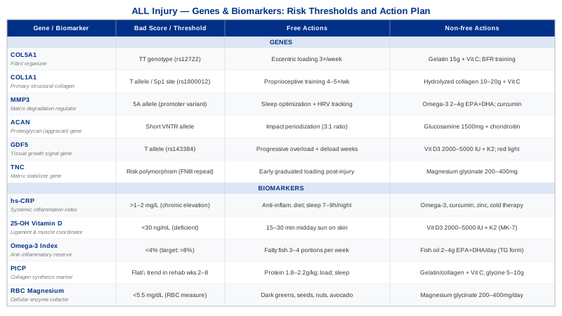
Biomarkers That Reveal Your Ligament Health Before an Injury Does
Genetic variants give you a fixed structural map — information about predispositions that do not change. Biomarkers give you the live version: what your connective tissue environment looks like right now, shaped by your diet, sleep, training load, and genetics together. The five biomarkers below are measurable, directly relevant to ALL health, and — importantly — all modifiable. If your genes load the gun, your biomarker levels often determine whether it fires.
1. hs-CRP — The Systemic Inflammation Index
High-sensitivity C-reactive protein is the most validated general marker of low-grade systemic inflammation. For ligament health, persistently elevated hs-CRP (above 1–2 mg/L) is a warning signal: it indicates an environment where matrix metalloproteinase enzymes are chronically upregulated, collagen remodeling is disrupted, and the normal repair cycle is running at a deficit. You can train consistently while hs-CRP quietly accelerates structural degradation in the background.
How to measure it: Standard serum blood test, available at most general practices and diagnostic labs. Cost: $10–$40 USD as a standalone test; frequently included in cardiovascular risk panels. Request the high-sensitivity version — standard CRP is not sensitive enough to detect low-grade inflammation.
Optimal target: Below 1 mg/L is the goal for connective tissue protection. Between 1–3 mg/L indicates moderate inflammatory burden worth addressing. Above 3 mg/L in the absence of acute illness warrants investigation.
If the score is bad — the plan without supplements
Dietary change is the most powerful free lever. Eliminate ultra-processed foods, refined seed oils (soybean, sunflower, canola), and high-glycemic refined carbohydrates. Replace with whole foods rich in polyphenols (berries, dark greens, extra-virgin olive oil), fatty fish two to three times per week, and adequate protein (1.6–2g/kg body weight daily). Prioritize 7–9 hours of sleep nightly — chronic sleep restriction below 6 hours significantly elevates hs-CRP independent of diet. Reduce psychological stress load, which elevates inflammatory markers through cortisol-mediated pathways.
If the score is bad — the plan with supplements or equipment
- Omega-3 (EPA+DHA): 2–4g/day with meals; no cycling required; benefits build over 8–12 weeks - Bioavailable curcumin: 500–1000mg/day, 8 weeks on / 2 weeks off as a maintenance cycle - Zinc: 15–30mg/day if dietary intake is low; take for 4–6 weeks, then reassess; doses above 40mg/day over extended periods suppress copper absorption — co-supplement with 1–2mg copper if using higher zinc doses long-term - Cold exposure (cold shower or immersion): 2–4 minutes at 10–15°C, 3 times per week; reduces systemic inflammatory markers with consistent practice over 4–8 weeks; side effects are minimal with gradual adaptation; avoid immediately post-strength training if muscle protein synthesis is the priority
2. Serum 25-OH Vitamin D — The Ligament and Muscle Coordinator
Vitamin D deficiency is common across populations and directly relevant to musculoskeletal injury risk. Vitamin D receptors are expressed in ligament fibroblasts, and the active form of vitamin D (1,25-OH₂D) regulates collagen gene expression, muscle fiber type composition, and proprioceptive neuromuscular signaling — three interconnected factors in ALL injury prevention and recovery. Low vitamin D impairs both the structural quality of the ligament and the neuromuscular speed of the protective response.
How to measure it: Serum 25-OH vitamin D blood test, available at any diagnostic lab. Cost: $30–$80 standalone; frequently included in comprehensive wellness panels. Test in late winter or early spring for the most clinically informative reading of your baseline — levels are naturally highest in late summer and lowest in late winter.
Optimal target: 40–70 ng/mL (100–175 nmol/L) for musculoskeletal protection. Below 30 ng/mL is clinically deficient; 30–40 ng/mL is functionally insufficient for an athlete under significant connective tissue stress.
If the score is bad — the plan without supplements
Daily midday sun exposure — 15–30 minutes depending on skin tone and latitude — to the arms and legs can produce 1,000–4,000 IU of vitamin D3 synthesis in the skin. This is the most natural approach, requires no cost, and has no toxicity risk. It is seasonal and latitude-dependent; above 50° north, meaningful synthesis is not possible between October and April.
If the score is bad — the plan with supplements or equipment
- Vitamin D3: 2,000–5,000 IU daily, depending on baseline measurement - Vitamin K2 (MK-7): 90–200mcg daily alongside D3 to direct calcium away from soft tissue - Retest serum 25-OH vitamin D after 3 months to adjust dose - Frequency: daily, year-round; no cycling required - Side effects: rare at these doses; toxicity threshold is well above 5,000 IU/day for most individuals; do not supplement above 4,000 IU/day without a serum test confirming deficiency
3. Omega-3 Index — The Anti-Inflammatory Reserve
The Omega-3 Index measures the percentage of EPA and DHA in red blood cell membranes, providing a 3-month average of omega-3 status that is far more reliable than a single serum measurement. An index below 4% is associated with elevated inflammatory burden and impaired tissue repair. Optimal connective tissue protection is associated with an index consistently above 8%.
For the ALL specifically, EPA and DHA directly suppress prostaglandin E2 and leukotriene B4 pathways that activate matrix metalloproteinase enzymes during and after mechanical loading. This is not a peripheral effect — it operates at the level of the ligament fibroblast's own inflammatory response to mechanical stress.
How to measure it: Specialized dried blood spot test requiring only a finger prick, offered by labs such as OmegaQuant. Cost: $50–$100 USD. Not included in standard panels — must be ordered directly by the patient or through a functional medicine provider.
Optimal target: 8–12% for combined connective tissue and cardiovascular protection, as referenced by practitioners including Peter Attia and Thomas Dayspring in their lipid and metabolic health frameworks.
If the score is bad — the plan without supplements
Increase dietary fatty fish to 3–4 portions per week — salmon, sardines, mackerel, herring, and anchovies are the highest-yield sources. At this intake, the Omega-3 Index can meaningfully rise over 8–12 weeks without any supplementation. Cost is the primary variable.
If the score is bad — the plan with supplements or equipment
- Triglyceride-form fish oil or algae-based EPA+DHA: 2–4g combined EPA+DHA daily, taken with a fat-containing meal for optimal absorption - Retest Omega-3 Index after 12 weeks to confirm response - Frequency: daily; no cycling required - Side effects: mild fishy aftertaste (mitigated by enteric-coated forms); mild anticoagulant effect at doses above 3g/day — discuss with a physician if you are on anticoagulant medication
4. PICP — The Active Collagen Synthesis Marker
Procollagen Type I C-terminal Propeptide (PICP) is cleaved from type I procollagen during fibril assembly and enters the bloodstream, making it a direct marker of new type I collagen synthesis. This is distinctly different from inflammatory markers or nutritional status indicators — PICP tells you whether your body is actually building new collagen matrix, or whether it is falling short despite training and supplementation.
Suppressed PICP in an actively training athlete — especially during rehabilitation — may indicate a synthetic deficit that explains why structural recovery is slower than expected. This is an advanced marker, but it provides information no general panel can.
How to measure it: Serum ELISA assay available through specialty and functional medicine labs. Cost: $80–$200 USD. Rarely ordered through standard GP panels; request specifically or access through a sports medicine or functional medicine physician.
Optimal pattern: Normal adult reference ranges vary by lab. In a recovering athlete, the desirable pattern is rising PICP during weeks 2–8 of early rehabilitation — a flat or suppressed trend during this window suggests inadequate substrate, signaling, or recovery.
If the score is bad — the plan without supplements
Three foundational free factors drive collagen synthesis: adequate protein intake (1.8–2.2g/kg body weight daily), appropriate mechanical loading (which provides the mechanical signal to initiate synthesis), and sufficient sleep (growth hormone release during slow-wave sleep is the primary endogenous driver of collagen fibroblast activity). Systematically auditing all three before adding supplementation is the rational first step.
If the score is bad — the plan with supplements or equipment
- Gelatin or hydrolyzed collagen: 15–20g daily, 30–60 minutes before training - Vitamin C: 100–500mg alongside - Glycine: 5–10g/day — the most abundant amino acid in collagen; additional dietary glycine may enhance synthesis rate in states of elevated demand; generally safe with no cycling requirement - Frequency: daily on training days; minimum 12 weeks continuous - Side effects: minimal; glycine at very high doses (30g+/day, well above these recommendations) may cause mild sedation
5. RBC Magnesium — The Cellular Enzyme Cofactor
Standard serum magnesium is a poor indicator of true magnesium status; only about 1% of body magnesium circulates in serum, meaning the serum level remains stable until depletion is severe. Red blood cell (RBC) magnesium reflects intracellular stores far more accurately and is significantly more useful for athletic populations.
Magnesium is a cofactor for over 300 enzymatic reactions including those involved in connective tissue synthesis pathways, ATP production in fibroblasts, and the regulation of inflammatory cytokine transcription. Athletes are particularly prone to magnesium depletion through sweat losses and high metabolic demand, and functional insufficiency can impair the enzymatic quality of collagen synthesis and recovery without triggering any obvious symptoms.
How to measure it: RBC magnesium is available through functional medicine labs and some specialty diagnostic services. Cost: $30–$80 USD. Standard serum magnesium is widely available and inexpensive but less informative; request RBC magnesium specifically for meaningful data.
Optimal target (RBC magnesium): 5.5–6.5 mg/dL is the functional range referenced by integrative medicine practitioners including Peter Attia; standard lab reference ranges are broader and often miss functional insufficiency in high-demand athletes.
If the score is bad — the plan without supplements
Increase dietary magnesium through dark leafy greens (spinach, Swiss chard), pumpkin seeds, almonds, avocado, legumes, and dark chocolate. Reduce factors that accelerate magnesium depletion: excess alcohol consumption, very high-dose isolated vitamin D supplementation without co-factors, and chronic psychological stress. Athletes in high training volumes may struggle to meet needs through diet alone.
If the score is bad — the plan with supplements or equipment
- Magnesium glycinate or magnesium malate: 200–400mg elemental magnesium daily, taken in the evening - Avoid magnesium oxide — poor absorption (approximately 4%) and high laxative effect - Frequency: daily; no cycling required for general use; ongoing supplementation is appropriate for athletes with confirmed insufficiency - Side effects: loose stools if dose is increased too quickly; begin at 100mg and titrate up over 2 weeks; glycinate form is significantly better tolerated than citrate or oxide in most individuals
The Keith Baar Research Framework — 10 Things That Could Change How You Approach Ligament Health
Keith Baar, Professor of Molecular Exercise Physiology at UC Davis, has spent over a decade studying how nutrition and loading interact to strengthen tendons, ligaments, and cartilage at the cellular level. His work, discussed in detail in the Huberman Lab podcast episode on connective tissue optimization and applied by performance practitioners across elite sport, fundamentally challenges the idea that ligaments are largely passive structures that just need time to heal. The central argument is that they respond to highly specific mechanical and nutritional inputs — and the window for doing so is narrow, precise, and routinely missed by standard rehabilitation.
1. Ligaments Have a Synthesis Window — and It Closes Within 24 Hours
Collagen synthesis in ligament tissue peaks approximately 6 hours after a loading session and returns to baseline within 24–36 hours. Rest days do not build collagen. Only the hours after an appropriate loading bout do. This means training frequency and timing — not just volume — directly determine how much structural adaptation accumulates.
2. The Gelatin + Vitamin C Finding Is the Single Most Actionable Protocol in Connective Tissue Science
In a controlled study, 15g of gelatin combined with 50mg of vitamin C, taken 60 minutes before a loading session, approximately doubled collagen synthesis markers compared to placebo. This is inexpensive (gelatin costs cents per serving), accessible, and backed by direct tissue-level data — not proxy measures. Hydrolyzed collagen at the same dose appears equivalent.
3. Vitamin C Is Not Optional
Proline and lysine hydroxylation — the steps that create stable collagen triple helices — require vitamin C as a non-substitutable cofactor. Even moderate insufficiency (not clinical scurvy, just suboptimal levels) impairs structural collagen quality. Periworkout vitamin C at 250–500mg is a negligible cost with direct mechanistic relevance.
4. Ligaments Need More Frequent Loading Than Muscles
Unlike muscle, which adapts with 48–72 hours between sessions, ligaments benefit from shorter, more frequent loading bouts. During active rehabilitation, 2 short loading sessions per day (6–8 hours apart) produces greater collagen synthesis than one longer session. Most rehabilitation protocols are designed for muscle, not connective tissue, and are too infrequent for optimal ligament adaptation.
5. Intermittent Loading Outperforms Continuous Loading for Collagen Synthesis
Baar's cell-culture research showed that intermittent loading (short bouts with rest intervals) produced significantly greater collagen upregulation than continuous loading of the same total duration. This has direct implications for session design: shorter, intermittent work sets with adequate rest between them are more effective for ligament adaptation than prolonged sustained loading.
6. Estrogen's Effect on Collagen Is a Critical but Under-Discussed Variable
High estrogen levels — occurring around ovulation in the menstrual cycle — measurably suppress collagen synthesis and increase ligament laxity. This contributes significantly to the well-documented higher soft tissue injury rate in female athletes and directly affects the ALL. Training structure, particularly high-rotational-load and deceleration work, may warrant adjustment during the pre-ovulatory phase in female athletes.
7. Standard Rehabilitation Under-Loads Connective Tissue
Standard post-injury protocols prioritize tissue protection and pain management over structural remodeling. The result is ligament that heals but does not fully remodel — achieving closure without restoring the original mechanical properties. Baar's research argues for graduated loading that begins earlier, progresses more deliberately, and treats collagen synthesis as an active target rather than a passive byproduct of rest.
8. Heavy Isometrics Are the Safest High-Signal Loading Stimulus
Isometric contractions at high joint angles generate the greatest connective tissue mechanical strain without joint movement — making them the safest option for early loading of injured tissue and one of the most effective stimuli for structural adaptation. Heavy isometrics (70–80% maximum voluntary contraction) held for 30–45 seconds are recommended early in both injury rehabilitation and ligament strengthening programs.
9. Sleep Is the Primary Recovery Driver That No Supplement Replicates
Growth hormone, released predominantly during deep slow-wave sleep, is the primary endogenous signal for connective tissue synthesis and remodeling. Chronic sleep restriction below 7 hours per night suppresses this release and disrupts the collagen cycle in ways that no supplement protocol meaningfully compensates for. Sleep is the platform; everything else is built on it.
10. Creatine's Role in Connective Tissue Is Emerging and Underappreciated
Emerging evidence suggests creatine monohydrate may support connective tissue health through its effects on cellular energy availability in fibroblasts — not just the satellite cell mechanisms relevant to muscle. While this remains an evolving area, the safety profile and concurrent performance benefits make it a reasonable addition for athletes focused on ligament resilience. Standard dose: 3–5g/day without a loading phase; no cycling required.
Other Evidence-Based Approaches Worth Exploring
The following modalities have meaningful human clinical evidence relevant to ligament injury recovery or prevention. None replaces the structural work — loading, nutrition, and genetics-informed intervention — but each has a defined role that can complement a comprehensive program.
Low-Level Laser Therapy (Photobiomodulation)
Photobiomodulation (PBM) uses specific wavelengths of red and near-infrared light (typically 630–850nm) to stimulate mitochondrial activity in cells, reduce inflammation, and accelerate tissue repair. For ligament tissue, PBM has been shown to upregulate collagen synthesis, reduce local inflammatory cytokines, and improve fibroblast proliferation in the injury repair zone — mechanisms directly relevant to ALL recovery.
A systematic review published in the journal Photomedicine and Laser Surgery (2012) examined low-level laser therapy for soft tissue injuries including ligament sprains and found consistent evidence of reduced healing time and improved functional outcomes. Effect sizes were moderate but reproducible across multiple randomized controlled studies.
Practical application: Use a class 3B or class 4 device targeting the lateral knee at 2–4 J/cm² per session. Sessions of 10–15 minutes, 4–5 times per week during acute and early subacute phases (weeks 1–8), are the typical evidence-based protocol. Home devices in the 650–850nm range are commercially available. Side effects at recommended parameters are minimal; avoid direct eye exposure and do not use directly over suspected malignancy.
Yoga for Proprioceptive and Neuromuscular Rehabilitation
Yoga's combination of controlled movement, single-leg balance demands, and deliberate proprioceptive challenge makes it directly relevant to anterolateral ligament rehabilitation and prevention. The ALL's primary function — rotational control at the knee — depends on both the passive ligamentous restraint and the active neuromuscular system that responds to joint position signals. Yoga specifically trains the latter.
A randomized controlled trial published in the Journal of Strength and Conditioning Research (2011) found that an 8-week yoga intervention significantly improved proprioceptive accuracy, balance, and knee stability metrics in recreationally active adults compared to controls — outcomes directly relevant to ALL injury prevention.
Practical application: Begin with a foundation-focused hatha or Iyengar yoga sequence emphasizing standing balance postures (Warrior III, Tree Pose, Half Moon) 3 sessions per week. Avoid deep rotational postures (Revolved Triangle, deep Pigeon variations) in the first 6 weeks post-injury or until lateral knee stability is confirmed. Progress to more dynamic vinyasa flows once single-leg stability is solid. Side effects: Minor muscle soreness is expected; avoid any posture that generates lateral knee pain or instability — modify with a block or strap before loading further.
Biofeedback for Neuromuscular Re-Education
Biofeedback uses real-time physiological signals — most commonly electromyography (EMG) of the muscles surrounding the knee — to help patients consciously retrain muscle activation patterns that were disrupted by injury. After an ALL injury, altered recruitment of the posterolateral rotator structures and biceps femoris is common and significantly increases re-injury risk. Biofeedback gives the nervous system the moment-to-moment data needed to correct those patterns.
Research in ACL rehabilitation has consistently demonstrated that EMG biofeedback accelerates the restoration of correct quadriceps-to-hamstring co-contraction ratios and significantly reduces compensatory movement patterns that load the anterolateral structures inappropriately. A review in Physical Therapy in Sport identified EMG biofeedback as a Grade B recommendation for knee ligament rehabilitation based on pooled RCT evidence.
Practical application: Work with a physical therapist or sports physiotherapist who uses surface EMG biofeedback during early gait retraining and functional movement assessment (weeks 3–8 post-injury). Home neuromuscular electrical stimulation (NMES) devices can supplement clinic-based biofeedback for daily quad and hamstring activation maintenance. Sessions of 20–30 minutes, 4–5 days per week, during the active rehabilitation phase. Side effects: Mild skin irritation under electrode pads in some individuals; electrical stimulation is contraindicated over open wounds or in patients with pacemakers.
Massage Therapy for Lateral Chain Function and Recovery
Sports massage and manual soft tissue therapy do not directly strengthen the ALL, but they address a secondary consequence of lateral knee injury that can perpetuate dysfunction: compensatory tension patterns in the iliotibial band, biceps femoris, and lateral gastrocnemius that alter joint mechanics and increase residual load on the healing ligament. Reducing these tensions improves both recovery quality and the effectiveness of subsequent loading sessions.
A randomized study examining sports massage in knee ligament rehabilitation found significant improvements in perceived stiffness, range of motion, and functional movement scores in the treatment group versus standard physiotherapy alone, particularly in the 4–8 week post-injury window. Evidence for direct structural ligament effects is not claimed — the mechanism is muscle tension normalization and improved local circulation.
Practical application: 1–2 sessions per week from a sports massage therapist during the active recovery phase (weeks 2–12), focusing on the iliotibial band, posterolateral knee region, and hamstring-biceps femoris complex. Avoid direct pressure over the acute injury site in the first 2 weeks. Light foam rolling of the iliotibial band (2–3 minutes each side, daily) is a cost-free self-care complement. Side effects: Post-session soreness is normal; deep work should be avoided over areas of active bruising or acute inflammation.
Conclusion
The anterolateral ligament does not operate in isolation from the biology of the person it is part of. Six well-studied gene variants can meaningfully shift structural risk — affecting collagen architecture, matrix degradation rates, and adaptive signaling — and five measurable biomarkers can tell you in real time whether your connective tissue environment is working for you or against you. Neither angle requires exotic technology or expensive protocols to begin addressing.
The next smart step depends on where you are. If you have access to genetic testing, start there — it gives you the fixed map. If you want immediate traction, test hs-CRP, 25-OH vitamin D, and the Omega-3 Index first — these three are affordable, widely available, and give you a concrete starting point. If you are in rehabilitation, apply the Keith Baar collagen timing protocol today. Whatever your starting point, bring these findings to a sports medicine physician or qualified physiotherapist who can contextualize them for your specific injury history, training demands, and health profile. Better information is the beginning, not the end.
Musculoskeletal: Joint Conditions Tendon & Ligament Conditions Sports Injuries
Autoimmune: Inflammatory Conditions Connective Tissue Conditions