This article contains AI generated content.
Anteromedial Knee Impingement - 4 Genes And 7 Biomarkers To Track
If you've been told your knee X-ray "looks fine" while you still feel a sharp pinch at the front-inner part of your knee every time you go down stairs, kneel, or straighten your leg fully, you already know how frustrating that gap between imaging and symptoms can be. Anteromedial knee impingement — usually driven by a thickened medial plica, an irritated infrapatellar (Hoffa's) fat pad, or scar tissue catching between the femur and tibia — is a mechanical problem, and mechanical problems don't always show up on standard blood work or even standard X-rays.
Generic advice tends to stop at "rest it, ice it, see how it feels." That's not wrong, but it treats every knee the same, ignoring that two people with an identical plica on MRI can have wildly different pain, healing speed, and recurrence risk. The difference often lives in the surrounding biology: how inflamed the tissue runs at baseline, how well cartilage is holding up under the extra mechanical stress, how efficiently the body repairs collagen, and how sensitized the nervous system has become to that specific pinch of pain.
This article goes past "rest and ice" and into the measurable biology that shapes how anteromedial knee impingement behaves in your particular body. None of this replaces a physical exam, imaging, or a conversation with an orthopedic specialist or physical therapist — impingement is fundamentally a structural issue that sometimes needs hands-on assessment or arthroscopy. But blood work and, to a lesser extent, genetic information can tell you why the same mechanical problem might be flaring for one person and staying quiet in another.
The goal here is grounded, not miraculous: better information about your inflammatory load, cartilage turnover, and tissue-repair capacity gives you and your clinician sharper levers to pull. The sections below walk through the biomarkers worth tracking first, then the genetic variants that add useful context, a mobility-focused framework worth knowing about, and a set of complementary approaches with real supporting evidence.
Summary
Anteromedial knee impingement is a mechanical, soft-tissue problem — but the body's internal chemistry still decides how much it hurts, how fast it heals, and how likely it is to come back. Below, you'll find seven blood-based biomarkers (from a simple hs-CRP to the more specialized urinary CTX-II) that reveal whether your inflammation, cartilage turnover, vitamin D status, and metabolic health are working for or against your knee, each with real-world testing costs and a two-track fix — one with no supplements, one that adds them or specific equipment. After that, four gene variants (GDF5, COL5A1, COMT, VDR) explain why identical impingement can feel and heal so differently between two people, with the same practical, dosed action plans. A mobility-first framework popularized on the Huberman Lab podcast challenges the passive "just rest it" model, and a final section covers the complementary approaches — from Tai Chi to EMG biofeedback — that have actual clinical trial support in knee conditions. Read on to see exactly what to test, what the numbers mean, and what to do about each one.
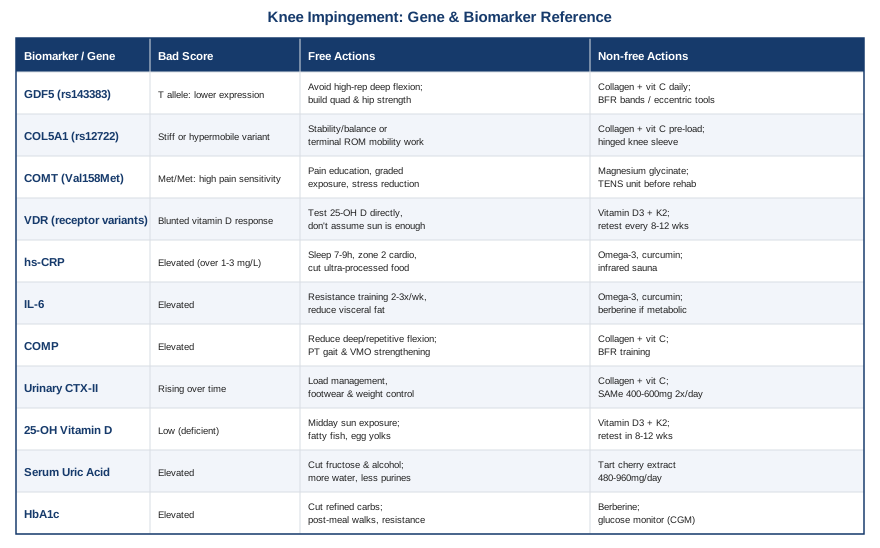
Before getting into the numbers, it helps to understand what's mechanically happening: in most anteromedial impingement cases, a synovial plica or the medial edge of the infrapatellar fat pad gets caught between the femoral condyle and tibia during terminal extension or deep flexion, producing localized synovitis and sometimes secondary cartilage irritation, as described in case reviews of Hoffa's fat pad and plica impingement (posterior Hoffa's fat pad impingement case review) and broader differential reviews of anterior knee pain (anterior knee pain differential diagnosis review). That localized synovitis is exactly where systemic biomarkers start to matter.
The Seven Biomarkers Worth Tracking For Anteromedial Knee Impingement
None of these biomarkers will show up on a knee MRI report, and none of them diagnose impingement on their own. What they do is tell you whether the tissue environment around the impinged plica or fat pad is primed to calm down quickly or to stay chronically irritated — and several of them are also the same panels Peter Attia and Thomas Dayspring push patients to track for whole-body inflammatory and metabolic risk, which happens to overlap heavily with joint health.
hs-CRP (High-Sensitivity C-Reactive Protein)
hs-CRP is the most accessible marker of low-grade systemic inflammation, and it's been directly linked to local synovial inflammation in osteoarthritic knees — patients with histologic synovial infiltrates had measurably higher CRP than those without, and CRP tracked with the degree of that infiltrate (elevated hs-CRP and local inflammatory findings in osteoarthritis). For anteromedial impingement, a chronically elevated hs-CRP suggests your baseline inflammatory "floor" is higher than it needs to be, which can keep an irritated plica or fat pad from settling down between flare-ups.
How to measure it: a standard hs-CRP blood draw, available through any lab (Quest, LabCorp, or direct-to-consumer services), typically 10 to 25 dollars out of pocket, no fasting required.
If the score is bad, the plan without supplements: prioritize 7 to 9 hours of sleep, address gum disease or other hidden low-grade infections, reduce ultra-processed food and added sugar, and add 150 minutes per week of zone 2 cardio, which reliably lowers hs-CRP over 8 to 12 weeks in most people.
If the score is bad, the plan with supplements or equipment: omega-3 (EPA/DHA) at 2 to 3 grams per day, and curcumin with piperine at 500 to 1000mg per day, both taken with food. Cycle 8 to 12 weeks, then retest hs-CRP before continuing. Side effects: omega-3 at this dose can mildly increase bleeding risk (caution if on blood thinners), and curcumin can cause GI upset or interact with certain medications — check with a physician if you take anticoagulants. Infrared sauna, 3 to 4 sessions per week for 15 to 20 minutes, has modest supporting evidence for lowering inflammatory load and can be added as equipment-based support.
IL-6 (Interleukin-6)
IL-6 is produced locally by synoviocytes, osteoblasts, and the fat within the infrapatellar (Hoffa's) fat pad itself — the exact tissue implicated in anteromedial impingement — and systemic IL-6 correlates with both synovial fluid IL-6 and the degree of synovial inflammation in knee osteoarthritis (systemic inflammation markers and local knee synovitis). Elevated IL-6 is a stronger signal than hs-CRP for fat-pad-driven irritation specifically.
How to measure it: less commonly ordered than hs-CRP; available through specialty or functional medicine labs, typically 50 to 90 dollars, sometimes bundled into broader inflammatory panels.
If the score is bad, the plan without supplements: resistance training 2 to 3 times per week (chronic, regular training lowers resting IL-6 despite transient post-workout spikes), reducing visceral fat if elevated, and consistent sleep timing.
If the score is bad, the plan with supplements or equipment: the same omega-3 and curcumin protocol above applies, plus considering berberine (500mg, 2 to 3 times daily, cycled 3 months on and 4 weeks off) if metabolic dysfunction is also present. Side effects: berberine can cause GI discomfort and shouldn't be combined with glucose-lowering medication without medical supervision.
COMP (Cartilage Oligomeric Matrix Protein)
COMP is released when cartilage matrix turns over faster than normal, and a large case-control study found serum COMP was strongly elevated in knee osteoarthritis patients, correlating with radiographic severity, pain, and disability, with very high diagnostic accuracy in that cohort (COMP as a biomarker for knee osteoarthritis). In impingement, chronically elevated COMP can flag that repetitive catching is starting to stress the adjacent cartilage, not just the soft tissue.
How to measure it: not a routine clinical test; available via specialty or research-oriented labs as an ELISA assay, typically 100 dollars or more, and worth discussing with a sports medicine physician rather than ordering blind.
If the score is bad, the plan without supplements: reduce high-impact repetitive knee flexion (deep squats under fatigue, repeated stair descent), get a gait and movement assessment from a physical therapist, and strengthen the quadriceps (especially VMO) to improve patellar tracking and reduce focal loading.
If the score is bad, the plan with supplements or equipment: collagen peptides (10 to 15g per day) paired with vitamin C (50 to 100mg, taken together to support collagen synthesis), and consider blood flow restriction (BFR) training equipment to build strength at lower joint loads. Cycle collagen for 3 months, then retest. Side effects are minimal (mild GI upset in some), but BFR should be introduced under professional guidance to avoid excessive cuff pressure.
Urinary CTX-II (C-Telopeptide of Type II Collagen)
Urinary CTX-II reflects direct breakdown of type II collagen, the main structural protein in articular cartilage, and longitudinal data show that rising CTX-II (often alongside COMP) predicts knee osteoarthritis severity and joint stiffness over a 10-year window (serum COMP and urinary CTX-II predicting knee OA severity). It's one of the more consistent biochemical predictors of joint degeneration available.
How to measure it: mostly a research-grade test rather than a routine clinical order; specialty labs offer it as an ELISA, typically 100 to 150 dollars, and it's worth pairing with COMP testing rather than ordering alone.
If the score is bad, the plan without supplements: the same load-management and biomechanics correction as COMP — reduce repetitive high-impact flexion, address footwear and single-leg control, and manage body weight to reduce joint force.
If the score is bad, the plan with supplements or equipment: collagen peptides plus vitamin C as above; some clinicians also consider SAMe (400 to 600mg twice daily) for cartilage-supportive effects, cycled for 8 to 12 weeks with a GI-tolerance check, since SAMe can cause nausea or mild agitation in some people at higher doses.
25-Hydroxyvitamin D (25-OH D)
Vitamin D deficiency is associated with faster knee osteoarthritis progression, more pain, greater functional impairment, and higher circulating TNF-α and IL-6 (vitamin D deficiency and progression of knee osteoarthritis). Beyond bone health, vitamin D has direct effects on cartilage turnover and pain sensitization, both relevant to a chronically irritated plica or fat pad.
How to measure it: a standard 25-OH D blood test, widely available, typically 30 to 50 dollars, often included in broader wellness panels.
If the score is bad, the plan without supplements: 15 to 20 minutes of midday sun exposure on exposed skin several times per week (season and latitude dependent), and increasing dietary intake of fatty fish and egg yolks.
If the score is bad, the plan with supplements or equipment: vitamin D3 at 2000 to 5000 IU per day paired with vitamin K2 (100 to 200mcg) to support proper calcium handling, retested after 8 to 12 weeks to adjust dose. Side effects: excessive dosing without monitoring can raise serum calcium — don't supplement blind long-term without periodic blood checks.
Serum Uric Acid
Serum urate has a genuinely reciprocal relationship with osteoarthritis: it can predict progression of joint space narrowing, and at higher concentrations can crystallize and activate the NLRP3 inflammasome, amplifying local inflammation independent of classic gout (urate and osteoarthritis reciprocal relationship). For anteromedial impingement, elevated uric acid is worth knowing because it can compound synovial irritation on top of the mechanical catching.
How to measure it: part of most basic or comprehensive metabolic panels, typically 10 to 20 dollars.
If the score is bad, the plan without supplements: reduce fructose (especially sugar-sweetened beverages), moderate alcohol (particularly beer), lower purine-heavy foods (organ meats, certain shellfish), and increase water intake.
If the score is bad, the plan with supplements or equipment: tart cherry extract (480 to 960mg per day) has modest evidence for lowering urate and inflammatory markers; cycle for 6 to 8 weeks and reassess. If uric acid is very high, this is a case where medical evaluation for gout risk should come first — supplements are a complement, not a substitute for that workup.
HbA1c (and Fasting Glucose)
Chronic hyperglycemia drives accumulation of advanced glycation end-products (AGEs) in joint tissue, and a mechanistic study showed hyperglycemia-induced AGEs accumulating in fibroblast-like synoviocytes directly promotes knee osteoarthritis-type changes (hyperglycemia-induced AGEs and knee osteoarthritis). Stiffened, glycated cartilage and synovium heal more slowly and tolerate repetitive impingement forces worse.
How to measure it: standard HbA1c blood test, typically 15 to 25 dollars, no fasting required (a fasting glucose panel is a reasonable cheaper add-on).
If the score is bad, the plan without supplements: reduce refined carbohydrates, take a 10 to 15 minute walk after meals, add 2 to 3 resistance training sessions weekly, and prioritize sleep consistency.
If the score is bad, the plan with supplements or equipment: berberine (500mg, 2 to 3 times daily, cycled 3 months on and 4 weeks off) has glucose-lowering evidence comparable in some trials to metformin; a continuous glucose monitor (equipment, 1 to 2 months of wear) can identify which specific foods spike your glucose the most. Side effects: berberine GI upset, and do not combine with diabetes medication without physician oversight.
What Your Genes May Be Telling You About Knee Impingement Risk
Genetics won't tell you whether you have a plica or a fat pad impingement — only imaging and a clinical exam can do that. But a handful of variants help explain why some people build more resilient cartilage and connective tissue, recover from synovitis faster, or feel pain more intensely from the same mechanical irritation. Ali Torkamani's work on genomic risk prediction and Gary Brecka's popularization of actionable biomarker-and-gene panels are both reasonable starting points if you want to pull your own raw genetic data (from a service like 23andMe) and look up these specific variants yourself.
GDF5 (rs143383)
The T allele of this well-studied variant sits in the regulatory region of GDF5, a gene central to cartilage growth and joint development, and reduces GDF5 expression — a variant consistently associated with osteoarthritis susceptibility across multiple populations (functional GDF5 polymorphism and osteoarthritis susceptibility). Carriers may have somewhat less resilient cartilage repair capacity, which matters when a plica or fat pad is repeatedly nicking the same patch of cartilage.
If the gene looks unfavorable, the plan without supplements: lean harder on load management than the average person would — avoid high-repetition deep knee flexion under fatigue, and build quadriceps and hip strength to offload the joint rather than relying on cartilage to absorb repetitive stress.
If the gene looks unfavorable, the plan with supplements or equipment: collagen peptides with vitamin C daily, and eccentric strengthening tools or BFR bands to build capacity at lower joint loads. Cycle collagen 3 months at a time; no meaningful side effects at standard doses.
COL5A1 (rs12722)
This variant affects Type V collagen, which regulates the structure of Type I collagen fibrils in tendon and ligament, and specific allelic combinations are linked to ACL injury risk and altered range of motion, with certain genotypes tending toward stiffer tissue and others toward more compliant, hypermobile tissue (COL5A1 allelic combination and ACL injury risk). Tissue compliance around the knee affects how well the plica and surrounding capsule tolerate impingement forces during terminal extension.
If the gene looks unfavorable (hypermobile-leaning), the plan without supplements: prioritize stability and strength training over additional stretching, and add proprioceptive/balance work to compensate for looser tissue.
If the gene looks unfavorable (stiff-leaning), the plan without supplements: prioritize consistent mobility work for terminal knee extension specifically, since impingement often worsens with restricted end-range motion.
The plan with supplements or equipment (either genotype): collagen peptides with vitamin C taken 30 to 60 minutes before loaded mobility or strength work, timed to support collagen synthesis; a hinged knee sleeve during high-load activity can add proprioceptive feedback. No significant side effects at standard collagen doses.
COMT (Val158Met, rs4680)
COMT breaks down catecholamines involved in pain modulation; the Met/Met genotype has three to four times lower enzymatic activity than Val/Val, and is linked to heightened pain sensitivity and altered opioid-system response to pain (COMT variants and pain sensitivity). This matters because two people with an identical degree of plical impingement on MRI can report very different pain intensity, and Met/Met carriers may be dealing with amplified central pain processing on top of the local mechanical issue.
If the gene looks unfavorable, the plan without supplements: pain neuroscience education (understanding that pain intensity isn't a direct readout of tissue damage), graded exposure to activity rather than total avoidance, consistent sleep, and stress-reduction practices that lower general nervous system reactivity.
If the gene looks unfavorable, the plan with supplements or equipment: magnesium glycinate (200 to 400mg) before bed to support sleep and pain modulation, and a TENS unit used before rehab exercises to reduce pain signaling during movement. Side effects: magnesium can cause loose stools at higher doses; start low and titrate.
VDR (Vitamin D Receptor Variants)
VDR polymorphisms determine how effectively cells actually respond to circulating vitamin D, and one clinical study found VDR genotype directly modulated the clinical and radiological response to vitamin D supplementation in knee osteoarthritis patients (VDR gene polymorphisms and response to vitamin D supplementation in knee OA). This means some people need a normal serum vitamin D level to feel a benefit, while others with less efficient VDR signaling may need higher maintenance levels and more frequent monitoring to get the same tissue effect.
If the gene looks unfavorable, the plan without supplements: don't assume dietary or casual sun exposure is enough — test serum 25-OH D directly rather than guessing.
If the gene looks unfavorable, the plan with supplements or equipment: vitamin D3 with K2 as described above, but retest every 8 to 12 weeks initially rather than annually, since blunted receptor response means you may need to adjust dosing more actively than someone with typical VDR function.
A Mobility-First Framework Worth Knowing About
Beyond bloodwork and genetics, one recent conversation worth understanding is Dr. Kelly Starrett's appearance on the Huberman Lab podcast, "How to Improve Your Mobility, Posture & Flexibility." Starrett, a physical therapist known for challenging passive rest-based injury protocols, argues that most joint pain — including anterior and mechanical knee pain — improves faster with restored range of motion and tissue capacity than with prolonged avoidance. Here are ten of the most useful ideas from that conversation and Starrett's broader body of work, applied cautiously to a mechanical issue like anteromedial impingement.
Terminal range of motion is where problems hide
Starrett's core argument is that most people lose the very last degrees of joint motion first, and that's exactly where impingement-type pain tends to show up — full knee extension is precisely the range where a plica or fat pad is most likely to get pinched.
Restoring motion often matters more than strengthening alone
Strength training on top of a restricted joint range can reinforce the restriction; Starrett advocates working the edges of your available range before loading it heavily.
Ten minutes a day beats one long session a week
Short, frequent mobility exposure (Starrett's "10 minutes a day" framework) produces more durable range-of-motion change than infrequent long stretching sessions.
Default posture during the other 23 hours matters
How you sit, stand, and hold your knee throughout the day shapes tissue tone more than the one hour you spend exercising — chronically locked-out or chronically flexed knee postures can both aggravate anteromedial structures.
Foam rolling and fascial work are inputs, not fixes
Starrett frames self-myofascial release as a way to temporarily quiet down guarded tissue enough to move better, not as a standalone treatment — useful before mobility work, not a substitute for it.Heat before movement, cold after intense loading
Consistent with general tissue physiology, Starrett recommends warming stiff or guarded tissue before working on range of motion, and reserving cold primarily for acute flare management rather than routine daily use.
Zero-cost tools go a long way
A large share of Starrett's protocols use nothing more than a wall, a doorway, or a strap — useful reassurance that meaningful mobility work doesn't require a fully equipped gym.
Movement quality under load prevents re-injury
Correcting how the knee tracks during a squat or step-down, not just how far it can passively bend, is what actually protects the impinged tissue during daily activity.
Pain doesn't always mean stop
Starrett pushes back against total activity avoidance, arguing that some graded, pain-informed loading (staying below a clear pain threshold rather than avoiding all discomfort) speeds recovery more than complete rest — a view that lines up with modern graded-exposure approaches to musculoskeletal pain, though it should be applied cautiously and adjusted with a clinician if swelling or mechanical locking is present.
Track range of motion the way you'd track a lab value
Measuring terminal knee extension and flexion with a simple goniometer or phone app periodically gives you an objective number to improve, the same way you'd track hs-CRP or vitamin D.
Complementary Approaches That Can Support Recovery
These are not replacements for a structural diagnosis or, when needed, arthroscopic correction of a symptomatic plica — but each has real clinical evidence in knee conditions and can be reasonably layered on top of the biomarker and mobility work above.
Tai Chi
Tai Chi combines slow, controlled range-of-motion work with balance and light muscular loading, which makes it a reasonable fit for a condition where terminal knee extension and controlled loading are the exact areas needing attention. Because movements are slow and self-paced, it also allows people to stay well below a painful impingement threshold while still training strength and control.
In a well-known 52-week randomized trial from Tufts Medical Center, Tai Chi (twice weekly) was compared directly against standard physical therapy for knee osteoarthritis and produced equivalent improvements in pain and physical function, as summarized by the NIH's National Center for Complementary and Integrative Health (Tai Chi versus physical therapy for knee osteoarthritis).
A realistic approach is a beginner Tai Chi class or instructor-led video, two sessions per week for at least 12 weeks before judging results, treating it as a substitute for one physical therapy session per week rather than an addition on top of an already full rehab schedule.
Yoga
Yoga's combination of controlled range-of-motion poses and isometric holds can help rebuild tolerance to knee positions that provoke impingement, particularly when a program is biomechanically adapted rather than generic.
A randomized controlled trial of a biomechanically-based yoga program in women with knee osteoarthritis found meaningful improvements in pain, self-reported function, and mobility performance compared to a no-exercise control (biomechanically-based yoga exercise program in knee osteoarthritis).
Because certain classic yoga poses (deep kneeling, full flexion holds) can directly provoke anteromedial impingement, the realistic approach is working with an instructor familiar with knee modifications, avoiding end-range flexion poses initially, and progressing only as terminal extension work (from the mobility framework above) improves.
Massage Therapy
Massage can reduce guarding and muscular tension around an irritated joint, which may indirectly reduce the compressive forces contributing to impingement, though its effect is more about symptom modulation than structural correction.
A randomized controlled trial of Swedish massage in knee osteoarthritis patients found meaningful improvements in pain and function versus a delayed-treatment control, though a later dose-finding follow-up found that benefits required ongoing weekly sessions rather than a one-time course (massage therapy for knee osteoarthritis, randomized trial commentary).
A practical approach is a focused course of weekly sessions targeting the quadriceps, IT band, and hip flexors for 6 to 8 weeks, reassessing pain and function before committing to ongoing maintenance sessions, since evidence suggests the benefit fades without continued treatment.
Biofeedback (EMG-Guided Quadriceps Retraining)
After knee injury or persistent irritation, the quadriceps often develop arthrogenic muscle inhibition (AMI) — a reflexive shutdown of muscle activation that isn't a strength problem but a neural one, and it's especially relevant to anteromedial impingement where altered quad firing changes how the patella and fat pad track during extension.
A randomized controlled trial of EMG biofeedback for knee extension after ACL reconstruction found it improved quadriceps activation and functional outcomes compared to standard rehab alone (EMG biofeedback and knee extension following ACL reconstruction).
A realistic approach is working with a physical therapist who has surface EMG equipment, using it during the first 4 to 6 weeks of quad-focused rehab specifically to correct firing patterns, rather than as a long-term standalone tool.
Low-Level Laser Therapy (Photobiomodulation)
Photobiomodulation delivers specific light wavelengths to reduce local inflammation and pain, which fits a condition where a chronically irritated plica or fat pad is the main pain generator rather than a purely mechanical block.
A recent network meta-analysis on the optimal wavelength of low-level light therapy for knee osteoarthritis found it superior to sham treatment for pain relief, though effects on function and stiffness were less consistent, and some guideline bodies remain more cautious than individual trials suggest (network meta-analysis of optimal LLLT wavelength for knee osteoarthritis).
A practical approach is a course of 8 to 12 sessions, 2 to 3 times weekly, using devices in the 785 to 860nm or 904nm range at clinically studied doses, treated as an adjunct for pain control during the weeks when mobility and strength work are being rebuilt, not as a standalone fix.
Conclusion
Anteromedial knee impingement is a structural problem first, but the biomarkers and genetic variants covered here explain why identical mechanical irritation can behave so differently from one person to the next — why one knee calms down in weeks and another stays inflamed for months. Tracking hs-CRP, IL-6, COMP, urinary CTX-II, vitamin D, uric acid, and HbA1c gives you a concrete read on the inflammatory and metabolic terrain your knee is healing in, while genetic context on GDF5, COL5A1, COMT, and VDR helps explain your baseline tissue resilience and pain sensitivity. Layered with a mobility-first rehab approach and evidence-supported complementary care, this is a far more precise starting point than generic rest-and-ice advice.
None of this replaces an actual physical exam or imaging if the pinching, locking, or swelling is persistent — that conversation belongs with an orthopedic specialist or sports medicine physician. The next smart step is a practical one: order the basic panel (hs-CRP, vitamin D, HbA1c, and uric acid are inexpensive and available almost anywhere), track your terminal knee extension over the next month, and bring both sets of numbers to whoever is helping you manage the knee directly.
Musculoskeletal: Bone Conditions Tendon & Ligament Conditions