This article contains AI generated content.
· UpdatedAnticoagulant-Related Hemarthrosis — 6 Genes and 7 Biomarkers to Track
Introduction
Joint bleeding while on anticoagulant therapy places you in a narrow corridor: the medication is protecting you from a potentially life-threatening clot, yet the same drug raises your risk of bleeding into the dense, pressure-sensitive tissue of a joint. If you have experienced the rapid swelling, warmth, and deep ache of hemarthrosis — or watched it happen repeatedly — you already sense that the standard guidance about keeping your INR "between two and three" is not telling the whole story.
The uncomfortable truth is that two people on an identical anticoagulant regimen can have vastly different bleeding profiles. One person's joints stay quiet for years; another bleeds after a minor stumble or for no obvious reason at all. Genetics shape how quickly each person metabolizes warfarin or clears a DOAC. Background joint inflammation sets the baseline fragility of the synovial vasculature. Platelet function, fibrinogen reserves, and even local enzyme activity in the synovium all contribute to whether a bleed starts and how fast it stops.
Generic dosing protocols cannot capture that individual biology. They are population averages, and hemarthrosis risk sits in the tail of those distributions. The people who bleed most often are frequently those whose specific metabolic profile was never fully mapped — not because the information is unavailable, but because it requires a more targeted approach to measurement and testing than a routine clinic visit allows.
This article approaches the topic from two complementary angles. The primary section identifies seven biomarkers that are measurable today, explain risk and severity, and point toward specific actions when a value is off. A second section maps the six most clinically relevant genetic variants that alter how anticoagulants behave in your body, with practical steps for each. Additional sections address a book that reframes how clinicians and patients think about clotting, and evidence-supported physical modalities for managing joint health alongside ongoing anticoagulation. More precise information does not replace your physician — but it can make every conversation with one significantly more productive.
Summary
This article covers 7 biomarkers — including INR, Anti-Xa activity, fibrinogen, platelet function, hsCRP, D-dimer, and MMP-3 — and 6 genes — CYP2C9, VKORC1, CYP4F2, Factor V Leiden, VWF, and PROS1 — that together explain much of the individual variation in anticoagulant-related joint bleeding. For each biomarker, you will find what it measures, how to get it tested, what an abnormal result means in practice, and specific plans with and without supplements. The genetics section shows why two people on the same warfarin dose can have completely different bleeding risks, and what to do about it. After that, a summary of Malcolm Kendrick's landmark work on clotting physiology and four evidence-supported complementary modalities round out the picture. If you have ever wondered why your joint bleeds when your INR looks "fine," this article is where to start.
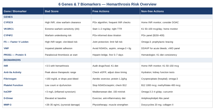
7 Biomarkers to Track for Anticoagulant-Related Hemarthrosis
Biomarkers for anticoagulant-related hemarthrosis fall into two overlapping categories: those that measure how anticoagulated you are, and those that measure how inflamed and vulnerable your joints are. Both matter. A person with a perfect INR can still bleed repeatedly if their synovium is chronically inflamed. Conversely, a person with pristine joint tissue can bleed catastrophically if their anticoagulation swings out of range. The seven markers below span both categories.
Biomarker 1 — INR (International Normalized Ratio)
Why it matters
The INR is the oldest and most universally tracked marker for patients on vitamin K antagonists such as warfarin or acenocoumarol. It standardizes prothrombin time across different laboratory reagents, allowing clinicians worldwide to compare results. For most indications, the therapeutic target is 2.0–3.0. For mechanical heart valves, it rises to 2.5–3.5. Every point above 3.0 roughly doubles the risk of a major bleeding event, including hemarthrosis, without proportionally increasing thrombotic protection. Time in therapeutic range (TTR) — the percentage of readings within target — is arguably more predictive of bleeding outcomes than any single INR value. Patients with a TTR below 65% have substantially higher rates of both bleeding and clotting events.
How to measure it
INR is measured on a venous blood draw (PT/INR panel) and costs $15–40 at most clinical labs. Home point-of-care monitors such as the CoaguChek XS or InRatio2 run $100–200 for the device and around $3–8 per test strip. Multiple randomized trials have shown that patient self-monitoring with a home device achieves TTR comparable to or better than clinic-based monitoring (published evidence includes the HAMLET and THINRS trials, both documented in the peer-reviewed literature).
If the score is bad — the plan without supplements
If your INR is persistently above 3.5 and you are experiencing hemarthrosis, the immediate priorities are: confirm the reading with a venous draw rather than a finger-stick, audit every food and drug interaction in the preceding week (antibiotics, antifungals, and NSAIDs are common culprits), and contact your anticoagulation clinic for a dose reduction. Increase the frequency of monitoring to every 3–5 days until stability is re-established. A dietary audit targeting vitamin K intake consistency — not elimination — is often more stabilizing than dose changes. Leafy greens consumed in consistent rather than variable amounts flatten INR swings significantly.
If the score is bad — the plan with supplements or equipment
For INR instability linked to vitamin K insufficiency or dietary inconsistency, low-dose vitamin K1 supplementation (50–150 mcg daily) has been shown in multiple small trials to reduce INR variability without pulling the INR below therapeutic range. The mechanism is straightforward: a small daily baseline flattens the effect of dietary fluctuations. Do not use K2 MK-4 or MK-7 at higher doses without specialist guidance, as MK-7 has a longer half-life and can shift INR unpredictably. Home INR monitoring (CoaguChek or equivalent) is the single most impactful equipment intervention, reducing both out-of-range events and emergency visits in controlled trials.
Biomarker 2 — Anti-Xa Activity
Why it matters
For patients on direct oral anticoagulants (DOACs) — rivaroxaban, apixaban, edoxaban — or low-molecular-weight heparin (LMWH), INR is not the relevant measurement. Anti-Xa activity directly measures the inhibitory effect on Factor Xa, the final common step before thrombin generation. Standard DOACs do not require routine monitoring in most patients, but people with hemarthrosis events "on-therapy" are precisely the population where knowing the actual drug level matters. High peak Anti-Xa levels correlate with bleeding risk; low trough levels may indicate rapid clearance through renal excretion, especially relevant in patients with fluctuating creatinine.
How to measure it
Anti-Xa is a chromogenic assay on a venous blood draw, costing $40–100 depending on the lab and payer. Timing matters: draw at peak (2–4 hours post-dose for rivaroxaban and apixaban) or trough (just before the next dose) and always specify the drug being used so the lab applies the correct calibration curve. This test must be explicitly ordered — it is not included in standard coagulation panels.
If the score is bad — the plan without supplements
A peak Anti-Xa dramatically above expected range (e.g., rivaroxaban peak above 341 ng/mL or apixaban peak above 321 ng/mL) while experiencing hemarthrosis usually points to one of three causes: renal clearance reduction, drug-drug interaction, or a dosing error (taking double doses unknowingly). Kidney function review (eGFR, creatinine) is the priority. Many DOACs require dose reduction when eGFR drops below 30–50 mL/min. Timing the dose with or without food also affects peak levels — rivaroxaban bioavailability drops significantly when taken without food.
If the score is bad — the plan with supplements or equipment
No supplement improves high Anti-Xa levels — this is a pharmacokinetic issue, not a nutritional deficiency. However, kidney-supportive habits (adequate hydration, avoiding nephrotoxic NSAIDs, managing blood pressure) protect renal clearance and stabilize DOAC levels over time. If hemarthrosis is recurrent despite appropriate Anti-Xa levels, the conversation shifts from anticoagulation adjustment to joint-specific inflammation management, which the CRP and MMP-3 biomarkers below address.
Biomarker 3 — Fibrinogen
Why it matters
Fibrinogen is the soluble precursor to fibrin, the protein mesh that physically seals a wound. It is both a clotting factor and an acute-phase protein — it rises with inflammation and falls when consumed during a bleeding event. In anticoagulated patients with hemarthrosis, fibrinogen levels are relevant in two scenarios: first, when baseline fibrinogen is constitutionally low (hypofibrinogenemia), the clot that forms in a joint bleed is structurally weak and the bleed may not resolve without intervention; second, after a large hemarthrosis episode, fibrinogen can drop transiently, making subsequent bleeds in the following 24–72 hours more dangerous. Levels below 150–200 mg/dL represent functional impairment in most clinical contexts.
How to measure it
Fibrinogen is measured by the Clauss method on a citrated blood sample, typically included in a comprehensive coagulation panel. Cost ranges $20–60. Testing is most informative within a few hours of a significant hemarthrosis event, or as a baseline when recurrent joint bleeds occur at seemingly normal anticoagulation levels.
If the score is bad — the plan without supplements
Constitutionally low fibrinogen in the context of anticoagulant-related hemarthrosis is a serious signal. Inform your hematologist before any procedure, and carry documentation for emergency settings. If fibrinogen drops acutely after a large joint bleed, cryoprecipitate or fibrinogen concentrate may be necessary in a hospital setting. For outpatient management, regular aerobic exercise at moderate intensity has consistently elevated fibrinogen in population studies — this applies to the low-fibrinogen context, not the high one (chronically elevated fibrinogen above 400 mg/dL is independently associated with cardiovascular risk).
If the score is bad — the plan with supplements or equipment
There is no well-validated supplement to raise pathologically low fibrinogen safely in anticoagulated patients. Protein adequacy matters — fibrinogen is a protein, and severe dietary protein restriction impairs synthesis. A dietary protein target of 1.2–1.6 g per kg of body weight per day, consistent with current sports medicine and longevity-focused guidelines, supports hepatic fibrinogen production. Omega-3 fatty acids at doses above 3 g EPA+DHA daily have fibrinogen-lowering effects in people with elevated levels, which is relevant for the opposite problem (high fibrinogen driving inflammation and thrombosis risk).
Biomarker 4 — Platelet Count and Function
Why it matters
Platelets form the primary plug at the site of vascular injury before the coagulation cascade reinforces it with fibrin. In anticoagulant-related hemarthrosis, platelet issues compound bleeding risk in two ways: thrombocytopenia (low count, under 100,000/μL) physically reduces the density of the primary plug, and platelet dysfunction — even at normal counts — means the plug that forms is fragile. Heparin-induced thrombocytopenia (HIT) is a specific, serious complication of heparin-based anticoagulants that can paradoxically cause both clotting and bleeding. Concurrent aspirin or NSAID use in an anticoagulated patient significantly impairs platelet function and is one of the most common reversible causes of unexplained hemarthrosis.
How to measure it
Platelet count is part of any standard complete blood count (CBC), costing $15–30. Platelet function requires specific assays: the PFA-100 (platelet function analyzer) costs $50–150 and measures closure time under shear stress. VerifyNow is used to assess response to aspirin or P2Y12 inhibitors in specialized settings ($100–200). If HIT is suspected, a PF4/heparin antibody assay (immunoassay) followed by a functional serotonin release assay is required — cost $100–400 depending on setting.
If the score is bad — the plan without supplements
If platelet count is low, the first step is identifying the cause: drug-induced (HIT, chemotherapy, some antibiotics), immune thrombocytopenic purpura (ITP), liver disease, or B12/folate deficiency. Remove the offending agent where possible. If aspirin or an NSAID is contributing to dysfunction, discuss with your cardiologist whether the aspirin can be safely discontinued — in many contemporary anticoagulated patients without recent ACS, aspirin adds bleeding risk without meaningful additional thrombotic protection.
If the score is bad — the plan with supplements or equipment
Vitamin B12 and folate deficiency are overlooked causes of both thrombocytopenia and platelet dysfunction. Testing serum B12, methylmalonic acid, and red blood cell folate before assuming a platelet problem is intrinsic is worthwhile. Supplementation is inexpensive and low-risk: methylcobalamin 1000 mcg daily for B12 (particularly important in patients on long-term metformin or proton pump inhibitors, which deplete B12); methylfolate 400–800 mcg daily for folate. Cycling is not required — these are water-soluble vitamins. High-dose omega-3 supplementation (above 3g/day) moderately inhibits platelet aggregation and should be disclosed to your prescriber in the context of anticoagulant use.
Biomarker 5 — High-Sensitivity CRP (hsCRP)
Why it matters
High-sensitivity CRP measures systemic and joint inflammation at clinically significant low concentrations. Its relevance to anticoagulant-related hemarthrosis is often underappreciated: inflamed synovial tissue is hyper-vascular, with fragile new blood vessel growth (angiogenesis) that bleeds more easily than mature vasculature. Patients with inflammatory joint conditions — rheumatoid arthritis, psoriatic arthritis, or even osteoarthritis with significant synovitis — who are placed on anticoagulation face a compounded risk because their joints were already prone to microbleeding before the drug was added. Chronically elevated hsCRP (above 3 mg/L) is a marker of this ongoing synovial vulnerability and predicts both initial hemarthrosis events and delayed resolution.
How to measure it
hsCRP is measured on a standard blood draw, costing $20–50. It should be measured at baseline before starting anticoagulation in patients with known inflammatory joint disease, then serially when hemarthrosis episodes cluster. Interpret in the context of infection — any acute illness will transiently spike CRP regardless of joint status.
If the score is bad — the plan without supplements
Reducing synovial inflammation is the structural answer. For patients with identifiable inflammatory arthritis, optimizing disease-modifying therapy (DMARDs for RA, for example) reduces joint fragility. For metabolic-pattern inflammation (elevated BMI, sedentary lifestyle, high glycemic diet), the evidence for lifestyle modification reducing hsCRP is robust. A Mediterranean-pattern diet consistently reduces hsCRP by 20–35% in randomized controlled trials. Moderate-intensity aerobic exercise — 150 minutes per week — has independent CRP-lowering effects across multiple meta-analyses. Both interventions have additive effects on joint tissue quality.
If the score is bad — the plan with supplements or equipment
Omega-3 fatty acids (EPA+DHA, 2–4 g/day) have the most consistent human evidence for CRP reduction among supplements, with effects appearing after 6–8 weeks of use. Important note: at doses above 3 g/day, platelet inhibition becomes relevant in anticoagulated patients — disclose this to your prescriber. Curcumin with piperine (500–1000 mg of standardized curcuminoids daily, with 5 mg piperine for absorption) shows CRP reductions in osteoarthritis and general inflammatory conditions across several small RCTs; the effect size is modest but the safety profile in anticoagulated patients appears acceptable at these doses, though curcumin has mild anticoagulant properties that warrant monitoring. Cycle omega-3 supplementation: 12 weeks on, 4 weeks off if high-dose, with INR or Anti-Xa checks at week 4 of each cycle.
Biomarker 6 — D-Dimer
Why it matters
D-dimer is a fibrin degradation product released when a clot is broken down. In anticoagulant-related hemarthrosis, its utility is two-directional. Elevated D-dimer following a joint bleed confirms that active fibrinolysis is occurring — the body is dissolving the clot in the joint — and persistently elevated levels suggest the process is incomplete or ongoing. On the other side, recurrent unexplained hemarthrosis episodes in a patient with elevated baseline D-dimer may indicate a prothrombotic state that is generating simultaneous microthrombi and reactive fibrinolysis, consuming clotting resources and making the joint vasculature unstable. This is not rare in patients with cancer-associated coagulopathy or antiphospholipid syndrome on anticoagulation.
How to measure it
D-dimer is measured on a venous sample, costing $30–80. In the context of hemarthrosis monitoring, it is most useful drawn at baseline (stable state, no acute bleed), then compared against a value taken during or shortly after an episode. An isolated high D-dimer without clinical context is not actionable — it must be interpreted alongside clinical findings and the timing of the bleed.
If the score is bad — the plan without supplements
Persistently elevated D-dimer outside an acute bleed episode should prompt a workup for underlying prothrombotic states: antiphospholipid antibodies, malignancy screen, inherited thrombophilias. If antiphospholipid syndrome is confirmed, the anticoagulation strategy itself may need revision — DOACs are generally not preferred in triple-positive antiphospholipid syndrome, and warfarin with tight INR control may be more appropriate. This changes the entire biomarker monitoring picture.
If the score is bad — the plan with supplements or equipment
No supplement directly reduces D-dimer. Addressing root causes (inflammation, metabolic syndrome, sedentary behavior) that drive ongoing low-grade fibrin deposition and dissolution is the practical approach. The omega-3, exercise, and dietary strategies that lower CRP also reduce the background fibrin turnover rate over time, with D-dimer following downward at 8–12 weeks. Compression garments on affected joints during the recovery phase after hemarthrosis may reduce local fibrin instability, though direct D-dimer impact has not been studied in this specific population.
Biomarker 7 — MMP-3 (Matrix Metalloproteinase-3)
Why it matters
MMP-3, or stromelysin-1, is an enzyme produced by synovial fibroblasts and macrophages in response to joint inflammation. It degrades collagen, proteoglycans, and the extracellular matrix of synovial blood vessel walls, making them structurally weaker. In patients with recurrent hemarthrosis — particularly those with hemophilia, where the synovial destruction cascade is well-characterized — elevated MMP-3 is one of the strongest predictors of irreversible joint damage and chronic synovitis. For anticoagulated patients who bleed repeatedly into the same joint, the same MMP-3-driven spiral can begin: each bleed triggers more synovial inflammation, which elevates MMP-3, which weakens the vasculature, which makes the next bleed more likely. This is the mechanism behind target joint formation.
How to measure it
MMP-3 is measured from a serum sample. It is not part of standard coagulation panels and must be specifically requested, typically through a rheumatology or specialized coagulation laboratory. Cost ranges $80–200. Normal serum levels are generally below 28–35 ng/mL in most assays, but reference ranges vary by laboratory. Synovial fluid MMP-3 is even more informative about local joint pathology but requires arthrocentesis.
If the score is bad — the plan without supplements
Elevated MMP-3 in the context of anticoagulant-related hemarthrosis is a strong signal to pursue joint-specific anti-inflammatory management aggressively, not just to adjust anticoagulation. Intra-articular corticosteroid injections (under the guidance of a rheumatologist, with appropriate pause in anticoagulation if needed) reduce synovial inflammation and can break the MMP-3-driven spiral. Radiosynovectomy — injection of a radioisotope into the joint to ablate the inflamed synovium — is used in hemophilia-related target joint disease and may be relevant in refractory anticoagulant-related cases. Physical therapy focused on muscle strengthening around the joint (quadriceps for knee, rotator cuff for shoulder) reduces mechanical stress on the synovium and downstream MMP-3 production.
If the score is bad — the plan with supplements or equipment
Doxycycline at sub-antimicrobial doses (20 mg twice daily, marketed as Periostat) is an MMP inhibitor with human evidence in periodontal disease and early osteoarthritis contexts — it requires a prescription and physician oversight, but is worth discussing in cases of recurrent target joint formation. Among supplements, type II collagen (undenatured, 40 mcg daily — not the high-dose hydrolyzed form) has shown reductions in joint pain and inflammatory markers including MMP-3 in several small OA trials. Frequency: daily, with effects appearing at 12 weeks. Side effects are minimal. Boswellic acids (AKBA fraction, 100–200 mg daily) inhibit leukotriene synthesis and have plausible MMP-3 modulating effects, though direct MMP-3 measurement in anticoagulant-related hemarthrosis studies does not yet exist.
Moving from what can be measured in blood to what is encoded in your DNA, the following section addresses the genetic layer that determines much of the variation in how anticoagulants behave — and bleed — from one person to the next.
6 Key Genes Behind Anticoagulant Sensitivity and Hemarthrosis Risk
Pharmacogenomics has transformed how we understand warfarin dosing, and the field is extending to DOACs and bleeding risk prediction. The six variants below explain a substantial portion of why some individuals are dramatically more sensitive to anticoagulants — and therefore at higher risk of hemarthrosis — than population averages suggest.
Gene 1 — CYP2C9: The Warfarin Metabolism Engine
CYP2C9 encodes the primary hepatic enzyme that metabolizes S-warfarin, the more potent enantiomer. The *2 variant (rs1799853, amino acid change R144C) reduces enzyme activity by roughly 30%; the *3 variant (rs1057910, I359L) reduces it by 80–90%. Additional rare variants (*5, *6, *8, *11) contribute further risk in populations of African ancestry where *2 and *3 testing alone can miss significant metabolizer status. Poor metabolizers (those carrying two reduced-function alleles) require dramatically lower warfarin doses — sometimes 30–50% of the standard starting dose — to reach therapeutic INR. The consequence of missing this: supratherapeutic anticoagulation in early therapy, precisely when hemarthrosis risk is highest.
If the gene is bad — the plan without supplements
If pharmacogenomic testing reveals CYP2C9 *2/*2, *2/*3, or *3/*3 status, the clinical action is straightforward: request that your physician use a pharmacogenomics-informed warfarin dosing algorithm (the IWPC algorithm or the EU-PACT algorithm, both published in major peer-reviewed journals) rather than standard empirical initiation. Start INR monitoring at day 3 rather than day 5. Consider more frequent monitoring (every 3 days) for the first month rather than weekly. A TTR target above 75% becomes even more important because oscillation in a slow metabolizer carries larger absolute INR swings.
If the score is bad — the plan with supplements or equipment
Home INR monitoring is especially high-value for CYP2C9 poor metabolizers because INR can shift rapidly with dietary changes or drug interactions that affect the already-reduced metabolic capacity. The CoaguChek XS or similar device (cost $100–200) allows daily or every-other-day readings during dietary changes or illness, catching supratherapeutic INR before a bleed occurs. For CYP2C9 poor metabolizers, a DOAC rather than warfarin may be a simpler long-term solution, since DOACs do not depend on CYP2C9 metabolism for their anticoagulant effect — this is a genuine pharmacogenomic rationale for drug class selection worth raising with your prescriber.
Gene 2 — VKORC1: The Warfarin Sensitivity Switch
VKORC1 encodes vitamin K epoxide reductase complex subunit 1, the direct target of warfarin. The -1639G>A variant (rs9923231) reduces VKORC1 expression, meaning less enzyme is present to recycle vitamin K. People with the AA genotype (approximately 30% of Europeans, higher in East Asians) need 30–50% less warfarin than GG carriers to achieve the same INR. Together, CYP2C9 and VKORC1 variants explain over 50% of the population variance in warfarin dose requirements — a fact embedded in the FDA-approved label revisions for warfarin since 2010.
If the gene is bad — the plan without supplements
VKORC1 AA status in combination with CYP2C9 reduced function is the highest-risk pharmacogenomic profile for supratherapeutic anticoagulation. If you have both, your initial therapeutic dose may be as low as 1–2 mg/day rather than the 5 mg/day population average. This information, properly communicated to prescribers, prevents the classic pattern of hospitalization in week one or two of warfarin therapy due to unexpected INR elevation and joint or soft-tissue bleeding.
If the score is bad — the plan with supplements or equipment
Vitamin K1 consistency supplementation (50–100 mcg daily), which stabilizes INR by providing a consistent substrate baseline, is particularly valuable in VKORC1 AA carriers because their buffering capacity for dietary vitamin K fluctuation is reduced. The mechanism: with less VKORC1 enzyme, a dietary vitamin K spike or deficit causes larger INR swings. A small, consistent daily supplement blunts these fluctuations without pulling INR out of therapeutic range — an effect supported by small RCTs in unstable warfarin patients.
Gene 3 — CYP4F2: The Dose Modifier
CYP4F2 encodes an enzyme that oxidizes and inactivates vitamin K1 in the liver. The V433M variant (rs2108622) reduces this inactivation, meaning more vitamin K1 accumulates and a higher warfarin dose is needed to overcome it. CYP4F2 *3 carriers therefore require more warfarin than average — the opposite pharmacogenomic pressure from CYP2C9 poor metabolizers. Alone, the effect is modest (approximately 5–7% dose increase), but in combination with VKORC1 GG status (low sensitivity to warfarin), CYP4F2 *3 can push required doses toward the upper end of typical ranges.
If the gene is bad — the plan without supplements
For CYP4F2 *3 carriers who are also VKORC1 GG, underdosing is the clinical risk — and undertreated anticoagulation is associated with therapeutic failure, not hemarthrosis. The hemarthrosis relevance is indirect: when prescribers finally optimize dosing upward for these individuals, titrating carefully with frequent monitoring avoids overshooting. Being aware of the CYP4F2 *3 status prevents prolonged subtherapeutic anticoagulation followed by aggressive dose escalation that creates INR instability.
If the score is bad — the plan with supplements or equipment
No specific supplement modifies CYP4F2 activity meaningfully. The clinical tool is pharmacogenomic-informed dosing. Testing (Genoptix, Genomind, or equivalent pharmacogenomics panel) costs $150–400 and is increasingly covered by insurance when ordered for anticoagulation management.
Gene 4 — F5 (Factor V Leiden, R506Q)
Factor V Leiden is the most common inherited thrombophilia in populations of European ancestry, carried by approximately 5% as heterozygotes and 0.02–0.06% as homozygotes. The R506Q variant makes Factor V resistant to inactivation by activated Protein C, creating a prothrombotic state. The reason it is relevant to anticoagulant-related hemarthrosis is counterintuitive: many patients on anticoagulation are there because they have Factor V Leiden, and the management tension is different. Their underlying thrombotic drive means that anticoagulation levels that are appropriate for average patients may leave them inadequately protected, prompting prescribers toward higher target INRs — which inherently increases hemorrhagic risk including hemarthrosis. Homozygous carriers are at particularly high risk of this tension.
If the gene is bad — the plan without supplements
The key clinical action is knowing your Factor V Leiden status when recurrent thrombosis or hemarthrosis occurs, since the therapeutic target may need individualization. Homozygous carriers with recurrent venous thromboembolism are typically maintained on indefinite anticoagulation with a higher target range. In this context, the joint-protective biomarkers — MMP-3, hsCRP, synovial inflammation management — become even more important because the anticoagulant dose cannot simply be reduced to prevent bleeding.
If the score is bad — the plan with supplements or equipment
For Factor V Leiden carriers who experience recurrent hemarthrosis, joint protection strategies take priority alongside anticoagulation optimization. This includes prophylactic bracing for high-risk joints, avoiding contact sports and high-fall-risk activities, and addressing modifiable inflammatory biomarkers (hsCRP, MMP-3) as described above. No supplement "corrects" the Factor V Leiden mutation — it is a permanent genetic variant — but reducing the background prothrombotic milieu through omega-3 fatty acids, exercise, and smoking cessation reduces the overall hemostatic burden.
Gene 5 — VWF Gene Variants (Von Willebrand Factor)
The VWF gene encodes von Willebrand factor, a multimeric protein that anchors platelets to damaged vessel walls and serves as a carrier for Factor VIII in the circulation. Variants that reduce VWF quantity or function — collectively called von Willebrand disease (vWD) — range from mild (Type 1, present in 1–2% of the population as a bleeding tendency) to severe (Type 3). In anticoagulated patients, even mild, undiagnosed Type 1 vWD can transform what would be a minor capsular bleed into a significant hemarthrosis, because the first line of vascular plugging (platelet adhesion via VWF) is impaired before the anticoagulant even acts.
If the gene is bad — the plan without supplements
Undiagnosed vWD in an anticoagulated patient is one of the more serious and correctable situations in this category. Testing involves VWF antigen, VWF activity (ristocetin cofactor), and Factor VIII activity — ordered as a vWD panel, costing $80–200. If Type 1 vWD is identified, the anticoagulant dose and target INR may need reassessment. Desmopressin (DDAVP) is the first-line treatment for Type 1 vWD bleeding episodes — it releases stored VWF from endothelial cells and is used in the acute hemarthrosis setting with specialist guidance.
If the score is bad — the plan with supplements or equipment
Avoiding agents that further impair VWF-mediated platelet function is the practical daily action: NSAIDs, aspirin, and high-dose omega-3 (above 3g/day) all inhibit platelet-VWF interaction. For mild Type 1 vWD, some evidence supports that moderate aerobic exercise acutely raises VWF antigen levels by 30–60% transiently — this is physiologically relevant before activities that carry fall or trauma risk. No over-the-counter supplement reliably raises VWF activity.
Gene 6 — PROS1 (Protein S)
Protein S, encoded by PROS1, is a natural anticoagulant co-factor for activated Protein C. Hereditary Protein S deficiency increases thrombotic risk and is found in about 1% of the population. Anticoagulated patients with PROS1 loss-of-function variants face a complex situation: their deficiency increases the VTE risk that originally indicated anticoagulation, but the mutations can also be associated with abnormal vascular biology that makes joint microvessels less resilient. Additionally, warfarin itself temporarily lowers Protein S levels early in therapy (through its vitamin K antagonism on Protein S synthesis), which can paradoxically worsen the thrombotic state in the first days of therapy — a phenomenon clinically recognized as warfarin-induced skin necrosis in severe cases, but also potentially manifesting as joint microthrombosis with reactive hemorrhage.
If the gene is bad — the plan without supplements
If Protein S deficiency is documented, warfarin initiation should always be covered by a parenteral anticoagulant (heparin or LMWH) for the first 5–7 days until stable anticoagulation is established. This standard-of-care intervention is specifically to prevent the paradoxical early prothrombotic state. For long-term management, the anticoagulant choice (warfarin vs. DOAC) should factor in the deficiency, and a hematologist familiar with inherited thrombophilias is the appropriate specialist.
If the score is bad — the plan with supplements or equipment
Protein S is a vitamin K-dependent protein, which means vitamin K availability influences its synthesis. Unlike the procoagulant factors that warfarin suppresses to therapeutic effect, Protein S suppression in warfarin therapy is a side effect. Consistent vitamin K intake (not high-dose supplementation, which counteracts warfarin's therapeutic effect) supports baseline Protein S levels. This reinforces the rationale for dietary vitamin K consistency rather than restriction in warfarin patients.
Tracking the Right Labs: What Malcolm Kendrick's "The Clot Thickens" Reveals About Hemostasis
The Clot Thickens: The Enduring Mystery of Heart Disease (2021) by Dr. Malcolm Kendrick, a Scottish physician and independent researcher, is one of the more intellectually challenging books in recent cardiovascular medicine. Kendrick argues, with extensive citation, that the dominant focus on cholesterol and arterial plaques has obscured the central role of blood clotting and thrombus formation in cardiovascular disease initiation and progression. His thesis — that cardiovascular disease is fundamentally a disease of the clotting system, not of lipid accumulation — has direct relevance for anyone managing anticoagulation and its bleeding consequences.
10 Key Ideas From The Clot Thickens That Change How You Think About Joint Bleeding
1. The endothelium is the primary protection
Kendrick argues that a healthy, intact endothelial lining is the primary defense against both arterial clotting and inappropriate bleeding. Damaged endothelium — from smoking, chronic stress, elevated blood glucose, or physical trauma — is the root trigger of the clotting cascade, not simply a passive surface. For joint bleeds, this reframes the conversation: synovial vascular endothelial health is the upstream target, not just the anticoagulant dose.
2. Repeated thrombosis and lysis cycles build up damage
The book describes how microthrombi form, are dissolved, and leave behind organized scar tissue. In the joint, this is the mechanism by which recurrent hemarthrosis events build toward chronic synovitis and cartilage erosion. Each resolved bleed is not a clean slate — it leaves behind fibrin residue and inflammatory mediators that prime the next event.
3. Psychological stress directly activates the clotting cascade
Kendrick documents the pathway from HPA axis activation (stress response) to fibrinogen elevation, von Willebrand factor release, and platelet hyperreactivity. This is not metaphorical — it is a direct, measurable physiological connection. For anticoagulated patients, acute psychological stress can transiently shift hemostatic balance.
4. Fibrinogen is a better cardiovascular risk marker than LDL
The book presents substantial evidence that fibrinogen is more consistently predictive of adverse cardiovascular events than LDL cholesterol. For readers of this article, this reinforces why fibrinogen belongs in a hemarthrosis risk panel, not just a cardiac one.
5. Vitamin K2 is undervalued in vascular biology
Kendrick highlights vitamin K2's role in activating matrix GLA protein, which prevents vascular calcification. Soft-tissue and vascular calcification increases vessel fragility — mechanistically relevant to why chronically vitamin K2-insufficient joint vasculature may be more prone to bleeding under mechanical stress.
6. Sunlight and nitric oxide protect the endothelium
UV exposure releases nitric oxide from skin stores, which vasodilates arteries and reduces platelet aggregation. Kendrick cites geographic and seasonal data showing that reduced sun exposure correlates with increased cardiovascular and clotting events. The practical implication: outdoor physical activity (beyond its fitness benefits) may have endothelium-protective effects via the nitric oxide pathway.
7. Statins' benefits may be partly anti-thrombotic, not just lipid-lowering
The book documents that statins reduce fibrinogen, CRP, and Von Willebrand factor independently of their cholesterol effects. This challenges the assumption that lipid-lowering is the primary mechanism of benefit — and raises the question of whether anti-inflammatory, anti-thrombotic effects are the actual drivers. For patients simultaneously on statins and anticoagulants, understanding this interaction matters for bleeding risk assessment.
8. Diet affects clotting through multiple pathways
Ultra-processed foods elevate CRP, fibrinogen, and platelet reactivity — all measurable in the biomarker panel above. Kendrick draws the line from dietary pattern to hemostatic function more explicitly than most cardiovascular textbooks, using population studies across multiple countries.
9. Exercise lowers clotting risk acutely and chronically
Physical activity increases tPA (tissue plasminogen activator, the body's natural clot-dissolver), reduces fibrinogen over time, and improves endothelial function. The acute exercise-induced increase in VWF is transient and balanced by the longer-term reduction in clotting activation — a net protective effect in most contexts.
10. The medical focus on single-factor solutions misses the system
Kendrick's central message — that hemostasis is a system of interacting signals, not a single pathway — is directly applicable to anticoagulant-related hemarthrosis management. Adjusting only the anticoagulant dose while ignoring fibrinogen, platelet function, CRP, MMP-3, and genetic metabolizer status is analogous to fixing one instrument in a poorly tuned orchestra.
Complementary Approaches for Joint Health on Anticoagulant Therapy
The following modalities have meaningful human evidence relevant to joint bleeding risk, inflammation, or pain management in the context of anticoagulant therapy. Each is presented with a specific protocol, a supporting study or clinical guideline, and realistic application guidance.
Tai Chi — Fall Prevention and Joint Stability
Tai chi is a slow, controlled movement practice originating in Chinese martial arts that trains balance, coordination, proprioception, and lower limb strength simultaneously. For anticoagulated patients, its primary relevance is fall prevention: the leading trigger of acute hemarthrosis in patients on anticoagulation is not spontaneous bleeding but trauma from minor falls that, in a healthy person, would cause only a bruise. Stronger proprioception and neuromuscular control dramatically reduce fall frequency, thereby reducing the mechanical trigger for joint bleeds.
A landmark randomized controlled trial published in the Journal of the American Geriatrics Society demonstrated that 15 weeks of Tai Chi reduced fall rates by 47.5% in community-dwelling older adults compared to a balance training control group. A Cochrane review of fall prevention interventions (Campbell and Robertson, 2007, Cochrane Database) confirmed Tai Chi among the most effective single modalities. These effects are documented in populations that overlap heavily with anticoagulated patients: older adults, those with cardiovascular conditions, and those on polypharmacy.
Practical application: begin with a group Tai Chi class (Yang short form, 24 movements) twice weekly for at least 12 weeks before reducing to once weekly maintenance. Chair-modified versions exist for patients with existing joint damage from prior hemarthrosis. A home practice of 20–30 minutes three times weekly sustains the proprioceptive benefit. The only caution: transitions from floor-level positions should be performed slowly to avoid orthostatic changes that increase fall risk in patients on antihypertensives.
Low-Level Laser Therapy (LLLT / Photobiomodulation) — Synovial Inflammation Reduction
Low-level laser therapy, also called photobiomodulation, applies near-infrared or red-light wavelengths (typically 630–1064 nm) at non-thermal doses to target tissue. In joint biology, the proposed mechanism is stimulation of mitochondrial cytochrome c oxidase in synovial cells, increasing ATP production, reducing oxidative stress, and downregulating inflammatory cytokines including IL-1β and TNF-α. This is directly relevant to the MMP-3-driven synovial inflammation cycle that amplifies recurrent hemarthrosis.
A systematic review and meta-analysis by Bjordal et al., published in Physiotherapy (2003), examined LLLT for rheumatoid arthritis and knee osteoarthritis, finding statistically significant reductions in pain, morning stiffness, and joint tenderness compared to sham, with an effect size similar to NSAIDs — without NSAID-associated gastrointestinal or platelet effects. A subsequent review by Huang et al. (2015) in Lasers in Medical Science confirmed these findings across musculoskeletal conditions. Crucially, LLLT does not interact with anticoagulation and carries no bleeding risk, making it particularly appropriate in this population.
Protocol for application: devices used in clinical physiotherapy deliver 4–8 J/cm² per session over affected joints, 3 times per week for 4–6 weeks. Class III B lasers (650–905 nm, 5–500 mW) are the evidence-supported category. Home devices in the 630–850 nm range (such as clinical-grade near-infrared panels) are available at $200–600 and can be used on affected joints for 10–20 minutes daily. In patients with active hemarthrosis, LLLT should be applied after the acute bleed has stabilized (72+ hours post-onset) rather than during active bleeding, where any increase in local blood flow is undesirable.
Mindfulness Meditation and MBSR — Chronic Pain and Systemic Inflammation
Mindfulness-Based Stress Reduction (MBSR), developed by Jon Kabat-Zinn, is an 8-week structured program combining body scan meditation, mindful movement, and sitting meditation. Its relevance to anticoagulant-related hemarthrosis operates on two levels: first, it reduces psychological stress — and as Kendrick's work and the HPA-axis literature both document, stress directly elevates fibrinogen, CRP, and platelet reactivity; second, MBSR has demonstrated direct reductions in hsCRP in randomized trials, including a well-cited trial by Creswell et al. (2012, Brain, Behavior, and Immunity) showing significant reductions in CRP and IL-6 in chronically stressed adults after 8 weeks of MBSR.
A 2016 meta-analysis by Creswell and Lindsay in Current Directions in Psychological Science confirmed that mindfulness interventions reduce inflammatory biomarkers including CRP across multiple populations, with effects persisting at 3-month follow-up. For patients managing chronic joint pain from recurrent hemarthrosis, the pain catastrophizing reduction component of MBSR is additionally important — catastrophizing increases pain perception and reduces adherence to protective movement strategies.
Application protocol: the standard 8-week MBSR course (available through hospital wellness programs, community mental health centers, or online platforms) includes one 2.5-hour session per week and 45 minutes of daily home practice. For patients who cannot commit to this format, 10–20 minutes daily of guided mindfulness meditation using apps such as Insight Timer (free) or Waking Up has shown measurable CRP reductions in 8-week adherence studies. Frequency: daily. Side effects: none known at recommended dose. There is no drug interaction with anticoagulants.
Biofeedback — Pain Regulation and Autonomic Stabilization
Biofeedback uses real-time physiological data — heart rate variability (HRV), skin conductance, muscle tension, or skin temperature — to train individuals to consciously regulate autonomic nervous system responses. For anticoagulated patients with recurrent hemarthrosis, its relevance is primarily in chronic pain management (the pain that persists between acute bleeds due to synovial damage and sensitization) and secondarily in autonomic regulation — normalizing HRV reduces the sympathetic overdrive that contributes to platelet hyperreactivity and inflammatory cytokine release.
A Cochrane review by Greenhalgh et al. on biofeedback for chronic pain found moderate evidence supporting reduction in pain intensity and analgesic use in musculoskeletal conditions. In the hemophilia literature (which models the chronic joint pain experienced in severe recurrent hemarthrosis), biofeedback-assisted relaxation has shown statistically significant reductions in pain scores and improvements in joint function in small controlled studies. The most relevant published evidence comes from rheumatology populations where biofeedback for fibromyalgia and inflammatory arthritis pain has been studied in controlled trials.
For practical application, HRV biofeedback using a Polar H10 chest strap with the Elite HRV or Welltory app ($50–100 for hardware) is accessible without a clinical setting. Protocol: 20 minutes of HRV biofeedback training (resonance breathing at 5–6 breaths/minute, guided by real-time HRV feedback) daily for 8 weeks. Afterward, maintain with 3–4 sessions per week. Neurofeedback (EEG-based) requires a trained clinician and costs $75–150 per session. For most patients, HRV biofeedback provides an accessible starting point with meaningful autonomic and inflammatory downstream effects.
Conclusion
Anticoagulant-related hemarthrosis is not random. It emerges from the intersection of drug metabolism shaped by CYP2C9 and VKORC1 variants, joint inflammation tracked by hsCRP and MMP-3, coagulation factor availability measured through fibrinogen and platelet function, and drug exposure quantified by INR and Anti-Xa activity. When all of these are monitored and understood together — rather than in isolation — the picture becomes actionable rather than overwhelming.
The complementary approaches in this article — Tai Chi to reduce fall-triggered bleeds, LLLT to quiet synovial inflammation, MBSR to reduce stress-driven coagulation activation, and biofeedback to stabilize chronic pain — are not alternatives to medical anticoagulation management. They are the layer of precision that works alongside it.
The next smart step is concrete: review which of the seven biomarkers you have never had tested, identify the gap, and bring this list to your next appointment with your hematologist, rheumatologist, or anticoagulation clinic. Ask about pharmacogenomic testing if you are on warfarin and your INR is unstable. Measure what has been unmeasured. The biology is specific — your management can be too.
Musculoskeletal: Joint Conditions
Cardiovascular: Vascular Conditions
Autoimmune: Inflammatory Conditions