This article contains AI generated content.
· UpdatedAntiphospholipid Syndrome Genes and Biomarkers: 6 Genes and 7 Biomarkers to Track
Introduction
If you or someone you care about has been diagnosed with antiphospholipid syndrome (APS), you already know how disorienting the experience can be. Clotting events, pregnancy losses, fatigue that doesn't match what tests can explain — the condition rarely follows a predictable path, and the standard response is often limited to anticoagulation and monitoring. That's necessary, but it doesn't capture the full picture of what's happening at the biological level.
APS is not simply a clotting disorder. It is an autoimmune condition in which the immune system produces antibodies that attack phospholipid-binding proteins, disrupting the delicate balance between clot formation and clot dissolution. The downstream effects touch inflammation, complement activation, endothelial function, and more. When clinicians focus only on INR targets or aspirin compliance, important drivers of disease activity often go unexamined.
That's where a deeper look at biomarkers and genetics becomes genuinely useful — not to replace medical care, but to make it more informed and more precise. Knowing which markers are elevated, which have normalized, and which genetic variants you carry can shape decisions about monitoring frequency, lifestyle modifications, and targeted interventions worth discussing with your specialist.
This article covers two complementary angles. The primary section walks through seven biomarkers that provide the most actionable information across different phases of APS — with practical detail on how to measure them, what results mean, and what you can actually do if a number is off. A second section then explores the genetic side: six genes with meaningful associations to APS risk and severity, including what each one affects and how lifestyle and supplementation approaches may support better outcomes. Following those, you'll find a curated book-based strategy and a section on complementary modalities with real clinical support.
Summary
This article examines antiphospholipid syndrome through two precise lenses most patients never hear about from their doctors. First: seven key biomarkers — including three antiphospholipid antibodies, complement levels, homocysteine, hsCRP, and D-dimer — with exact measurement methods, cost ranges, and step-by-step action plans for when results are abnormal (both with and without supplements). Second: six genes — HLA-DQB1, PTPN22, IRF5, MTHFR, Factor V Leiden, and STAT4 — with explanations of what each gene does to APS risk and specific compensatory plans with dosing, cycling, and side effect notes. Beyond those two core strategies, the article includes a full summary of Amy Myers' The Autoimmune Solution and its ten most impactful insights for autoimmune conditions like APS, plus four complementary approaches — including the Autoimmune Protocol, MBSR, microbiome therapy, and breathing-based techniques — grounded in human clinical evidence. If you've felt like standard APS management was leaving key questions unanswered, this article is built specifically for that gap.
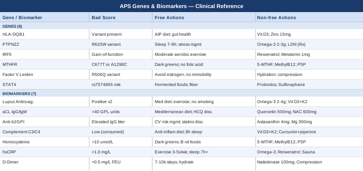
7 Biomarkers to Track If You Have Antiphospholipid Syndrome
The three classic antiphospholipid antibodies used for diagnosis are only the beginning. Once you have a confirmed APS diagnosis — or are trying to understand your risk — a broader panel of biomarkers gives you the data to monitor disease activity, cardiovascular risk, inflammation, and thrombotic pressure in real time. The following seven markers represent the most clinically meaningful and practically trackable options, informed by rheumatology, cardiology, and functional medicine perspectives including frameworks developed by Peter Attia, Thomas Dayspring, and Allan Sniderman on cardiovascular biomarker strategy.
Biomarker 1: Lupus Anticoagulant (LA)
Why it matters and what it reveals
Lupus anticoagulant is a functional antibody that paradoxically prolongs clotting time in the laboratory while dramatically increasing clotting risk in the body. Of the three main antiphospholipid antibodies, it carries the highest thrombotic risk and is the most powerful predictor of both venous and arterial events. It is also the most technically sensitive to measure — anticoagulation therapy, acute infection, and sample handling errors can all affect results.
The 2006 updated Sapporo (Sydney) criteria, authored by Miyakis et al. and widely cited in rheumatology practice, require LA positivity on at least two occasions at least 12 weeks apart to count as a classification criterion, precisely because transient positivity (often triggered by infection or medication) is common.
How to measure it
LA is detected through functional coagulation assays — most commonly the dilute Russell viper venom time (dRVVT) and a silica clotting time (SCT) or APTT-based assay. Both are required per international guidelines, and both must show a phospholipid-dependent prolongation that fails to correct on mixing with normal plasma. Cost: typically $40–$120 depending on laboratory and insurance coverage. It cannot be reliably interpreted in patients actively receiving anticoagulants, particularly direct oral anticoagulants (DOACs), and requires clinical interpretation by a specialist.
If the result is positive: the plan without supplements
Confirm with a repeat test after a minimum of 12 weeks. In the interim, focus on reducing all modifiable thrombotic risk factors: eliminate smoking completely, optimize blood pressure to below 130/80 mmHg, pursue consistent moderate aerobic exercise (150 minutes per week), maintain a healthy weight, and address sleep quality. Dietary emphasis on a Mediterranean-style anti-inflammatory pattern (olive oil, oily fish, vegetables, minimal ultra-processed food) reduces endothelial activation and platelet reactivity. Avoid prolonged immobility, including long-haul flights without compression stockings and movement breaks.
If the result is positive: the plan with supplements or equipment
In consultation with your physician, omega-3 fatty acids (EPA + DHA) at 2–4 g/day may modestly reduce platelet aggregation and endothelial inflammation. Frequency: daily with meals. Cycling: not typically required, though some practitioners take a 1-week break every 3 months to assess. Side effects: possible increase in bleeding time — important to disclose to your prescribing physician if on anticoagulation. Vitamin D3 (2,000–5,000 IU/day with K2 MK-7 at 100–200 mcg/day) supports immune regulation and may reduce autoantibody production in autoimmune conditions broadly. Side effects: hypercalcemia at very high doses; test 25-OH vitamin D before supplementing.
Biomarker 2: Anticardiolipin Antibodies (aCL IgG and IgM)
Why it matters and what it reveals
Anticardiolipin antibodies, particularly the IgG isotype at medium or high titer (>40 GPL or MPL units), are strongly associated with arterial and venous thrombosis and with pregnancy morbidity. IgM isotype positivity is more weakly associated but still clinically significant when the titer is high. Together with anti-β2GPI antibodies, aCL antibodies define the serological hallmark of APS. Higher titers correlate with greater risk; low-titer positivity, especially IgM, can be transient and non-pathological.
How to measure it
Measured by ELISA (enzyme-linked immunosorbent assay) in serum or plasma. Cost: $50–$150 per isotype. Standardization across laboratories has improved but remains imperfect, so repeat testing at the same laboratory is preferable for trend monitoring. Results must be confirmed at least 12 weeks apart.
If the titer is elevated: the plan without supplements
High-titer aCL positivity in the absence of a clotting event still warrants serious lifestyle attention. The Mediterranean diet has been studied specifically in APS and lupus patients and is associated with lower inflammatory burden and better endothelial function. Hydroxychloroquine (Plaquenil) — a prescription medication — is recommended by the European Alliance of Associations for Rheumatology (EULAR) for all patients with APS and lupus, and is worth discussing with your rheumatologist even in primary APS. It modulates autoantibody production and reduces thrombotic risk.
If the titer is elevated: the plan with supplements or equipment
Quercetin (500–1000 mg/day) has anti-inflammatory properties and may reduce oxidative stress driving endothelial activation. Frequency: daily with meals. Cycling: consider a 1-month break every 3 months. Side effects: generally well tolerated; rare headache or GI discomfort. NAC (N-acetyl cysteine, 600–1200 mg/day) supports glutathione production and reduces oxidative burden. Side effects: GI sensitivity at higher doses; avoid if asthma history without physician guidance. Wearables like continuous heart rate variability monitors (Garmin, Apple Watch, Whoop) can guide activity intensity and recovery, helping avoid the overtraining that transiently increases inflammatory and procoagulant stress.
Biomarker 3: Anti-Beta2 Glycoprotein I Antibodies (Anti-β2GPI IgG and IgM)
Why it matters and what it reveals
Anti-β2GPI antibodies are now considered the most specific of the three antiphospholipid antibodies and are especially associated with arterial thrombosis and with the triple-positive profile (LA + aCL + anti-β2GPI) — the highest-risk serological pattern in APS. Beta-2 glycoprotein I is a natural anticoagulant protein; when antibodies bind and activate it, they trigger complement activation, endothelial disruption, and platelet hyper-activation. The IgG isotype at medium-high titer has the strongest evidence base for clinical risk.
How to measure it
Measured by ELISA. Cost: $50–$150. Increasingly included in APS antibody panels alongside aCL and LA. Some advanced commercial panels also include anti-phosphatidylserine/prothrombin (aPS/PT) antibodies, which are not in the official criteria but may add risk stratification value in ambiguous cases.
If the titer is elevated: the plan without supplements
Confirm triple positivity status with your physician — this carries significantly different management implications than single-antibody positivity. If triple positive, most current guidelines recommend long-term anticoagulation after a first event. Lifestyle: strict cardiovascular risk factor management is critical — statins may be worth discussing even at borderline LDL levels, as they have pleiotropic anti-inflammatory and endothelial-protective effects beyond cholesterol lowering.
If the titer is elevated: the plan with supplements or equipment
Astaxanthin (4–12 mg/day) is a potent antioxidant carotenoid with evidence for reducing oxidative stress and endothelial inflammation. Frequency: daily with a fat-containing meal. Side effects: skin may take on a slight orange tint at very high doses; otherwise well tolerated. Magnesium glycinate (300–400 mg/night) supports vascular smooth muscle relaxation and sleep quality, both of which are clinically relevant in autoimmune conditions. Side effects: loose stool at high doses — glycinate form is better tolerated than oxide or citrate for most people.
Biomarker 4: Complement C3 and C4
Why it matters and what it reveals
The complement system is a cascade of immune proteins that bridges innate and adaptive immunity. In APS, complement activation — particularly through the classical pathway — plays a direct mechanistic role in placental inflammation and thrombosis. Low C3 and C4 levels indicate active complement consumption, often reflecting higher disease activity or coexisting lupus. Elevated complement can also occur in acute inflammatory states. For APS patients, especially those with pregnancy complications or arterial events, tracking C3/C4 alongside antibody titers provides a more complete picture of immune system engagement.
How to measure it
Standard serum complement assay. Cost: $30–$80 total for both C3 and C4. Often included in comprehensive autoimmune panels. Ideally measured in a stable state, not during an acute illness.
If levels are low: the plan without supplements
Low complement levels in APS often reflect ongoing immune activation. The primary non-pharmacological strategy is aggressive inflammatory load reduction: removing ultra-processed foods, seed oils, and refined carbohydrates from the diet; prioritizing 7–9 hours of quality sleep; and managing psychological stress through structured approaches (see the MBSR section below). Work with your rheumatologist to assess whether your autoimmune disease is in a sufficiently active phase to warrant a change in pharmacological management.
If levels are low: the plan with supplements or equipment
Vitamin D3 + K2 (as described above) has evidence for modulating complement activity and broader immune tolerance in autoimmune conditions. Curcumin with piperine (500–1000 mg/day) may reduce NF-κB activation and downstream complement triggering. Frequency: daily with meals. Cycling: 8 weeks on, 2 weeks off is a common practice to prevent tolerance. Side effects: GI sensitivity at high doses; piperine enhances absorption but may interact with some medications (CYP3A4 inhibition — check with prescriber).
Biomarker 5: Homocysteine
Why it matters and what it reveals
Elevated homocysteine is an independent risk factor for thrombosis — arterial and venous — and operates through mechanisms that overlap with APS: endothelial injury, oxidative stress, and impaired anticoagulant pathways. In APS patients, even moderately elevated homocysteine (above 10–12 µmol/L) meaningfully compounds clotting risk. Homocysteine is largely driven by methylation pathway efficiency, which depends on adequate B12, folate, and B6. The MTHFR gene variant (covered in the genetics section) directly impairs this pathway.
Peter Attia has consistently emphasized homocysteine as an underused marker in cardiovascular risk assessment, noting that many patients with elevated levels are never tested because it is not part of standard metabolic panels.
How to measure it
Fasting plasma homocysteine. Cost: $30–$60. Optimal target: below 9 µmol/L. Elevated above 15 µmol/L is considered hyperhomocysteinemia requiring active intervention. Available through most reference labs and can be ordered by a primary care physician.
If homocysteine is elevated: the plan without supplements
Dietary sources of B vitamins are the foundational intervention. Increase intake of dark leafy greens (natural folate, not folic acid), legumes, eggs, meat, and seafood (B12). Reduce alcohol, which depletes B vitamins and impairs methylation. Avoid smoking, which raises homocysteine independently. Adequate protein intake ensures sufficient methionine cycling.
If homocysteine is elevated: the plan with supplements or equipment
The most evidence-backed combination is: Methylfolate (5-MTHF) at 400–1000 mcg/day (preferred over folic acid, especially if MTHFR variant is present), Methylcobalamin B12 at 500–1000 mcg/day, and Pyridoxal-5-phosphate (P5P, the active B6) at 25–50 mg/day. For significantly elevated levels, adding Betaine/TMG (trimethylglycine) at 500–2000 mg/day provides an alternative methylation route (the betaine-homocysteine methyltransferase pathway). Frequency: daily with meals. Cycling: can be maintained continuously; recheck homocysteine every 3 months. Side effects: B6 at doses above 100 mg/day over extended periods can cause peripheral neuropathy — stay at or below 50 mg/day. Betaine is generally well tolerated; occasional GI discomfort.
Biomarker 6: High-Sensitivity C-Reactive Protein (hsCRP)
Why it matters and what it reveals
hsCRP is produced by the liver in response to IL-6 and other pro-inflammatory cytokines. In APS, elevated hsCRP reflects active endothelial inflammation and immune activation — both of which drive the procoagulant environment that makes events more likely. Peter Attia and Thomas Dayspring include hsCRP in their cardiovascular risk stratification frameworks not as a standalone marker but as part of a pattern: when elevated alongside other markers (homocysteine, Lp(a), apoB), it substantially shifts the risk picture.
Target for most adults: below 1.0 mg/L. Above 3.0 mg/L in a stable state is a red flag warranting investigation and intervention. Transient elevations from infection or injury are expected and should not be interpreted in isolation.
How to measure it
Standard blood test requiring hsCRP (not standard CRP — they are different assays). Cost: $20–$50. Order when free from acute infection. Can be tracked every 3–6 months in active disease or when implementing anti-inflammatory interventions.
If hsCRP is elevated: the plan without supplements
The anti-inflammatory dietary pattern is the most evidence-backed lifestyle intervention for reducing hsCRP: Mediterranean diet (extra virgin olive oil, oily fish 2–3 times/week, vegetables, minimal sugar), combined with time-restricted eating if metabolically indicated. Regular moderate aerobic exercise at 3–5 times/week reduces CRP significantly in most populations — more than any single supplement in head-to-head comparisons. Sleep below 6 hours per night reliably raises CRP; fixing sleep is non-negotiable.
If hsCRP is elevated: the plan with supplements or equipment
High-dose omega-3 EPA/DHA (2–4 g/day) consistently reduces hsCRP in controlled trials. Resveratrol (250–500 mg/day) activates SIRT1 and reduces NF-κB-driven inflammation. Cycling: 8 weeks on, 2 weeks off. Side effects: mild GI sensitivity; may interact with anticoagulants at high doses — disclose to prescriber. Sauna use (3–5 sessions/week, 15–20 minutes at 80°C) has emerging evidence for reducing systemic inflammation and improving endothelial function — notable if you have access to a Finnish-style sauna, as the data from Finnish cohort studies is compelling. Side effects: avoid immediately after acute illness or if blood pressure is poorly controlled.
Biomarker 7: D-Dimer
Why it matters and what it reveals
D-dimer is a fibrin degradation product — a direct signal that the coagulation cascade has been active and that clot breakdown is occurring. In a healthy state with low APS disease activity, D-dimer should be low. Persistently elevated D-dimer in an APS patient outside of an acute clotting event suggests ongoing subclinical coagulation activation — a warning sign worth acting on before a symptomatic event occurs. It is not specific enough to diagnose thrombosis alone, but as a longitudinal tracking tool it can reveal trends.
How to measure it
Blood test (quantitative ELISA or latex turbidimetric assay). Cost: $30–$80. Normal range varies by assay; most labs use a cutoff of 0.5 mg/L FEU. Elevated values in stable patients — not attributable to inflammation, surgery, or pregnancy — should prompt imaging discussion with your physician.
If D-dimer is persistently elevated: the plan without supplements
Discuss with your anticoagulation team whether therapy adjustment is warranted. Non-pharmacologically: reduce all triggers of chronic low-grade coagulation activation — prolonged sitting, dehydration, high-dose estrogen (oral contraceptives in particular warrant discussion for women with APS), and obesity. Daily walking of at least 7,000–10,000 steps improves venous return and reduces stasis-driven clotting tendency.
If D-dimer is persistently elevated: the plan with supplements or equipment
Nattokinase (100–200 mg/day, providing 2,000 FU) is a fibrinolytic enzyme derived from fermented soybeans with emerging evidence for reducing D-dimer and supporting fibrinolysis. Important: nattokinase has blood-thinning effects and should only be used under physician supervision in APS patients on anticoagulation due to significant interaction risk. Cycling: typically 4–8 weeks on, assess with repeat D-dimer. Compression stockings (medical grade 20–30 mmHg) reduce venous stasis and lower the risk of DVT in high-risk periods — an inexpensive, zero-side-effect tool. Hydration tracking via a smart water bottle or phone app is deceptively effective: even mild dehydration increases blood viscosity and D-dimer.
The Genetic Side of APS: 6 Genes Worth Knowing
Genetics alone do not determine whether someone develops APS or how severe their disease will be. But for people who want to understand their biological vulnerability and find precision leverage points, genetic data adds a layer of understanding that biomarkers alone cannot provide. The following six genes have meaningful evidence for influencing APS susceptibility, antibody production, or thrombotic risk.
Gene 1: HLA-DQB1 and HLA-DR4
What the gene does in APS context
The human leukocyte antigen (HLA) region on chromosome 6 is the strongest genetic determinant of autoimmune disease susceptibility across many conditions. In APS, HLA-DQB1*0302 (linked to DR4) and several other HLA haplotypes are associated with increased production of antiphospholipid antibodies. These genes shape how the immune system presents antigens to T-cells — when the HLA variant creates an environment where self-phospholipid-protein complexes look immunogenic, autoantibody production becomes more likely.
If the gene is unfavorable: the plan without supplements
HLA variants cannot be modified, but the environment in which they operate can be significantly shifted. Antigen load reduction is the core principle: minimizing gut permeability, chronic infections, and molecular mimicry triggers reduces the chance that the immune system mounts cross-reactive responses. The autoimmune protocol diet (see Strategy 4) is specifically designed to address this. Avoiding unnecessary antibiotic use and supporting a diverse gut microbiome further reduces inappropriate immune activation.
If the gene is unfavorable: the plan with supplements or equipment
Vitamin D3 (2,000–5,000 IU/day) is uniquely relevant here because vitamin D receptors are expressed on antigen-presenting cells and T-cells that interact with HLA molecules. Low vitamin D is associated with increased autoantibody production in HLA-susceptible individuals. Side effects: test serum 25-OH D before and after. Target 50–80 ng/mL. Zinc (15–25 mg/day) supports thymic T-cell development and helps maintain immune tolerance. Cycling: 5 days on, 2 days off to avoid copper depletion; pair with 1–2 mg copper if taking long-term.
Gene 2: PTPN22 (Protein Tyrosine Phosphatase Non-Receptor Type 22)
What the gene does in APS context
The PTPN22 R620W variant (rs2476601) is one of the most replicated genetic risk factors across multiple autoimmune diseases — rheumatoid arthritis, lupus, type 1 diabetes, and APS. This variant reduces the ability of Lyp (the protein it encodes) to properly regulate T-cell activation thresholds. The result is a lower threshold for T-cell activation, making self-reactive T-cell expansion more likely. For APS, this translates to a more permissive environment for B-cells to produce antiphospholipid antibodies.
If the gene is unfavorable: the plan without supplements
T-cell over-activation is highly sensitive to two lifestyle factors: sleep quality and psychological stress. Chronic sleep deprivation and elevated cortisol both lower the activation threshold further, compounding the genetic predisposition. Structured stress management (see MBSR section) and consistent 7–9 hour sleep should be treated as non-negotiable interventions for PTPN22 variant carriers. Cold exposure (cold showers, cryotherapy) may support Treg expansion, though evidence in autoimmune patients specifically is limited.
If the gene is unfavorable: the plan with supplements or equipment
Omega-3 EPA/DHA (2–3 g/day) modulates T-cell signaling and reduces inflammatory cytokine production downstream of T-cell activation. Low-dose naltrexone (LDN, 1.5–4.5 mg/night) is an off-label prescription approach used by integrative physicians for autoimmune modulation, with preliminary evidence suggesting it upregulates endogenous opioid signaling and reduces pro-inflammatory cytokines. Note: requires physician prescription and monitoring; not widely used in standard rheumatology but worth discussing with an open-minded specialist. Side effects: vivid dreams in the first weeks; rare GI effects.
Gene 3: IRF5 (Interferon Regulatory Factor 5)
What the gene does in APS context
IRF5 is a transcription factor that regulates type I interferon (IFN-α/β) production. Variants in IRF5 are strongly associated with systemic lupus erythematosus and appear in APS GWAS data, particularly in patients with secondary APS. Upregulation of the type I interferon pathway — sometimes called the "interferon signature" — is a key driver of autoimmune B-cell activation and antiphospholipid antibody production. IRF5 gain-of-function variants tip the immune system toward chronic interferon elevation.
If the gene is unfavorable: the plan without supplements
Interferon signaling is amplified by viral infections, gut dysbiosis, and ultraviolet radiation stress. Practical priorities include regular but moderate exercise (intense training transiently raises interferons — moderate aerobic exercise at 60–70% max heart rate is the optimal intensity), minimizing UV overexposure, and treating any chronic infections or dental pathology promptly. An anti-inflammatory, whole-food diet reduces TLR activation that feeds into IRF5-mediated interferon production.
If the gene is unfavorable: the plan with supplements or equipment
Hydroxychloroquine (prescription) works in part by blocking endosomal TLR7/9 signaling that activates IRF5 — this is why it is so useful in lupus and APS. For non-prescription options, resveratrol (250–500 mg/day) modulates interferon regulatory networks and has demonstrated anti-inflammatory action in autoimmune-adjacent research. Melatonin (1–3 mg/night) has immunomodulatory properties beyond sleep and may dampen interferon overactivation. Side effects: drowsiness if taken too late; lower doses (0.5–1 mg) are increasingly recommended over high doses.
Gene 4: MTHFR (Methylenetetrahydrofolate Reductase)
What the gene does in APS context
MTHFR C677T and A1298C are the two most common variants that reduce the efficiency of converting folate to its active methylated form. This impairs the methylation cycle, raising homocysteine levels (see Biomarker 5) and reducing the body's capacity for DNA repair, neurotransmitter synthesis, and detoxification. In APS, MTHFR variants compound thrombotic risk through homocysteine-mediated endothelial damage. Homozygous C677T (TT genotype) can reduce enzyme activity by up to 70%. Gary Brecka, who has popularized genetic-based nutrition, frequently highlights this variant as a modifiable driver of cardiovascular and autoimmune risk.
If the gene is unfavorable: the plan without supplements
Optimize dietary sources of natural folate: dark leafy greens, liver, legumes, and eggs are the highest-density sources. Avoid folic acid (the synthetic form) in fortified foods and supplements — MTHFR variants impair its conversion and unmetabolized folic acid can accumulate. Limit alcohol, which depletes folate stores.
If the gene is unfavorable: the plan with supplements or equipment
The most targeted intervention is 5-MTHF (methylfolate, 400–800 mcg/day) plus methylcobalamin B12 (500–1000 mcg/day) and P5P (active B6, 25–50 mg/day) — all in their already-activated forms that bypass the enzymatic conversion step MTHFR controls. Adding TMG/betaine (1–3 g/day) provides a MTHFR-independent methylation route. Frequency: daily with meals. Side effects: some people experience anxiety or overmethylation symptoms (irritability, insomnia) when starting — begin at the lower end and titrate. Recheck homocysteine at 3 months.
Gene 5: Factor V Leiden (F5 Gene, R506Q Variant)
What the gene does in APS context
Factor V Leiden is a mutation in the coagulation Factor V gene that makes Factor Va resistant to inactivation by activated Protein C — a natural anticoagulant mechanism. Heterozygous carriers have 4–8x increased venous thrombosis risk; homozygous carriers up to 80x. In APS, Factor V Leiden co-occurrence creates a compounding thrombotic risk — two independent pro-coagulant mechanisms operating simultaneously. Studies have shown that APS patients who also carry Factor V Leiden have significantly higher rates of recurrent thrombosis.
If the gene is unfavorable: the plan without supplements
Combined APS + Factor V Leiden warrants specific discussion with a hematologist about anticoagulation intensity and duration. Non-pharmacologically, avoid all additional thrombotic triggers: estrogen-containing contraceptives, prolonged immobility, dehydration, and obesity. Leg exercise and compression during long-haul travel are not optional in this context.
If the gene is unfavorable: the plan with supplements or equipment
Given the high-risk nature of this combination, supplements with any anticoagulant effect (omega-3, nattokinase, high-dose vitamin E) require explicit physician approval before use — the interaction with therapeutic anticoagulation is clinically significant. Safe adjuncts include flavonoid-rich foods (berries, citrus, dark chocolate) which mildly support endothelial function. Hydration tracking tools (smart bottle or daily water reminders) and a standing desk or under-desk pedal device for sedentary work are practical equipment interventions that meaningfully reduce venous stasis.
Gene 6: STAT4 (Signal Transducer and Activator of Transcription 4)
What the gene does in APS context
STAT4 is a transcription factor activated by IL-12 and IL-23, driving Th1 and Th17 immune responses. The STAT4 risk variant (rs7574865) is among the most replicated genetic associations with lupus and APS — it promotes a pro-inflammatory immune environment that facilitates antiphospholipid antibody production and increases the risk of vascular events in APS patients. In practical terms, STAT4 risk variants tend to create a state of Th1/Th17 dominance, which amplifies the type of autoimmune attack seen in APS.
If the gene is unfavorable: the plan without supplements
Th17-dominant immune states are particularly sensitive to dietary intervention: a diet high in fermented foods, fiber, and polyphenols — and low in refined carbohydrates and saturated fats — supports regulatory T-cell (Treg) expansion and dampens Th17 activity. Intermittent fasting (16:8 protocol) has shown preliminary evidence for reducing IL-17A and Th17-related inflammation, though APS patients on anticoagulation should ensure consistent meal timing to avoid interactions with medication absorption.
If the gene is unfavorable: the plan with supplements or equipment
Probiotics (Lactobacillus rhamnosus + Bifidobacterium longum combination) support regulatory T-cell development through gut-immune axis modulation — a mechanism directly relevant to STAT4-driven Th17 excess. Frequency: daily. Cycling: continuous use is common; some practitioners take a break monthly to assess. Side effects: mild bloating initially. Sulforaphane (from broccoli sprout extract, 20–40 mg/day standardized) activates Nrf2 and modulates STAT4-related NF-κB signaling. Side effects: GI sensitivity at high doses; take with meals.
The Autoimmune Solution: What Amy Myers' Work Changes About APS Management
Amy Myers, MD is a functional medicine physician and author of The Autoimmune Solution (2015) and The Thyroid Connection. Her work draws on hundreds of peer-reviewed studies and a decade of clinical practice treating autoimmune conditions. What makes her framework particularly relevant for APS is its focus on the upstream drivers that conventional care rarely addresses: gut health, toxin burden, infections, diet, and stress. The following are the ten most impactful ideas from her approach, applied to APS specifically.
1. Leaky Gut Is the Gateway to Autoimmunity
Myers argues — drawing on Alessio Fasano's zonulin research — that intestinal permeability allows partially digested food proteins and bacterial fragments to enter the bloodstream, triggering the kind of cross-reactive immune activation that may initiate or perpetuate antiphospholipid antibody production. Healing the gut lining with L-glutamine, bone broth, and elimination of dietary triggers is her foundational first step.
2. Gluten Is a Major Trigger for Autoimmune Patients
She presents evidence that gliadin (a component of gluten) directly activates zonulin and increases gut permeability independently of celiac disease. In her clinical experience, a strict 30-day gluten elimination is one of the highest-yield interventions for reducing autoimmune activity. She cites studies showing antibody titer reductions in non-celiac autoimmune patients who remove gluten entirely.
3. Toxins Directly Activate Autoimmunity
Environmental toxins — heavy metals, pesticides, mold toxins — can act as molecular mimics or immune adjuvants that lower the threshold for autoantibody production in genetically susceptible individuals. Myers recommends routine testing for mercury, lead, and mold exposure, plus daily sweating (sauna or exercise) and cruciferous vegetable consumption to upregulate detoxification pathways.
4. Infections Are Often Hidden Drivers
Chronic low-grade infections — including EBV reactivation, H. pylori, and Candida overgrowth — can perpetuate immune dysregulation through molecular mimicry. Myers recommends working with a physician to test for and address chronic infections as part of autoimmune management, not separately from it.
5. The Autoimmune Spectrum Is Reversible in Early Stages
One of Myers' most challenging-but-important assertions is that the autoimmune process exists on a spectrum, and that early intervention — addressing root causes before full tissue damage — can significantly reduce antibody titers. She cites cases of significant biomarker improvement in patients who implemented dietary and lifestyle changes before progressing to severe disease.
6. Stress Is Biochemically Specific, Not Just Psychological
Cortisol dysfunction — especially the blunted cortisol awakening response seen in chronic stress — directly impairs immune regulation. Myers emphasizes adaptogenic herbs (ashwagandha, rhodiola) alongside lifestyle changes, noting their evidence for HPA axis normalization. Side effects: ashwagandha can be mildly sedating; start at 300 mg/day.
7. Thyroid Function Is Closely Linked to Autoimmune Activity
Hashimoto's and APS frequently co-occur, and uncontrolled thyroid dysfunction — even subclinical hypothyroidism — increases inflammatory burden and antibody activity. Myers recommends comprehensive thyroid panels (TSH, free T3, free T4, TPO antibodies) for all autoimmune patients, not just TSH alone.
8. A High-Nutrient, Low-Inflammatory Diet Is Dose-Dependent
Myers' dietary template eliminates not just gluten but all grains, legumes, nightshades, dairy, eggs, nuts, seeds, and refined sugar in the first phase (similar to Sarah Ballantyne's AIP — see Strategy 4). She emphasizes that half-measures produce half-results, and that the most dramatic clinical improvements come from full elimination followed by systematic reintroduction.
9. Gut Microbiome Repair Requires More Than Probiotics Alone
Her protocol includes prebiotic fiber, digestive enzymes, gut-soothing nutrients (zinc carnosine, deglycyrrhizinated licorice), and targeted antifungal or antimicrobial support when dysbiosis is identified. She views probiotics alone as insufficient to repair the gut environment in active autoimmune disease.
10. Functional Medicine Labs Reveal What Standard Tests Miss
Myers recommends testing that most standard rheumatology workups exclude: organic acids (for mitochondrial function), stool microbiome analysis, salivary cortisol curves, comprehensive thyroid panels, and heavy metal testing. These give a multi-system picture of what's driving immune dysregulation — and what can be improved.
Complementary Approaches With Evidence for Autoimmune Conditions
The following four modalities have meaningful human clinical evidence applicable to APS or closely related autoimmune conditions. None is a replacement for medical management, and each is most useful when integrated alongside it.
The Autoimmune Protocol (AIP) by Sarah Ballantyne
The Autoimmune Protocol, developed by Dr. Sarah Ballantyne (a PhD research scientist and author of The Paleo Approach), is an evidence-informed dietary and lifestyle framework specifically designed for autoimmune conditions. It addresses APS directly by targeting the gut permeability, nutrient deficiencies, and inflammatory triggers that drive autoantibody production. In its dietary phase, AIP eliminates all grains, legumes, nightshades, eggs, dairy, nuts, seeds, alcohol, NSAIDs, refined sugars, and food additives. The lifestyle component emphasizes sleep optimization, stress management, gentle movement, and social connection.
A pilot study by Abbott et al. (2019) on inflammatory bowel disease patients following AIP demonstrated significant reductions in disease activity scores and C-reactive protein within 6 weeks — with all participants achieving clinical remission. While direct APS trials are lacking, the mechanistic overlap (gut permeability, systemic inflammation, immune activation) supports its relevance. Ballantyne's protocol cites over 1,200 peer-reviewed sources in its development.
For APS patients, a realistic application begins with a strict 30–90 day elimination phase, then systematic reintroduction of foods one at a time to identify individual triggers. The lifestyle components — particularly 8–9 hours of sleep and circadian rhythm protection — are not optional additions but integral parts of the protocol. Tracking biomarkers (hsCRP, aCL titers, complement) before and after provides objective feedback. Ballantyne's The Paleo Approach is the most complete reference for this protocol.
Mindfulness-Based Stress Reduction (MBSR)
MBSR is an 8-week structured program developed by Jon Kabat-Zinn at the University of Massachusetts Medical School, combining meditation, body scan, and mindful movement. Its relevance to APS lies in the bidirectional relationship between psychological stress and immune dysregulation: elevated cortisol and catecholamines directly activate NF-κB-driven inflammation and can raise antiphospholipid antibody production in susceptible individuals. Chronic stress is not a soft driver of autoimmune activity — it operates through specific, measurable inflammatory pathways.
A meta-analysis published in Annals of the New York Academy of Sciences (Bower & Irwin, 2016) found that mind-body interventions including MBSR significantly reduced markers of inflammation including IL-6 and CRP in immune-related conditions. A randomized controlled trial in rheumatoid arthritis patients (a closely related autoimmune condition) demonstrated significant reductions in disease activity and inflammatory markers after 8 weeks of MBSR.
For APS patients, MBSR is most practically accessed through online platforms (the original UMass curriculum is available through Palouse Mindfulness online, free of charge). A 45-minute daily practice is the full program standard; for those with limited time, 15–20 minutes of focused breath-based meditation daily still shows measurable HRV and cortisol benefits. Consistency matters more than session duration. Tracking resting heart rate variability with a wearable provides biofeedback on whether the practice is shifting your nervous system toward parasympathetic dominance.
Microbiome-Directed Therapies
The gut microbiome is not a peripheral player in autoimmune disease — it is a core regulator of immune tolerance. In APS, dysbiosis (imbalanced gut bacterial communities) can perpetuate systemic inflammation, impair regulatory T-cell function, and increase intestinal permeability, all of which feed autoantibody production. Research in lupus patients (a close APS relative) has documented significant microbiome differences compared to healthy controls, including reduced Lactobacillus and Bifidobacterium and elevated pro-inflammatory species.
A randomized controlled trial by Azad et al. (2018) demonstrated that probiotic supplementation in patients with autoimmune thyroid disease significantly reduced inflammatory antibody titers over 12 weeks. Animal models of APS specifically have shown that microbiome manipulation can alter antiphospholipid antibody levels and thrombotic outcomes, suggesting the connection is mechanistically direct rather than incidental.
Practical implementation combines three approaches: a high-fiber, polyphenol-rich diet to feed beneficial bacteria (aiming for 30+ different plant foods per week), daily fermented foods (2–4 tablespoons of sauerkraut, kimchi, or kefir), and targeted probiotic supplementation (multi-strain Lactobacillus and Bifidobacterium, 10–50 billion CFU/day). For more significant dysbiosis, comprehensive stool testing (GI-MAP or equivalent) followed by practitioner-guided microbiome restoration is the higher-level intervention. Prebiotic fibers (inulin, arabinogalactan) are worth adding, but introduce gradually — too much too quickly causes significant bloating.
Breathing-Based Therapies
Structured breathing practices modulate the autonomic nervous system in a way that directly affects immune function. Slow diaphragmatic breathing at 5–6 breaths per minute (approximately 5 seconds in, 5 seconds out) maximally stimulates the baroreflex and increases heart rate variability — a marker of parasympathetic nervous system activity that is associated with reduced inflammatory cytokine production. For APS patients, where chronic stress amplifies immune dysregulation, breathing is one of the most accessible and cost-free interventions available.
A randomized trial published in Psychosomatic Medicine demonstrated that slow-paced breathing at 6 breaths/minute significantly increased HRV and reduced cortisol and salivary IL-6 compared to controls. Wim Hof method breathing (cyclic hyperventilation followed by breath holds) has shown immune-modulatory effects in a landmark study published in PNAS (Kox et al., 2014), where trained practitioners demonstrated measurable attenuation of the inflammatory response to endotoxin exposure. For APS patients specifically, however, the hyperventilation component can transiently affect blood viscosity and is best approached cautiously and with physician awareness.
For practical application: begin with 5–5 diaphragmatic breathing for 10 minutes daily, ideally in the morning before other activities. Biofeedback apps (HeartMath Inner Balance, Polar H10 + HRV4Training) provide real-time feedback on HRV resonance frequency. Over 4–6 weeks, most people reach a stable resonant breathing frequency and see measurable HRV improvement — which can be tracked alongside hsCRP and complement levels as an integrated picture of inflammatory load reduction.
Conclusion
Antiphospholipid syndrome is a condition where the gap between standard medical management and what's actually driving disease activity can be significant. Monitoring the three classical antibodies is necessary — but tracking homocysteine, complement levels, D-dimer, and hsCRP alongside them provides a far richer picture of risk, disease activity, and response to intervention. Understanding whether you carry MTHFR, Factor V Leiden, PTPN22, or IRF5 variants adds another layer of precision, turning vague lifestyle recommendations into targeted ones.
No single biomarker tells the whole story, and no genetic variant is destiny. What this data does is shift the conversation from reactive (responding to events after they happen) to proactive (managing the terrain that makes events more or less likely). The approaches outlined in this article — dietary, supplemental, genetic-informed, and complementary — are not alternatives to anticoagulation or rheumatological care. They are the parts of the picture your specialist may not have time to address, and that you can, in partnership with your medical team, begin to act on today.
The next smart step is specific: choose one biomarker from this list that you have not yet tested, discuss it with your physician at your next appointment, and request it added to your panel. Build from there.
Women's Health Cardiovascular Autoimmune
Autoimmune: Inflammatory Conditions Connective Tissue Conditions
Women's Health: Pregnancy Conditions