This article contains AI generated content.
· UpdatedArcuate Ligament Avulsion Fracture - 5 Genes And 6 Biomarkers To Track
Introduction
An arcuate ligament avulsion fracture sits at the intersection of trauma and biology. The injury itself — a small fragment of the fibular head pulled away by the arcuate ligament complex when the posterolateral corner of the knee fails — is well-defined mechanically. What is far less discussed is why two people with the same fracture, treated by the same surgeon, following the same rehabilitation plan, can end up with dramatically different outcomes. One heals cleanly within four to six months. The other faces persistent instability, delayed bone consolidation, or recurrent inflammation that drags recovery well past a year.
The mechanics of surgery or bracing do not explain this gap. The biological environment in which healing unfolds does. Bone repair requires an active, well-resourced cascade involving mineralization signals, collagen synthesis, inflammatory resolution, and hormonal support. Any one of those processes can be quietly impaired — by nutritional deficits, suboptimal hormones, or genetic variants that skew your baseline physiology — without you knowing it unless you look.
Generic post-fracture advice rarely looks. It prescribes calcium and rest, maybe physiotherapy, and assumes biology will handle the rest. For some people, it does. For others, the missing piece is a specific deficiency or a measurable imbalance that nobody has checked.
This article takes a more grounded approach. The first and main section walks through six biomarkers that directly or indirectly shape bone healing and soft tissue recovery after an arcuate ligament avulsion fracture — what each one reveals, how to measure it affordably, and what to do if your numbers are outside the optimal range. A second section covers five genetic variants relevant to collagen integrity, bone metabolism, and tissue remodeling, with practical plans for each. Beyond that, you will find a curated podcast summary with a fresh lens on the topic, plus a look at complementary therapies with real clinical evidence behind them. The goal throughout is the same: more specific information, more useful decisions.
Summary
This article unpacks the biology behind arcuate ligament avulsion fracture recovery — the part that standard treatment plans rarely address. Six key biomarkers can reveal whether your bone-building machinery, collagen synthesis, inflammation response, and hormonal environment are actually supporting healing or working against it. Each one comes with a practical protocol: what to measure, what the numbers mean, and what to do if they are suboptimal — both with and without supplements. A genetics section adds another layer, identifying five variants (COL1A1, VDR, MTHFR, MMP3, ACTN3) that may explain why your baseline recovery capacity differs from average — with specific compensation plans for each. Beyond biology, the article includes a distilled Andrew Huberman framework on bone health and injury recovery, plus three evidence-backed complementary therapies. If you have been following standard advice and still feel stuck, the answers may be in a lab report you have never ordered.
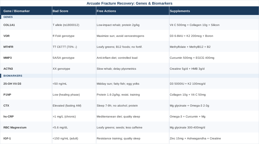
6 Biomarkers to Track for Arcuate Ligament Avulsion Fracture Recovery
Biomarkers are not a replacement for imaging or clinical assessment. They are a complement — a way to see what the fracture site cannot show you. The six biomarkers below were chosen because they either directly regulate bone repair biology, modulate collagen metabolism in ligament tissue, or govern the inflammatory environment that can accelerate or sabotage healing. Together they give you a functional read on whether your body's recovery systems are adequately resourced.
Biomarker 1: 25-OH Vitamin D3
Why it matters for this injury
Vitamin D is not just a bone mineral. It is a steroid hormone that regulates calcium absorption, osteoblast differentiation, and fracture callus formation — the biologically active tissue bridge that forms between fracture fragments during healing. Deficiency does not prevent bone union, but it significantly slows it and reduces the mechanical quality of the callus. For an avulsion fracture at the fibular head, where the fragment needs to reintegrate fully to restore posterolateral corner stability, callus quality matters.
Beyond the fracture site itself, vitamin D also influences ligamentous tissue. VDR (vitamin D receptor) expression has been detected in human ligament fibroblasts, meaning the arcuate ligament complex that was stressed or partially disrupted in this injury also responds to D status. There is growing evidence that low vitamin D correlates with increased ligament laxity and slower soft tissue remodeling.
How to measure it
A standard serum 25-OH vitamin D test is available at most labs and through direct-to-consumer testing panels. Cost range: $30–$80 as a standalone test, often included in comprehensive metabolic or wellness panels. Aim to test in late winter or early spring when levels are seasonally lowest. Optimal range for fracture recovery context: 50–80 ng/mL (125–200 nmol/L), not just the clinical sufficiency cutoff of 30 ng/mL. Peter Attia has consistently emphasized that the clinical cutoff for "deficiency" does not equal the level needed for optimal bone metabolism.
If the score is low — the plan without supplements
Maximize sun exposure: 15–30 minutes of midday sun on large body surface areas (legs, torso) daily, without sunscreen for that window. This approach works best in summer at latitudes below 40° and for lighter skin types. In practical terms, most people with a deficiency — especially post-injury with limited mobility — cannot correct low D through sun alone. Dietary sources (fatty fish, egg yolks, liver) contribute modestly. For severe deficiency in an acute fracture context, sunlight is a support measure, not a correction.
If the score is low — the plan with supplements
Vitamin D3: 5,000 IU daily with the largest meal, always combined with Vitamin K2 (MK-7) at 100–200 mcg/day to direct calcium into bone rather than arterial tissue. Duration: ongoing during fracture recovery, with retesting at 8–12 weeks to adjust. Always take D3 with fat for absorption. Cycling: no cycling necessary, but reassess dose when levels stabilize between 50–70 ng/mL. Side effects at these doses are rare but include hypercalcemia with very prolonged high-dose use — hence the retesting requirement. If you start below 20 ng/mL, a supervised loading protocol (10,000 IU for 8–12 weeks, then maintenance) may be warranted. Discuss with a physician if you have granulomatous conditions (sarcoidosis, TB) where D can dysregulate.
Biomarker 2: P1NP — Procollagen Type I N-terminal Propeptide
Why it matters for this injury
P1NP is the gold-standard bone formation marker recommended by the International Osteoporosis Foundation and IFCC. When osteoblasts are actively building new bone matrix — exactly what must happen at the fracture site — they produce procollagen Type I and release P1NP as a byproduct into circulation. A rising P1NP in the weeks after a fracture indicates that bone-forming activity is occurring. A persistently low P1NP in a person months post-fracture who should be remodeling is a red flag for impaired bone formation.
This marker is particularly valuable for monitoring, not just diagnosing. Checking it at baseline (as soon as safe post-injury), then at 6–8 week intervals, gives you a dynamic read on whether your fracture is biologically active in terms of repair.
How to measure it
P1NP is measured via blood draw (serum or EDTA plasma). It is included in some advanced bone health panels. Cost: $60–$120 standalone. Reference ranges vary by lab but general adult reference is 20–76 µg/L for women and 13–43 µg/L for men, though in an acute fracture healing context, levels are expected to be higher than baseline. The pattern over time matters more than any single value.
If the score is persistently low — the plan without supplements
Low P1NP in a fracture recovery context often signals inadequate caloric intake, particularly inadequate protein. Osteoblasts cannot build collagen matrix without amino acid substrate. Prioritize protein intake: 1.6–2.0 g per kilogram of body weight per day during healing, distributed across three or more meals. Include collagen-rich foods: bone broth, slow-cooked cartilaginous cuts of meat, and high-glycine sources. Resistance exercise of the non-injured limbs and upper body is the second lever — mechanical loading drives osteoblast activity systemically. Even during knee immobilization, upper body resistance training elevates systemic bone turnover markers.
If the score is persistently low — the plan with supplements
Collagen peptides (hydrolyzed Type I and II): 10–15 g/day taken 30–60 minutes before any weight-bearing rehabilitation exercise, with 50 mg of Vitamin C to support prolyl hydroxylase activity (the enzyme that cross-links collagen strands). This specific protocol was studied in a Shaw et al. 2017 randomized trial showing improved collagen synthesis markers in athletes. Cycle: use throughout active fracture healing phase (typically 12–20 weeks post-injury). Side effects are minimal; some people report mild digestive bloating at higher doses — splitting doses into two helps. Add silicon (orthosilicic acid) at 5–10 mg/day as a cofactor for collagen stability; early human data is promising though not yet definitive for fractures specifically.
Biomarker 3: CTX — C-terminal Telopeptide of Type I Collagen
Why it matters for this injury
CTX is the mirror biomarker to P1NP. Where P1NP measures bone building, CTX measures bone breakdown (resorption). After a fracture, some resorption is normal and expected — osteoclasts clean up damaged bone tissue to allow new formation. But excessive or prolonged CTX elevation, combined with low P1NP, signals a catabolic state where bone is being broken down faster than it is being rebuilt. This imbalance delays union and can compromise the structural integrity of the healed fragment.
Elevated CTX is also associated with the acute stress response, poor sleep, high cortisol, and chronic inflammation — factors often present in the post-surgical or post-injury period. Tracking CTX alongside P1NP gives you the P1NP:CTX ratio, a functional read on whether bone metabolism is net anabolic or catabolic.
How to measure it
CTX is measured in fasting morning blood (serum); levels spike after eating and with physical activity, so consistent morning fasting conditions are essential for meaningful results. Cost: $60–$100. For an optimal healing window, you want CTX to be within or slightly above the normal reference range, while P1NP is elevated above baseline (indicating active formation). Most standard labs provide CTX in beta-CTX form.
If CTX is elevated — the plan without supplements
Three non-supplement levers move CTX significantly: sleep quality (cortisol elevation from poor sleep directly drives osteoclast activity — prioritize 7–9 hours in a dark, cool room), protein sufficiency (low protein paradoxically increases resorption markers), and weight-bearing activity even in partial form. Anti-catabolic dietary patterns — adequate caloric intake, meal timing with a protein-containing breakfast — blunt the fasting-state CTX spike. Eliminate excessive alcohol and smoking entirely during the healing window; both elevate CTX and suppress P1NP.
If CTX is elevated — the plan with supplements
Magnesium glycinate 300–400 mg before bed (see also Biomarker 5) lowers cortisol and improves sleep quality, indirectly reducing CTX. Omega-3 fatty acids (EPA + DHA, 2–3 g/day from fish oil) have demonstrated modest anti-resorptive effects in human trials by reducing RANKL-driven osteoclastogenesis. Cycling: take continuously during fracture healing (12–20 weeks), then reassess. Side effects: fish oil at these doses may thin blood slightly — pause 5–7 days before any surgical procedures. If CTX remains elevated despite lifestyle correction, investigate cortisol dysregulation through a 4-point salivary cortisol test and discuss with an endocrinologist before pursuing pharmacological options.
Biomarker 4: High-Sensitivity CRP (hs-CRP)
Why it matters for this injury
Some inflammation immediately post-fracture is required — the acute inflammatory cascade recruits macrophages, growth factors, and stem cells to the injury site. The problem arises when inflammation does not resolve on schedule. Chronic low-grade systemic inflammation, reflected in persistently elevated hs-CRP, interferes with the remodeling phase of healing, disrupts the P1NP/CTX balance, and extends recovery timelines significantly.
hs-CRP is also a proxy for metabolic health and lifestyle factors that are independently relevant to fracture healing: visceral fat, sleep quality, diet quality, and psychological stress all drive chronic CRP elevation. Monitoring it gives you a broad-stroke read on whether your systemic environment is pro-healing or pro-inflammatory.
How to measure it
hs-CRP is a standard, inexpensive blood test available at virtually any lab. Cost: $20–$50. Optimal for recovery: below 1.0 mg/L. Values of 1–3 mg/L indicate moderate systemic inflammation. Above 3 mg/L consistently (when not in the acute injury phase) signals a significant inflammatory load that is likely impairing tissue repair quality. Note that hs-CRP will be naturally elevated in the first 2–4 weeks post-fracture — meaningful tracking starts after this acute window.
If hs-CRP is elevated — plan without supplements
Dietary pattern is the most powerful lever: shift toward a Mediterranean-style diet emphasizing olive oil, fatty fish, leafy vegetables, and minimal ultra-processed food. Eliminate trans fats and reduce refined carbohydrate load. Sleep quality is the second lever — hs-CRP drops substantially with consistent 7–9 hour sleep in clinical studies. Moderate-intensity exercise (above what is restricted by the injury) also resolves chronic CRP over weeks to months. Stress management matters: sustained psychological stress is one of the strongest CRP drivers.
If hs-CRP is elevated — plan with supplements
Omega-3 fatty acids (EPA + DHA, 2–3 g/day): among the most replicated anti-inflammatory supplement interventions. Curcumin with piperine: 500–1000 mg/day of a high-bioavailability form (e.g., Meriva or Longvida); evidence for reducing CRP in musculoskeletal contexts is solid. Magnesium: addresses multiple drivers simultaneously. Cycling for omega-3: use continuously during recovery, then reassess. Curcumin: cycle on for 8–12 weeks, off for 4 weeks. Side effects: curcumin at high doses may interact with anticoagulants (relevant if on warfarin post-surgery); consult your physician.
Biomarker 5: RBC Magnesium
Why it matters for this injury
Magnesium is involved in over 300 enzymatic reactions, including several directly relevant to bone healing: alkaline phosphatase activity (required for bone matrix mineralization), Vitamin D activation (the conversion of 25-OH to 1,25-OH requires a magnesium-dependent enzyme), and muscle function at the knee during rehabilitation. Serum magnesium is a poor test because the body maintains serum levels by pulling magnesium from intracellular stores — you can be intracellularly deficient with a normal serum reading. RBC (red blood cell) magnesium is the preferred test for identifying functional deficiency.
Estimates suggest 50–70% of the Western population has suboptimal magnesium status. For someone recovering from a surgical or acute knee injury — with physiological stress and potentially altered eating patterns — this number is likely higher.
How to measure it
RBC magnesium is offered by specialty labs and some broader wellness panels. Cost: $40–$80. Optimal range: 5.6–6.8 mg/dL for RBC magnesium (vs. standard serum reference which is lower). If only serum magnesium is available, optimal is 2.0–2.5 mg/dL (upper half of the reference range).
If RBC magnesium is low — plan without supplements
Food-first approach: dark leafy greens (spinach, Swiss chard), pumpkin seeds, almonds, dark chocolate (>85%), and legumes are the richest dietary sources. Eliminate or reduce the main dietary magnesium antagonists: alcohol, high sugar intake, and excess caffeine all increase urinary magnesium excretion. Stress reduction is relevant: adrenaline and cortisol both deplete intracellular magnesium. Cooking with filtered water matters less than food quality, but heavily processed water softeners can reduce dietary magnesium intake marginally.
If RBC magnesium is low — plan with supplements
Magnesium glycinate or magnesium threonate: 300–400 mg/day elemental magnesium, taken in the evening (it also supports sleep quality and parasympathetic tone). Magnesium threonate is the preferred form for neurological effects and has slightly better CNS penetration; magnesium glycinate is gentler on digestion. Avoid magnesium oxide — it has poor bioavailability. Cycling: no cycling needed at physiological doses; maintain throughout the healing window and beyond if dietary intake remains inadequate. Side effects: loose stools at doses above 400–500 mg/day (this is actually the dose-limiting mechanism — back off by 50 mg if this occurs). Retesting at 8–12 weeks to confirm normalization.
Biomarker 6: IGF-1 (Insulin-like Growth Factor 1)
Why it matters for this injury
IGF-1 is produced primarily in the liver in response to growth hormone (GH) signaling and is the main anabolic signal driving bone matrix synthesis, osteoblast proliferation, and muscle repair. In fracture healing specifically, IGF-1 has been shown to stimulate periosteal osteoblasts — the cells most responsible for callus formation — and to enhance collagen Type I gene expression. Low IGF-1 is associated with delayed fracture union, reduced bone density, and slower soft tissue regeneration.
IGF-1 declines with age, chronic sleep deprivation, caloric restriction, excessive alcohol, and high cortisol states. For middle-aged and older patients recovering from an avulsion fracture, IGF-1 deficiency is a plausible but under-tested contributor to prolonged healing timelines.
How to measure it
IGF-1 is measured in serum and is available at most standard labs as part of hormonal panels. Cost: $50–$100. Optimal range for active healing: age-adjusted upper-normal — roughly 150–300 ng/mL for adults under 50, declining with age. Peter Attia and others tracking longevity biomarkers tend to target the upper quartile of the age-adjusted range for tissue maintenance. Note: very high IGF-1 (over 350–400 ng/mL) has been associated with increased cancer risk in some epidemiological data, so the goal is optimization, not maximization.
If IGF-1 is low — plan without supplements
The most evidence-based non-pharmacological drivers of IGF-1 are: resistance training (the non-injured body segments can be trained — upper body and contralateral leg), sleep quality (GH is secreted in pulsatile bursts during slow-wave sleep; poor sleep is one of the strongest suppressors), adequate protein intake (low protein directly suppresses hepatic IGF-1 production), and intermittent fasting used cautiously (short-term fasting elevates GH, but extended restriction suppresses IGF-1 — during fracture recovery, moderate time-restricted eating rather than aggressive fasting is appropriate).
If IGF-1 is low — plan with supplements
Zinc: 15–30 mg/day zinc bisglycinate is a cofactor for GH axis function; deficiency suppresses IGF-1 production. Ashwagandha (KSM-66): 300–600 mg/day has demonstrated modest increases in testosterone and IGF-1 in human trials, likely via cortisol reduction. Cycling: 8 weeks on, 4 weeks off. Creatine monohydrate: 3–5 g/day — evidence for supporting IGF-1 pathway signaling during strength training and potentially supporting bone density. Side effects of creatine: mild water retention intracellularly, benign for most people. If IGF-1 remains very low despite lifestyle optimization, discuss evaluation for growth hormone deficiency with an endocrinologist; therapeutic GH or peptides (such as ipamorelin/CJC-1295) exist but require medical supervision and are not first-line.
Tracking these six biomarkers at baseline, then at 6–8 week intervals during recovery, transforms a passive wait-and-see approach into an active feedback loop. Most can be ordered directly without a specialist referral and provide information your surgeon's follow-up appointment never will.
The Genetics Behind Bone and Ligament Healing: 5 Variants Worth Knowing
Genetics does not determine your outcome. But a handful of variants meaningfully shift your baseline — the speed of collagen cross-linking, how efficiently you absorb Vitamin D, how aggressively your body remodels tissue after injury. Knowing your variants lets you compensate upstream rather than discover the hard way that standard protocols are not working for your biology.
Gene 1: COL1A1 — The Collagen Architecture Gene
COL1A1 encodes the alpha-1 chain of Type I collagen — the most abundant structural protein in both bone matrix and ligaments, including the arcuate ligament complex. The Sp1 polymorphism (rs1800012, G to T substitution) is the best-studied variant in this gene. The TT genotype is associated with reduced collagen fibril tensile strength, increased fracture risk, and slower callus consolidation. This variant has been replicated across multiple human cohorts including the GENOMOS consortium.
If the gene is suboptimal (T allele carrier): plan without supplements — load management is the non-negotiable intervention. The T allele means collagen structure is inherently more fragile; this is not fixable by nutrition alone, but the degenerative signal can be slowed. Prioritize lower-impact rehabilitation progression (avoid early high-impact loading even when pain allows), invest in eccentric strengthening of the posterolateral knee musculature, and ensure protein intake is at the higher end of the recommendation range (2.0 g/kg/day).
If the gene is suboptimal: plan with supplements — Vitamin C (500–1000 mg/day): required for prolyl hydroxylase to cross-link collagen strands; collagen with suboptimal cross-linking from COL1A1 variants needs maximal enzymatic support. Collagen peptides (10–15 g/day) + Vitamin C before exercise. Silicon as orthosilicic acid (5 mg/day): promotes collagen Type I synthesis at the gene expression level in human cell studies. Cycling: use Vitamin C continuously (it is water-soluble and safe at these doses); collagen during active rehab phases. Side effects: Vitamin C above 2 g/day can cause loose stools and may increase oxalate kidney stone risk — stay below 1 g/day if you have a history of calcium oxalate stones.
Gene 2: VDR — Vitamin D Receptor Variants
Even if your 25-OH Vitamin D levels are adequate, a poorly functional VDR gene means the signal does not land properly in bone cells, muscle cells, or ligament fibroblasts. The most clinically relevant variants are FokI (rs2228570) and BsmI (rs1544410). The FokI "ff" genotype produces a longer VDR protein with reduced transcriptional efficiency. Studies in fracture populations show ff individuals have lower bone mineral density regardless of serum Vitamin D levels.
If the VDR variant is present: plan without supplements — because D signaling is inefficient, you need to maximize every upstream lever: solar UVB exposure, fat-soluble meal timing for dietary D, and reducing VDR-antagonizing factors (xenoestrogens, some pesticides, and cadmium from cigarette smoke all impair VDR function).
If the VDR variant is present: plan with supplements — target the upper end of the optimal D range (70–80 ng/mL) rather than just sufficiency (30 ng/mL). This typically requires 6,000–8,000 IU/day of D3 in an ff individual to achieve what a wildtype individual achieves at 3,000 IU. Always pair with K2 (MK-7, 200 mcg/day). Magnesium is essential alongside — without adequate magnesium, VDR cannot function regardless of genotype. Retest at 8 weeks to dial in your personalized dose. Note: individuals with VDR variants should also consider boron (3–9 mg/day), which has been shown to upregulate VDR expression in human studies, partially compensating for receptor inefficiency.
Gene 3: MTHFR — The Methylation and Collagen Cross-linking Gene
MTHFR (methylenetetrahydrofolate reductase) is widely known for its role in folate metabolism and homocysteine clearance. Less commonly discussed is its direct relevance to connective tissue: elevated homocysteine (a consequence of impaired MTHFR activity) interferes with lysyl oxidase — the enzyme responsible for cross-linking collagen and elastin fibers into their mature, load-bearing forms. In bone, elevated homocysteine has been associated with increased fracture risk independent of bone density in multiple human prospective studies.
The C677T variant (rs1801133) is the most common; TT homozygotes have roughly 70% reduced enzyme activity compared to CC. This is relatively straightforward to identify on any genetic panel that includes MTHFR.
If MTHFR is impaired: plan without supplements — reduce dietary homocysteine load: minimize methionine-heavy foods in excess (lean red meat is fine; processed meats are not), maximize riboflavin-rich foods (cofactor for MTHFR), leafy greens (dietary folate), and B12-rich animal protein. Avoid high folic acid intake from fortified foods (synthetic folic acid can paradoxically block methylfolate receptors in TT individuals).
If MTHFR is impaired: plan with supplements — Methylfolate (5-MTHF): 400–800 mcg/day (not folic acid); Methylcobalamin (B12): 1,000 mcg/day; Riboflavin (B2): 50–100 mg/day (drives MTHFR activity where residual function exists). Cycling: these are safe long-term; reassess homocysteine levels at 8–12 weeks to confirm you are driving homocysteine below 9 µmol/L. Side effects: methylfolate can trigger anxiety or headaches in some people with certain COMT variants — start low (200 mcg) and increase gradually. B12 at these doses has essentially no toxicity.
Gene 4: MMP3 — Matrix Metalloproteinase and Tissue Remodeling
MMP3 (stromelysin-1) is a matrix metalloproteinase involved in the degradation and remodeling of extracellular matrix components — including collagen, fibronectin, and proteoglycans — after injury. A key promoter polymorphism, the 5A/6A variant (rs3025058), affects MMP3 gene expression levels. The 5A/5A genotype is associated with higher MMP3 expression and more aggressive tissue remodeling, which may sound beneficial but in a ligament/bone context can lead to excessive matrix degradation during the inflammatory phase, delayed stabilization, and potentially weaker scar tissue in repaired ligamentous structures.
If MMP3 is overactive (5A carrier): plan without supplements — anti-inflammatory dietary patterns are especially important because systemic inflammation amplifies MMP3 expression further. Omega-3 rich foods, polyphenol-rich fruits and vegetables, and controlled rehabilitation (avoiding excessive mechanical stress in the early remodeling phase) are the primary levers.
If MMP3 is overactive: plan with supplements — Curcumin (BCM-95 or Meriva): 500–1000 mg/day; curcumin has been shown to downregulate MMP-3 gene expression in multiple human cell and animal models. Green tea extract (EGCG): 400–600 mg/day; a natural MMP inhibitor with human safety data. Cycling: 8 weeks on, 4 weeks off for curcumin; EGCG can be used more continuously. Side effects: EGCG on an empty stomach may cause nausea — always take with food. Curcumin interacts with anticoagulants (relevant in post-surgical context); confirm timing with your prescribing physician.
Gene 5: ACTN3 — The Power and Recovery Gene
ACTN3 encodes alpha-actinin-3, a structural protein in fast-twitch muscle fibers. The R577X variant (rs1815739) results in a complete absence of alpha-actinin-3 protein in the XX genotype — a state present in roughly 18% of the general population. While this genotype has no dramatic effect on endurance activity, it is associated with reduced muscle power output, slower post-exercise recovery, and potentially reduced protective muscle co-activation around joints during dynamic loading. For the posterolateral corner of the knee — which depends heavily on the popliteus, biceps femoris, and lateral gastrocnemius for dynamic stability — muscle power and responsiveness matter significantly in preventing re-injury.
If ACTN3 is XX: plan without supplements — the rehabilitation loading strategy should emphasize lower-velocity, higher-time-under-tension resistance training over explosive plyometric progressions, particularly in the first 6 months. The XX genotype responds better to endurance-style strength training and takes longer to rebuild power in fast-twitch fiber populations. Do not rush the plyometric progression phase that many ACL/posterolateral reconstruction protocols include — the muscle responsiveness may not yet be there.
If ACTN3 is XX: plan with supplements — Creatine monohydrate (5 g/day) has been shown to partially compensate for reduced fast-twitch fiber output by increasing phosphocreatine availability. HMB (beta-hydroxy beta-methylbutyrate): 3 g/day during the early rehabilitation phase when muscle atrophy is a risk; evidence for anti-catabolic effects in non-trained individuals during immobilization. Cycling: creatine is safe long-term; HMB is most useful during the first 8–12 weeks of post-immobilization rebuilding. Side effects: creatine causes benign intracellular water retention; HMB has a minimal side effect profile at 3 g/day.
What Andrew Huberman's Framework on Bone Health and Injury Recovery Gets Right
The Andrew Huberman Lab podcast episodes on bone health, injury recovery, and growth factors represent one of the most science-referenced popular frameworks on musculoskeletal biology available without a medical degree. Drawing from multiple episodes — including those on cortisol, growth hormone, sleep, and bone density — here are the ten most impactful principles for arcuate ligament avulsion fracture recovery:
1. Sleep is the most underrated fracture healing intervention
Huberman consistently cites data showing that 80–90% of growth hormone (GH) secretion occurs during slow-wave sleep. GH drives IGF-1 production and bone matrix synthesis. Missing even one night of quality sleep measurably suppresses the GH pulse. During fracture recovery, sleep is not rest — it is the primary anabolic window.
2. Cortisol's timing determines whether it heals or harms
A sharp morning cortisol spike (driven by brief cold exposure, morning sunlight, or light exercise) is beneficial and anti-inflammatory in its timing context. Sustained elevated cortisol — from chronic stress, poor sleep, or excessive caloric restriction — directly suppresses osteoblast activity and elevates CTX. Managing cortisol architecture is more nuanced than simply "reducing stress."
3. Morning sunlight is not optional for recovery
Early morning light exposure (10–30 minutes within an hour of waking) anchors the circadian rhythm, improves sleep quality downstream, and regulates the GH secretion cycle. Huberman frames this as a foundational tool, not a wellness luxury. For a recovering patient with limited mobility, this is one of the most accessible interventions available.
4. Resistance training of non-injured body parts has systemic anabolic effects
Exercise-induced IGF-1 and testosterone responses are systemic. Training the upper body and contralateral leg during knee immobilization measurably reduces bone and muscle atrophy in the injured limb through systemic hormonal mechanisms — not just through neural crosstalk. This is a well-replicated finding that challenges the traditional "rest the injured area completely" approach.
5. Omega-3s are not primarily anti-inflammatory — they are pro-resolution
Huberman distinguishes between blocking inflammation (which can impair healing if done too early or too aggressively with NSAIDs) and resolving inflammation. EPA and DHA produce specialized pro-resolving mediators (SPMs) that actively resolve the inflammatory cascade. This is why omega-3s are appropriate throughout fracture recovery, while NSAIDs should be used cautiously and time-limited in the early healing phase.
6. Collagen synthesis peaks with heat, not cold
While cold therapy has a role in acute injury (first 24–72 hours), prolonged cryotherapy suppresses the inflammatory signaling needed for collagen production and fracture callus formation. Huberman cites data suggesting that transitioning to heat therapy during the remodeling phase (from approximately week 3–4 onward) supports collagen fiber alignment and tissue maturation.
7. Protein distribution matters as much as total intake
Leucine, the amino acid that triggers mTOR-driven protein synthesis, requires a threshold dose (roughly 2.5–3 g per meal) to maximally stimulate anabolic signaling. Spreading protein across three to four meals rather than loading it in one or two meals maintains a more consistent anabolic environment for tissue repair. This is relevant for muscle rebuilding in the injured knee and for IGF-1 production.
8. The bone-brain axis is bidirectional
Huberman references emerging research showing that osteocalcin — released by active osteoblasts — exerts direct signaling effects on memory, stress response, and autonomic regulation. Bone health is not isolated from nervous system function. Chronic pain states, fear of re-injury, and psychological stress all have documented suppressive effects on bone turnover markers. Psychological recovery is part of the biology.
9. Deliberate heat (sauna) upregulates growth hormone transiently
Huberman cites Finnish and Laukkanen data showing that sauna protocols (approximately 80°C, 20-minute sessions, repeated 2–3 times per week) produce acute GH spikes. This is a practical tool for non-pharmacological GH support during the rehabilitation window, available to most patients once the acute injury phase has resolved. Caution: avoid if post-surgical wounds are not fully healed.
10. Zone 2 aerobic exercise drives mitochondrial and vascular health without cortisol excess
Low-intensity sustained aerobic exercise (Zone 2: nasal breathing possible, light conversation possible) improves capillary density and blood supply to healing tissue without triggering the cortisol elevation of high-intensity training. For a knee injury patient with restricted weight-bearing, upper-body cycling or swimming provides the Zone 2 benefit. Huberman emphasizes 150–200 minutes per week of Zone 2 as a baseline, calibrated to injury restrictions.
Complementary Approaches With Clinical Evidence
Beyond biomarkers and genetics, a small number of evidence-backed modalities have meaningful clinical data relevant to fracture and soft tissue recovery. The three below were selected because their evidence is most directly applicable to this injury type — not as alternatives to medical care, but as adjuncts worth discussing with your care team.
Low-Level Laser Therapy / Photobiomodulation
Photobiomodulation (PBM) uses specific wavelengths of red and near-infrared light (typically 630–1000 nm) to stimulate cellular mitochondria, increase ATP production, and upregulate collagen synthesis. It has been investigated specifically in fracture healing contexts, where it appears to accelerate callus formation and increase the mechanical strength of healing bone in both animal models and early human trials. For a fibular head avulsion fracture, the injury site is relatively superficial — ideal for PBM penetration.
A systematic review published by Enyinnaya Chukwuemeka Obi and colleagues, along with earlier meta-analyses on biphasic dose response in low level laser therapy, supports its application for musculoskeletal tissue repair at appropriate doses. The Arndt-Schulz principle (biphasic dose response) means both underdosing and overdosing reduce efficacy — the therapeutic window matters.
Realistic application: seek a physiotherapy clinic with a Class 3B or Class 4 laser device certified for musculoskeletal use. Typically 6–12 sessions are delivered over 3–4 weeks during the early remodeling phase. Home-use red light therapy panels (660 nm + 850 nm) are a lower-intensity but accessible option for daily self-application over the lateral knee area. Ensure direct skin contact (remove compression bracing for sessions) and consistent frequency (daily or every other day for home devices). Evidence remains stronger for soft tissue conditions than for avulsion fractures specifically — approach as a supportive adjunct, not a primary treatment.
Massage Therapy
Therapeutic massage is relevant to arcuate ligament avulsion fracture recovery primarily through its effects on surrounding musculature and the local vascular environment, not on the fracture site itself. Post-surgical or post-immobilization tightness in the biceps femoris, popliteus, and posterior knee capsule creates altered joint mechanics that increase stress on the healing ligament complex. Massage targeting these structures reduces mechanical compensation patterns and supports symmetrical rehabilitation progression.
Research on massage and musculoskeletal recovery — including a 2015 clinical review on soft tissue mobilization — supports its role in improving local circulation, reducing myofascial restrictions, and accelerating return to function timelines. Evidence specific to posterolateral knee reconstruction is limited; extrapolation from general knee surgery recovery data is appropriate with appropriate clinical supervision.
Practical application: begin with light effleurage and petrissage of the posterior and lateral thigh and calf once wound integrity is confirmed (typically 4–6 weeks post-surgery). Progress to deeper cross-fiber friction techniques on the biceps femoris and iliotibial band as rehabilitation advances. Frequency: 1–2 sessions per week during active rehabilitation. Avoid direct pressure over the fracture site or any areas of active swelling. A licensed sports massage therapist or physiotherapist trained in knee rehabilitation is preferable to a general spa setting.
Breathing-Based Therapies
Structured breathing interventions are not the first thing most people associate with fracture healing, but their mechanism is physiologically direct: controlled diaphragmatic breathing activates the parasympathetic nervous system, reduces cortisol levels, and lowers hs-CRP in clinical studies. Since both cortisol and chronic inflammation are documented suppressors of bone formation markers (P1NP) and collagen synthesis, breathing practices represent a zero-cost, accessible intervention with a legitimate mechanistic path to improved recovery biology.
The most evidence-based protocol for this purpose is slow-paced breathing at 5–6 breaths per minute (approximately 5 seconds inhale, 5 seconds exhale), practiced for 10–20 minutes twice daily. A Lehrer et al. study on heart rate variability biofeedback — which uses the same breath pace — demonstrated measurable autonomic and cortisol effects. The Wim Hof method (hyperventilation-based) is less appropriate in the acute recovery phase due to its strong sympathetic activation component.
Realistic daily integration: practice morning and evening slow breathing in a supine or supported seated position. Apps such as Resonance or simple visual timers can guide the pace. For patients experiencing post-surgical anxiety or pain-related hypervigilance — which is common and itself elevates CRP — the combination of slow breathing with progressive muscle relaxation of the non-injured limbs offers additional benefit. Minimal contraindications at the breath rates described; consult a physician if you have uncontrolled respiratory conditions.
Conclusion
An arcuate ligament avulsion fracture is a mechanical event, but recovery is a biological process — one that unfolds differently depending on your Vitamin D status, your bone formation markers, your genetic collagen blueprint, and dozens of other factors most standard follow-up appointments never examine. The biomarkers in this article can be ordered through a primary care physician or directly, and they give you a meaningful picture of whether your body's healing systems are adequately resourced or quietly running on fumes.
The genetics section adds another layer of self-knowledge — not to predict doom, but to explain baseline differences and create targeted compensation strategies rather than generic ones. Combined with the sleep, nutrition, and anabolic support principles drawn from current research, these tools shift your recovery from passive compliance to active optimization.
The next smart step is straightforward: get a baseline panel. At minimum, start with 25-OH Vitamin D, hs-CRP, and P1NP — the three most actionable markers for this injury context — and bring the results to your next clinical appointment. Better information leads to better decisions. That is not a promise of a faster recovery, but it is a meaningful improvement on waiting and hoping.
Musculoskeletal: Bone Conditions Joint Conditions Tendon & Ligament Conditions Sports Injuries
Autoimmune: Inflammatory Conditions