This article contains AI generated content.
· UpdatedArthrofibrosis – 5 Genes and 6 Biomarkers to Track
Introduction
If you have been managing joint stiffness after surgery or injury and keep hitting the same wall — stretching, physical therapy, pain management, repeat — you already know that the standard protocol has limits. Arthrofibrosis, the abnormal accumulation of scar tissue inside or around a joint, is one of the most frustrating outcomes in orthopedic medicine precisely because it does not respond uniformly across patients. Two people can undergo identical ACL reconstructions or total knee replacements, follow identical rehabilitation, and land in completely different places six months later. That gap is not random.
Generic advice for arthrofibrosis — stretch more, move early, reduce inflammation — is not wrong, but it is incomplete. It treats everyone as though they are working with the same biological hardware, when in reality the inflammatory response, collagen remodeling dynamics, and tissue signaling pathways involved in arthrofibrosis vary meaningfully from person to person. Those differences are partly genetic, partly reflected in measurable blood markers, and partly modifiable through the right interventions — if you know which levers to pull.
This article takes a more precise approach. It examines both the biomarkers that can tell you what your body is actively doing right now — how much inflammatory signaling is running, how fibrosis-promoting your tissue environment is, and how efficiently your body is breaking down or building collagen — and the genetic variants that may explain your underlying predisposition. Neither layer offers a cure. But together they point toward smarter, more individualized strategies that go well beyond ice packs and generic anti-inflammatories.
The goal here is grounded, practical hope. Six measurable biomarkers can give you a real-time picture of your inflammatory and fibrotic load, with clear action plans for improving each one. Five genes can explain your baseline risk profile and suggest targeted compensations. And beyond those two layers, there are complementary tools and lifestyle frameworks backed by human evidence that most clinicians never mention. Better information does not guarantee better outcomes — but it meaningfully shifts the odds.
6 Biomarkers Worth Tracking in Arthrofibrosis
Understanding what is happening biochemically is one of the most underused tools in managing arthrofibrosis. While imaging and clinical assessments capture structural outcomes, blood and tissue biomarkers reveal the underlying biological activity — the inflammation, the fibrotic signaling, the collagen turnover — that determines whether things will improve or worsen over time. The six markers below are the most clinically relevant based on current research in joint fibrosis, post-surgical inflammation, and connective tissue remodeling.
High-Sensitivity CRP (hs-CRP)
Why it matters
CRP is the most accessible inflammation marker in clinical practice, but standard CRP misses low-grade chronic inflammation. High-sensitivity CRP (hs-CRP) is the version that matters for conditions like arthrofibrosis, where inflammation may smolder at levels that standard panels do not detect. Sustained low-grade inflammation drives fibroblast activation and collagen deposition — the precise mechanism underlying arthrofibrosis. Peter Attia consistently highlights hs-CRP as one of the foundational biomarkers for any chronic inflammatory condition, and that recommendation is especially relevant here.
An hs-CRP below 0.5 mg/L is generally considered optimal. Levels between 1 and 3 mg/L indicate moderate inflammation; above 3 mg/L signals significant inflammatory burden that requires active management.
How to measure it
hs-CRP is ordered through most standard labs. It is typically covered by insurance with clinical justification, or available for approximately $20–40 as a standalone out-of-pocket test through labs like Quest Diagnostics or LabCorp. Results are returned within 24–48 hours. Repeat every 3 months while actively managing arthrofibrosis or post-surgical recovery to track trends, not just single values.
If the score is bad, the plan without supplements
Prioritize sleep quality first — disrupted or insufficient sleep is one of the most reliable drivers of elevated hs-CRP. Aim for 7–9 hours in a cool, dark environment with a consistent sleep-wake schedule. Eliminate ultra-processed foods, refined seed oils, and excess added sugar, as these provide direct inflammatory inputs through multiple pathways. Low-impact aerobic exercise (walking, swimming, cycling) at a moderate pace for 30 minutes daily consistently reduces CRP over 8–12 weeks. Prolonged sitting independently elevates inflammatory markers and compounds the problem in people already dealing with joint stiffness; structured movement breaks every 60–90 minutes are a simple corrective.
If the score is bad, the plan with supplements or equipment
Omega-3 fatty acids (EPA/DHA) at 2–4 grams daily have the strongest evidence for reducing hs-CRP among commercially available supplements. Use a purified, molecularly distilled fish oil or algae-based DHA/EPA; check for oxidation testing from the manufacturer. This is a long-term intervention — no cycling needed. Curcumin with piperine (500–1000 mg daily) has shown modest CRP-lowering effects in multiple meta-analyses; cycle 8 weeks on, 2 weeks off to assess individual response. Magnesium glycinate (200–400 mg nightly) reduces inflammatory signaling and supports the sleep quality that further drives CRP down. A portable infrared sauna (15–20 minutes, 3–4 times per week) has demonstrated lowering of systemic inflammatory markers in multiple controlled trials over 8–12 weeks. Cost for quality home units: $400–1,200.
Serum Interleukin-6 (IL-6)
Why it matters
IL-6 is a cytokine that sits at the intersection of acute inflammation and fibrotic signaling. In arthrofibrosis, elevated IL-6 drives the transition from normal wound healing to excessive scarring — it stimulates fibroblast proliferation, inhibits collagen degradation, and promotes fibrotic capsule formation. Serum IL-6 is also a more dynamic marker than CRP, rising and falling faster in response to interventions, which makes it particularly useful for monitoring whether your approach is actually working.
Research in post-surgical joint fibrosis, including studies in patients following total knee arthroplasty, consistently identifies elevated IL-6 as a predictor of restricted range-of-motion outcomes. Optimal serum IL-6 is generally below 2 pg/mL; levels above 5–7 pg/mL represent meaningful inflammatory excess in a stable, non-acutely ill individual.
How to measure it
Serum IL-6 is available through major reference labs for approximately $50–120 out of pocket. It requires a clinician's order in most jurisdictions and careful timing: draw during a stable period, not within a week of illness, vaccination, or high-intensity training, as acute events will temporarily elevate results. For tracking purposes, repeat every 3 months alongside hs-CRP.
If the score is bad, the plan without supplements
Reducing visceral fat is the most powerful lifestyle intervention for chronically elevated IL-6, because adipose tissue itself is a major IL-6 source. A time-restricted eating window (10–12 hour feeding window, such as 8am–6pm) combined with regular resistance training consistently reduces visceral fat and therefore IL-6 over 12–16 weeks. Address psychological stress deliberately — cortisol dysregulation from chronic stress upregulates IL-6 through the HPA axis independently of body composition. Cold water immersion (10–15°C, 5–10 minutes, 2–3 times per week) has demonstrated robust IL-6 reduction in controlled trials, though this is contraindicated during acute joint inflammation.
If the score is bad, the plan with supplements or equipment
Quercetin (500–1000 mg daily with food) is one of the better-studied flavonoids for IL-6 suppression, with evidence from both in vitro work and human trials showing effects through NF-κB pathway inhibition. Take consistently for 8–12 weeks, then assess. Berberine (500 mg twice daily with meals) reduces inflammatory cytokines including IL-6 through AMPK pathway activation; cycle 2 months on, 1 month off to minimize gut microbiome disruption, which is a real consideration with extended berberine use. Photobiomodulation devices (red/near-infrared, 630–850 nm) applied directly over affected joints 3–4 times per week have shown direct reduction in IL-6 at the local tissue level in controlled studies — addressing both the systemic and local fibrotic environment simultaneously.
Plasma TGF-β1 (Transforming Growth Factor Beta 1)
Why it matters
TGF-β1 is arguably the single most important molecule in the biology of arthrofibrosis. It is the master switch for fibroblast-to-myofibroblast transition — the cellular event that drives excessive collagen deposition in joint tissue. Elevated TGF-β1 is consistently found in fibrotic joint tissue from patients with arthrofibrosis, and serum TGF-β1 levels have been examined as a potential indicator of fibrotic activity in multiple tissue contexts. While plasma TGF-β1 is not yet a standard clinical ordering for arthrofibrosis, it is measurable, and patterns of elevation carry real clinical meaning when interpreted alongside other inflammatory markers.
Reference ranges vary by lab, but plasma TGF-β1 above 20 ng/mL is generally considered elevated in the context of chronic inflammatory and fibrotic conditions. A single measurement is less informative than a trend — the direction of change over 3–6 months matters as much as any single value.
How to measure it
Plasma TGF-β1 is available through specialty and functional medicine labs. Cost typically ranges from $80–200 depending on the lab and whether a panel or standalone test is ordered. The assay requires plasma (not serum) in some formats — verify with your ordering provider to avoid sample handling errors. Repeat every 3–6 months while implementing interventions.
If the score is bad, the plan without supplements
Resistance training at moderate intensity has been shown to modulate TGF-β1 signaling without chronically elevating baseline levels — in contrast to extreme training loads, which can transiently spike it. Regular heat exposure via dry sauna (80–90°C, 15–20 minutes, 3–4 times per week) has demonstrated influence on TGF-β1 regulation in muscle and connective tissue contexts. Critically: reducing dietary advanced glycation end-products (AGEs) lowers TGF-β1 signaling, because AGEs are direct activators of this pathway. AGEs form primarily through high-heat cooking: charring, frying, grilling at extreme temperatures. Switching to slow cooking, steaming, poaching, and lower-temperature roasting is a meaningful dietary shift for TGF-β1-high individuals.
If the score is bad, the plan with supplements or equipment
Vitamin D3 at therapeutic doses (targeting serum 25-OH-D between 50–70 ng/mL) downregulates TGF-β1 signaling through nuclear receptor pathways — this connection is well-supported in fibrosis literature across multiple tissue types and represents one of the most cost-effective TGF-β1 interventions available. Always pair with K2 (MK-7 form) and magnesium. N-acetylcysteine (NAC, 600–1200 mg daily) reduces the oxidative stress that drives TGF-β1 upregulation; monitor liver enzymes with extended use above 1200 mg daily. Resveratrol or pterostilbene (250–500 mg daily with food; pterostilbene has better bioavailability) has demonstrated TGF-β1 modulating effects in early human studies and animal models — cycle 8 weeks on, 4 weeks off and assess response. For refractory or severe cases, losartan (an angiotensin receptor blocker, prescription only) has documented TGF-β1 inhibitory effects studied in clinical fibrosis contexts — discuss specifically with a physician managing your case.
25-Hydroxyvitamin D (25-OH-D)
Why it matters
Vitamin D is not just a bone metabolism hormone. The vitamin D receptor (VDR) is expressed in fibroblasts, immune cells, synovial tissue, and joint capsule cells — all key players in arthrofibrosis. Low vitamin D status impairs the resolution of inflammation, reduces the expression of anti-fibrotic genes, and is associated with worse outcomes in post-surgical joint recovery. Thomas Dayspring has consistently highlighted vitamin D as one of the most underappreciated modifiers of inflammatory and fibrotic conditions across medical specialties.
Multiple studies have found that patients with arthrofibrosis or significant post-surgical joint stiffness have lower mean 25-OH-D levels than matched controls without these complications. The optimal range for anti-inflammatory and anti-fibrotic effects is generally 40–60 ng/mL, though some researchers in fibrosis and autoimmunity argue for 50–80 ng/mL during active conditions — the target should be individualized based on measured response.
How to measure it
25-OH-D is one of the most commonly ordered blood tests globally. Through insurance it is frequently covered; out of pocket it runs $30–70 at most standard labs. Measure it twice yearly — once in winter when levels are typically lowest and once in late summer after natural sun exposure — and adjust supplementation based on actual results, not assumptions. Results vary significantly by season, latitude, skin tone, and body weight.
If the score is bad, the plan without supplements
Safe direct sun exposure remains the most physiologically natural source of vitamin D3. 15–25 minutes of midday sun on arms and legs (without sunscreen application) 3–4 times per week during summer months can help maintain adequate levels in lighter-skinned individuals in sunny latitudes. This approach is generally insufficient for individuals in northern latitudes above 40°N during winter months, those with darker skin tones, those who primarily work indoors, or those who are overweight (adipose tissue sequesters vitamin D). Sun exposure should be pursued thoughtfully, not as a sole strategy for deficient individuals.
If the score is bad, the plan with supplements or equipment
Vitamin D3 supplementation at 4,000–6,000 IU daily is appropriate for most deficient adults, but individual response varies significantly based on body weight, fat mass, gut absorption efficiency, and VDR gene variants. Always co-supplement with vitamin K2 (MK-7 form, 100–200 mcg daily) to prevent calcium dysregulation as D3 levels rise — this pairing is essential, not optional. Magnesium glycinate (300–400 mg daily) is also required for D3 conversion to its active form; without adequate magnesium, supplemental D3 activates poorly. Re-test 25-OH-D after 90 days of consistent supplementation to calibrate your personal dose. No cycling needed — maintain year-round at target range. This is a maintenance intervention, not a short-term fix.
Ferritin
Why it matters
Ferritin is commonly ordered as an iron storage marker, but at elevated levels it is also a significant acute-phase inflammatory reactant. In arthrofibrosis and other fibrotic conditions, chronically elevated ferritin reflects ongoing inflammatory burden and is associated with increased reactive oxygen species — a key driver of fibroblast activation and collagen overproduction. At the same time, low ferritin impairs tissue repair and oxygen delivery to healing joint structures, creating a different but equally problematic state for recovery.
The clinically useful range is nuanced. Per Peter Attia's framework for metabolic and inflammatory health, ferritin between 50–150 ng/mL is generally considered optimal for tissue repair and energy metabolism, while levels above 200–300 ng/mL in the absence of acute illness suggest chronic inflammatory burden. Below 30 ng/mL may signal iron insufficiency that impairs healing capacity.
How to measure it
Ferritin is included in many comprehensive metabolic panels or available as a standalone test for $20–50 out of pocket. It is widely covered by insurance. For proper interpretation, always run it alongside a complete iron panel (serum iron, total iron-binding capacity, transferrin saturation) to distinguish between inflammatory elevation of ferritin and actual iron-deficiency states.
If the score is bad (elevated), the plan without supplements
When ferritin is elevated due to chronic inflammation rather than hemochromatosis, the focus shifts to reducing the inflammatory load driving it — which maps directly to all the interventions listed under hs-CRP, since these conditions share common upstream drivers. Reduce or eliminate alcohol, which independently elevates ferritin through liver stress pathways. For individuals with ferritin above 200 ng/mL and no contraindication to blood donation, voluntary blood donation is one of the most underappreciated iron management strategies — it reliably reduces ferritin within weeks and comes with added cardiovascular benefits in evidence from multiple cohort studies.
If the score is bad (low), the plan with supplements
For low ferritin: iron bisglycinate at 25–50 mg elemental iron, taken 2–3 times per week rather than daily, improves absorption by avoiding hepcidin upregulation that blunts daily dosing. Take with vitamin C (250–500 mg at the same time) to enhance non-heme iron absorption. Take on an empty stomach if tolerated; with food if gastrointestinal upset occurs. The every-other-day or biweekly dosing protocol is now preferred over daily supplementation based on current hepcidin regulation research. Retest after 60 days of consistent supplementation.
Serum MMP-9 (Matrix Metalloproteinase-9)
Why it matters
Matrix metalloproteinases are enzymes responsible for breaking down components of the extracellular matrix — the structural scaffolding within and around joints. In a healthy joint, MMPs and their inhibitors (TIMPs) maintain a working balance. In arthrofibrosis, this balance is disrupted: TGF-β1 and inflammatory cytokines suppress MMP activity while upregulating TIMPs, shifting the net outcome toward collagen accumulation and scar tissue progression. Elevated serum MMP-9 reflects active tissue remodeling, but interpretation requires context — it can indicate either beneficial breakdown of fibrotic tissue or active inflammatory degradation of healthy matrix. Tracking it over time, alongside TGF-β1 and CRP, provides a much clearer picture of whether tissue remodeling is moving in the right direction.
Serum MMP-9 above 670 ng/mL is generally considered elevated, though ranges vary by assay platform.
How to measure it
MMP-9 is a specialty biomarker not included in standard panels. It is available through functional medicine and specialty labs for approximately $80–180 out of pocket. Order it alongside CRP, IL-6, and TGF-β1 — results from this marker in isolation carry limited meaning, but as part of the full inflammatory and fibrotic biomarker picture, they provide actionable signal. Repeat every 3–6 months.
If the score is bad, the plan without supplements
Moderate aerobic exercise — particularly walking or swimming 30–45 minutes daily — has been shown to normalize MMP-9 imbalances in inflammatory conditions by reducing IL-6 and TNF-α, which are upstream drivers of dysregulated MMP-9 expression. Sleep quality directly regulates MMP-9: poor sleep chronically elevates it through inflammatory pathway activation. Address sleep hygiene as a first-line tool, not an afterthought. Critically: avoid prolonged joint immobilization, which paradoxically worsens the MMP/TIMP balance in affected joints by removing the mechanical signals that regulate normal matrix remodeling.
If the score is bad, the plan with supplements or equipment
EGCG from green tea extract (400–800 mg daily of a standardized extract with at least 50% EGCG) modulates MMP-9 activity through multiple pathways and has human-grade evidence for connective tissue benefits; cycle 8 weeks on, 4 weeks off. Serrapeptase (a proteolytic enzyme, 20,000–60,000 IU daily on an empty stomach 30 minutes before a meal) has been used clinically to support breakdown of fibrotic tissue; human evidence for arthrofibrosis specifically remains limited, but broader scar tissue and inflammatory reduction evidence is available. Avoid combining multiple proteolytic enzymes without medical guidance, particularly if on anticoagulants, as bleeding risk increases. Doxycycline, a prescription antibiotic with a well-documented off-label role as an MMP inhibitor, has been studied specifically in arthrofibrosis management contexts — this requires physician oversight due to antibiotic risks including gut dysbiosis, resistance selection, and photosensitivity with extended use.
With these six biomarkers mapped, the question naturally becomes: why do some people generate this kind of inflammatory and fibrotic load in the first place? Part of that answer lives in the genome.
What Your DNA May Reveal About Joint Fibrosis
Biomarkers show you what is happening now. Genes help explain why — and in some cases, why certain people develop arthrofibrosis after the same surgical procedure while others do not. The genetic layer does not override lifestyle or environment, but it creates a baseline predisposition that shapes how aggressively your body responds to joint injury and surgical trauma. Gary Brecka has made the case compellingly in precision health contexts that understanding specific genetic variants is one of the most underused tools for individualized health strategy, and Ali Torkamani's work in genomics at Scripps reinforces the same point: population-level advice is often calibrated for average genetics, which means it undershoots for high-risk variants and overcomplicates things for low-risk ones.
Consumer genetic testing through platforms like 23andMe, AncestryDNA, or more detailed functional genomics reports from platforms like Strategene (developed by Dr. Ben Lynch) or NutraHacker allows identification of relevant variants. Whole genome sequencing through Nebula Genomics or clinical genomics labs provides greater depth. The five genes below are the most mechanistically relevant to arthrofibrosis based on fibrosis pathway research.
TGFB1 – The Master Fibrosis Switch
What it does
The TGFB1 gene encodes the TGF-β1 protein — the central driver of fibroblast-to-myofibroblast transition and the most consistently upregulated signaling molecule in arthrofibrotic joint tissue. Variants in this gene, particularly rs1800469 (the C-509T polymorphism) and rs1982073 (T869C), are associated with significantly altered TGF-β1 expression levels. High-expression variants at these loci are consistently linked to increased fibrotic responses across lung, kidney, skin, and joint tissue in human studies.
If you carry high-expression TGFB1 variants, your body likely produces more TGF-β1 in response to injury or surgical trauma, tilting the healing process toward excessive scar tissue formation from the start. This explains why some patients develop arthrofibrosis even when their surgical technique and rehabilitation are by every objective measure appropriate.
If the gene is bad, the plan without supplements
The most effective lifestyle intervention for high-expression TGFB1 carriers is eliminating AGE-generating dietary practices: replace grilled, fried, and charred foods with steamed, poached, and slow-cooked preparations. AGEs directly activate TGF-β1 signaling at the receptor level and provide one of the most modifiable inputs to this pathway. Avoid chronic sleep deprivation — even mild sleep restriction has been shown to upregulate TGFB1 expression in leukocytes. Regular infrared sauna use (20 minutes, 3 times per week) has demonstrated connective tissue benefits that may partly operate through TGF-β1 pathway modulation and heat shock protein activation.
If the gene is bad, the plan with supplements or equipment
Vitamin D3 at therapeutic doses (maintaining 25-OH-D at 50–70 ng/mL) is supported by strong mechanistic evidence as a direct downregulator of TGF-β1 — particularly important if you also carry VDR variants that reduce receptor sensitivity, since both issues compound each other. NAC (600–1200 mg daily), pterostilbene (250–500 mg daily), and curcumin with piperine (500 mg twice daily) each target oxidative stress and inflammatory pathways that amplify TGFB1 expression — these can be stacked with care. Cycle curcumin 8 weeks on, 2 weeks off. Monitor liver enzymes periodically if using high-dose NAC (above 1200 mg) for extended periods. Omega-3 fatty acids (EPA/DHA, 2–4 g daily) further support resolution signaling that counteracts TGF-β1-driven fibrosis.
MMP3 – The Collagen Clearance Gene
What it does
MMP3 encodes matrix metalloproteinase-3, an enzyme that degrades multiple extracellular matrix components and plays a central role in collagen turnover. The well-studied 5A/6A polymorphism at rs3025058 significantly affects enzyme expression: the 6A/6A genotype is associated with reduced MMP-3 expression and therefore reduced collagen breakdown capacity. The rs679620 variant also influences MMP-3 activity in connective tissue.
In arthrofibrosis, a low-activity MMP3 variant means the body is less efficient at clearing the collagen formed during healing. Normal wound healing produces collagen as scaffolding; the collagen should then be selectively remodeled and partially broken down as healing completes. With impaired MMP-3 activity, this remodeling step is blunted, and collagen accumulates rather than resolves — directly driving fibrotic joint restriction.
If the gene is bad, the plan without supplements
Moderate dynamic joint loading — gentle cycling, aqua therapy, or progressive physical therapy incorporating active range-of-motion exercises — is the primary mechanical tool for stimulating MMP activity in joint tissue. Joint loading activates mechanotransduction pathways that upregulate MMP expression in fibroblasts and chondrocytes, and this mechanical signal is essential for those with genetically low MMP-3 baseline activity. Conversely, immobilization profoundly worsens MMP/TIMP balance. For low-activity MMP3 variant carriers, aggressive early mobilization after joint surgery — guided carefully by a physical therapist — may carry outsized importance compared to the general post-surgical population.
If the gene is bad, the plan with supplements or equipment
EGCG from green tea extract (400–800 mg daily standardized) modulates MMP expression in connective tissue through multiple pathways; cycle 6–8 weeks on. Bromelain (500–1000 mg between meals, not with food) is a plant-derived protease from pineapple with some evidence for supporting post-surgical tissue remodeling by complementing the matrix breakdown that low-activity MMP3 reduces. Serrapeptase (20,000–40,000 IU on an empty stomach) supports proteolytic activity in fibrotic tissue, though human evidence for arthrofibrosis specifically requires further development. Do not combine multiple proteolytic enzymes without clinical guidance, particularly if on blood thinners — the interaction risk is real.
COL1A1 – The Collagen Structure Gene
What it does
COL1A1 encodes the alpha-1 chain of type I collagen — the most abundant structural protein in tendons, ligaments, joint capsules, and periarticular connective tissue. The rs1800012 Sp1 binding site polymorphism alters the mechanical properties and structural integrity of type I collagen: the T allele at this site produces collagen with altered characteristics that behave differently under stress and repair conditions.
While COL1A1 variants are more extensively studied in ligament injury risk and osteoporosis contexts, their relevance to arthrofibrosis lies in how collagen quality affects scar tissue properties. Poor-quality or abnormally crosslinked collagen behaves more rigidly and forms adhesions more readily than healthy, properly organized collagen fibers — contributing directly to the loss of joint flexibility that defines arthrofibrosis.
If the gene is bad, the plan without supplements
Dietary protein adequacy is foundational — 1.6–2.0 g of protein per kilogram of body weight daily, distributed evenly across three to four meals. Collagen synthesis specifically requires glycine, proline, and hydroxyproline — amino acids present in higher concentrations in animal connective tissue, slow-cooked bone broth, and gelatin. Including these food sources regularly provides the raw materials that COL1A1 gene variants may not produce as efficiently.
If the gene is bad, the plan with supplements or equipment
Vitamin C (500–1000 mg daily) is an essential cofactor for prolyl and lysyl hydroxylation — the chemical reactions that make collagen structurally stable. Without adequate vitamin C, even ample dietary protein cannot produce well-formed collagen. Collagen peptides (10–15 g daily, taken with vitamin C at the same time) have demonstrated in multiple clinical trials the ability to improve joint collagen density with 12–24 weeks of consistent use, including post-surgical joint contexts. Copper (1–2 mg daily, from food sources or supplement) is a required cofactor for lysyl oxidase, which crosslinks collagen fibers for structural strength — do not exceed 3 mg daily total from all sources. Silica from horsetail extract or orthosilicic acid supports collagen formation, though evidence is more limited compared to vitamin C and collagen peptides.
IL6 – The Inflammatory Responder Gene
What it does
The IL6 gene encodes interleukin-6, the same cytokine tracked in the biomarker section. Genetic variants in the IL6 promoter — particularly rs1800795 (the -174 G/C polymorphism) — significantly influence how much IL-6 is produced in response to inflammatory signals. The GG genotype at rs1800795 is associated with substantially higher IL-6 production; the CC genotype with lower. High-IL6 genotypes create a more robust and prolonged inflammatory response after joint surgery or injury, increasing the probability that the inflammatory phase transitions into fibrosis rather than resolving normally.
This is one of the strongest genotype-to-phenotype links in post-surgical outcome research, with multiple studies connecting IL6 promoter variants to differential outcomes in joint arthroplasty, wound healing, and inflammatory disease progression.
If the gene is bad, the plan without supplements
All interventions listed under the serum IL-6 biomarker section apply directly here, because the genetic predisposition produces the exact same elevated serum IL-6 state — it simply means that maintaining low IL-6 requires more consistent lifestyle management than for individuals without the variant. Prioritize visceral fat reduction as the highest-leverage single target. A diet centered on whole foods with minimal refined carbohydrates and abundant polyphenols (berries, dark leafy greens, olive oil, cruciferous vegetables) consistently reduces IL-6 expression at the transcriptional level. Moderate-intensity aerobic exercise 5 times per week — not excessive training, which acutely spikes IL-6 — is the most reliable non-supplemental regulator.
If the gene is bad, the plan with supplements or equipment
Quercetin (500 mg twice daily with meals) directly inhibits IL-6 production through NF-κB suppression and deserves particular priority in GG-genotype individuals. Berberine (500 mg twice daily, 2 months on / 1 month off) and magnesium glycinate (300 mg nightly) each reduce IL-6 through distinct mechanisms and can be combined safely. High-expression IL6 variant carriers should consider quarterly hs-CRP and serum IL-6 monitoring as a baseline, with more frequent testing (every 6 weeks) during periods of physical stress, recovery from surgery, or illness.
CTGF (CCN2) – The Fibrosis Amplifier Gene
What it does
CTGF, now officially named CCN2, encodes connective tissue growth factor — a downstream mediator of TGF-β1 signaling that amplifies its fibrotic effects. CTGF promotes fibroblast proliferation, extracellular matrix production, and the sustained formation of fibrotic tissue that characterizes advanced arthrofibrosis. When TGF-β1 activity is elevated — whether from the TGFB1 gene variant or from inflammatory signals — CTGF amplifies and extends that fibrotic signal.
Variants that increase CTGF expression are associated with more aggressive fibrotic responses across multiple tissue types. In arthrofibrotic joint tissue specifically, CTGF is consistently overexpressed, and its levels correlate with the degree of capsular thickening observed both histologically and clinically. CTGF represents an emerging target in arthrofibrosis research, and genetic profiling of this locus adds a meaningful layer to understanding individual fibrotic risk.
If the gene is bad, the plan without supplements
The same low-AGE dietary strategy that targets TGF-β1 directly also reduces CTGF activity, since CTGF is downstream of TGF-β1 — the two interventions reinforce each other. A consistently low-AGE diet combined with regular aerobic exercise and optimized sleep addresses both signaling nodes simultaneously with no additional cost or complexity. Avoid high-impact, high-load exercise during active arthrofibrosis flares: mechanical trauma to an inflamed joint upregulates both TGF-β1 and CTGF in susceptible tissue, worsening the fibrotic environment.
If the gene is bad, the plan with supplements or equipment
The combination of curcumin (1000 mg daily with piperine), NAC (600–1200 mg daily), and vitamin D3 at therapeutic levels each suppress CTGF expression through overlapping nodes of the fibrotic signaling cascade. Stacking these three with intentionality — and with periodic monitoring of liver enzymes during high-dose NAC use — targets the pathway more comprehensively than any single compound alone. For severe or refractory arthrofibrosis, pirfenidone — a prescription anti-fibrotic medication with strong evidence in pulmonary fibrosis that inhibits both TGF-β1 and CTGF — represents a pharmacological option worth discussing with a physician managing complex cases, though it is not yet standard of care for arthrofibrosis specifically.
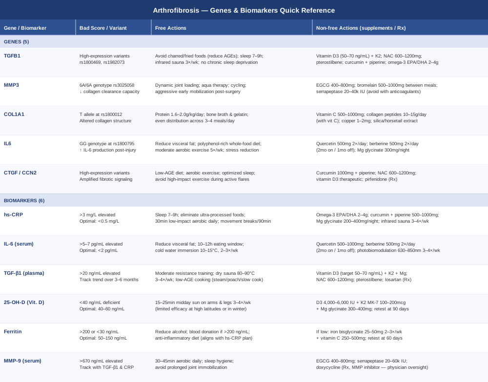
The table below consolidates all five genes and six biomarkers with their key action items for quick reference.
The Huberman Lab Perspective on Inflammation, Tissue Repair, and Recovery
The Huberman Lab podcast, hosted by Stanford neuroscientist Andrew Huberman, has produced some of the most accessible and research-grounded content on the mechanisms of inflammation, tissue remodeling, and post-injury recovery available to the general public. While no episode addresses arthrofibrosis directly, the biology Huberman covers in episodes on inflammation resolution, cold therapy, sauna, sleep, and tissue repair maps precisely onto the fibrotic pathways described in this article. The following synthesis draws from across his body of work to extract the ten most impactful insights for anyone managing arthrofibrosis.
1. Inflammation Is Not the Enemy — Its Duration Is
Huberman consistently distinguishes between acute inflammation (biologically necessary for healing) and chronic inflammation (the driver of pathological fibrosis). The first 72 hours after injury or surgery involve an inflammatory response that is essential — suppressing it too aggressively with NSAIDs or corticosteroids during this window may paradoxically impair long-term tissue repair by blunting the signals that initiate proper healing. The therapeutic goal is to support the resolution of inflammation, not to prevent it from occurring at all.
2. Cold Exposure Timing Changes Everything
Ice and cold therapy reduce acute pain — but used in the first 24–48 hours after surgery or significant joint injury, they may blunt the adaptive inflammatory signal needed for proper healing initiation. Drawing from work by exercise researchers, Huberman recommends waiting at least 48–72 hours before using therapeutic cold immersion. After that window, cold water immersion (10–15°C, 5–10 minutes) 2–3 times per week is a potent and well-evidenced tool for systemic anti-inflammatory effect, with particular impact on IL-6 and TNF-α levels.
3. Sleep Is Active Anti-Fibrotic Biology
Across multiple episodes, Huberman returns to sleep as the single highest-leverage recovery intervention available. During deep slow-wave sleep, growth hormone secretion peaks, anti-inflammatory cytokines are released, and collagen synthesis is regulated. Chronic poor sleep directly elevates TGF-β1, IL-6, and CRP — exactly the triad that drives arthrofibrosis progression. Seven to nine hours consistently is not passive recovery; it is active tissue regulation at the molecular level, and for arthrofibrosis patients it deserves the same priority as physical therapy sessions.
4. Morning Sunlight Regulates the Inflammatory Clock
Huberman's well-known morning sunlight protocol — 10–30 minutes of outdoor natural light within an hour of waking — regulates the circadian cortisol rhythm, which in turn governs inflammatory gene expression throughout the day. When cortisol peaks appropriately in the morning, it functions as a natural anti-inflammatory. Chronic disruption of this circadian cortisol pattern leads to sustained low-grade inflammatory signaling through the rest of the day. This is a zero-cost intervention with consistent mechanistic support and no side effects.
5. Zone 2 Cardio Is the Most Underused Anti-Inflammatory Tool
Huberman emphasizes zone 2 aerobic exercise — conversational pace, approximately 60–70% of maximum heart rate — for 45–60 minutes, three to four times per week, as one of the most consistent and durable reducers of systemic inflammatory markers over time. This intensity reduces IL-6, TNF-α, and CRP without generating excessive mechanical stress on recovering joints. For arthrofibrosis patients, aqua aerobics, stationary cycling, or swimming can deliver zone 2 benefits with minimal joint loading. This is not a substitute for physical therapy — it is a metabolic and anti-inflammatory complement to it.
6. Sauna Activates Protective Heat Shock Proteins
Regular sauna use (dry Finnish sauna, 80–90°C, 15–20 minutes, 3–4 times per week) activates heat shock proteins (HSPs), which function as molecular chaperones — they protect proteins from misfolding and aggregation under stress conditions. In the context of joint fibrosis, HSPs may help regulate TGF-β1 pathway activity and support normal protein turnover in connective tissue. Huberman cites dose-dependent cardiovascular and inflammatory benefits from sauna use; cardiovascular contraindications should be assessed before beginning, but for most individuals this represents one of the highest benefit-to-effort interventions available.
7. Breathing Protocols Directly Modulate NF-κB
Huberman has covered controlled breathing practices including box breathing, physiological sighing, and slow diaphragmatic breathing in depth. Slow controlled breathing activates the parasympathetic nervous system, which downregulates NF-κB — the master transcription factor that controls IL-6, TNF-α, and TGF-β1 expression. Five to ten minutes daily of slow diaphragmatic breathing (4 counts in, hold 4, 6 counts out) measurably shifts the autonomic balance and reduces inflammatory cytokine levels in contexts from chronic pain to inflammatory disease — and it costs nothing.
8. The Post-Exercise Protein and Collagen Timing Window
Drawing on work from collagen researcher Keith Baar and protein metabolism researchers, Huberman emphasizes consuming 15–40 g of protein or collagen hydrolysate within 30 minutes after rehabilitative exercise or physical therapy for joint conditions. This timing aligns with a peak in collagen synthesis signaling in connective tissue. Vitamin C (500 mg taken simultaneously) enhances hydroxylation of proline residues, improving the quality of collagen formed. For arthrofibrosis patients in rehabilitation, this is a practical, low-cost intervention that most physical therapy protocols never mention.
9. Omega-3s as a Biological Resolution Switch
EPA and DHA from omega-3 sources are enzymatically converted to resolvins and protectins — molecules that actively terminate inflammatory cascades rather than merely suppressing them. Huberman references research showing that omega-3 supplementation at 2–4 g EPA/DHA daily significantly reduces the duration of post-injury inflammation and supports the transition to the proliferative repair phase. In arthrofibrosis, where inflammation chronically fails to resolve, omega-3s at therapeutic doses deserve priority alongside any other intervention.
10. Psychological Stress Is a Direct Upstream Fibrosis Driver
Through detailed episodes on the HPA axis and stress physiology, Huberman makes explicit that psychological stress upregulates the same fibrotic pathways — TGF-β1, IL-6, NF-κB — as physical trauma. Chronic stress can worsen fibrotic outcomes by keeping the body in a state of sustained inflammatory signaling, independent of diet, exercise, or supplementation. Social connection, nature exposure, structured relaxation, and purposeful activity are not soft add-ons to an arthrofibrosis management plan — they are biological regulators of the inflammatory cascade and deserve to be treated as such.
Evidence-Based Complementary Approaches
The approaches below were selected specifically for arthrofibrosis based on their mechanistic relevance to joint fibrosis and their support from human clinical evidence. Evidence quality and condition-specificity vary across these modalities, and that variation is noted honestly.
Low-Level Laser Therapy (Photobiomodulation)
Photobiomodulation (PBM) uses specific wavelengths of red and near-infrared light — typically 630–1000 nm — to penetrate tissue and modulate cellular energy production, inflammation, and collagen synthesis at the mitochondrial level. In the context of arthrofibrosis, PBM is particularly relevant because it targets fibroblast activity, reduces pro-inflammatory cytokines at the local tissue level, and has shown the ability to support matrix remodeling without the mechanical risks of aggressive physical therapy during sensitive stages of recovery.
Multiple randomized controlled trials and systematic reviews have investigated LLLT for joint conditions including post-surgical knee recovery, where inflammatory and fibrotic mechanisms overlap substantially with arthrofibrosis. A systematic review published in the Cochrane database and multiple RCTs have confirmed PBM's anti-inflammatory efficacy and safety profile in musculoskeletal conditions. While arthrofibrosis-specific RCTs remain limited, the mechanistic rationale and overlapping inflammatory pathways support its use as a complementary tool. This is an area where the evidence base is growing, and current findings are encouraging rather than definitive.
Practical protocol: use a device delivering 50–200 mW at 810–850 nm applied directly over the affected joint for 5–10 minutes per site, three to four times per week. FDA-cleared home devices are available from manufacturers like Joovv, Mito Red, and Kineon at price points of $200–600. Allow at least 24 hours between sessions on the same joint. Avoid applying over areas with active infection, malignancy, or during acute inflammatory flare-ups severe enough to warrant rest.
Massage Therapy and Soft Tissue Mobilization
Soft tissue mobilization targets the periarticular adhesions, fascial restrictions, and capsular thickening that are the structural hallmarks of arthrofibrosis. Skilled massage therapy — particularly myofascial release techniques and instrument-assisted soft tissue mobilization (IASTM) — can mechanically disrupt collagen crosslinks in superficial layers of scar tissue, stimulate mechanoreceptors that modify the local fibroblast environment, and improve circulation to tissue that may be hypoxic due to fibrotic capsule compression.
Published systematic reviews and clinical trials in post-surgical joint fibrosis contexts have found that manual therapy, including targeted soft tissue techniques, produced clinically meaningful improvements in range of motion and pain scores when integrated with physical rehabilitation. Manual lymphatic drainage is particularly relevant in cases where post-surgical edema has contributed significantly to fibrotic capsule formation, as chronic lymphatic congestion maintains the inflammatory environment that sustains TGF-β1 and CTGF signaling.
In practical terms, this requires working with a licensed massage therapist or physical therapist specifically trained in post-surgical manual techniques — general wellness massage is not equivalent. Sessions one to two times per week for 8–12 weeks represents a typical therapeutic protocol for arthrofibrosis. Percussion massagers and foam rollers can supplement between sessions but cannot replicate the anatomical specificity of skilled manual therapy. Avoid aggressive deep tissue work during acute inflammatory flares, as excessive mechanical force on inflamed tissue can worsen cytokine release.
Mindfulness-Based Stress Reduction (MBSR)
MBSR is an 8-week structured program combining formal mindfulness meditation, body scan practices, and gentle movement, originally developed by Jon Kabat-Zinn at the University of Massachusetts. Its relevance to arthrofibrosis extends beyond pain management — though that evidence is robust across chronic musculoskeletal conditions — to its direct, measurable effects on inflammatory signaling. As described earlier in this article, chronic psychological stress upregulates TGF-β1, IL-6, and NF-κB activity, all of which drive fibrotic progression. MBSR directly addresses that stress-to-inflammation pathway.
Multiple meta-analyses have found that MBSR produces consistent, statistically significant reductions in inflammatory biomarkers including CRP and IL-6 across chronic inflammatory and pain conditions. The effect sizes are most pronounced in individuals with high baseline inflammatory burden — precisely the population most at risk for progressive or refractory arthrofibrosis. A 2016 RCT published in Brain, Behavior, and Immunity demonstrated that MBSR produced measurable reductions in inflammatory gene expression in peripheral blood cells, suggesting an effect that goes beyond stress-reporting to actual biological modulation.
The standard MBSR protocol involves 2.5 hours per week in structured sessions plus 45 minutes of daily home practice for 8 weeks. Many certified online programs are now available through accredited institutions, including the Center for Mindfulness at UMass Medical School, making access more practical. For arthrofibrosis, MBSR functions best as an adjunct to physical rehabilitation and medical management — not as a replacement for either. The combination of reduced inflammatory signaling and improved pain tolerance can make physical therapy sessions more productive and recovery more sustainable.
Breathing-Based Therapies
Structured breathing practices — including slow diaphragmatic breathing, box breathing, and the Buteyko method — modulate autonomic nervous system balance in ways that have measurable downstream anti-inflammatory effects. The mechanism connects directly to the arthrofibrosis biology described throughout this article: slow breathing activates the vagal nerve's anti-inflammatory arm, suppressing NF-κB-driven cytokine production and reducing circulating levels of IL-6, TNF-α, and related fibrosis-promoting signals.
Controlled studies in chronic pain populations and inflammatory conditions have consistently found that slow breathing at 5–6 breaths per minute significantly increases heart rate variability (HRV) — a reliable proxy for parasympathetic tone — and reduces inflammatory markers over consistent 8–12 week practice periods. For arthrofibrosis patients, the additional benefit of improved pain regulation through central sensitization pathways makes this a multi-mechanism tool: it addresses both the inflammatory biology and the chronic pain experience simultaneously.
A practical daily protocol: 10 minutes twice daily of box breathing (4 counts in, hold 4, out 4, hold 4) or slow diaphragmatic breathing (4 in, 6 out). This can be practiced supine — an advantage for those with limited joint mobility who cannot easily maintain seated posture. Guided breathing apps including Breathwrk, Othership, and the free DARE app offer accessible protocols. Consistent practice over 8–12 weeks is needed to observe measurable HRV and inflammatory improvements; the intervention asks for nothing more than time and attention.
Conclusion
Arthrofibrosis is not simply bad luck or a failure of rehabilitation. It is a biologically specific process driven by measurable inflammatory signals, fibrotic growth factors, and in some cases genetic predispositions that create outsized fibrotic responses even to standard surgical and rehabilitative inputs. The six biomarkers explored here — hs-CRP, IL-6, plasma TGF-β1, 25-hydroxyvitamin D, ferritin, and MMP-9 — give you a functional, real-time window into what your body is doing at the molecular level, and each one points toward specific, practical interventions that can meaningfully shift that biology. The five genes — TGFB1, MMP3, COL1A1, IL6, and CTGF — explain the underlying architecture that makes some individuals more vulnerable to fibrotic outcomes and point toward compensations tailored to that biology rather than to population averages.
The most useful next step is straightforward: assemble a panel that includes hs-CRP, serum IL-6, 25-hydroxyvitamin D, and ferritin at minimum. Add TGF-β1 and MMP-9 if you can access them through a functional medicine provider. Review those results with a clinician who understands joint fibrosis and can contextualize them alongside your clinical picture. If you have access to genetic testing, the TGFB1 and IL6 variants in particular are worth checking. Then build an individualized response — not one designed for an average patient, but one calibrated to what your specific biology is actually doing.
Musculoskeletal: Joint Conditions Tendon & Ligament Conditions Sports Injuries
Autoimmune: Inflammatory Conditions Connective Tissue Conditions