This article contains AI generated content.
· UpdatedArthrogryposis Multiplex Congenita Genes Biomarkers - 7 Genes And 5 Biomarkers To Track
Introduction
If you or someone in your family has a diagnosis of arthrogryposis multiplex congenita, one of the first things that becomes clear is how differently the condition presents from person to person. The number of joints involved, the pattern of contractures, whether muscle weakness is part of the picture, how much function can be recovered through early therapy — all of it varies in ways that standard descriptions of the condition rarely address. That variability is not arbitrary. In most cases, it traces back to specific genetic biology that differs from individual to individual, and that modern testing is increasingly able to identify.
Most clinical conversations about AMC still center on physical therapy, serial casting, and surgical options. These are genuinely important and often effective. But they were designed before genetic testing could reliably identify which gene — out of dozens of possible candidates — is driving the condition in any particular person. Whole-exome sequencing and targeted gene panels can now identify a pathogenic cause in 30 to 60 percent of AMC cases, and that number continues to grow as sequencing panels expand. Different gene variants carry different prognoses, different associated risks, and different responses to targeted interventions.
This article focuses on that precision. The genetics section covers seven of the most clinically significant genes implicated in AMC — what each gene does, what goes wrong when it mutates, and what the research supports in terms of targeted management, with specific plans for each variant. The biomarker section covers five measurable values that provide ongoing insight into muscle health, inflammation, and nutritional status, each with interpretation guidance and practical action plans. Beyond those two frameworks, the article includes a neuroplasticity-focused breakdown of research that changes how rehabilitation is approached, plus four complementary modalities with meaningful clinical evidence.
Better biology data does not guarantee better outcomes, but it consistently leads to better questions — and better questions lead to more targeted clinical conversations. The sections that follow are built to give you that foundation.
Summary
This article breaks down arthrogryposis multiplex congenita with more precision than most general health resources. The genetics section profiles 7 specific genes — PIEZO2, MYH3, RYR1, TPM2, TNNT3, TNNI2, and CHRNG — explaining what each does in fetal muscle and joint development, what happens when it mutates, and exactly what to do when a pathogenic variant is found (with and without supplements). The biomarker section covers 5 trackable lab values — creatine kinase, hsCRP, aldolase, 25-OH vitamin D, and protein markers — with cost ranges, interpretation guides, and action plans for when results fall outside optimal range. Beyond the biology, this article includes a neuroscience research breakdown with 10 insights that challenge standard rehabilitation thinking, plus four complementary approaches — massage therapy, photobiomodulation, biofeedback, and mindfulness — each grounded in real clinical evidence. If you have ever wondered why AMC looks so different from case to case, the genetics section is where that answer begins. The biomarker section shows you what to measure to know whether the body is holding up over time.
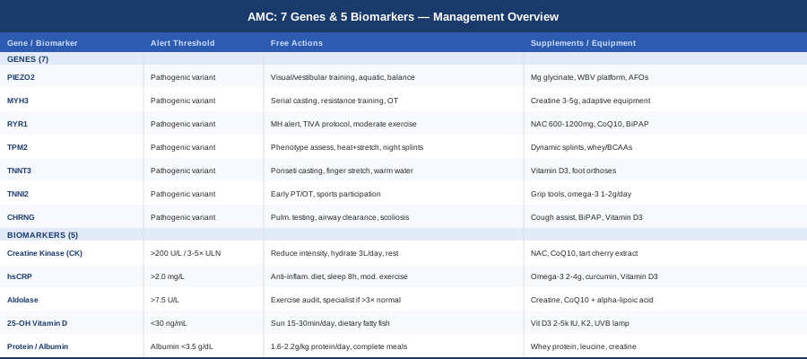
7 Genes Linked to Arthrogryposis Multiplex Congenita: What the Research Shows
AMC is not a single disease. It is a clinical descriptor — multiple congenital joint contractures — that can be produced by dozens of distinct genetic causes. As the StatPearls clinical overview details, the genetic architecture of AMC is highly heterogeneous, and the probability of identifying a pathogenic variant with whole-exome sequencing has risen sharply over the past decade. The seven genes below represent the most frequently implicated and best-characterized causes, with the most actionable evidence for targeted management.
For each gene, two paths are outlined: interventions that do not require supplements (the foundational work — movement, positioning, rehabilitation, and surveillance that produces the majority of the benefit) and those that add targeted supplements or equipment. Supplementation is an amplifier, not a foundation.
PIEZO2: The Proprioception Gene
PIEZO2 encodes a mechanosensitive ion channel that plays two simultaneous roles: it mediates proprioception — the sense of joint position and body location — in sensory neurons throughout the body, and it drives the mechanotransduction signals that guide joint morphogenesis during fetal development. When PIEZO2 is dysfunctional, both roles are disrupted at once: fetal joint development proceeds without the mechanical feedback that normally shapes it, and the postnatal nervous system lacks its primary channel for sensing limb position.
Studies published in the American Journal of Human Genetics identified pathogenic PIEZO2 variants in individuals with Gordon syndrome, Marden-Walker syndrome, and distal arthrogryposis type 5 — conditions that share joint contractures combined with progressive proprioceptive ataxia, scoliosis, and diminished vibration sense. This combination distinguishes PIEZO2-related AMC from purely myopathic forms and has direct, practical implications for how rehabilitation should be structured.
If PIEZO2 has a pathogenic variant — plan without supplements
The central strategy is to build compensatory sensory pathways using visual and vestibular systems to substitute for impaired proprioceptive input.
- Visual feedback training: Balance exercises performed in front of a mirror or video monitor, 20–30 minutes daily. The nervous system uses visual input to compensate for reduced joint position sense. Begin with supported standing and progress toward single-leg balance as stability improves. - Aquatic therapy: 2–3 sessions weekly in warm water (33–35°C). Water reduces fall risk and joint load while providing consistent sensory feedback and resistance for proprioceptive retraining. - Vestibular stimulation: Balance board training (Bosu ball, wobble board), 15 minutes daily, progressing from supported to unsupported positions. Strengthens vestibular-cerebellar circuits that can partially substitute for PIEZO2-dependent sensory input. - Scoliosis surveillance: Annual spinal imaging from age 3. PIEZO2-related scoliosis can progress quickly; early bracing significantly reduces the probability of surgical intervention. - Gait analysis every 1–2 years in ambulatory individuals to detect subtle deterioration before functional decline becomes apparent.
If PIEZO2 has a pathogenic variant — plan with supplements or equipment
No supplement directly restores PIEZO2 channel function. The following support the surrounding neuromuscular context.
- Magnesium glycinate: 200–400 mg/day for adults (weight-adjusted for children with physician guidance). Supports neuromuscular excitability and reduces cramping common in proprioceptive disorders. Daily with food; no cycling required. Dose-dependent loose stools are the primary side effect at higher doses. - Whole-body vibration (WBV) platforms: 20–50 Hz, 10–15 minutes per session, 3–5 times weekly. Stimulates muscle spindles and sensory afferents, partially compensating for reduced PIEZO2-mediated mechanosensing. Evidence in related proprioceptive disorders is emerging; AMC-specific data remain limited but the mechanism is sound. - Custom ankle-foot orthoses (AFOs): Provide external joint positioning stability, substituting for the missing proprioceptive anchoring. Fitting by a certified orthotist; review every 6–12 months in growing children.
MYH3: The Embryonic Myosin Gene
MYH3 encodes embryonic myosin heavy chain, the motor protein driving fetal muscle contractions during the critical window of joint morphogenesis. Pathogenic variants in MYH3 cause some of the most common and clinically recognizable forms of distal arthrogryposis: Freeman-Sheldon syndrome (DA2A) and Sheldon-Hall syndrome (DA2B). MYH3 variants are among the most frequently identified genetic causes in familial AMC.
The key clinical insight about MYH3 is developmental restriction. After birth, muscle fibers transition to adult myosin isoforms (MYH1 and MYH2), making postnatal contractile function largely intact. The contractures at birth reflect a fetal-period deficit, not an ongoing one. This is why MYH3-related AMC typically shows the best response to early rehabilitation among all genetic subtypes — the machinery for postnatal muscle function is fundamentally sound.
If MYH3 has a pathogenic variant — plan without supplements
Because postnatal muscle function is largely preserved, early aggressive rehabilitation can achieve outcomes that would not be possible in conditions with continuing myopathy.
- Serial casting: Begin within the first weeks of life; change casts every 1–2 weeks. The Ponseti method for clubfoot achieves correction in over 90% of cases when started early and maintained with 3–4 years of nighttime bracing. - Resistance training: From toddler age, age-appropriate functional resistance exercises 3–4 times weekly emphasizing hand intrinsics, wrist extensors, and ankle dorsiflexors. Functional tasks — picking up objects, pushing, pulling — are more effective than isolated joint contractions. - Occupational therapy: Fine motor skill development beginning at 6 months if possible. Adaptive tools, grip strengthening, and 6-monthly hand function evaluation through the first decade. - Surgical review at age 3–5 for persistent contractures limiting function; tendon lengthening procedures produce excellent outcomes in MYH3-related AMC specifically.
If MYH3 has a pathogenic variant — plan with supplements or equipment
- Creatine monohydrate: 3–5 g/day in adults; reserved for age 8+ in children with physician oversight. Supports ATP regeneration during intensive rehabilitation. No loading phase required. Ensure 500 ml extra water daily. Side effects are minimal. - Adaptive equipment assessment: Universal cuffs, built-up utensil handles, custom keyboard and mouse configurations via an occupational therapist, reviewed every 2–3 years as needs evolve.
RYR1: The Ryanodine Receptor Gene
RYR1 encodes the ryanodine receptor type 1, the primary calcium release channel of the sarcoplasmic reticulum in skeletal muscle. When a motor nerve fires, RYR1 channels open and release calcium that triggers contraction. Pathogenic RYR1 variants are the most common cause of congenital myopathy and account for a meaningful proportion of AMC cases, particularly those presenting with hypotonia, proximal weakness, and abnormal muscle biopsy findings such as central core or multiminicore disease.
The most clinically urgent aspect of confirmed RYR1 pathogenic variants is malignant hyperthermia (MH) susceptibility — a life-threatening hypermetabolic crisis triggered by halogenated volatile anesthetics (halothane, sevoflurane, desflurane) and the depolarizing muscle relaxant succinylcholine. Every individual with a confirmed pathogenic RYR1 variant must be treated as MH-susceptible, and this information must reach every member of the clinical team, including dentists and proceduralists.
If RYR1 has a pathogenic variant — plan without supplements
- Medical alert documentation: Carry a written card stating the RYR1 variant and MH susceptibility. Register with the Malignant Hyperthermia Association of the United States (MHAUS). Provide written documentation to every anesthesiologist in advance of every procedure. This step cannot wait. - Anesthesia protocol: Total intravenous anesthesia (TIVA) or a fully non-triggering protocol for every procedure. Confirm dantrolene availability at the facility beforehand. - Moderate-intensity aerobic exercise: Cycling, swimming, and elliptical at perceived effort 4–6/10, 20–30 minutes 4–5 times weekly. High-intensity work triggers calcium dysregulation in RYR1-affected muscle, causing rhabdomyolysis. Moderate intensity is both safer and more effective long-term. - Annual pulmonary function testing: FVC, FEV1, and maximal inspiratory pressure. Diaphragmatic weakness in RYR1 myopathy can progress silently; proactive monitoring allows respiratory support before a crisis develops. - Avoid prolonged fasting before procedures and manage body temperature carefully during exercise in warm environments.
If RYR1 has a pathogenic variant — plan with supplements or equipment
- N-acetylcysteine (NAC): 600–1200 mg/day in adults, divided into 2 doses with meals. Preliminary human evidence from the RYR1 Foundation research network suggests NAC reduces oxidative stress in RYR1-affected muscle fibers. Cycle: 8 weeks on, 2 weeks off. Side effects: occasional mild nausea at higher doses. - Coenzyme Q10: 200–300 mg/day with a fatty meal. Supports mitochondrial function and reduces secondary muscle fatigue. No cycling required; broadly used in RYR1 myopathy communities with a favorable safety profile. - Overnight pulse oximetry and home BiPAP: For FVC below 50–60% predicted, establish overnight monitoring. If saturation regularly falls below 93%, non-invasive ventilation initiated with pulmonology supervision dramatically improves sleep quality and CO2 management.
TPM2: The Beta-Tropomyosin Gene
TPM2 encodes beta-tropomyosin, a thin-filament regulatory protein that controls when myosin heads are permitted to bind actin and generate force. Pathogenic variants produce a phenotypic range spanning nemaline myopathy, cap disease, and distal arthrogryposis, depending on the specific variant and its molecular effect. Gain-of-function variants that increase the filament's calcium sensitivity tend to produce prominent contractures and joint stiffness. Dominant-negative variants that reduce calcium sensitivity produce more weakness-dominant presentations.
This distinction between hypercontractile and hypotonic presentations is clinically critical because it directs the rehabilitation approach in opposite directions. A hypertonic TPM2 presentation calls for stretching, heat application, and soft-tissue release; a hypotonic one calls for progressive resistance loading. Without identifying the functional variant class, therapy can work against the patient's actual biology.
If TPM2 has a pathogenic variant — plan without supplements
- Phenotype-directed assessment first: Determine whether the presentation is primarily hypercontractile or hypocontractile before designing the exercise program. A neuromuscular specialty center can clarify this through functional calcium sensitivity studies or detailed clinical assessment. - Thermotherapy before stretching: 10–15 minutes of moist heat applied to the affected joint immediately before stretching reduces passive tissue resistance by approximately 20–30% and significantly improves the degree of stretch achievable. - Daily sustained stretching: Holds of 30–60 seconds per position, designed by a physical therapist, performed daily with specific focus on hands, wrists, and any other involved joints. - Night splinting: Static night splints maintaining joints at maximum corrected position. Fitted by a hand therapist and reviewed every 6–12 months as correction progresses.
If TPM2 has a pathogenic variant — plan with supplements or equipment
- Dynamic day splinting: Spring-loaded or elastic orthoses applying gentle, continuous stretch during waking hours. Combined with static night splints, this is the most evidence-based approach to managing hand and wrist contractures in thin filament myopathies. - Whey protein isolate or BCAAs: 25–40 g protein daily for adults, taken within 2 hours post-exercise. Supports muscle protein synthesis and reduces atrophy in thin filament myopathies. No cycling required.
TNNT3: The Fast Troponin T Gene
TNNT3 encodes troponin T type 3, the fast-twitch skeletal muscle isoform responsible for anchoring the troponin complex to tropomyosin along the thin filament. Pathogenic variants cause distal arthrogryposis, typically presenting with features of Sheldon-Hall syndrome: camptodactyly, clubfoot, mild ptosis, and short stature. Inheritance is autosomal dominant with notable variable expressivity — meaning an apparently unaffected parent may carry the same variant as a significantly affected child, a pattern that complicates family recognition but provides useful prognostic information.
TNNT3 variants alter the calcium sensitivity of muscle activation, reducing the force of fetal contractions during the critical window of joint formation. Because postnatal muscle function is relatively preserved, early intervention — particularly for foot and hand contractures — has an excellent evidence base in this subtype.
If TNNT3 has a pathogenic variant — plan without supplements
- Ponseti method for clubfoot: Weekly serial casting from the first weeks of life. Achilles tenotomy in the majority of cases, followed by Denis Browne bar bracing worn full-time until walking age and nightly for 3–4 years. Relapse rate with full bracing compliance is consistently below 10%. - Passive finger extension stretching: 5–10 repetitions, 3 times daily from infancy. Technique taught to parents by a hand therapist; fits naturally into diaper changes and daily care. - Daily warm water activity: 20–30 minutes in warm water reduces joint stiffness and encourages spontaneous hand and foot movement. Both therapeutic and enjoyable for infants and young children. - Annual hand therapy review through adolescence; contracture relapse during growth spurts is common and responds to a brief intensive therapy course.
If TNNT3 has a pathogenic variant — plan with supplements or equipment
- Vitamin D3: 1000–2000 IU/day for children, 2000–4000 IU/day for adults. Foot contractures alter mechanical loading and increase fracture risk; bone density support is worth maintaining. Recheck 25-OH-D every 6 months. - Custom foot orthoses and high-top supportive footwear: Essential for preventing contracture recurrence after casting. Pediatric orthotics clinic fitting; revisited every 6–12 months in growing children.
TNNI2: The Fast Troponin I Gene
TNNI2 encodes the inhibitory subunit of the troponin complex in fast skeletal muscle. Variants in TNNI2 cause distal arthrogryposis type 1 (DA1), the most common and typically mildest form of the distal arthrogryposis spectrum — primarily camptodactyly and clubfoot without significant systemic involvement. The prognosis for independent ambulation and functional hand use with appropriate early intervention is excellent among all AMC genetic subtypes.
Variable expressivity is a defining characteristic of TNNI2-related AMC. Family surveys consistently document that the same pathogenic variant can produce markedly different severity across relatives — from clearly affected individuals to those who would never seek medical attention. This variability complicates diagnosis but also offers a reassuring implication: the genotype does not fully determine the outcome.
If TNNI2 has a pathogenic variant — plan without supplements
- Early physical and occupational therapy: Investment in the first 2 years of life, when neuroplasticity and tissue compliance are highest, produces the best measurable long-term outcomes. Most children with DA1 treated early achieve independent ambulation and good hand function. - Sports participation: Children with DA1 can participate in most sports. Swimming, cycling, and modified gymnastics build strength and coordination without excessive joint stress. Forced exclusion from physical activity is counterproductive in the majority of cases. - Annual occupational therapy review through adolescence to identify and address any functional regression during growth spurts. - Genetic counseling for the family; cascade testing of potentially affected relatives is worth offering, as early identification benefits those identified in infancy.
If TNNI2 has a pathogenic variant — plan with supplements or equipment
- Grip strengthening tools: Therapy putty, hand exercisers, and resistance bands used 10–15 minutes daily. Expect 3–6 months of consistent use before significant improvement is measurable. - Omega-3 fatty acids (EPA + DHA): 1–2 g/day with food. Anti-inflammatory and muscle-supportive effects; no cycling required. Particularly useful when hsCRP is elevated or stiffness is a limiting factor.
CHRNG: The Fetal Acetylcholine Receptor Gene
CHRNG encodes the gamma subunit of the fetal-specific nicotinic acetylcholine receptor at the neuromuscular junction. This receptor — which substitutes a gamma subunit for the adult epsilon subunit — is expressed specifically during fetal development to transmit nerve signals to developing muscle. Loss-of-function mutations in CHRNG cause Escobar syndrome (non-lethal multiple pterygium syndrome), a severe form of AMC characterized by joint contractures, skin webbing across joints (pterygia), scoliosis, and frequently significant respiratory compromise.
The mechanism is fetal neuromuscular transmission failure: without functional gamma-subunit receptors, nerve-to-muscle signaling during fetal life is severely impaired, fetal movement is drastically reduced, and the structural consequences — contractures and pterygium formation — are established before birth. Adult neuromuscular transmission is intact because the adult epsilon subunit is unaffected. The damage is the legacy of fetal immobility, and it is permanent, which makes proactive long-term management the only meaningful strategy.
If CHRNG has a pathogenic variant — plan without supplements
Respiratory management is the highest immediate priority, given the frequency and severity of respiratory muscle weakness and restrictive lung disease from progressive scoliosis.
- Pulmonary function testing at baseline and annually: FVC, FEV1, and maximal inspiratory pressure. Diaphragm ultrasound for non-invasive assessment of diaphragmatic excursion. - Airway clearance techniques: High-frequency chest wall oscillation (vest device) or mechanical insufflator-exsufflator (cough assist). Daily in those with FVC below 50% predicted; during respiratory illness for those with higher function. - Scoliosis monitoring and intervention: Spinal X-ray every 6 months in growing children; bracing initiated at Cobb angle 20–25 degrees; surgical evaluation when curves threaten respiratory function. - Multidisciplinary care coordinated through a neuromuscular specialty center: pulmonology, orthopedics, and respiratory therapy as a minimum core team.
If CHRNG has a pathogenic variant — plan with supplements or equipment
- Cough assist device (e.g., CoughAssist E70): 3–5 cycles, 2–4 times daily during respiratory illness; once daily for maintenance when FVC falls below 60% predicted. Substantially reduces hospitalization risk during viral respiratory illness. - Vitamin D3: 2000–4000 IU/day with physician monitoring. Reduced mobility and limited outdoor time make vitamin D deficiency both common and consequential; bone density protection matters given the surgical exposure and fracture risk in this population. - Nighttime BiPAP: Initiated when FVC falls below 50% predicted or when overnight oximetry shows saturation below 93%. Managed with pulmonology; dramatically improves sleep quality and reduces morning CO2 retention.
Understanding which gene is involved gives you the map. Knowing what to measure over time gives you a way to track whether the terrain is changing — whether interventions are working, whether complications are developing silently, and whether the body's nutritional and metabolic foundations are intact. The five biomarkers below are worth monitoring across AMC subtypes.
5 Biomarkers to Monitor in AMC: What to Test and Why
Biomarkers do not change the underlying genetics, but they provide an ongoing read of how the body is functioning in response to disease, rehabilitation, and the demands of daily life. For AMC, where muscle, nerve, and connective tissue involvement varies by genetic cause, a targeted biomarker panel can reveal whether muscle integrity is being maintained, whether inflammation is silently limiting rehabilitation gains, and whether nutritional deficiencies are holding outcomes back.
Creatine Kinase (CK)
Creatine kinase (CK) is the most direct blood marker of skeletal muscle integrity. When muscle fibers are damaged or under excessive physiological stress, CK leaks from cells into the bloodstream. In AMC, monitoring CK helps distinguish forms with active ongoing muscle fiber pathology — such as RYR1-related myopathy and some TPM2 variants — from the more structurally driven contractures without meaningful muscle fiber turnover seen in most MYH3, TNNI2, and TNNT3 cases. This distinction matters for how aggressively exercise intensity can be pushed.
How to measure CK
Standard serum blood draw, available at any laboratory. Cost: $15–50 without insurance; typically included in or added to a standard metabolic workup. Normal range: 20–200 U/L in most adults (women generally run lower). Draw should follow at least 24 hours without strenuous exercise to avoid post-exercise elevation masking the baseline value. Recommended frequency: every 6–12 months, or whenever a new exercise program is initiated.
If CK is elevated — plan without supplements
Resting CK persistently above 3–5 times the upper limit of normal should prompt specialist review and reassessment of the genetic cause.
- Reduce exercise intensity to the level at which CK normalizes; for RYR1 cases, this typically means keeping perceived exertion below 6/10. - Increase recovery time between exercise sessions; add scheduled rest days to the weekly structure. - Maintain hydration at a minimum of 3 liters daily to reduce renal risk from elevated circulating myoglobin. - Avoid routine NSAID use; at regular doses, NSAIDs impair muscle repair signaling and may worsen longer-term outcomes.
If CK is elevated — plan with supplements or equipment
- NAC (600–1200 mg/day in divided doses): Most appropriate when a RYR1 mutation is confirmed; reduces the oxidative stress driving fiber membrane leakage. Cycle: 8 weeks on, 2 weeks off. - Coenzyme Q10 (200–300 mg/day with a fatty meal): Supports mitochondrial function and reduces secondary muscle damage. No cycling required; well tolerated. - Tart cherry extract (standardized to anthocyanins): Taken daily. Multiple studies document meaningful reductions in post-exercise CK elevation and muscle soreness. No cycling required; safe for long-term use.
High-Sensitivity CRP (hsCRP)
High-sensitivity C-reactive protein is among the most sensitive blood markers of systemic low-grade inflammation. In AMC, chronic inflammation can develop from abnormal joint mechanics creating repeated microtrauma, from the reduced physical activity that increases inflammatory baseline, or from subclinical respiratory or other secondary complications. Persistently elevated hsCRP impairs rehabilitation gains, contributes to fatigue, and is associated with accelerated joint degradation over the long term — making it worth tracking even when it is not immediately symptomatic.
How to measure hsCRP
Standard serum blood draw; specifically request high-sensitivity CRP rather than standard CRP, which misses the low-level chronic elevation that is most clinically relevant. Cost: $20–60 without insurance. Optimal level: below 1.0 mg/L, consistent with the threshold recommended by longevity-focused clinicians including Peter Attia. Concerning: above 2.0 mg/L chronically; clearly elevated: above 3.0 mg/L. Frequency: annually as a baseline; every 3–6 months if previously elevated or if rehabilitation program has recently changed significantly.
If hsCRP is elevated — plan without supplements
- Address mechanical contributors: Abnormal joint loading from contractures or irregular gait patterns creates repeated microtrauma that drives chronic inflammation. Orthoses, gait optimization, and joint protection techniques systematically reduce this source. - Anti-inflammatory dietary pattern: Fatty fish 3 servings weekly, increased colorful vegetables, reduction of ultra-processed foods and refined sugars. Dietary changes produce measurable CRP reductions within 4–6 weeks of consistent change. - Sleep optimization: 7–9 hours nightly. Sleep deprivation is one of the most powerful drivers of elevated CRP. Pain, positioning challenges, and respiratory issues can all disrupt sleep in AMC; address them proactively. - Moderate aerobic exercise 4–5 times weekly: Consistently and dose-dependently reduces hsCRP. Excessive training raises it; the dose-response curve is non-linear, which makes the moderate zone especially important.
If hsCRP is elevated — plan with supplements or equipment
- Omega-3 fatty acids (EPA + DHA, 2–4 g/day): The most evidence-based supplement for reducing hsCRP across conditions. Taken with a fatty meal for absorption; no cycling required. - Bioavailable curcumin (500–1000 mg/day) with piperine: Multiple meta-analyses confirm significant CRP reductions. Cycle: 12 weeks on, 4 weeks off. Rare GI side effects; caution with high doses in gallstone disease. - Vitamin D3: Correct any deficiency (target 40–60 ng/mL of 25-OH-D). Low vitamin D independently predicts elevated CRP; addressing deficiency often produces meaningful CRP reduction before any other intervention is added.
Aldolase
Aldolase is a glycolytic enzyme found in high concentrations in skeletal muscle. It provides complementary information to CK about muscle fiber integrity — in some myopathies affecting fast-twitch oxidative fibers (particularly RYR1-related forms), aldolase may be more sensitively elevated than CK and can reveal muscle cell stress that CK misses. Interpreted alongside CK, aldolase helps characterize the pattern and metabolic severity of muscle fiber turnover.
How to measure aldolase
Standard serum blood test, ordered as part of a muscle enzyme panel or independently. Cost: $30–80 without insurance. Normal range: 1.0–7.5 U/L in most adults; slightly higher in children. Best interpreted alongside CK: disproportionate aldolase elevation relative to CK may indicate fast-twitch fiber–predominant pathology, which warrants specialist attention. Frequency: annually or whenever CK values change significantly.
If aldolase is elevated — plan without supplements
- Review the CK-to-aldolase ratio with a neuromuscular specialist to characterize the pattern; this can meaningfully guide investigation and management. - Audit the exercise program: high-intensity eccentric work elevates both markers. Reduce intensity if values are elevated following a program change and reassess after 4 weeks. - Referral for neuromuscular specialist evaluation if aldolase exceeds 3x normal at rest despite activity reduction.
If aldolase is elevated — plan with supplements or equipment
- Creatine monohydrate (3–5 g/day): Supports ATP regeneration in muscle fibers, reducing the metabolic stress that drives enzyme leakage. Most relevant when aldolase elevation accompanies exercise intolerance or early fatigue. - CoQ10 (200–300 mg/day) combined with alpha-lipoic acid (300–600 mg/day): Together address oxidative damage to mitochondria in muscle fibers. Both well tolerated at these doses; no cycling required.
25-Hydroxyvitamin D (25-OH-D)
Serum 25-hydroxyvitamin D is the standard measure of vitamin D status, and deficiency is both common and underappreciated in AMC. Reduced mobility, limited outdoor access, and restricted sun exposure combine to make deficiency nearly universal in this population if not actively managed. The consequences are tangible and directly relevant to AMC: impaired bone mineral density increases fracture risk from abnormal loading patterns, vitamin D receptors expressed in skeletal muscle mean that deficiency reduces muscle force production, and low vitamin D independently predicts elevated inflammatory markers.
How to measure 25-OH-D
Standard serum blood test; specify 25-hydroxyvitamin D (not 1,25-dihydroxyvitamin D, which reflects a different part of vitamin D metabolism). Cost: $30–80 without insurance. Optimal functional range: 40–60 ng/mL, consistent with the thresholds recommended by Peter Attia and other longevity-focused clinicians. Below 30 ng/mL is deficient; below 20 ng/mL is severely deficient. Test twice annually — fall and spring — to capture seasonal variation.
If 25-OH-D is low — plan without supplements
- Maximize outdoor sun exposure: 15–30 minutes of midday sun to arms and face without sunscreen on most clear days meaningfully raises vitamin D levels through summer months. Schedule outdoor time as a structured part of the weekly routine. - Remove accessibility barriers to outdoor mobility: accessible outdoor environments, adapted transportation, and scheduled outdoor activity periods. - Dietary sources (fatty fish, egg yolks, fortified foods) provide modest contributions — worth maximizing but insufficient alone for correcting significant deficiency.
If 25-OH-D is low — plan with supplements or equipment
- Vitamin D3 (cholecalciferol): 2000–5000 IU/day with a fatty meal. Recheck 25-OH-D in 3 months to confirm response. Most individuals with AMC require 3000–4000 IU/day year-round to maintain optimal levels. - Vitamin K2 (MK-7 form, 100–200 mcg/day): Directs calcium to bone rather than soft tissues, amplifying D3's bone-protective effect. No cycling; negligible side effects at these doses. - UVB lamp: For high-latitude residents or those unable to access outdoor sun regularly. A D-bulb UVB lamp used for 15–30 minutes of skin exposure 3–4 times weekly through winter months is a practical cost-effective alternative.
Serum Protein and Amino Acid Status
Protein adequacy is the nutritional foundation of muscle repair and rehabilitation progress — and it is one of the most frequently overlooked limiters of outcome in AMC. In growing children and in adults undergoing intensive rehabilitation, insufficient protein means the stimulus from exercise cannot be fully converted into structural muscle improvement. Serum albumin, prealbumin (transthyretin), and a full amino acid panel together provide a comprehensive picture of protein nutritional status across different timescales.
How to measure protein status
- Serum albumin: Included in the comprehensive metabolic panel (CMP). Cost: $25–60 as part of CMP. Optimal: 4.0–5.0 g/dL. Below 3.5 g/dL indicates protein depletion; this marker reflects average status over weeks to months. - Prealbumin (transthyretin): More sensitive to recent dietary changes than albumin. Cost: $30–60 standalone. Normal: 18–45 mg/dL. More responsive to acute-onset nutritional decline. - Full amino acid panel: Identifies specific deficiencies (leucine, methionine, glutamine, others). Cost: $200–400 through specialty labs. Order once at baseline; repeat if major dietary changes are made or rehabilitation progress stalls without clear explanation.
If protein status is low — plan without supplements
- Target 1.6–2.2 g of protein per kilogram of body weight per day during active rehabilitation; 1.2–1.6 g/kg/day as an ongoing maintenance minimum. - Prioritize complete protein at every meal: eggs, fish, poultry, Greek yogurt, legumes combined with grains. - Work with a pediatric or sports dietitian to meet targets without caloric excess — reduced mobility in AMC lowers total energy requirements, and overfeeding has its own consequences. - Use a 3–7 day dietary diary to establish a baseline; most people significantly underestimate actual protein intake when assessing it mentally.
If protein status is low — plan with supplements or equipment
- Whey protein isolate: 25–40 g per serving within 2 hours post-exercise or as a between-meal supplement. Complete essential amino acid profile; very low lactose in isolate form. For dairy intolerance: pea protein isolate is the most complete plant alternative. - Leucine (2–3 g per meal): The principal trigger of muscle protein synthesis signaling. If a full amino acid panel shows leucine insufficiency specifically, targeted supplementation corrects this efficiently. Available as a standalone supplement or in leucine-enriched BCAA products. - Creatine monohydrate (3–5 g/day) works synergistically with adequate protein intake to maximize muscle protein accretion; the combination consistently outperforms either intervention alone.
The genetics and biomarker frameworks above provide the biology of AMC in actionable form. The section that follows draws from neuroscience research that directly challenges many of the standard assumptions about how rehabilitation in conditions like this should be approached — and offers a framework that has changed outcomes for many people working with neuromuscular and musculoskeletal conditions.
Neuroscience Research That Changes How You Think About AMC Rehabilitation
Andrew Huberman, professor of neurobiology and ophthalmology at Stanford University School of Medicine, has produced one of the most rigorously research-referenced public health series available — drawing from peer-reviewed neuroscience and physiology across hundreds of hours of content. His podcast systematically challenges conventional wisdom on physical rehabilitation, including topics directly relevant to AMC management. What follows are the ten most applicable insights from that body of work, synthesized for the specific challenges of arthrogryposis.
1. Short, Deliberate Stretch Intervals Outperform Long, Passive Sessions
Huberman's deep dive into the neuroscience of stretching consolidates multiple studies showing that 5 repetitions of 30-second stretches performed at moderate discomfort (not pain) produce greater range-of-motion gains than longer, relaxed passive stretching sessions. The mechanism involves activating the golgi tendon organ, which inhibits muscle spindle resistance and allows genuine tissue elongation rather than just relaxation reflex. For AMC contractures, this reframes daily stretching: shorter, more deliberate, and appropriately uncomfortable sessions are more productive than longer passive ones.
2. Tissue Temperature Has a Measurable Effect on Stretch Effectiveness
Research synthesized in Huberman's training and recovery episodes confirms that moist heat applied for 10–15 minutes before a stretch session reduces passive connective tissue stiffness by a meaningful degree and consistently improves the range of motion achievable in that session. Cold application after resistance training (ice pack or cold water for 10–15 minutes) reduces post-exercise inflammatory signaling and accelerates recovery. These bookends are underused in AMC rehabilitation programs and add measurable, low-cost benefit.
3. Motor Neuroplasticity Does Not Have a Developmental Cutoff
A consistent thread across Huberman's neuroplasticity episodes is that the motor cortex and motor learning circuits remain plastic throughout adult life. The most intense plasticity occurs in early development, but deliberate, attention-focused motor practice at any age produces structural changes in motor cortex and cerebellar circuitry. For adults with AMC who received limited early intervention, this is clinically meaningful: intensive rehabilitation in adulthood continues to produce gains, and the concept of a permanent developmental ceiling is not supported by current neuroscience.
4. Chronic Stress Directly Degrades Motor Learning Capacity
Huberman covers the neurological pathway through which chronic stress impairs motor skill acquisition: sustained high cortisol suppresses hippocampal and motor cortex plasticity, slowing the consolidation of new movement patterns. For families managing the psychological burden of AMC — the clinical coordination, functional limitations, and social complexity involved — this is directly relevant. Stress management is not separate from rehabilitation success; it is a biological precondition for it. MBSR, sleep optimization, and social support all produce measurable changes in stress biology that affect rehabilitation outcomes.
5. Sleep Is the Mechanism of Motor Skill Consolidation
Among the most consistently supported findings in Huberman's episodes on learning and memory: motor skill consolidation occurs primarily during slow-wave and REM sleep, not during practice itself. Practice triggers the plasticity; sleep encodes the new patterns. For AMC rehabilitation, this elevates sleep from a recovery concern to a primary therapeutic variable. Eight hours of consistent, quality sleep should be treated as a non-negotiable component of the rehabilitation program, as important as any scheduled therapy session.
6. Cold Exposure Provides a Low-Cost Anti-Inflammatory Tool
Huberman covers the research on deliberate cold exposure (10–20°C water, 1–3 minutes) as a tool for reducing systemic inflammatory cytokines and increasing norepinephrine, which modulates pain and improves mood and focus. For individuals with elevated hsCRP from chronic joint microtrauma in AMC, structured cold exposure provides an accessible, low-cost anti-inflammatory intervention with a favorable safety profile for most people. Cold showers ending with 60–90 seconds fully cold achieve a meaningful fraction of the immersion benefit.
7. Effort-Level Drives the Most Durable Strength Adaptations
Huberman's synthesis of resistance training research consistently points to the same finding: the highest-effort repetitions, nearest to volitional failure, produce the most potent signal for muscle adaptation. For AMC rehabilitation, which often defaults to low-load, high-repetition protocols out of excessive caution, this suggests meaningful recalibration. In forms where muscle pathology is absent or minimal — MYH3, TNNI2, TNNT3 — working to near-failure within safe joint range (6–12 repetitions) produces more durable strength gains than cautious, low-effort training.
8. Proprioceptive Training Produces Central Nervous System Remodeling
Huberman's material on sensory systems confirms that balance and proprioceptive training produce measurable structural changes in sensory cortex and cerebellum, not just at peripheral nerve level. For PIEZO2-related AMC, where the primary mechanosensing channel at the periphery is absent, this has significant implications: intensive balance training physically remodels the central representation of body position over time. The compensatory rewiring is real and is not limited to an early developmental window.
9. Chronic Pain and Tissue Damage Are Neurobiologically Different Problems
Huberman's pain neuroscience episodes address a point consistently underemphasized in AMC clinical care: chronic pain in musculoskeletal conditions is substantially maintained by central sensitization — the brain amplifying threat signals independently of ongoing tissue damage. Anti-inflammatory interventions target tissue damage; central sensitization requires different tools: graded sensory exposure, mindfulness practice, sleep optimization, and in some cases low-dose naltrexone under medical supervision. Conflating the two leads to intervention mismatch.
10. Zone 2 Aerobic Training Provides the Largest Systemic Benefits With the Lowest Joint Cost
The zone 2 aerobic training framework Huberman presents — sustained, conversational-pace aerobic work for 30–45 minutes, 4–5 days per week — produces disproportionate gains in mitochondrial density, anti-inflammatory signaling, and metabolic flexibility. For AMC, where high-impact and high-load exercise must be carefully managed, zone 2 via stationary cycling, aquatic jogging, or elliptical training maximizes systemic health benefits while staying reliably within safe joint loading parameters. It is the single most accessible high-yield cardiovascular and anti-inflammatory tool in this population.
These neuroscientific insights work best when integrated with the evidence-based complementary approaches that follow — each of which addresses a different facet of the AMC experience that genetics, biomarkers, and standard physical therapy alone do not fully reach.
Complementary Approaches With Clinical Evidence for Arthrogryposis
The following modalities are selected because they have meaningful human clinical evidence and a plausible mechanism of action for the specific challenges of AMC — joint stiffness, contracture management, motor rehabilitation, and the psychological burden of long-term disability. None of them cure anything; all of them add measurable value when integrated thoughtfully with the core medical program.
Massage Therapy
Massage therapy in AMC targets the soft tissue component of contracture: the secondary stiffness, fibrotic change, and compensatory muscle tension that accumulate around affected joints over time. The underlying genetic mutation cannot be addressed through manual contact, but the periarticular tissue changes that worsen contractures and reduce the effectiveness of casting and splinting can be meaningfully improved. Improved tissue extensibility makes subsequent stretching, casting, and orthotic management more effective and may reduce the total intervention burden over time.
A clinical technique particularly relevant to congenital contractures is myofascial release (MFR) applied to the joint capsule, tendon sheaths, and surrounding connective tissue. Pediatric rehabilitation literature documents that structured soft tissue mobilization applied before serial casting sessions reduces mechanical resistance to correction and may decrease the total number of casting cycles required to achieve functional range of motion. The mechanism involves sustained, low-load elongation of fibrotic connective tissue, gradually reducing its resistance to passive joint movement.
In practice, massage and MFR for AMC should be delivered by a therapist with specific pediatric neuromuscular experience. Sessions of 30–45 minutes, 2–3 times weekly, coordinated with the rehabilitation team, produce the most consistent benefit. Parents can be taught simplified home mobilization techniques — 10 minutes of gentle soft-tissue work before daily stretching — extending the therapeutic window without requiring daily clinic attendance. Avoid vigorous techniques near recent surgical sites until healing is fully confirmed.
Low-Level Laser Therapy (Photobiomodulation)
Low-level laser therapy uses specific wavelengths of red and near-infrared light (typically 630–1000 nm) to stimulate cytochrome c oxidase in the mitochondrial respiratory chain, increasing cellular ATP production in targeted tissue. In musculoskeletal applications, this translates clinically into reduced joint stiffness, improved connective tissue repair, and pain modulation — effects directly applicable to the periarticular fibrosis and joint stiffness that characterize AMC contractures. It is non-invasive, painless, and produces no tissue heating at therapeutic doses.
Multiple systematic reviews examining LLLT across musculoskeletal joint conditions have found significant reductions in pain and improvements in joint mobility with protocols using 780–980 nm wavelengths at 2–8 J/cm² applied to periarticular tissue. While no study has specifically examined LLLT in AMC to date, the connective tissue stiffness and periarticular fibrosis mechanisms studied in conditions such as chronic tendinopathy and inflammatory arthropathies are mechanistically comparable to those driving contracture maintenance in AMC.
Practical application in AMC involves LLLT treatment of affected joint capsules, 2–3 times weekly for an initial 4–6 week course, followed by functional reassessment. Treatment is available through physiotherapy clinics and some rehabilitation centers. Home devices exist and have improved in quality, but require guidance on dosing parameters from a qualified practitioner before independent use. LLLT is most appropriately positioned as an adjunct to, not a replacement for, established physical therapy. Evidence specific to AMC remains limited, and realistic expectations should be maintained when introducing it.
Biofeedback
Electromyographic (EMG) biofeedback uses real-time electrical signals from surface electrodes placed on skin overlying target muscles to provide a visible or audible representation of muscle activity. In AMC, where sensory feedback loops between joints and the nervous system may be impaired — particularly in PIEZO2-related forms — biofeedback provides an external substitute for the internal proprioceptive cues that normally guide motor learning. The patient can see or hear their own muscle activation patterns in real time and learn to consciously modify them.
EMG biofeedback–assisted neuromuscular rehabilitation has been examined in multiple studies of children and adults with acquired and congenital motor conditions. Results consistently demonstrate improvements in targeted muscle activation accuracy and motor coordination beyond what conventional therapy alone produces. A standard protocol involves 30–45 minute sessions, 2–3 times weekly, with a therapist guiding the patient to activate specific muscle groups within the joint's safe range while real-time EMG feedback confirms the correct activation pattern is being achieved.
EMG biofeedback equipment is available in university-affiliated rehabilitation centers and specialized physiotherapy clinics. For AMC, the primary application is re-establishing or building new muscle activation patterns around contracted joints — particularly valuable after surgical correction when the therapeutic goal is maintaining the corrected joint position through appropriate muscle recruitment. Sessions are non-invasive, completely pain-free, and are appropriate for children from approximately age 5 onward with guidance from an experienced therapist.
Mindfulness Meditation (MBSR)
Mindfulness-Based Stress Reduction (MBSR) is a structured 8-week program teaching moment-to-moment, non-judgmental awareness of physical and psychological experience. In AMC — which carries significant psychological burden, including chronic pain, functional limitations, caregiver fatigue, and the psychological weight of navigating a rare condition in clinical settings that often lack familiarity with it — MBSR addresses dimensions that physical rehabilitation cannot reach. Neurologically, consistent mindfulness practice reduces activity in the brain's default mode network and in the central sensitization circuits that amplify chronic pain independent of ongoing tissue damage.
A widely cited meta-analysis by Goyal and colleagues, published in JAMA Internal Medicine (2014), examined 47 randomized controlled trials across chronic conditions and found moderate-strength evidence that mindfulness meditation programs significantly reduce pain, anxiety, and depression — with effects that hold at follow-up and are not attributable to expectation or placebo alone. For AMC, this evidence applies most directly to adolescents and adults managing chronic joint pain and the ongoing psychological demands of long-term disability.
MBSR programs are available through hospital-affiliated integrative medicine centers, community programs, and high-quality digital platforms. The full protocol involves weekly 2.5-hour group sessions over 8 weeks plus a single half-day retreat. Daily home practice of 20–45 minutes using guided audio bridges sessions together. For individuals with AMC, modifications for limited mobility are standard practice in any experienced MBSR delivery. Formal program completion consistently produces larger and more durable effects than informal self-directed practice, and the evidence favors the full 8-week format over shorter adaptations.
Music Therapy
Music therapy, delivered by a board-certified music therapist, uses rhythmic, melodic, and improvisational musical activity to support rehabilitation goals. In pediatric AMC, it has a specific and practical role: children who disengage from repetitive conventional exercises participate significantly more consistently in movement tasks embedded within music-based play. Rhythmic auditory stimulation (RAS) — the entrainment of movement timing to an external musical beat — activates motor cortex and basal ganglia circuits via auditory-motor coupling, improving movement consistency and coordination beyond what voluntary effort alone produces.
Randomized controlled trials examining RAS in children with neuromotor conditions have documented significant improvements in gait parameters, upper-limb reaching accuracy, and motor task completion compared to conventional therapy without rhythmic scaffolding. For AMC, rhythm-based movement therapy has been applied to hand function training (adapted percussion instruments, keyboard touch), gait training, and upper-limb reaching exercises — capitalizing on motor entrainment to drive more consistent and coordinated movement within the available range of motion.
In practice, music therapy sessions for AMC are typically 30–45 minutes, once or twice weekly, integrated into the rehabilitation program rather than replacing it. For children, instrument play on adapted instruments (electronic keyboards, hand percussion, modified stringed instruments) serves simultaneously as hand strengthening, wrist range-of-motion work, and fine motor development in a motivationally engaging format. For families not near a certified music therapist, structured rhythmic exercise using a metronome or specifically timed music playlists can partially replicate the motor entrainment effect.
Conclusion
AMC is a condition where the diagnostic label tells you relatively little about the individual. Which gene is involved, what specific risks that variant creates, how the body responds to intervention, and what the long-term trajectory looks like are all shaped by biology that varies significantly from person to person. The genetic framework and biomarker approach in this article are tools for navigating that specificity more precisely — not as replacements for clinical care, but as a more informed foundation for it.
The clearest actionable next step, if not already taken, is pursuing whole-exome sequencing or a targeted AMC gene panel through a neuromuscular specialty center. If a pathogenic variant is already identified, reviewing the management implications in this article alongside a knowledgeable clinical team is the productive follow-up. And regardless of genetic status, the five biomarkers described here — CK, hsCRP, aldolase, 25-OH-D, and protein markers — are affordable, accessible, and informative enough to track as routine components of ongoing care.
There is no genetic shortcut that corrects contractures already established at birth. But the space between genetic cause and functional outcome across a lifetime is large, and what happens in that space is substantially within influence. Better genetic understanding, consistent biomarker tracking, and targeted rehabilitation approaches can meaningfully shift what that space looks like over decades. That is worth pursuing with the most precise information currently available.
Musculoskeletal: Bone Conditions Joint Conditions Muscle Conditions Spine Conditions
Neurological: Nerve Conditions