This article contains AI generated content.
· UpdatedAtopic Dermatitis Genes And Biomarkers — 5 Genes And 7 Biomarkers To Track
Introduction
Living with atopic dermatitis means learning to read your own skin — noticing when it flares, guessing which trigger matters this time, and wondering why the same routine that worked last month suddenly fails. It is an exhausting loop, and it is made harder by the fact that most generic advice treats eczema as one condition with one solution, when the research increasingly shows otherwise.
Atopic dermatitis is not simply sensitive skin or an allergic overreaction. It involves a complex interplay between a compromised skin barrier, a Th2-skewed immune system, specific genetic predispositions, and environmental exposures — all of which vary significantly from person to person. Someone with a filaggrin gene mutation has a fundamentally different biological problem than someone whose main driver is elevated IL-31 signaling with no barrier defect. Managing them identically is part of why so many people cycle through topical treatments without ever addressing the root.
What has changed in the last decade is access to information. Genetic testing, functional blood panels, and inflammatory biomarkers once reserved for clinical trials are now accessible enough to guide real decisions. The goal is not to replace dermatological care but to add a layer of precision — knowing your specific vulnerabilities allows you to address them rather than treating every flare as a mystery.
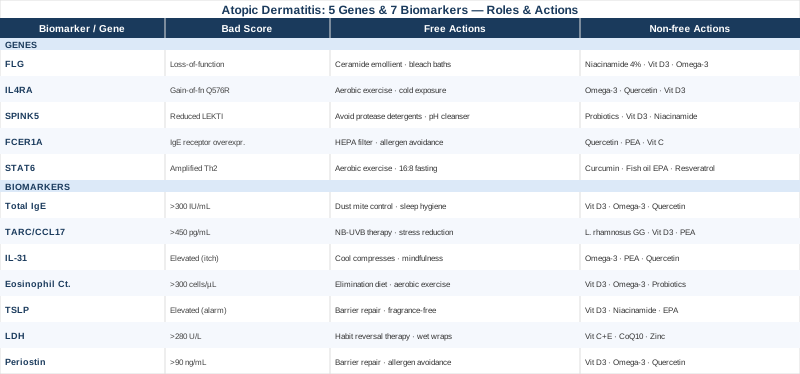
This article explores two complementary frameworks based on current research. The first covers 7 measurable blood biomarkers that track the state of your immune system and inflammation — each one actionable with or without supplementation. The second examines 5 key genes identified in atopic dermatitis genetics research, explaining what each variant means and what practical steps may help compensate for it. Beyond these, you will find a summary of a book that challenges conventional dietary management of eczema, and a review of four complementary therapies with credible clinical support specifically for this condition.
Summary
This article covers 7 blood biomarkers and 5 genes directly linked to atopic dermatitis severity, barrier function, and immune dysregulation. For each biomarker — Total IgE, TARC/CCL17, IL-31, Eosinophil Count, TSLP, LDH, and Periostin — you will find how to measure it (with cost ranges), what an abnormal result reveals about your immune state, and concrete action plans both with and without supplementation. For each gene — FLG, IL4RA, SPINK5, FCER1A, and STAT6 — you will find how the variant shifts your skin biology and what compensatory strategies have the best evidence.
The article also summarizes ten key insights from The Eczema Detox by Karen Fischer, which reframes dietary triggers in ways that challenge what most eczema patients are typically told. And it reviews microbiome-directed therapy, the Autoimmune Protocol, MBSR, narrowband UVB, and Chinese herbal medicine — selected specifically for the strength of their evidence in atopic dermatitis, not generic inflammation.
If you have been told that eczema is a lifelong condition to manage but not address at its roots, the biomarker and genetic data here may offer a more specific map.
7 Biomarkers to Track if You Have Atopic Dermatitis
Blood biomarkers offer something that skin examinations alone cannot: a window into the systemic immune processes driving inflammation before it becomes visible on the surface. These seven markers are among the best-validated in atopic dermatitis research, ranging from tests available in any standard panel to more specialized immunoassays. Tracking them over time — not just at a single point — is where the most useful signal emerges.
1. Total IgE
Why it matters: Immunoglobulin E is the antibody class most associated with allergic sensitization. In atopic dermatitis, elevated total IgE reflects a Th2-skewed immune environment where mast cells and basophils are chronically primed for overreaction. High IgE is present in roughly 80% of AD patients, though a normal result does not rule out the condition. Its particular value lies in correlating with allergic comorbidities — asthma, food allergies, rhinitis — that frequently accompany AD and can amplify skin inflammation through systemic immune activation. A level above 300 IU/mL in an adult with eczema is clinically meaningful; above 1000 IU/mL indicates significant systemic sensitization.
How to measure it: Standard ELISA blood draw at any clinical lab. Cost: approximately $20–60 USD. Available through primary care, allergists, or direct-to-consumer lab services. Request specific IgE allergen panels alongside total IgE to identify what your immune system has sensitized against.
If the result is high — plan without supplements: Rigorous environmental allergen reduction is the most evidence-backed first step. House dust mite exposure is a primary IgE driver in AD: HEPA filtration in the bedroom, allergen-proof mattress and pillow encasements, and washing bedding weekly at 60°C address this directly. A structured elimination diet trial — removing the top six allergens (milk, egg, wheat, soy, peanut, tree nut) for 4–6 weeks with systematic reintroduction — identifies IgE-mediated food contributions. Sleep extension matters here too: partial sleep deprivation measurably elevates IgE and histamine release, compounding baseline sensitization.
If the result is high — plan with supplements or equipment: Vitamin D3 at 2000–4000 IU/day (based on a tested 25-OH vitamin D baseline) consistently reduces total IgE in AD patients across multiple randomized trials; pair with vitamin K2 at 100–200 mcg for transport synergy. Omega-3 fatty acids at 2–3g combined EPA+DHA daily help shift the Th2/Th1 balance; take with a fat-containing meal for absorption; at doses above 3g, mild blood-thinning effects are relevant if you take anticoagulants. Quercetin at 500mg twice daily with meals acts as a natural mast cell stabilizer; cycle 8 weeks on, 4 weeks off; mild GI discomfort is possible at higher doses. Reassess IgE after 12 weeks before adjusting.
2. TARC / CCL17 (Thymus and Activation-Regulated Chemokine)
Why it matters: TARC, also known as CCL17, is currently regarded as the most sensitive serum marker for atopic dermatitis disease activity. Unlike total IgE, which reflects sensitization history, TARC tracks the state of ongoing skin inflammation — it rises during flares and falls with effective treatment. Produced by keratinocytes and dendritic cells, TARC recruits Th2 lymphocytes into inflamed skin and sustains the inflammatory cycle. Studies consistently show it correlates with SCORAD severity scores more closely than eosinophil count or IgE, making it particularly useful for monitoring whether a treatment or lifestyle intervention is actually working over time.
How to measure it: Specialized immunoassay (ELISA); available through reference labs and some academic medical centers. Cost: approximately $50–150 USD. In Japan, TARC testing for AD is reimbursed by national insurance — reflecting its status as a validated clinical tool. Normal reference: below approximately 450 pg/mL. Active moderate-to-severe AD commonly produces levels of 2000–10000 pg/mL.
If the result is high — plan without supplements: Narrowband UVB phototherapy is among the most effective non-pharmaceutical interventions that directly reduces TARC by modulating skin-resident dendritic cell activity. Reduce psychological stress: cortisol directly stimulates keratinocyte TARC production, which is why flares often track closely with high-stress periods. Consistent fragrance-free emollient use within 3 minutes of a lukewarm shower maintains barrier integrity and reduces the keratinocyte stress that drives TARC secretion. Twice-weekly dilute sodium hypochlorite baths (one teaspoon of 5–6% bleach per 4 liters of water, 5–10 minutes) reduce Staphylococcus aureus colonization, which independently amplifies TARC production.
If the result is high — plan with supplements or equipment: Lactobacillus rhamnosus GG at 10 billion CFU/day has demonstrated TARC-reducing effects in AD patients in multiple clinical trials; cycle 3 months on, 1 month off; avoid if immunocompromised. Vitamin D3 supplementation targeting a serum 25-OH vitamin D above 40 ng/mL has been shown to reduce chemokine expression in AD keratinocytes across several controlled studies. Palmitoylethanolamide (PEA) at 600mg twice daily with meals reduces skin inflammation through PPAR-alpha activation and is very well tolerated; assess effect after 8–12 weeks minimum before deciding whether to continue.
3. IL-31 (The Itch Cytokine)
Why it matters: IL-31 is uniquely important for one reason: it is the primary molecular driver of chronic itch in atopic dermatitis. Produced by Th2 lymphocytes, IL-31 binds to receptors on sensory neurons and keratinocytes, triggering both the itch signal and further barrier breakdown simultaneously. Elevated IL-31 correlates specifically with itch intensity rather than just general flare severity — patients with high IL-31 tend toward compulsive scratching that perpetuates lesion formation independent of allergen exposure. Early research characterizing this cytokine's role in AD established the foundation for anti-IL-31 therapy (nemolizumab), which has since shown dramatic itch reduction in phase 3 trials.
How to measure it: Serum ELISA through specialized or research reference labs; not yet widely available in standard commercial panels. Cost: $100–200 USD. For most patients, tracking itch severity using a validated numerical rating scale (NRS-itch, 0–10 daily log) alongside blood eosinophil count serves as a practical proxy while IL-31 testing becomes more broadly accessible.
If the result is high — plan without supplements: Cooling strategies directly interrupt the IL-31 neural signaling loop: cool compresses on affected areas for 10–15 minutes, keeping the sleeping environment below 19°C (66°F), and wearing lightweight natural-fiber clothing prevent the thermal amplification of itch-nerve firing. Mindfulness-based itch management has shown benefit in several controlled trials — 15–20 minutes of body scan meditation daily reduces the cortical response to itch signals without suppressing the sensation itself. Habitualize the "competing response" technique: applying firm cold pressure to a non-itchy skin area when the urge to scratch arises disrupts the behavioral loop without causing tissue damage.
If the result is high — plan with supplements or equipment: Omega-3 fatty acids at 2–3g combined EPA+DHA daily reduce IL-31 production by modulating Th2 cytokine output; takes 6–8 weeks for measurable effect. PEA (palmitoylethanolamide) at 600mg twice daily is particularly relevant here as it acts on the sensory neuron signaling pathway through PPAR-alpha and TRPV1 modulation — mechanisms that sit directly on the IL-31 itch circuit; cycle 12 weeks on, 4 weeks off; very well tolerated. Quercetin at 500mg twice daily reduces Th2 lymphocyte IL-31 secretion; take with vitamin C for enhanced absorption; mild digestive discomfort at higher doses. Topical application: colloidal oatmeal (1% in emollient base, twice daily) has direct anti-itch signaling effects at the IL-31 receptor level in the epidermis with no meaningful side effects.
4. Blood Eosinophil Count
Why it matters: Eosinophils are white blood cells that accumulate in the skin during AD inflammation, contributing to tissue damage, itch amplification, and barrier disruption. A blood count above 300–500 cells/µL is frequently seen in moderate AD; counts above 1000 cells/µL suggest significant systemic Th2 activation. Beyond its value as an AD marker, an elevated eosinophil count flags potential contributions from parasitic infections, drug reactions, and food sensitivities — all of which can masquerade as or worsen atopic dermatitis in ways that topical treatments cannot address. Tracking this number over time reveals whether systemic immune activation is being controlled or intensifying.
How to measure it: Part of the Complete Blood Count (CBC) with differential — the most accessible test on this list. Cost: approximately $15–30 USD, included in most routine annual labs. Specifically request the differential breakdown, which separates white cell subtypes including eosinophils; a CBC without differential will not report this.
If the result is high — plan without supplements: A structured elimination diet guided by a registered dietitian with allergy training — removing the top sensitizing foods for 4–6 weeks with careful individual reintroduction — is the most practical first intervention. In patients who have traveled internationally, ruling out intestinal parasites through a stool ova and parasites test is a clinically underused step that is directly relevant to unexplained eosinophilia. Regular moderate aerobic exercise at 60–70% of maximum heart rate for 30–40 minutes, five days per week, has demonstrated eosinophil-lowering effects through normalization of cortisol rhythms and anti-inflammatory cytokine induction.
If the result is high — plan with supplements or equipment: Vitamin D3 at 2000–4000 IU/day based on tested baseline is consistently associated with reduced eosinophilic tissue inflammation across allergy and AD studies. Omega-3 EPA+DHA at 2–3g/day reduces eosinophil trafficking into tissue; minimum 8 weeks to assess effect. Probiotics, particularly Lactobacillus rhamnosus GG at 10 billion CFU/day, reduce eosinophil counts in allergic disease in multiple randomized trials; run a 12-week course with 4-week breaks. If eosinophil count persists above 1000 cells/µL despite 12 weeks of dietary, lifestyle, and supplement intervention, specialist evaluation for hypereosinophilic syndrome or systemic causes is warranted before escalating supplementation.
5. TSLP (Thymic Stromal Lymphopoietin)
Why it matters: TSLP is the primary "alarm cytokine" produced by skin epithelial cells in response to barrier damage, irritants, and allergen contact. When the skin barrier is compromised — whether through genetic variants, scratching, or detergent exposure — keratinocytes release TSLP, which then instructs dendritic cells to drive a Th2 immune response. This makes TSLP arguably the most upstream measurable driver of the entire AD inflammatory cascade. Blocking TSLP (tezepelumab) is now a licensed treatment for severe asthma, and AD-specific TSLP inhibitor research is active. An elevated serum TSLP indicates that your skin is persistently sending danger signals to your immune system regardless of which surface triggers are currently visible.
How to measure it: Serum ELISA through specialized reference labs. Cost: approximately $100–200 USD. Less commonly available than other markers on this list, but access is growing alongside pharmaceutical interest in the target. Some academic dermatology centers measure TSLP as part of phenotyping protocols for patients considering biological therapies.
If the result is high — plan without supplements: Aggressive barrier repair is the most direct TSLP-reduction strategy. Plain petrolatum applied to thinned or broken areas twice daily creates an immediate physical block to allergen and irritant penetration. Eliminate any product containing synthetic fragrance, alcohol, or methylisothiazolinone from skincare, cleaning, and laundry routines — these are direct TSLP inducers in the epidermis. A bedroom humidifier maintaining 45–55% relative humidity reduces trans-epidermal water loss overnight and lowers basal TSLP secretion. Replace hot showers (a major thermal keratinocyte stressor and TSLP amplifier) with lukewarm water limited to 5–10 minutes.
If the result is high — plan with supplements or equipment: Vitamin D3 is the most consistently studied TSLP-suppressing supplement, shown in multiple studies to reduce TSLP gene expression in keratinocytes through vitamin D response elements; target serum 25-OH vitamin D above 40 ng/mL (typically 2000–5000 IU/day based on tested baseline). Topical niacinamide (vitamin B3) at 4–5% concentration increases ceramide and free fatty acid production in the stratum corneum, directly reducing the barrier compromise that induces TSLP; use niacinamide-containing moisturizers daily with no significant side effects in most skin types. Oral EPA supplementation specifically (1.5–2g/day from a high-EPA fish oil product) reduces NF-κB activity in keratinocytes — one of the primary transcription pathways driving TSLP expression; minimum 8-week course.
6. LDH (Lactate Dehydrogenase)
Why it matters: Serum LDH is an older but persistently useful atopic dermatitis severity marker, routinely used in Japanese dermatology as part of standard AD assessment. LDH is released from damaged cells — in the context of AD, it reflects the degree of keratinocyte destruction occurring in actively inflamed skin. While it is not specific to AD (elevated LDH also appears in liver disease, hemolysis, and muscle damage), in a person with known AD and normal liver function, rising LDH reliably correlates with disease activity and can serve as a practical longitudinal marker. Its main advantage is universal accessibility: it is inexpensive, available everywhere, and included in standard metabolic panels.
How to measure it: Standard comprehensive metabolic panel (CMP) or standalone test at any clinical lab. Cost: approximately $15–40 USD. Normal adult range: approximately 140–280 U/L (varies slightly by laboratory). Active moderate-to-severe AD commonly produces values of 300–600 U/L; severe presentations may exceed 600 U/L. When using LDH as an AD marker, ensure liver function tests are normal to avoid misattributing hepatic elevation to skin disease.
If the result is high — plan without supplements: The dominant driver of high LDH in AD is the mechanical tissue damage caused by scratching — reducing scratch behavior is therefore the most direct intervention. Habit Reversal Training (HRT), a behavioral therapy approach specifically studied in dermatological conditions including AD, has clinical evidence for measurable scratch reduction; a therapist trained in dermatology-focused cognitive behavioral therapy can guide this process. Wet-wrap therapy — applying damp cotton wraps over medicated or plain emollient-covered skin for 2–4 hours or overnight — reduces acute inflammation and allows skin tissue to recover without further damage. Cooling the sleep environment below 19°C and using 100% cotton bedding reduces nocturnal scratching intensity.
If the result is high — plan with supplements or equipment: Antioxidants that address keratinocyte oxidative stress are the most mechanistically relevant supplement category: Vitamin C at 500–1000mg/day with food (reduce to 500mg if GI symptoms arise), Vitamin E at 200–400 IU/day with a fat-containing meal. Coenzyme Q10 at 100mg/day with food supports mitochondrial integrity in stressed and damaged keratinocytes; well tolerated at this dose. Zinc picolinate at 15–30mg/day with food supports tissue repair signaling — take with food to avoid nausea; do not exceed 40mg/day long-term without medical guidance; supplement copper at 1–2mg/day separately to prevent depletion from sustained zinc use. Suggested cycle: zinc and antioxidant protocol for 12 weeks, retest LDH, then adjust.
7. Periostin
Why it matters: Periostin is an extracellular matrix protein whose serum levels serve as a specific marker for active type 2 (Th2-driven) inflammation — the precise immune subtype that dominates in classical atopic dermatitis. Unlike IgE or eosinophil count, which can be elevated across multiple immune contexts, periostin specifically reflects ongoing IL-4 and IL-13 signaling activity in tissue. This makes it particularly useful for confirming whether a person's AD is driven by the canonical Th2 pathway — making them a biologically appropriate candidate for dupilumab or tralokinumab — versus a less common alternate mechanism. Elevated periostin is also seen in eosinophilic asthma and chronic sinusitis, pointing to the broader type 2 inflammatory phenotype that often underlies what is called the "atopic march."
How to measure it: Serum ELISA through specialized reference labs. Cost: approximately $100–200 USD. In clinical AD and asthma trials, elevated periostin (generally above 90 ng/mL in the relevant assays used) has been used to select and monitor biological therapy candidates. Increasingly available through allergy-immunology centers.
If the result is high — plan without supplements: Addressing the Th2 environment systemically mirrors the interventions described for TARC and TSLP. Barrier repair reduces the allergen penetration that continuously triggers Th2-activating signals; comprehensive environmental allergen avoidance removes the upstream inputs driving periostin-inducing IL-4 and IL-13 production; and moderate aerobic exercise five days per week has a modest but documented Th2-tempering effect through adrenergic and anti-inflammatory mechanisms operating independently of pharmaceutical intervention.
If the result is high — plan with supplements or equipment: The same core protocol applies as for the broader Th2-skewed phenotype: Vitamin D3 targeting serum levels above 40 ng/mL, Omega-3 EPA+DHA at 2–3g/day, and Quercetin 500mg twice daily. One additional consideration: if periostin remains persistently elevated after 3–6 months of optimized lifestyle and supplementation, a referral to an allergist-immunologist for evaluation of biological therapy is not only appropriate but strongly evidence-based. Persistently high periostin is one of the clearest signals that systemic Th2 activation has reached a threshold that topical and supplement strategies are unlikely to resolve alone.
The biomarkers above provide a running map of immune state — something a skin examination alone cannot offer. Understanding the genetic factors that created your specific vulnerabilities in the first place adds another layer of context, one that shapes which biomarkers are most likely to be elevated and which interventions are most likely to be relevant for the long term.
5 Key Genes Linked to Atopic Dermatitis Risk
Genetic variants do not cause atopic dermatitis in isolation — they shift probabilities and set biological constraints that determine how a person's skin responds to exposures that others tolerate without issue. The five genes below represent the most replicated findings across AD genetics research, from skin barrier discoveries to immune-pathway variants that sustain chronic Th2 activation.
FLG — The Skin Barrier Gene
What it does: Filaggrin is a structural protein essential for forming the outermost layer of the skin and for maintaining the acidic pH of the skin surface that inhibits bacterial colonization. Loss-of-function mutations in the FLG gene — the most common in Europeans being R501X and 2282del4 — are found in approximately 10% of the general population and in 25–30% of people with atopic dermatitis. The 2006 discovery by Palmer and colleagues that FLG mutations are the single strongest known genetic risk factor for AD was a turning point in understanding the condition as primarily a barrier disease rather than purely an immune one.
Carriers of FLG loss-of-function variants have measurably higher trans-epidermal water loss (TEWL), lower natural moisturizing factor (NMF) in the stratum corneum, and elevated susceptibility to allergen sensitization through the skin rather than through gut or airway exposure. This is why FLG mutation carriers tend to develop multiple allergies over time — the structurally leaky barrier repeatedly exposes skin-resident immune cells to unprocessed environmental allergens.
If the gene is bad — the plan without supplements: Consistent, high-quality barrier restoration is the central strategy. Apply fragrance-free ceramide-containing emollient (products containing ceramide 1, 3, and 6-II in physiological ratios) within 3 minutes of bathing — the "soak and seal" method locks moisture before TEWL can accelerate. Use only lukewarm water below 37°C and limit showers to 5–10 minutes. Replace soap with a gentle syndet (synthetic detergent) cleanser at neutral pH. Dilute bleach baths twice weekly (one teaspoon of 5–6% bleach per gallon of water; soak 5–10 minutes) reduce Staphylococcus aureus colonization, which thrives on FLG-deficient skin and drives secondary inflammation. Maintain bedroom humidity at 45–55% with a humidifier. Wear 100% cotton fabrics; avoid wool and synthetic materials in direct skin contact.
If the gene is bad — the plan with supplements or equipment: Topical niacinamide (vitamin B3) at 4–5% is the most evidence-based topical supplement for FLG-deficient skin, stimulating ceramide synthesis through a pathway independent of filaggrin; apply daily, well tolerated across most skin types. Oral Vitamin D3 at 2000–4000 IU/day (test blood levels first) has been shown in keratinocyte studies to upregulate FLG gene expression — it cannot repair a complete null mutation but may benefit heterozygous carriers with residual FLG activity; pair with K2 at 100 mcg. Oral collagen peptides at 10g/day for a minimum of 12 weeks support stratum corneum hydration through extracellular matrix enrichment; no significant side effects at this dose. Omega-3 EPA+DHA at 2g/day reduces TEWL by incorporating into epidermal phospholipids; assess effect after 8 weeks. Equipment: a home narrowband UVB lamp (311nm) used under dermatologist prescription and guidance — sessions of 3–5 minutes 3x/week, titrating based on skin response — supports skin barrier differentiation and reduces colonization in parallel.
IL4RA — The Allergic Signal Amplifier
What it does: The IL4RA gene encodes the alpha subunit of the IL-4 receptor, which is shared by both IL-4 and IL-13 signaling complexes. Specific variants — most notably the Q576R gain-of-function polymorphism — produce a receptor with enhanced downstream signaling, meaning that for the same amount of circulating IL-4 or IL-13, carriers generate a stronger Th2 immune response. This results in heightened IgE class switching, increased skin inflammation, and greater susceptibility to allergen sensitization. IL4RA variants are among the most replicated immune gene associations in atopic dermatitis across genome-wide association studies, and the therapeutic success of dupilumab (which blocks both IL-4 and IL-13 signaling through this receptor) provides indirect validation of its centrality in the disease.
If the gene is bad — the plan without supplements: Regular moderate-intensity aerobic exercise at 60–70% maximum heart rate for 30–40 minutes, five days per week, is the most accessible intervention for reducing resting IL-4 and IL-13 levels through adrenergic and anti-inflammatory mechanisms. Progressive cold water exposure — finishing showers cold for 2–3 minutes, building tolerance over 3–4 weeks — has been proposed to support Th1 responses and counterbalance Th2 dominance, though direct AD trial evidence remains limited to observational data. Dietary reduction of refined sugar and ultra-processed carbohydrates has a consistent, modest anti-Th2 effect across metabolic studies, likely mediated through insulin and NF-κB signaling. Stress management receives particular importance for IL4RA gain-of-function carriers because cortisol directly drives IL-4 production, amplifying the already-sensitized receptor's output.
If the gene is bad — the plan with supplements or equipment: Omega-3 EPA+DHA at 2–3g/day with meals reduces IL-4 and IL-13 production in Th2 cells and modulates IL-4 receptor membrane dynamics; 8–12 weeks minimum to assess effect; minor blood-thinning effect at doses above 3g. Quercetin at 500mg twice daily with food (8 weeks on, 4 weeks off) inhibits IL-4-induced downstream signaling, partially compensating for the gain-of-function amplification; mild GI sensitivity possible. Vitamin D3 targeting serum levels above 40 ng/mL regulates IL4RA expression through vitamin D response elements in the gene promoter. Astragalus root extract at 500mg/day standardized to 0.5% astragalosides, in short courses of 4–6 weeks only, may support Th1 counterbalance — avoid in autoimmune conditions, with immunosuppressants, or for long-term use without medical supervision.
SPINK5 — The Inflammation Brake
What it does: SPINK5 encodes LEKTI (lympho-epithelial Kazal-type-related inhibitor), a serine protease inhibitor that controls the activity of kallikrein enzymes in the skin. Kallikreins are responsible for controlled desquamation — the natural shedding of skin cells. When SPINK5 carries loss-of-function or reduced-function variants (the E420K variant being among the most studied in AD), kallikreins run less regulated, accelerating barrier breakdown, increasing proinflammatory cytokine release (including IL-1 and IL-18), and promoting sensitization to environmental allergens. The genetic link is demonstrated most dramatically by Netherton syndrome, where SPINK5 is completely absent — AD patients with milder SPINK5 variants sit on a less severe point of the same biological spectrum.
If the gene is bad — the plan without supplements: The most critical environmental modification is reducing exposure to exogenous serine proteases, which compound SPINK5 deficiency by directly activating the same kallikrein pathways LEKTI is meant to suppress. House dust mite allergens (Der p 1, Der f 1) are themselves serine proteases — making standard dust mite avoidance protocols proportionally more important for SPINK5 variant carriers than for the average AD patient. Avoid "bio" laundry detergents containing protease-based cleaning enzymes, which are added for stain removal and function as direct kallikrein amplifiers on skin. Use a pH-adjusted (pH 5.5) gentle cleanser to support the acid mantle that LEKTI helps maintain; standard alkaline soaps at pH 9–10 further impair an already-limited protease inhibition system.
If the gene is bad — the plan with supplements or equipment: Probiotic therapy addressing gut and skin microbiome balance reduces the inflammatory load on a SPINK5-insufficient barrier: a combination of Lactobacillus rhamnosus GG (10 billion CFU/day) and Bifidobacterium longum (5 billion CFU/day) for 12-week courses with 4-week breaks; avoid during immunosuppressive therapy. Vitamin D3 at therapeutic dosing (target above 40 ng/mL serum) supports tight junction integrity in epidermis independently of LEKTI function. Topical niacinamide 4% supports ceramide synthesis to partially compensate for the accelerated barrier degradation from unchecked kallikrein activity. Oral evening primrose oil at 500–1000mg GLA equivalent per day has clinical evidence for reducing AD severity in barrier-deficient phenotypes; cycle 12 weeks on, 4 weeks off; mild GI symptoms possible at higher doses.
FCER1A — The IgE Receptor Gene
What it does: FCER1A encodes the alpha subunit of the high-affinity IgE receptor (FcεRI) expressed on mast cells, basophils, and — particularly relevant in AD — Langerhans cells and inflammatory dendritic epidermal cells in the skin. Promoter variants in FCER1A increase receptor expression on these cells, meaning that even at moderate circulating IgE levels, skin immune cells are densely loaded with IgE-bearing receptors ready to respond. In practice, FCER1A gain-of-expression variants translate to enhanced mast cell degranulation, amplified histamine and prostaglandin release, and a substantially lower threshold for triggering allergic inflammation in skin from the same exposure level that would not elicit a reaction in non-carriers. This locus is among the consistently replicated findings in AD genome-wide association studies.
If the gene is bad — the plan without supplements: Comprehensive allergen avoidance is proportionally more important for FCER1A variant carriers because their skin mast cells will respond more intensely to any antigen they have IgE against — even at exposure levels that would not trigger reactions in others. A thorough approach: HEPA air purification in living spaces, allergen-proof bedding, regular washing of soft furnishings, and identification and removal of known food triggers. Avoid NSAIDs (ibuprofen, aspirin) when clinically possible — these shift arachidonic acid metabolism toward leukotriene production, compounding IgE-receptor-mediated inflammatory output. Maintaining a regular sleep schedule matters here: IgE receptor expression on mast cells has circadian regulation, and sleep disruption measurably increases receptor density.
If the gene is bad — the plan with supplements or equipment: Quercetin at 500–1000mg/day (taken as 500mg twice daily with meals) is the most directly relevant supplement as it stabilizes mast cell membranes and reduces FcεRI-triggered degranulation; cycle 8 weeks on, 3–4 weeks off; mild GI sensitivity at the higher dose. Vitamin C at 500–1000mg/day supports connective tissue around mast cells and contributes to direct histamine breakdown; split doses across the day to reduce GI effects. PEA (palmitoylethanolamide) at 600mg twice daily down-regulates mast cell activation and FcεRI receptor expression through PPAR-alpha signaling; among the best-tolerated compounds on this list; minimum 12-week course; can be taken continuously for extended periods if well tolerated. Freeze-dried stinging nettle leaf at 600mg twice daily has traditional and some preliminary clinical support as a natural antihistamine; 6–8 week short courses; mild diuretic effect.
STAT6 — The Th2 Master Switch
What it does: STAT6 (Signal Transducer and Activator of Transcription 6) is the principal transcription factor activated by both IL-4 and IL-13 receptor signaling. When these cytokines bind their receptors, STAT6 is phosphorylated and moves to the nucleus, driving expression of Th2-associated genes including IgE class-switching factors, eotaxin (the primary eosinophil recruiter), and periostin. Gain-of-function variants in STAT6 — including several single nucleotide polymorphisms in the exon 2 coding region — amplify this response, making the entire downstream Th2 cascade more sensitive to activation at lower cytokine concentrations. STAT6 variants have been replicated across multiple genome-wide association studies for both atopic dermatitis and asthma, consistent with its role as a central amplification node in type 2 immune programming.
If the gene is bad — the plan without supplements: STAT6 activation is tightly coupled to IL-4 and IL-13 availability, so reducing upstream cytokine production is the most direct non-supplement approach. Moderate aerobic exercise at 150 minutes per week consistently reduces circulating IL-4 and IL-13 in adults with allergic disease through adrenergic anti-inflammatory mechanisms. Time-restricted eating (16:8 intermittent fasting pattern) reduces pro-inflammatory cytokine baselines, including those feeding STAT6 activation. Sauna exposure at 70–90°C for 15–20 minutes, 3 times per week followed by a cool rinse, has immunomodulatory effects associated with reduced Th2 polarization over time — and Finnish epidemiological data links regular sauna use to lower asthma and allergy incidence. Consistent restorative sleep of 7–9 hours is essential; sleep deprivation specifically enhances STAT6 pathway sensitivity within 24 hours of insufficient rest.
If the gene is bad — the plan with supplements or equipment: Curcumin standardized extract at 500mg twice daily with black pepper piperine extract for bioavailability inhibits STAT6 phosphorylation in multiple cell-based and animal studies with modest anti-inflammatory clinical support; cycle 8–12 weeks on, 4 weeks off; avoid at high doses in gallbladder disease or with anticoagulant medication. Fish oil with a high-EPA ratio (targeting 1.5–2g EPA specifically per day) reduces STAT6 transcriptional activity through membrane phospholipid remodeling; 8 weeks minimum to assess. Resveratrol at 200–500mg/day with a meal (trans-resveratrol form preferred for absorption) has demonstrated STAT6-suppressive effects in preclinical models; cycle 8 weeks on, 4 weeks off; avoid with blood-thinning medications. L-theanine at 200mg/day — from green tea or supplement — reduces IL-4-mediated STAT6 activation with a strong safety profile and mild relaxation effect; can be used continuously.
Understanding your genetic landscape clarifies which biological pathways are structurally biased toward inflammation in your particular case. Alongside this map, the nutritional dimension of atopic dermatitis is one that mainstream dermatology has historically underserved — and the following book addresses this gap with more specificity than most clinical guidance offers.
The Eczema Detox by Karen Fischer — 10 Things Worth Knowing
Published in 2018, The Eczema Detox by Karen Fischer — a nutritionist whose own daughter had severe eczema — represents one of the most comprehensive dietary frameworks for atopic dermatitis written outside of academic literature. The book challenges the common framing of AD as a skin problem managed topically, arguing that internal dietary and metabolic factors are primary drivers that have to be addressed upstream. Several of its central arguments anticipate findings that later appeared in peer-reviewed research on histamine, the gut-skin axis, and barrier nutrition.
Total Histamine Load Matters More Than Any Single Trigger
Fischer's central thesis is that the relevant unit of measurement in eczema flares is not a single trigger food but the cumulative histamine burden across the entire diet. High-histamine foods (fermented products, aged cheese, wine, canned fish, vinegar) may not individually cause a reaction, but their combined load can exceed the capacity of the DAO enzyme to clear histamine — triggering skin inflammation that appears random when isolated foods are tested individually.
Salicylate Sensitivity Is Underdiagnosed and Paradoxically Worsened by Healthy Eating
Many foods categorized as health-promoting — berries, tomatoes, citrus, avocado, almonds — are among the highest in dietary salicylates. Fischer documents cases where patients pursuing "clean eating" significantly worsened their AD because their salicylate intake increased alongside their healthy food consumption. Testing for salicylate sensitivity through a structured elimination and challenge protocol is not standard practice in most dermatology offices, which means it goes unidentified in many patients.
The 2-Week Simultaneous Reboot Works Where Single Eliminations Fail
Rather than eliminating one food group at a time, Fischer proposes removing all high-histamine, high-salicylate, and additive-heavy foods simultaneously for two weeks, then reintroducing them systematically. The rationale is mechanistically sound: with individual eliminations, the cumulative burden may not drop below the symptom threshold — making single-food eliminations appear falsely negative when the combined load is the actual problem.
Nightshades Drive Non-IgE Inflammation Through Alkaloids
Tomatoes, peppers, eggplant, and potatoes contain solanine and related steroidal alkaloids that can trigger mast cell degranulation and histamine release through non-IgE-mediated mechanisms. They will not appear on an IgE blood test or skin prick test. For FCER1A variant carriers in particular — whose mast cells are already primed — this non-IgE pathway can be a significant undiscovered driver of persistent skin inflammation.
Topical Steroid Dependence Is a Real and Preventable Cycle
Fischer dedicates substantial space to what she calls topical steroid addiction — now increasingly recognized in dermatological literature as topical steroid withdrawal (TSW). She documents patient cases where chronic use of moderate-to-potent topical steroids resulted in rebound flares far more severe than the original condition, requiring months of careful tapering with significant interim discomfort. Her writing predates mainstream dermatological acknowledgment of TSW by several years.
The Liver's Histamine-Clearing Capacity Is Modifiable
DAO (diamine oxidase), which breaks down dietary histamine in the gut, and HNMT (histamine N-methyltransferase), which clears histamine systemically, are both influenced by liver metabolic capacity. Alcohol, acetaminophen overuse, low choline intake, and gut permeability all impair these clearing systems. Fischer frames a portion of eczema management as a liver support and gut integrity problem — a framing that has since gained traction in functional medicine approaches to allergic disease.
Synthetic Fragrance Is a Primary Hidden Driver That Allergen Tests Miss
Synthetic fragrance chemicals — present not just in perfume but in most mainstream lotions, shampoos, and laundry detergents — are among the most consistent hidden triggers in AD patients who otherwise have good allergen control. Regulatory labeling requires disclosure of 26 specific fragrance allergens in the EU, but hundreds of other sensitizing compounds remain listed only as "parfum" or "fragrance," invisible to standard patch test panels.
Zinc and Molybdenum Deficiencies Impair Histamine Metabolism
Both zinc and molybdenum are cofactors for enzymes involved in histamine clearance and skin repair. Zinc deficiency is documented in a subset of AD patients and correlates with severity. Molybdenum — needed to convert sulfites (common in wine and preserved foods) to less reactive sulfates — is almost never measured in standard labs, yet its deficiency may explain why some AD patients react to sulfite-containing foods without any detectable IgE-mediated allergy.
Vitamin B6 Deficiency Reduces DAO Activity and Amplifies Histamine Burden
Pyridoxal phosphate (the active form of vitamin B6) is a required cofactor for diamine oxidase. B6 insufficiency — common in people eating low-protein or heavily processed diets — directly reduces histamine clearance capacity at the intestinal level. Fischer points to this as one reason certain AD patients respond meaningfully to B-complex supplementation even when standard lab ranges suggest no deficiency, because standard cutoffs for B6 are not calibrated for enzyme-cofactor sufficiency.
The FID Protocol Is Diagnostic and Time-Limited, Not Permanent Restriction
Fischer's Foods for Itchy Dermatitis (FID) protocol is not intended as indefinite avoidance but as a structured diagnostic-therapeutic tool: two weeks of full elimination, then systematic reintroduction of food groups, then a sustainable long-term diet built around each person's specific identified triggers. This frames dietary investigation as a clinical process with a defined end point — significantly more actionable than the vague "avoid inflammatory foods" advice that most eczema patients receive and that rarely translates into meaningful changes.
With dietary and metabolic contributors addressed, there remains a body of evidence around complementary and mind-body approaches that deserves equal consideration for a condition as strongly stress-modulated as atopic dermatitis.
Complementary Approaches With Meaningful Clinical Evidence
The following modalities have human clinical evidence specific to atopic dermatitis — not just general inflammation or allergy. Each was selected because the proposed mechanism connects directly to the biology described above, and because the evidence meets a threshold above anecdote.
Microbiome-Directed Therapies
Atopic dermatitis has one of the most extensively studied microbiome connections in all of dermatology. The skin microbiome of AD patients is characterized by dramatic Staphylococcus aureus overgrowth — and this organism is not merely a colonizer but an active disease driver. S. aureus produces serine proteases (V8 protease, exfoliative toxins) that directly degrade filaggrin, amplify IL-31 and TSLP production, and trigger IgE-mediated sensitization simultaneously. The gut microbiome shows parallel dysbiosis, with reduced biodiversity, lower beneficial bacteria abundance, and increased intestinal permeability that delivers microbial antigens systemically.
A landmark study in Science Translational Medicine by Kong and colleagues characterized the AD skin microbiome in detail, demonstrating that S. aureus dominance correlates directly with flare severity and that microbiome diversity inversely predicts disease activity. This work has driven active research into live biotherapeutic products — including Roseomonas mucosa and Staphylococcus epidermidis transplants — as topical microbial competitors to S. aureus colonization.
Practically: dilute bleach baths twice weekly reduce S. aureus without broadly eliminating the beneficial microbiome. Oral probiotics — Lactobacillus rhamnosus GG (10 billion CFU/day) and Bifidobacterium breve (5 billion CFU/day) — have meta-analytic support for reducing AD severity in children and modest support in adults; run 12-week courses. Prebiotic dietary fiber from chicory, leek, and onion supports gut microbiome diversity; increase fiber intake gradually to avoid gas and bloating.
The Autoimmune Protocol from Sarah Ballantyne
Atopic dermatitis — which involves both barrier dysfunction and dysregulated immune activation with significant overlap with autoimmune mechanisms — is a condition where the Autoimmune Protocol (AIP) developed by Dr. Sarah Ballantyne warrants serious consideration. The protocol, detailed in The Paleo Approach, removes foods hypothesized to drive intestinal permeability and immune dysregulation: grains, legumes, dairy, eggs, nightshades, nuts, seeds, refined sugars, processed foods, NSAIDs, and alcohol. It emphasizes nutrient-dense animal proteins, organ meats, fermented vegetables, bone broth, and diverse colorful vegetables.
Direct AD-specific trial evidence for AIP is currently limited to patient-reported outcomes and case series, but several of its components have strong mechanistic support relevant to the biomarkers and genes above. Eliminating nightshades directly addresses the alkaloid-mast cell issue relevant to FCER1A variants; removing gluten and dairy reduces intestinal permeability markers in susceptible individuals and reduces systemic allergen exposure; the emphasis on organ meats addresses the zinc, vitamin A, and copper deficiencies documented in AD populations. A 2019 prospective study on AIP in inflammatory bowel disease — a related mucosal immune condition — reported clinical remission in 73% of participants within 6 weeks, providing a meaningful signal even in the absence of direct AD trial data.
Application: commit to the strict elimination phase for a minimum of 8 weeks before beginning reintroduction. Work with a registered dietitian to ensure adequate caloric and micronutrient intake throughout. Reintroduce one food group every 3–5 days and track AD severity against each reintroduction to build a personalized trigger profile.
Mindfulness Meditation and MBSR
Atopic dermatitis is one of the few inflammatory skin conditions where the bidirectional skin-brain axis has been systematically quantified. Psychological stress modulates AD through at least three parallel pathways: HPA axis activation increases cortisol, which drives Th2 cytokine production and raises skin permeability; neuropeptides released in stressed skin (substance P, CGRP) directly stimulate mast cell degranulation and IL-31 production; and behavioral consequences of stress (compulsive scratching, sleep disruption) amplify physical barrier damage independently of immune mechanisms.
Mindfulness-Based Stress Reduction — the 8-week structured protocol developed by Jon Kabat-Zinn — has been examined directly in AD in randomized controlled trials. A study published in Acta Dermato-Venereologica found that AD patients completing an MBSR course showed significantly reduced SCORAD scores and psychological distress compared to controls, with effects maintained at 6-month follow-up. The mechanism involves both cortisol normalization and reduced habitual scratching through improved moment-to-moment awareness of the itch-scratch response cycle.
Application: follow the standard 8-week MBSR program, which includes weekly 2.5-hour group sessions and daily 45-minute home practice. Body scan meditation — systematically attending to skin sensations without reacting — is proposed as the most AD-relevant component, directly addressing the itch-scratch cycle by decoupling the sensation from the behavioral response. Accessible through certified MBSR instructors, university-based programs, and structured apps.
Light Therapy — Narrowband UVB
Narrowband UVB phototherapy (NB-UVB, 311nm wavelength) is one of the best-evidenced non-pharmaceutical interventions for atopic dermatitis, with an evidence base that places it in international clinical treatment guidelines. Its mechanisms are directly relevant to the biology covered throughout this article: NB-UVB reduces Th2 cytokine production (IL-4, IL-13, TARC) in treated skin; suppresses IgE receptor expression on Langerhans cells (directly relevant to FCER1A variant carriers); normalizes TSLP secretion from keratinocytes; reduces S. aureus colonization through direct antimicrobial effects; and stimulates cutaneous vitamin D synthesis, contributing to barrier gene regulation including FLG expression.
Multiple randomized controlled trials and systematic reviews confirm NB-UVB superior to broadband UVB for AD, with response rates of 60–70% for significant improvement in SCORAD. The standard clinical protocol involves 3 sessions per week, with starting dose calculated from minimal erythema dose (MED) testing and carefully titrated over 8–12 weeks at a phototherapy clinic. Side effects include temporary skin redness and dryness (manageable with emollients) and cumulative UV exposure considerations with indefinite long-term use — most guidelines recommend periodic treatment breaks.
Home NB-UVB devices are available by prescription in many countries and have demonstrated equivalent efficacy to clinic-based delivery in AD trials. Regardless of whether treatment is clinic-based or home-based, dermatologist involvement in dosing, monitoring, and decision-making is strongly recommended.
Conclusion
Atopic dermatitis is increasingly understood not as a single condition but as a cluster of overlapping phenotypes — different people with the same diagnosis may be driven predominantly by barrier failure, Th2 hyperactivation, microbiome dysbiosis, histamine accumulation, or some combination of all of these. The biomarkers and gene variants covered here offer a way to determine which pathways are dominant in a specific individual, replacing generic management with something more targeted and more likely to produce lasting results.
The most practical starting point is to run the biomarkers already accessible in a standard blood panel — Total IgE, Eosinophil Count, and LDH — and consider adding TARC or Periostin when monitoring treatment response over time. If genetic testing is available, checking for FLG loss-of-function variants is particularly worthwhile given the magnitude of their effect on barrier function and the clear management implications.
None of the strategies here replace dermatological or allergist-immunologist evaluation, especially for moderate-to-severe disease. But the more specific the information brought into those conversations — tracked biomarkers, identified genetic variants, documented dietary patterns and supplement responses — the more productive those conversations become. Better data leads to better decisions. That remains the most useful thing any of this offers.