This article contains AI generated content.
· UpdatedAvascular Necrosis of the Hip - 5 Genes And 6 Biomarkers To Track
Introduction
Receiving a diagnosis of avascular necrosis (AVN) of the hip—also known as osteonecrosis of the femoral head—can feel like a sudden and silent betrayal by your own body. The primary bone in your hip joint, starved of blood, begins to suffer micro-fractures, initiating a cascade that can lead to joint collapse and severe osteoarthritis. In the medical mainstream, the conversation around this condition is often frustratingly brief, framing your path as a straight line from temporary pain management to eventual total joint replacement.
Standard advice for bone health, such as taking generic calcium supplements or engaging in low-impact walking, is simply inadequate for a condition rooted in microvascular failure. The femoral head is nourished by a fragile, terminal network of blood vessels, predominantly the lateral epiphyseal artery. When this microcirculation is blocked by micro-clots, fat emboli, or damaged by chronic inflammation, the bone marrow cells begin to die within hours. Solving this problem requires lookouts at the cellular and molecular levels rather than broad, generalized health tips.
This article takes a deeper, science-aware approach to joint preservation by focusing on the physiological bottlenecks that drive bone ischemia. By identifying and tracking specific vascular, metabolic, and hematological biomarkers, and understanding your underlying genetic and epigenetic risks, you can actively modify the factors that compromise your hip’s blood supply.
Grounded in clinical literature and vascular bone research, this guide offers practical protocols designed to support endothelial repair, normalize blood viscosity, and stimulate osteogenesis. Whether you are dealing with early-stage bone marrow edema or attempting to prevent the progression of established necrosis, understanding these pathways provides a clear, actionable roadmap to protect your joint structure.
To help you navigate this comprehensive guide, the following summary outlines the physiological connections between circulation, genetic risk, and the bone-building pathways that decide the fate of the femoral head.
Summary
Avascular necrosis of the hip is not a disease of the bone itself, but a localized circulatory failure. This article details the six primary circulating biomarkers that reflect your blood viscosity, arterial health, and bone turnover rate. It also explores five critical genetic predispositions, such as MTHFR and VEGFA polymorphisms, which determine your susceptibility to blood clotting and impaired vessel repair. By identifying these variables, you can implement targeted protocols—including microcirculation stimulants, endothelial supports, and bone-remodeling therapies—to bypass these biological bottlenecks. Below is a comprehensive visualization mapping how these genes and biomarkers interact within the hip microvasculature.
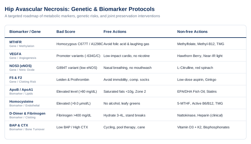
Armed with this overview of the structural and cellular pathways, we can now explore the specific blood markers that allow us to monitor these processes in real-time.
Vascular and Metabolic Biomarkers: The Core Metrics for Hip Microcirculation
To halt the progression of avascular necrosis, you must understand what is happening inside the microcapillaries of your femoral head. Standard blood tests rarely look deep enough into the mechanisms of lipid embolization, hypercoagulability, and bone remodeling. The following six biomarkers, frequently highlighted by preventative medicine specialists like Peter Attia and lipidologists like Thomas Dayspring, provide the quantitative data needed to assess your risk and track the success of your joint-preservation protocols.
Biomarker 1: Apolipoprotein B (ApoB) and the ApoB/ApoA1 Ratio
Why It Matters and What It Reveals
Apolipoprotein B (ApoB) measures the total number of atherogenic particles in your bloodstream, including LDL, VLDL, and IDL. In avascular necrosis, lipid-mediated microemboli are a major cause of capillary blockages in the bone marrow. This is especially true in corticosteroid-induced AVN, where steroids cause fat cells in the bone marrow to swell, raising pressure inside the rigid bone cavity and compressing blood vessels. High levels of ApoB indicate a high concentration of particles that can penetrate the vascular wall, cause endothelial damage, and lead to localized fat emboli in the femoral head.
How to Measure It and Cost Range
An ApoB test is a standard venipuncture blood draw that does not require fasting. You should also request Apolipoprotein A1 (ApoA1) to calculate the ApoB/ApoA1 ratio, which is a powerful predictor of vascular risk. The test is widely available through commercial labs and costs between $30 and $60.
If the Score is Bad: The Plan Without Supplements or Equipment
If your ApoB is above the optimal threshold of 80 mg/dL, adjust your diet to restrict saturated fats to under 10 grams per day. Saturated fats downregulate LDL receptors in the liver, leaving more ApoB particles in circulation. Replace these with monounsaturated fats like extra virgin olive oil. Engage in at least 150 minutes of Zone 2 aerobic exercise weekly (exercise at a pace where you can maintain a conversation but with some effort) to improve lipid clearance and endothelial function.
If the Score is Bad: The Plan With Supplements or Equipment
Take 4 grams of pharmaceutical-grade, highly purified EPA/DHA omega-3 fish oil daily to lower VLDL particles and reduce systemic inflammation. Side effects include mild gastrointestinal upset or a temporary thinning of the blood. If dietary changes and omega-3s do not bring ApoB below 80 mg/dL, discuss starting a low-dose hydrophilic statin, such as Rosuvastatin (5 mg daily), with your doctor. Clinical trials show that statins can prevent corticosteroid-induced osteonecrosis by stopping bone marrow fat accumulation and stabilizing blood vessels corticosteroid-induced osteonecrosis prevention. Monitor your liver enzymes and muscle health (creatine kinase) annually when taking statins.
Biomarker 2: Plasma Homocysteine
Why It Matters and What It Reveals
Homocysteine is a sulfur-containing amino acid produced during methionine metabolism. Elevated levels (hyperhomocysteinemia) are directly toxic to endothelial cells, the cells that line your blood vessels. High homocysteine impairs the production of nitric oxide, a natural vasodilator, causing blood vessels to constrict. It also activates platelets and clotting factors, creating a hypercoagulable state. For the terminal arteries supplying the hip joint, elevated homocysteine increases the risk of microvascular thrombosis (small blood clots), which can starve the bone of oxygen.
How to Measure It and Cost Range
This is measured via a fasting plasma homocysteine blood test. The cost typically ranges from $40 to $70.
If the Score is Bad: The Plan Without Supplements or Equipment
If your homocysteine is above 9.0 µmol/L, eliminate alcohol completely, as it interferes with the enzymes that break down homocysteine. Increase your intake of dietary riboflavin (vitamin B2), folate, and vitamin B6 by eating more leafy green vegetables, cruciferous vegetables, and organ meats. Ensure you stay well hydrated to help your kidneys flush metabolic waste.
If the Score is Bad: The Plan With Supplements or Equipment
Implement a targeted methylation protocol: take 1,000 mcg of L-methylfolate (5-MTHF), 1,000 mcg of methylcobalamin (B12), and 50 mg of pyridoxal-5-phosphate (active B6) daily. If your homocysteine remains elevated, add 500 mg of Trimethylglycine (TMG) daily to support the alternative conversion pathway in the liver. Re-test your levels every 8 weeks. Side effects of methylated B vitamins can include mild anxiety or sleep disturbances. If these occur, reduce the dose and cycle the supplement by taking a two-day break every weekend.
Biomarker 3: D-Dimer and Fibrinogen
Why It Matters and What It Reveals
D-Dimer is a protein fragment produced when a blood clot dissolves in the body. Fibrinogen is a plasma glycoprotein that is converted into fibrin during blood coagulation. Elevated levels of both markers show that your body is actively forming and breaking down blood clots. In avascular necrosis, subclinical hypercoagulability is often the primary driver of bone death. Measuring these markers helps determine if your blood is too prone to clotting, which can lead to micro-infarctions in the femoral head.
How to Measure It and Cost Range
Both are standard blood tests. A D-Dimer test costs about $30 to $60, and a Clauss Fibrinogen activity test costs between $40 and $80.
If the Score is Bad: The Plan Without Supplements or Equipment
If your D-Dimer is elevated or fibrinogen is above 400 mg/dL, focus on mechanical ways to improve blood flow. Avoid sitting for more than 45 minutes at a time. Use a standing desk or take short walking breaks. Drink 3 to 4 liters of water daily to maintain blood volume and reduce blood viscosity. Practice daily dry skin brushing from your extremities toward your heart to support lymphatic and venous return.
If the Score is Bad: The Plan With Supplements or Equipment
Take 2,000 to 4,000 FU (Fibrinolytic Units) of Nattokinase twice daily on an empty stomach to help break down fibrin and prevent clotting. Cycle this supplement by taking it for 8 weeks, followed by a 1-week break. Side effects can include mild bruising or prolonged bleeding from minor cuts. Do not use Nattokinase if you are taking prescription anticoagulants. If you have a diagnosed clotting disorder (thrombophilia), discuss a prescription protocol of low-molecular-weight heparin (such as Enoxaparin) with a hematologist to protect the hip joint prevention of osteonecrosis with thrombophilia.
Biomarker 4: Bone-Specific Alkaline Phosphatase (BAP) and C-Terminal Telopeptide (CTX)
Why It Matters and What It Reveals
Bone-Specific Alkaline Phosphatase (BAP) is a marker of osteoblastic bone formation, while C-terminal telopeptide of type I collagen (CTX) measures osteoclastic bone resorption. In AVN, osteocyte death triggers a strong inflammatory reaction that activates osteoclasts. These cells quickly resorb the dead bone, weakening the trabecular structure before osteoblasts can build new bone. Tracking BAP and CTX reveals whether your bone tissue is breaking down faster than it can rebuild, which is the primary cause of subchondral collapse.
How to Measure It and Cost Range
These are fasting morning blood tests. The combined cost of BAP and CTX ranges from $80 to $150.
If the Score is Bad: The Plan Without Supplements or Equipment
If CTX is elevated (showing rapid bone breakdown) and BAP is low (showing poor bone formation), protect the joint from mechanical overload. Use a cane or crutches during flare-ups to reduce weight-bearing stress. Perform zero-resistance stationary cycling or pool therapy daily. This stimulates synovial fluid movement and tells your osteoblasts to build bone, without the impact that can cause the weakened femoral head to collapse.
If the Score is Bad: The Plan With Supplements or Equipment
Take 5,000 IU of Vitamin D3 combined with 180 mcg of Vitamin K2 (MK-7) daily to ensure calcium is deposited into the bone matrix rather than soft tissues. Add 1,000 mg of Microcrystalline Hydroxyapatite Concentrate (MCHC) daily to provide the mineral building blocks for bone. If bone resorption remains high, discuss using a bisphosphonate (such as Alendronate) or a RANKL inhibitor (such as Denosumab) with an endocrinologist. These medications can temporarily halt osteoclast activity, preserving the bone structure while the joint revascularizes. Side effects of bisphosphonates include acid reflux and, rarely, atypical fractures; they require close medical supervision.
Biomarker 5: High-Sensitivity C-Reactive Protein (hs-CRP)
Why It Matters and What It Reveals
High-Sensitivity C-Reactive Protein (hs-CRP) is a protein synthesized by the liver in response to systemic inflammation. Chronic low-grade inflammation damages endothelial cells, impairs nitric oxide synthesis, and makes blood vessels more prone to spasm. In AVN, systemic inflammation hinders the body's ability to dilate blood vessels and deliver healing oxygen and nutrients to the ischemic femoral head.
How to Measure It and Cost Range
This is a standard blood test. The cost is low, typically between $20 and $40.
If the Score is Bad: The Plan Without Supplements or Equipment
If your hs-CRP is above 1.0 mg/L, focus on lifestyle factors that drive inflammation. Remove refined sugars, industrial seed oils, and ultra-processed foods from your diet. Prioritize 7 to 8 hours of sleep per night, as sleep deprivation raises inflammatory cytokines like IL-6 and TNF-alpha. Practice daily stress reduction, such as deep breathing or meditation, to help lower inflammatory signaling.
If the Score is Bad: The Plan With Supplements or Equipment
Take 500 mg of a highly bioavailable curcumin phytosome supplement twice daily with meals. Curcumin inhibits inflammatory pathways like NF-kB. You can also add 300 mg of Boswellia Serrata extract daily, which helps reduce joint inflammation. Take these continuously for 12 weeks, then re-test your hs-CRP. Side effects are rare but can include mild stomach upset.
Biomarker 6: Serum Vascular Endothelial Growth Factor (VEGF)
Why It Matters and What It Reveals
VEGF is a signal protein produced by cells to stimulate vasculogenesis and angiogenesis (the growth of new blood vessels). In early-stage AVN, your body tries to heal itself by producing VEGF to grow new capillaries into the ischemic bone. Tracking your systemic VEGF level provides insight into your body’s capacity for vascular repair. A low level suggests that your angiogenesis pathway is impaired, leaving the hip joint unable to recover from oxygen deprivation.
How to Measure It and Cost Range
This is a specialized ELISA blood test. It is less common than standard tests and typically costs between $150 and $250.
If the Score is Bad: The Plan Without Supplements or Equipment
If your VEGF is low, use physical stressors to trigger the body's natural angiogenesis pathways. Use a dry sauna at 140°F–170°F for 20 minutes, 3 times per week. The heat stress activates heat-shock proteins and hypoxia-inducible factor 1-alpha (HIF-1a), which upregulate the VEGFA gene. Ensure your diet includes foods rich in copper, such as sesame seeds and dark chocolate, as copper is an essential cofactor for VEGF expression.
If the Score is Bad: The Plan With Supplements or Equipment
Take 250 mg of trans-resveratrol and 250 mg of quercetin phytosome daily. These polyphenols help modulate angiogenesis and support endothelial cell survival. Cycle these supplements by taking them for 12 weeks, followed by a 2-week break. Side effects can include mild digestive changes or interactions with blood-thinning medications.
While blood biomarkers reflect your current physiological state, your genetic blueprint determines the underlying vulnerability of your vascular and bone remodeling systems, which we will analyze next.
Genetics and Epigenetics: Identifying Inherited Vulnerabilities to Bone Infarction
Your genes define the structural strength of your blood vessels, the efficiency of your methylation cycle, and how easily your blood clots. If you have inherited certain genetic variants, your risk of developing avascular necrosis of the hip rises significantly when you are exposed to environmental triggers like corticosteroids, alcohol, or physical trauma. Understanding these genetic factors, as discussed by genetics researchers like Ali Torkamani, allows you to customize your prevention strategy.
Gene 1: MTHFR (Methylenetetrahydrofolate Reductase)
The Genetic Mechanism and Human Evidence
The MTHFR gene encodes the enzyme responsible for converting dietary folate into its active form, 5-MTHF, which is crucial for recycling homocysteine back into methionine. Common polymorphisms, specifically C677T and A1298C, reduce enzyme efficiency by 35% to 70%. When this pathway is impaired, homocysteine levels rise, leading to endothelial damage and a higher risk of blood clots. Human clinical studies show a strong correlation between MTHFR mutations and the development of osteonecrosis of the femoral head MTHFR polymorphism and osteonecrosis.
If the Gene is Bad: The Plan Without Supplements or Equipment
If you carry homozygous MTHFR mutations (two copies of C677T or A1298C), avoid all foods fortified with synthetic folic acid, such as enriched flour, white rice, and packaged cereals. Unconverted folic acid can build up in your bloodstream and block the receptors for active folate. Additionally, avoid nitrous oxide (laughing gas) during medical or dental procedures, as it inactivates the cobalamin (B12) enzymes needed for methylation, which can cause a sudden spike in homocysteine.
If the Score is Bad: The Plan With Supplements or Equipment
Take 1,000 to 2,000 mcg of L-methylfolate (5-MTHF) and 1,000 mcg of methylcobalamin daily to bypass the defective MTHFR enzyme. Add 500 mg of Trimethylglycine (TMG) daily to support the body’s second pathway for processing homocysteine. Take these supplements daily, monitoring your levels every 2 months. If you experience muscle tension or irritability, reduce the dose.
Gene 2: VEGFA (Vascular Endothelial Growth Factor A)
The Genetic Mechanism and Human Evidence
The VEGFA gene provides instructions for making the VEGF protein, which stimulates new blood vessel growth. Genetic variants in the VEGFA promoter or untranslated regions (such as the -634G/C and +936C/T polymorphisms) can reduce the amount of VEGF your cells produce. If you have these variants, your hip joint will struggle to grow new capillaries to replace those blocked by injury or inflammation. Studies show that these polymorphisms are associated with an increased risk of hip osteonecrosis VEGF gene polymorphisms and hip osteonecrosis.
If the Gene is Bad: The Plan Without Supplements or Equipment
If you have a VEGFA variant, focus on physical triggers to stimulate vascular growth. Perform low-impact, cardiovascular exercises like swimming or water aerobics to increase blood flow and shear stress on your blood vessels, which naturally stimulates the body to grow new capillaries. Avoid nicotine completely, as it damages endothelial cells and interferes with angiogenesis.
If the Score is Bad: The Plan With Supplements or Equipment
Supplement with 450 mg of Hawthorn Berry extract daily to support microvascular circulation and blood flow. Use a low-level near-infrared light device (850 nm wavelength) over the hip and inguinal area for 15 minutes daily. The light penetrates deep into the tissue, stimulating mitochondrial activity and local blood vessel growth. Use the device for 12-week cycles.
Gene 3: NOS3 (Endothelial Nitric Oxide Synthase - eNOS)
The Genetic Mechanism and Human Evidence
The NOS3 gene produces endothelial nitric oxide synthase (eNOS), the enzyme that creates nitric oxide (NO) in the lining of your blood vessels. Nitric oxide keeps blood vessels relaxed and prevents platelets from sticking together. Polymorphisms like G894T reduce eNOS activity, leading to lower nitric oxide production, chronic blood vessel constriction, and a higher risk of micro-clots in the femoral head eNOS polymorphisms and osteonecrosis.
If the Gene is Bad: The Plan Without Supplements or Equipment
If you have a NOS3 variant, avoid using antiseptic mouthwashes containing chlorhexidine. These mouthwashes kill the beneficial bacteria in your mouth that convert dietary nitrates into nitric oxide. Practice slow, deep nasal breathing throughout the day, as nitric oxide is produced in the nasal passages and carried to the lungs and bloodstream when you inhale through your nose.
If the Score is Bad: The Plan With Supplements or Equipment
Take 1,500 mg of L-Citrulline twice daily on an empty stomach to provide the raw materials your body needs to produce nitric oxide. Citrulline is more effective at raising nitric oxide levels than L-arginine because it bypasses the digestive tract. You can also take 500 mg of red spinach or beetroot extract daily for a boost in dietary nitrates. If you have low blood pressure, monitor it closely as these supplements can lower it further.
Gene 4: F5 (Factor V Leiden) and F2 (Prothrombin G20210A)
The Genetic Mechanism and Human Evidence
The F5 gene encodes Factor V, a clotting factor. The Factor V Leiden mutation makes Factor V resistant to being inactivated by Activated Protein C, keeping the clotting process turned on longer than normal. The F2 gene encodes prothrombin, and the G20210A variant causes the body to produce too much of this clotting protein. Both mutations lead to hypercoagulability. Clinical evidence confirms that these mutations are significant risk factors for early-stage osteonecrosis of the femoral head osteonecrosis and thrombophilia.
If the Gene is Bad: The Plan Without Supplements or Equipment
If you carry the Factor V Leiden or Prothrombin mutation, avoid long periods of immobility. If you travel by plane or car for more than 2 hours, wear compression socks and perform ankle pumps every 30 minutes. Avoid hormone replacement therapy or birth control containing estrogen, as estrogen further increases the risk of blood clots.
If the Score is Bad: The Plan With Supplements or Equipment
Discuss taking a daily low-dose aspirin (81 mg) with your doctor to reduce platelet clumping. Alternatively, you can use 120 mg of Ginkgo Biloba extract daily to help improve microcirculation. Be aware that these options can increase the risk of bleeding or bruising, so discontinue them 10 days before any planned surgical or dental procedures.
Gene 5: SERPINE1 (Plasminogen Activator Inhibitor-1 / PAI-1)
The Genetic Mechanism and Human Evidence
The SERPINE1 gene regulates the production of Plasminogen Activator Inhibitor-1 (PAI-1), a protein that blocks tissue plasminogen activator (tPA) and stops your body from breaking down blood clots. The 4G/5G polymorphism affects how much PAI-1 your body makes. People with the 4G/4G genotype have higher PAI-1 levels, meaning their bodies struggle to dissolve micro-clots. This impaired clot breakdown is associated with a higher risk of avascular necrosis PAI-1 gene polymorphism in osteonecrosis.
If the Gene is Bad: The Plan Without Supplements or Equipment
If you have the 4G/4G genotype, focus on lifestyle changes to lower PAI-1 levels. High blood sugar and insulin levels stimulate the production of PAI-1, so follow a low-glycemic, anti-inflammatory diet. Avoid simple sugars and refined carbohydrates. If you are overweight, losing weight can also help lower PAI-1 levels.
If the Score is Bad: The Plan With Supplements or Equipment
Take 2,000 FU of Nattokinase twice daily to help break down fibrin and counteract the effects of high PAI-1. You can also take 250 mg of green tea extract (EGCG) daily to help downregulate PAI-1 expression. Use these supplements in 8-week cycles, followed by a 2-week break, and monitor for signs of increased bleeding or bruising.
Understanding both your blood chemistry and genetic risks allows you to implement highly targeted structural and metabolic protocols to rescue compromised bone tissue.
Science-Backed Protocols: 10 Impactful Interventions to Restore Hip Circulation
Standard medical management of hip avascular necrosis often focuses on wait-and-see protocols or pain control. However, modern research into microvascular health and bone biology offers several targeted interventions. The following ten strategies leverage clinical evidence to improve blood flow, reduce tissue pressure, and support bone repair.
1. Pulsed Electromagnetic Field (PEMF) Therapy to Stimulate Osteogenesis
Pulsed Electromagnetic Field (PEMF) therapy uses low-frequency electromagnetic fields to stimulate bone repair. PEMF increases the movement of calcium ions into bone cells, which upregulates bone morphogenetic proteins (BMPs) and vascular endothelial growth factor (VEGF). This process stimulates osteoblasts to build new bone and encourages new blood vessels to grow. Clinical studies show that PEMF therapy can reduce pain and delay the collapse of the femoral head in early-stage avascular necrosis PEMF in femoral head osteonecrosis. The standard protocol is applying a PEMF device directly to the hip for 2 hours daily for 6 to 12 months.
2. Hyperbaric Oxygen Therapy (HBOT) for Tissue Hypoxia Relief
Hyperbaric Oxygen Therapy (HBOT) involves breathing pure oxygen in a pressurized chamber, which increases the amount of oxygen dissolved in your blood plasma. This extra oxygen reaches tissues that have been starved of blood, reducing swelling inside the bone marrow and stimulating angiogenesis. Research shows that HBOT is effective for Stage I and II avascular necrosis, helping to reduce pain and prevent joint collapse hyperbaric oxygen for femoral head osteonecrosis. A typical protocol consists of 30 to 40 sessions at 2.0 to 2.4 atmospheres absolute (ATA) for 60 to 90 minutes per session.
3. Vascular Endothelial Growth Factor (VEGF) Regulation via Thermal Shock
Angiogenesis can be stimulated through controlled heat exposure. Using an infrared sauna at temperatures between 140°F and 170°F for 20 minutes, three times a week, induces mild thermal shock. This stress triggers the production of heat-shock proteins and activates hypoxia-inducible factor 1-alpha (HIF-1a), which stimulates the VEGFA gene to produce more VEGF. This process helps grow new blood vessels to bypass blocked capillaries in the hip.
4. Nitric Oxide Boosting via Dietary and Breathing Protocols
Nitric oxide is a natural gas that signals the smooth muscles in blood vessels to relax, improving blood flow. You can increase nitric oxide levels by eating foods high in natural nitrates, such as beets, arugula, and spinach. Additionally, practicing exclusive nasal breathing helps carry nitric oxide produced in the nasal sinuses down into the lungs and throughout the bloodstream. Avoid antibacterial mouthwashes, which kill the oral bacteria needed to convert dietary nitrates into nitric oxide.
5. Targeting the RANK/RANKL/OPG Pathway to Halt Bone Resorption
When bone cells die in avascular necrosis, the body initiates a rapid cleanup process by activating osteoclasts to resorb the damaged bone. If this resorption happens too quickly, the bone structure weakens and collapses. While medications like Denosumab are used to block this process, natural compounds like Genistein and Honokiol can also help balance the RANKL/OPG ratio, reducing excessive bone breakdown while new bone is being built.
6. Lipid-Lowering Therapy to Prevent Fat Emboli in Bone Marrow
Corticosteroids and metabolic issues can cause fat cells in the bone marrow to enlarge. Because the femoral head is enclosed in a rigid bone shell, swelling fat cells raise the pressure inside the bone, compressing blood vessels and starving the tissue of oxygen. Using low-dose hydrophilic statins or high-dose EPA/DHA omega-3 fatty acids helps lower circulating lipids, preventing fat emboli from forming and reducing pressure inside the bone to restore blood flow.
7. Anticoagulation for Thrombophilic Micro-Infarctions
For individuals with genetic clotting factors like Factor V Leiden, small blood clots can repeatedly block the tiny blood vessels in the hip. Taking daily low-dose aspirin (81 mg) or using natural enzymes like Nattokinase helps keep blood flowing smoothly, preventing these microvascular blockages and allowing the bone tissue to heal.
8. Cold Thermogenesis for Peripheral Blood Flow Redistribution
Brief exposure to cold water triggers a strong sympathetic nervous system response, causing blood vessels in the skin to constrict and redirecting blood to your internal organs and deeper tissues. Following this with a warm shower or natural rewarming causes a rebound widening of the blood vessels. This vasodilation improves blood flow through the microcapillaries of the hip. Practice this by taking a 2-minute cold shower at 50°F to 55°F daily.
9. Optimizing Vitamin D Receptor (VDR) Sensitivity and K2 Synergism
For bone repair, the body must direct calcium into the bone matrix rather than letting it build up in the blood vessels. Taking high-dose Vitamin D3 (5,000 to 10,000 IU daily) increases calcium absorption from your diet. Combining this with Vitamin K2 (MK-7, 180 to 360 mcg daily) activates osteocalcin, a protein that binds calcium to the bone mineral matrix, supporting structural recovery of the femoral head.
10. Non-Weight-Bearing Protocols Combined with Joint Decompression
Protecting the weakened subchondral bone from heavy physical impact is critical for preventing joint collapse. Using crutches or a cane during painful flare-ups reduces mechanical stress on the femoral head. Additionally, gentle pool traction exercises or professional hip decompression therapy can reduce joint pressure, promoting the circulation of synovial fluid and helping nutrients reach the bone tissue.
To support these advanced vascular protocols, incorporating select clinical and physical therapies can further improve systemic pain management and joint function.
Evidence-Based Complementary Therapies for Joint Preservation
Complementing your metabolic and vascular strategies with evidence-based physical and mind-body therapies can help manage pain, improve mobility, and reduce systemic stress. The following four modalities have demonstrated clinical benefits for joint health, circulation, and chronic pain management.
Low-Level Laser Therapy / Photobiomodulation
Low-level laser therapy (LLLT), also known as photobiomodulation (PBM), uses specific wavelengths of red and near-infrared light to stimulate cell recovery. For avascular necrosis of the hip, PBM penetrates deep into the tissue to target cytochrome c oxidase in the mitochondria of bone cells. This process increases cellular ATP production, reduces oxidative stress, and stimulates osteoblasts while supporting vascular growth factors.
Clinical protocols for deep-tissue bone healing typically use near-infrared wavelengths (800 to 980 nm) directed at the front of the hip and the outer thigh. A randomized trial studying bone repair demonstrated that near-infrared light therapy significantly accelerated bone healing and improved mineral density laser therapy and bone healing.
To apply this therapy safely, seek a practitioner experienced in deep-tissue laser therapy, or use a high-powered home near-infrared device (with an irradiance of at least 50 mW/cm²) for 10 to 15 minutes per session, 3 to 4 times a week. Avoid applying direct pressure to the painful joint, monitor the skin for heat, and do not use laser therapy if there is any concern about local cancer.
Mindfulness Meditation / MBSR
Mindfulness-Based Stress Reduction (MBSR) is a structured program that teaches non-judgmental awareness of bodily sensations. For avascular necrosis of the hip, chronic pain and the fear of joint collapse can trigger a constant sympathetic "fight-or-flight" response, which causes blood vessels to constrict. MBSR helps lower sympathetic nervous system activity, promoting blood vessel dilation and reducing muscle tension around the hip joint.
The MBSR protocol involves a daily 30- to 45-minute practice of body scan meditation, sitting meditation, and mindful movement. A meta-analysis of clinical trials on MBSR showed significant, long-term reductions in pain severity and improved coping strategies for patients with chronic musculoskeletal pain MBSR for chronic pain.
You can participate in an 8-week MBSR program, dedicating 20 minutes daily to mindfulness practice at home. Sit in a comfortable, supportive chair that minimizes hip bending and pressure on the hip joint. Use cushions under your thighs to keep the joint in a relaxed, neutral position.
Tai Chi
Tai Chi is a low-impact mind-body exercise that combines slow, controlled movements with deep breathing and mental focus. For hip preservation in AVN, maintaining joint mobility and strengthening the surrounding stabilizer muscles, like the gluteus medius, is vital. Tai Chi helps improve your range of motion and balance without subjecting the femoral head to high-impact forces.
Tai Chi protocols typically focus on gentle, modified Sun-style or Yang-style movements practiced for 45 to 60 minutes, two to three times a week. A clinical trial evaluating Tai Chi for hip and knee osteoarthritis reported significant improvements in physical function, joint stiffness, and balance Tai Chi for joint mobility.
Begin practicing with a certified instructor who can modify movements to avoid deep hip bending (greater than 90 degrees) or sudden twisting. Always practice on a flat, non-slip surface, and stop immediately if you feel sharp pain in your groin, which can indicate that the femoral head is being overloaded.
Microbiome-Directed Therapies
Microbiome-directed therapies focus on balancing the bacteria in your gut using specific prebiotics, probiotics, and dietary fiber. The gut-bone axis is a recognized biological pathway where an unbalanced gut microbiome (dysbiosis) can release inflammatory endotoxins into the bloodstream. These endotoxins can travel to the bones and activate osteoclasts, worsening bone loss in conditions like avascular necrosis.
Protocols typically involve a daily intake of multi-strain probiotics containing Lactobacillus reuteri and Bifidobacterium longum, combined with soluble prebiotic fibers like inulin. A scientific review of the gut microbiota and bone metabolism highlighted that balancing the microbiome helps lower inflammatory cytokines and improves calcium and magnesium absorption, supporting bone strength gut microbiota and bone metabolism.
To implement this therapy safely, introduce prebiotic fibers slowly (starting with 2 to 3 grams daily) to avoid gas or bloating, and take a probiotic supplement with breakfast. Monitor your digestive comfort, and consult your doctor before starting probiotics if you have a compromised immune system or severe gut issues.
By combining clinical monitoring, genetic insights, and targeted lifestyle modifications, we can build a long-term strategy for preserving hip function, which we will summarize in our final thoughts.
Conclusion: A Strategic Roadmap for Femoral Joint Preservation
Avascular necrosis of the hip is a complex condition, but a diagnosis does not mean joint collapse is inevitable. Preserving your hip requires moving beyond generic advice and focusing on the underlying vascular and metabolic factors. By monitoring key biomarkers like ApoB, Homocysteine, and D-Dimer, and understanding genetic risks such as MTHFR and VEGFA, you can address the specific issues affecting blood flow to your hip.
Combine these biological insights with targeted therapies like PEMF, hyperbaric oxygen, and gentle physical movement to support joint health. The next step is to work with your healthcare team to order the appropriate biomarker tests, review your risk factors, and build a personalized joint preservation plan.
Cardiovascular: Vascular Conditions
Autoimmune: Inflammatory Conditions