This article contains AI generated content.
· UpdatedAvascular Necrosis of the Knee: 7 Genes and 6 Biomarkers to Track
Introduction
Receiving a diagnosis of avascular necrosis of the knee — or watching symptoms progress without a clear answer — can feel disorienting. The pain is real, the imaging confirms bone compromise, and yet the standard advice often stops at management: rest, physical therapy, consider surgery if it progresses. That advice is not wrong, but it misses a substantial piece of the puzzle that many clinicians simply do not have time to explore.
Avascular necrosis, or osteonecrosis, happens when blood supply to subchondral bone is interrupted long enough that the tissue begins to die. In the knee, this most commonly affects the femoral condyles. But why that blood supply was interrupted varies enormously from person to person. For some, a coagulation disorder is silently creating microvascular thrombosis. For others, corticosteroid exposure altered fat metabolism in a way that led to fat emboli. For others still, a combination of genetic predispositions and lifestyle factors quietly converged into a crisis in that specific tissue.
Generic advice — eat well, avoid steroids, stay active — does not account for those differences. Two people with identical MRI findings may have completely different upstream drivers, and therefore need different interventions. Understanding which biological pathways are most compromised in your specific case is the difference between treating a symptom and addressing a cause.
This article explores two frameworks that can genuinely help. The first looks at six trackable biomarkers — blood-based signals you can measure, monitor, and influence — that reveal how your coagulation system, inflammatory load, vascular function, and bone metabolism are performing. The second examines seven genes that shape individual susceptibility to osteonecrosis, along with concrete plans to compensate. Together, they do not promise a cure. But they offer something more actionable than most people with this diagnosis are ever given: a map.
6 Biomarkers Worth Tracking If You Have Avascular Necrosis of the Knee
The biomarkers below were selected because they each connect to a known mechanism in avascular necrosis, are measurable through standard or near-standard lab panels, and have some evidence base for targeted intervention. None of them replace imaging, clinical evaluation, or orthopedic care. What they do is add a layer of biological context that makes decision-making smarter.
Biomarker 1: D-Dimer and Coagulation Panel (Fibrinogen, Protein C, Protein S)
Why it matters: The most consistent finding in the research on non-traumatic avascular necrosis is thrombophilia — a tendency toward abnormal clotting in small blood vessels. When microvascular thrombosis occurs in the subchondral bone of the femoral condyle, blood supply is blocked, intraosseous pressure rises, and the bone begins to die. D-dimer is a breakdown product of fibrin clots and is a useful flag for elevated coagulation activity. Fibrinogen is a clotting protein that, when elevated chronically, stiffens blood and increases clot risk. Protein C and Protein S are natural anticoagulants; deficiencies of either are well-documented risk factors for osteonecrosis. Research from the Cincinnati Cholesterol Center, published in journals including Clinical and Applied Thrombosis/Hemostasis, has repeatedly shown that a significant subset of AVN patients carry thrombophilic abnormalities that go undetected in standard care. See the PubMed literature on this association for an overview of this research area.
How to Measure It
D-dimer and fibrinogen are available through any standard blood draw and typically cost $30–80 depending on the laboratory and insurance status. A full thrombophilia panel — which includes Protein C activity, Protein S activity, antithrombin III, and antiphospholipid antibodies — runs $150–350 but provides a much more complete picture. This extended panel is worth requesting if your orthopedic team has not already ordered it.
If the Score Is Bad: The Plan Without Supplements
Elevated D-dimer, high fibrinogen, or a deficiency in Protein C or S calls for lifestyle changes that directly support fibrinolysis and reduce clot burden. Aerobic exercise is the single most potent non-pharmaceutical fibrinolytic stimulus. Even low-impact options like swimming, cycling, or water walking, performed for 30–45 minutes at moderate intensity five times per week, significantly improve the balance between clotting and clot dissolution. Avoiding prolonged sitting (especially after surgery or during flares) matters. Hydration is not trivial — blood viscosity rises in mild dehydration, increasing clot risk. A Mediterranean-style diet rich in polyphenols (berries, olive oil, leafy greens) reduces baseline fibrinogen. Frequency: these habits are long-term, not cyclical.
If the Score Is Bad: The Plan With Supplements or Equipment
Nattokinase (2,000–4,000 FU per day, taken on an empty stomach) has demonstrated fibrinolytic activity in human trials and is a reasonable first-line supplement for elevated fibrinogen or D-dimer. Cycle it for 8 weeks on, 4 weeks off and retest D-dimer at the end of each cycle. Side effects are mild but include slight blood thinning — do not combine with anticoagulant medications without medical supervision. Fish oil at 2–4g EPA+DHA daily reduces fibrinogen chronically and is generally safe for continuous use; monitor for bruising at higher doses. Compression therapy (graduated compression stockings or pneumatic compression devices) improves venous return and is especially useful post-surgery or during periods of reduced activity. If Protein C or S deficiency is confirmed, this is a clinical finding that warrants discussion with a hematologist, as anticoagulation may be appropriate.
Biomarker 2: Lipid Panel — LDL, Non-HDL Cholesterol, and Triglycerides
Why it matters: A second well-supported mechanism in avascular necrosis — particularly the corticosteroid-induced and alcohol-related types — is fat embolism. Elevated lipids, especially very-low-density lipoproteins (VLDL) carrying triglycerides, can cause fat emboli that lodge in the terminal vessels of subchondral bone. These emboli compress sinusoidal blood flow, raise intraosseous pressure, and initiate the necrotic cascade. Hyperlipidemia is disproportionately prevalent in case series of osteonecrosis patients, and some genetic studies identify fatty acid metabolism variants as upstream contributors. Tracking LDL particle count (LDL-P or apoB) rather than just LDL-C gives a more accurate picture of atherogenic burden and embolic risk. Published literature on hyperlipidemia and osteonecrosis supports this mechanism.
How to Measure It
A standard fasting lipid panel costs $15–50 and measures total cholesterol, HDL, LDL, and triglycerides. For more precision — as Peter Attia and Thomas Dayspring consistently recommend — request apolipoprotein B (apoB) as well, which costs an additional $15–40 and directly counts atherogenic particles. This is a significantly better predictor of vascular risk than LDL-C alone.
If the Score Is Bad: The Plan Without Supplements
High triglycerides (above 150 mg/dL) are dramatically responsive to dietary change — often more so than to medication. Reducing refined carbohydrates, added sugars, and alcohol typically brings triglycerides down by 30–50% within 6–12 weeks. Replacing processed fats with olive oil and increasing fiber intake (legumes, vegetables, whole grains) reduces LDL-C. Weight loss of even 5–10% of body weight reliably improves the full lipid panel. These are continuous, not cyclical, lifestyle changes.
If the Score Is Bad: The Plan With Supplements or Equipment
Omega-3 fatty acids at 2–4g EPA+DHA per day are the most evidence-backed supplement for triglyceride reduction, lowering levels by up to 30% in some studies. Use continuously; retest lipids at 12 weeks. Berberine (500mg twice daily with meals) has demonstrated meaningful LDL and triglyceride lowering in multiple human trials and works through an AMPK pathway similar to metformin. Cycle it for 12 weeks, then take 4 weeks off before restarting, as prolonged use may affect gut microbiome diversity. Psyllium husk fiber (5–10g daily) is inexpensive and reduces LDL by binding bile acids; it is appropriate for continuous use. If apoB remains elevated despite lifestyle changes, this is a conversation to have with a cardiologist or lipid specialist.
Biomarker 3: 25-Hydroxyvitamin D
Why it matters: Vitamin D is not just a bone mineral — it regulates more than 200 genes involved in immune function, cell differentiation, and vascular integrity. In the context of avascular necrosis, low vitamin D impairs the bone repair response that would otherwise help re-vascularize necrotic areas. It also appears in research as a modulator of corticosteroid-induced effects on bone. Patients on long-term corticosteroids — the most common pharmacological cause of secondary AVN — are especially prone to vitamin D deficiency, creating a compounding risk. Most functional medicine practitioners and researchers like Peter Attia recommend maintaining serum 25-OH vitamin D between 40–60 ng/mL (100–150 nmol/L). Research linking vitamin D status to osteonecrosis outcomes is growing and clinically relevant.
How to Measure It
Serum 25-hydroxyvitamin D is a routine blood test available everywhere, costing $30–60 without insurance. It should be tested at baseline and every 3–6 months when supplementing.
If the Score Is Bad: The Plan Without Supplements
Sun exposure remains the most efficient way to produce vitamin D — 15–30 minutes of direct midday sun on arms and legs generates 10,000–20,000 IU of vitamin D3 in fair-skinned individuals. This is not possible year-round in many climates. Dietary sources are limited (fatty fish, egg yolks, cod liver oil), but including them consistently provides a useful baseline contribution. Most people with AVN and low vitamin D levels will need supplementation to reach therapeutic ranges.
If the Score Is Bad: The Plan With Supplements or Equipment
Vitamin D3 at 2,000–5,000 IU per day taken with fat and always paired with vitamin K2 (MK-7 form, 100–200mcg) is the standard approach. K2 directs calcium into bones and away from arterial walls, which is critical when raising D3 chronically. Retest at 3 months. Side effects: vitamin D toxicity is rare below 10,000 IU/day but possible with prolonged high doses — always retest before increasing. Magnesium (200–400mg glycinate or malate form) is required as a cofactor for vitamin D activation; deficiency in magnesium blunts the D response.
Biomarker 4: Homocysteine
Why it matters: Homocysteine is an amino acid intermediate in the methylation cycle. When it accumulates — typically because of nutritional deficiencies or MTHFR gene variants — it damages the endothelium (the lining of blood vessels), activates coagulation, and increases thrombosis risk. This makes elevated homocysteine a potentially upstream driver of the microvascular injury that initiates AVN. It also directly impairs collagen cross-linking, weakening the structural matrix of bone tissue. Optimal homocysteine levels are generally considered to be below 8–10 µmol/L; anything above 15 µmol/L is classified as hyperhomocysteinemia and warrants active intervention. Published studies on homocysteine and osteonecrosis highlight this clinically relevant link.
How to Measure It
Homocysteine is a standard blood test costing $20–50. It requires a fasting blood draw in some labs. Request it alongside your standard metabolic panel or as part of a cardiovascular risk assessment.
If the Score Is Bad: The Plan Without Supplements
The most impactful dietary intervention is increasing dark leafy greens, legumes, eggs, and other foods rich in natural folate and B vitamins. These provide the methyl-group donors needed to convert homocysteine back to methionine or cysteine. Reducing methionine load from excessive red meat consumption has a modest but real effect. Kidney function matters here too: impaired kidneys raise homocysteine, making adequate hydration and metabolic health relevant. Removing alcohol reduces homocysteine meaningfully in regular drinkers.
If the Score Is Bad: The Plan With Supplements or Equipment
Methylfolate (5-MTHF, 400–800mcg daily) is the active form of folate and is especially important if you carry MTHFR variants that impair conversion of synthetic folic acid. Methylcobalamin (B12, 500–1,000mcg daily) and pyridoxal-5-phosphate (P5P, the active form of B6, 25–50mg daily) complete the triad. This combination is generally taken continuously and is well-tolerated. Rarely, pushing the methylation cycle too fast can cause temporary overstimulation in individuals with anxiety disorders — start low and increase gradually. Retest homocysteine at 8 weeks.
Biomarker 5: High-Sensitivity C-Reactive Protein (hs-CRP)
Why it matters: CRP is a hepatic protein released in response to systemic inflammation. While it is not specific to bone tissue, elevated hs-CRP in the context of AVN has two relevant implications. First, chronic low-grade inflammation impairs angiogenesis — the formation of new blood vessels — which is precisely the process needed for revascularization of necrotic bone. Second, elevated inflammation promotes endothelial dysfunction and increases coagulation activity, potentially worsening the same microvascular environment that initiated the condition. Peter Attia uses hs-CRP as part of his standard cardiovascular and longevity panel, targeting levels below 0.5–1.0 mg/L. For AVN patients, this threshold is equally relevant. Research linking inflammatory markers to osteonecrosis prognosis supports its relevance as a monitoring tool.
How to Measure It
hs-CRP (the high-sensitivity version, which is more precise at low concentrations) is a standard test, available for $15–40. Regular testing every 3–6 months gives a trend rather than a single snapshot, which is more clinically useful.
If the Score Is Bad: The Plan Without Supplements
Sleep is the most underappreciated anti-inflammatory intervention available — chronic sleep restriction of even one or two hours per night reliably elevates CRP. Prioritizing 7–9 hours of consistent, high-quality sleep has measurable effects on inflammatory markers within weeks. An anti-inflammatory dietary pattern — centered on vegetables, olive oil, fatty fish, and minimal processed food — directly lowers hs-CRP. Stress management matters: elevated cortisol from chronic stress increases systemic inflammation. Moderate aerobic exercise reduces CRP over time, though intense exercise briefly elevates it.
If the Score Is Bad: The Plan With Supplements or Equipment
Omega-3 fatty acids (2–4g EPA+DHA daily) have the broadest anti-inflammatory evidence base and are appropriate for continuous use. Curcumin (500–1,000mg with piperine or in a liposomal formulation) reduces NF-κB-driven inflammation and has shown hs-CRP lowering in randomized trials. Cycle it for 8–12 weeks, with a 4-week break, as chronic curcumin use may theoretically impair some adaptation processes. Boswellia serrata (300–500mg standardized to 65% boswellic acids) is a synergistic companion that inhibits the 5-lipoxygenase pathway. Side effects are generally mild (occasional GI discomfort). Use the same cycle as curcumin.
Biomarker 6: Bone Turnover Markers — CTX-I and P1NP
Why it matters: Bone is a dynamic tissue constantly remodeled by osteoclasts (breakdown) and osteoblasts (formation). In avascular necrosis, this remodeling balance is severely disrupted: breakdown outpaces formation in the necrotic zone, while the surrounding tissue struggles to compensate. CTX-I (C-terminal telopeptide of type I collagen) is a serum marker of bone resorption — a proxy for osteoclast activity. P1NP (procollagen type I N-terminal propeptide) is a marker of bone formation — a proxy for osteoblast activity. Together, they show whether the body is in a net bone-building or bone-degrading state. In AVN patients being managed conservatively or post-surgically, these markers help gauge whether the repair response is active. Research on bone turnover markers in osteonecrosis offers useful clinical context.
How to Measure It
Both CTX-I (fasting morning draw preferred) and P1NP are available through most specialty labs and increasingly through primary care. Cost is $50–150 each, or $100–200 as a combined panel. They should be retested every 3–6 months during active management.
If the Score Is Bad: The Plan Without Supplements
Low-impact weight-bearing activity is the fundamental stimulus for bone formation signaling. Walking, resistance training adapted to avoid joint overload, and pool-based resistance exercises generate the mechanical strain that upregulates osteoblast activity. Adequate protein intake (1.2–1.6g per kilogram of body weight per day) provides the raw material for collagen synthesis. Dietary calcium from whole food sources (dairy, leafy greens, sardines) supports the mineralization process. Avoiding smoking and alcohol is particularly relevant here, as both directly suppress osteoblast function.
If the Score Is Bad: The Plan With Supplements or Equipment
Vitamin D3 + K2 (as described above) is foundational. Collagen peptides (10g daily, taken with vitamin C) have shown in human trials to stimulate cartilage and bone matrix synthesis, particularly in load-bearing joints. Vitamin C (500mg, taken alongside the collagen peptides) is required for collagen cross-linking. Magnesium glycinate (200–400mg daily) is used continuously and supports both bone mineralization and vitamin D activation. Silicon as orthosilicic acid (5–10mg daily) has emerging evidence for supporting collagen synthesis in bone and connective tissue; this can be cycled at 12 weeks on, 4 weeks off. For high CTX-I (elevated resorption), under medical supervision, brief periods of bisphosphonate therapy are sometimes used to slow osteoclast activity — this is a clinical discussion, not a self-management strategy.
What Your Genes May Be Telling You About Osteonecrosis Risk
Genetics in avascular necrosis is not about determinism — it is about understanding which biological weaknesses you may carry and planning around them. Several genetic variants have been found at higher frequency in osteonecrosis patients than in the general population. Testing through a consumer DNA service (23andMe, AncestryDNA) or a clinical genetics panel gives you raw data; interpreting it requires cross-referencing with tools that analyze variants related to thrombophilia, methylation, and vascular biology. Ali Torkamani's work on clinical genomics and Gary Brecka's focus on functional methylation genetics both offer relevant frameworks.
Gene 1: MTHFR (C677T and A1298C)
What it affects: The MTHFR gene encodes an enzyme critical for converting folate into its active form (5-methyltetrahydrofolate), which donates methyl groups throughout the body. Variants — particularly the homozygous C677T (TT genotype) — reduce enzyme activity by up to 70%, leading to elevated homocysteine and impaired methylation. In AVN, the relevance is dual: elevated homocysteine promotes endothelial damage and coagulation, while impaired methylation disrupts gene expression programs involved in bone remodeling and inflammation. This is one of the most studied genetic associations in osteonecrosis, particularly steroid-induced osteonecrosis. MTHFR and osteonecrosis literature on PubMed.
If the Gene Is Bad: The Plan Without Supplements
Prioritize naturally occurring folate (not synthetic folic acid) from dark leafy greens, lentils, asparagus, and avocado. Avoid fortified foods that use folic acid — unconverted folic acid can accumulate when MTHFR is impaired. Limit alcohol, which depletes B vitamins. Sunlight supports B12 co-metabolism indirectly.
If the Gene Is Bad: The Plan With Supplements or Equipment
Methylfolate (5-MTHF, 400–1,000mcg daily), methylcobalamin (500–1,000mcg), and P5P (25–50mg) taken together are the core stack. Start low and increase gradually. This stack is appropriate for continuous use with retest of homocysteine at 8 weeks. Trimethylglycine (TMG, 500–1,000mg daily) provides additional methyl groups via the betaine-homocysteine methyltransferase pathway and is useful in homozygous TT individuals. Retest homocysteine to guide dosing.
Gene 2: Factor V Leiden (F5 G1691A)
What it affects: Factor V Leiden is the most common inherited thrombophilia in European populations, occurring in approximately 3–8% of individuals. The G1691A mutation makes Factor V resistant to inactivation by activated Protein C, resulting in a persistent pro-coagulant state. Carriers have a 4–8x increased risk of venous thrombosis; homozygous individuals face a 50–80x increase. In AVN, this translates to elevated risk of microvascular thrombosis in subchondral bone. Multiple studies link Factor V Leiden to non-traumatic osteonecrosis, particularly femoral head and condyle involvement. Research on Factor V Leiden and AVN is well-established.
If the Gene Is Bad: The Plan Without Supplements
Regular moderate aerobic exercise is the most powerful lifestyle tool for reducing thrombosis risk — it enhances fibrinolytic activity and improves endothelial function. Avoid prolonged immobility (long flights, post-surgical bed rest), stay well hydrated, and maintain a healthy weight. Avoid combined oral contraceptives if applicable, as they multiply Factor V Leiden thrombosis risk substantially.
If the Gene Is Bad: The Plan With Supplements or Equipment
Nattokinase (2,000 FU/day) and fish oil (2–4g EPA+DHA) provide fibrinolytic and anti-platelet support. This is not a substitute for anticoagulant therapy in someone who has had a thrombotic event — that is a medical decision. Compression therapy during long periods of inactivity is a simple, low-risk protective measure. Confirmed Factor V Leiden carriers with AVN should discuss with a hematologist whether prophylactic anticoagulation during high-risk periods (surgery, prolonged immobilization) is warranted.
Gene 3: Prothrombin G20210A (F2)
What it affects: This variant in the prothrombin gene leads to elevated levels of prothrombin (Factor II) in the blood, increasing the rate at which fibrin clots form. Like Factor V Leiden, it predisposes to venous thromboembolism and has been identified in osteonecrosis case series more frequently than in controls. The two variants (F5 and F2) have an additive effect when co-inherited.
If the Gene Is Bad: The Plan Without Supplements
The same behavioral framework as Factor V Leiden applies: regular movement, hydration, and avoiding prolonged stasis. The combination of F2 + F5 variants warrants particularly diligent monitoring of any conditions that increase clotting risk (dehydration, illness, surgery).
If the Gene Is Bad: The Plan With Supplements or Equipment
The same supplemental stack (nattokinase + fish oil) is applicable. If the prothrombin variant is confirmed and especially if combined with F5 Leiden, medical consultation for potential anticoagulant therapy during high-risk windows is important. Aspirin (81mg) is sometimes used in this context under physician direction.
Gene 4: PAI-1 (SERPINE1 4G/5G Polymorphism)
What it affects: Plasminogen activator inhibitor-1 (PAI-1) regulates fibrinolysis — the natural dissolution of blood clots. The 4G/4G genotype leads to higher PAI-1 expression, meaning clots form more easily and dissolve more slowly. The 5G/5G genotype is associated with lower PAI-1 and more active fibrinolysis. In AVN research — particularly in corticosteroid-related cases — elevated PAI-1 activity has been repeatedly associated with greater necrosis risk. PAI-1 and osteonecrosis research links this gene to the thrombotic mechanism.
If the Gene Is Bad: The Plan Without Supplements
Aerobic exercise is the most potent natural PAI-1 reducer — both acute bouts and regular training decrease PAI-1 activity. Avoiding weight gain around the abdomen is important, as visceral adiposity significantly upregulates PAI-1. A low-glycemic diet reduces PAI-1 expression through insulin signaling pathways.
If the Gene Is Bad: The Plan With Supplements or Equipment
Niacin (sustained-release or flush niacin, 500–1,500mg/day) — used with physician monitoring for liver enzymes — reduces PAI-1 in clinical settings, in addition to its well-known lipid effects. Fish oil reduces PAI-1 activity and is appropriate for continuous use. Nattokinase addresses the downstream effect of high PAI-1 by actively supporting fibrinolysis. Start niacin at 100mg and titrate upward; side effects include flushing and, at higher doses, potential liver stress with sustained-release forms.
Gene 5: eNOS (NOS3 Glu298Asp)
What it affects: The NOS3 gene encodes endothelial nitric oxide synthase, the enzyme responsible for producing nitric oxide (NO) in blood vessel walls. NO is the primary signal for vasodilation, endothelial health, and blood flow regulation. The Glu298Asp variant reduces eNOS activity, impairing NO production and contributing to vascular dysfunction. In bones, reduced NO signaling may impair the vascular responses needed to support osteoblast activity and revascularization following ischemic insult. eNOS, nitric oxide, and osteonecrosis are a growing area of vascular-bone research.
If the Gene Is Bad: The Plan Without Supplements
A diet rich in dietary nitrates — beets, arugula, spinach, celery — provides substrate that gut bacteria and salivary bacteria convert to nitric oxide, bypassing the need for functional eNOS. Zone 2 aerobic exercise (where you can still speak in sentences) is particularly effective at upregulating eNOS expression over time. Sunlight on the skin generates NO directly, independent of the enzymatic pathway.
If the Gene Is Bad: The Plan With Supplements or Equipment
L-citrulline (3–6g daily, more bioavailable than L-arginine) provides the substrate for eNOS. Aged garlic extract (600–1,200mg daily) has demonstrated eNOS-upregulating activity in human studies. Red light or near-infrared therapy devices targeted at large muscle groups can enhance local NO production through a photobiomodulation mechanism. L-citrulline is well-tolerated for continuous use; retest blood pressure as a proxy for improved vascular tone.
Gene 6: VEGF Polymorphisms (-634G>C, -2578C>A)
What it affects: Vascular endothelial growth factor (VEGF) is the master regulator of angiogenesis — the process of forming new blood vessels. In AVN, revascularization of the necrotic area is the key to natural repair, and VEGF is the upstream signal that initiates it. Variants that reduce VEGF expression may impair the body's ability to reperfuse damaged bone. Studies have found certain VEGF polymorphisms overrepresented in AVN patients, particularly those with poorer outcomes. VEGF and osteonecrosis research has explored this association.
If the Gene Is Bad: The Plan Without Supplements
Exercise is the most powerful physiological stimulus for VEGF upregulation. Particularly, endurance-type activity increases VEGF in muscle and bone tissue. Even low-impact daily walking produces meaningful VEGF signaling. Intermittent fasting has been shown to elevate VEGF through AMPK and autophagy pathways. Cold exposure may provide a stimulus as well, though the evidence for bone-specific VEGF induction in humans is preliminary.
If the Gene Is Bad: The Plan With Supplements or Equipment
There is no well-validated supplement that reliably upregulates VEGF in humans. Resveratrol (100–500mg trans-resveratrol daily) has shown pro-angiogenic activity in preclinical studies, with some human data. Green tea extract (EGCG, 400–800mg) has also shown VEGF modulation in preliminary research. These can be cycled for 12 weeks on, 4 weeks off given their phytochemical potency. Hyperbaric oxygen therapy, sometimes used clinically for AVN, mechanically increases VEGF expression — this is a medical procedure done under supervision, typically in 20–40 sessions.
Gene 7: CYP3A5 (*3 Allele)
What it affects: CYP3A5 is a liver enzyme involved in the metabolism of corticosteroids and many other drugs. Individuals carrying the *3 allele (especially *3/*3 homozygotes, which is the majority of non-African populations) have significantly reduced CYP3A5 activity. This matters for AVN because impaired steroid metabolism leads to higher systemic exposure to administered corticosteroids — and corticosteroid-induced AVN is the most common secondary cause. People with CYP3A5 *3/*3 genotype who are prescribed corticosteroids face a longer and more potent pharmacological exposure per dose, potentially raising AVN risk. CYP3A5 and corticosteroid-induced osteonecrosis is an actively studied pharmacogenomic area.
If the Gene Is Bad: The Plan Without Supplements
The most important non-pharmacological response is awareness: knowing you are a slow steroid metabolizer allows you to work with prescribing physicians to use the lowest effective corticosteroid dose for the shortest possible time, and to consider steroid-sparing alternatives. If corticosteroids cannot be avoided, prophylactic coagulation monitoring becomes more urgent. Inform your orthopedic surgeon and rheumatologist of your genotype.
If the Gene Is Bad: The Plan With Supplements or Equipment
Liver support through NAC (N-acetylcysteine, 600–1,200mg daily), milk thistle (silymarin, 140–420mg daily), and adequate B vitamins helps maintain optimal hepatic metabolic function. These are primarily supportive and do not directly accelerate CYP3A5 activity. Discuss with your physician whether any concurrent medications that inhibit CYP3A enzymes (certain antifungals, grapefruit compounds) should be avoided during steroid courses.
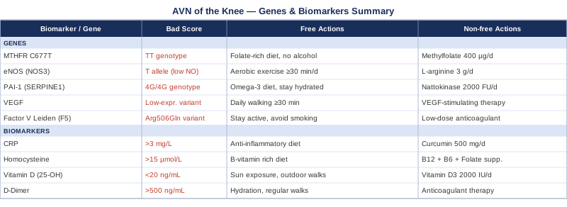
At a Glance: Genes, Biomarkers, and Action Plans
Andrew Huberman's Framework for Bone and Vascular Health: 10 Things Worth Knowing
If one scientific communicator has done the most to translate bone biology and vascular physiology into actionable human protocols, it is Andrew Huberman. His podcast, Huberman Lab, draws on peer-reviewed research and expert interviews to build practical frameworks that challenge some of the oversimplified advice commonly heard in clinical settings. For someone dealing with avascular necrosis of the knee, several of his recurring themes are directly relevant.
1. Impact Loading Is the Irreplaceable Signal for Bone Remodeling
Bone does not remodel in response to calcium intake alone. It remodels in response to mechanical stress. Huberman emphasizes research showing that short bouts of high-impact loading — jumping, landing, brisk walking — generate the piezoelectric and mechanotransduction signals that activate osteoblasts. For AVN patients, this is complicated by joint compromise, but low-impact alternatives (pool resistance, resistance training with safe loading patterns) can generate meaningful osteogenic signaling without excessive joint stress.
2. Nitric Oxide Is Not Optional for Vascular Health
Nitric oxide production — covered extensively in Huberman's cardiovascular health content — is the mechanism by which arteries dilate and maintain healthy blood flow in small vessels. Reduced NO in subchondral bone is a direct contributor to the ischemic environment of AVN. Huberman highlights dietary nitrates, zone 2 cardio, and nasal breathing as practical, non-pharmacological ways to support the NO system daily.
3. The Vitamin D–Calcium Cascade Requires Active Management
Huberman has repeatedly noted that optimizing vitamin D requires understanding the entire cascade: sunlight or supplementation raises serum 25-OH vitamin D, which requires magnesium to be hydroxylated to its active form, and vitamin K2 to direct the resulting calcium into bone rather than soft tissue. Treating only vitamin D in isolation is a common incomplete approach.
4. Sleep Is the Master Repair Window
The majority of growth hormone pulsation occurs during slow-wave sleep. Growth hormone is a key driver of tissue repair, including bone vascularization. Huberman cites research showing that chronic sleep restriction reduces GH secretion, bone density accrual, and healing capacity in connective tissue. For AVN patients hoping to maximize the body's natural repair response, sleep architecture matters as much as supplementation.
5. Cortisol Dysregulation Actively Destroys Bone
Huberman has covered the bidirectional relationship between chronic stress, HPA axis activation, and bone loss in detail. Elevated chronic cortisol suppresses osteoblasts and promotes osteoclast activity — literally accelerating bone resorption. This is the same mechanism that makes prolonged corticosteroid therapy so damaging to bone. Managing psychological stress is not a soft intervention; it has direct, measurable effects on bone metabolism.
6. Zone 2 Cardio Is Anti-Inflammatory at the Systemic Level
Regular moderate-intensity aerobic exercise — approximately 150–180 minutes per week of zone 2 activity — is one of the most powerful systemic anti-inflammatory interventions available. Huberman discusses how it reduces CRP, supports vascular endothelial function, improves fibrinolytic balance, and upregulates VEGF. These effects are all directly relevant to the vascular pathology of AVN.
7. Omega-3s Are a Non-Negotiable for Bone and Vascular Function
Huberman consistently places high-dose EPA and DHA in his personal supplement regimen because of their evidence base across cardiovascular, neurological, and musculoskeletal outcomes. For bone specifically, research suggests omega-3s promote osteoblast differentiation and inhibit osteoclast activity — a favorable balance for anyone with active osteonecrosis. The dose matters: 2–4g EPA+DHA daily is what the evidence supports, not the 300mg found in most supermarket fish oil capsules.
8. Magnesium Is the Overlooked Bone Mineral
Huberman has discussed the fact that approximately 40–50% of Americans are deficient in magnesium, yet most bone health conversations focus on calcium. Magnesium is required for vitamin D activation, for ATP-dependent bone remodeling processes, and for parathyroid hormone regulation. A deficiency silently blunts the effect of every other bone-supportive intervention.
9. Resistance Training Outperforms Stretching for Bone
Flexibility work has value for joint range of motion, but Huberman is clear that resistance training generates the specific mechanical tension and hormonal response (testosterone, IGF-1 locally) that drives bone density. For AVN patients, this requires working with a physical therapist to design loading patterns that strengthen supporting musculature without overloading the compromised joint surface — but the resistance training principle remains important.
10. The Gut-Bone Axis Is Emerging and Worth Watching
Huberman has touched on research showing that gut microbiome diversity influences bone mineral density through short-chain fatty acid production, systemic inflammation levels, and vitamin K synthesis. This is an early area, but the directionality of evidence is consistent: a healthy gut microbiome supports better bone outcomes. A diet rich in fermented foods (yogurt, kefir, kimchi), prebiotic fiber, and minimal antibiotics is the accessible starting point.
Complementary Approaches with Clinical Support for Avascular Necrosis of the Knee
The following modalities have enough clinical evidence in relevant areas — bone repair, pain management, vascular function, or knee joint function — to be worth discussing with your care team. None replace standard orthopaedic management, but several offer meaningful adjunctive support.
Low-Level Laser Therapy and Photobiomodulation
Low-level laser therapy (LLLT), now more commonly called photobiomodulation (PBM), uses specific wavelengths of red and near-infrared light to stimulate cellular energy production, reduce inflammation, and promote tissue repair. For avascular necrosis, the most relevant mechanisms are its effects on osteoblast activation, local VEGF upregulation, and reduction of oxidative stress in compromised tissue. Several in vitro and animal studies have shown that near-infrared light stimulates bone cell proliferation and angiogenesis — the exact processes needed in AVN recovery. Human clinical trials on photobiomodulation for knee pain and cartilage have demonstrated measurable reductions in pain and inflammatory markers. Evidence for AVN specifically is still emerging, but the mechanistic rationale is strong, and the safety profile is excellent.
A commonly used protocol for musculoskeletal conditions uses devices emitting light at 630–850nm wavelengths, applied directly to the joint area for 10–20 minutes per session, three to five times per week. A systematic review of LLLT in knee and bone conditions supports the anti-inflammatory and tissue-repair mechanisms. Devices range from clinical-grade panels ($300–1,500) to professional in-office units. Some physical therapy centers offer PBM sessions as part of their standard knee rehabilitation protocol.
To apply it cautiously: begin with shorter sessions (8–10 minutes) and increase to 15–20 minutes based on tolerance. Avoid directing the beam at the eyes. Do not apply over any active malignancy. If you have implanted metals in the area, verify device compatibility. The realistic expectation is a reduction in pain and inflammation over 6–12 weeks of consistent use, with potential support for the repair environment — not reversal of existing necrosis.
Tai Chi for Knee Stability and Circulation
Tai chi is a traditional Chinese movement practice characterized by slow, controlled, weight-shifting exercises and focused breathing. For avascular necrosis of the knee, it is notable for two reasons: it provides a form of low-impact joint loading that stimulates bone and connective tissue without the compressive stress of jogging or jumping, and its deep breathing and meditative components improve autonomic regulation and circulation. The slow weight transfer inherent in tai chi movements also strengthens the quadriceps and stabilizers of the knee without excessive stress on compromised subchondral bone.
Several randomized controlled trials have evaluated tai chi in knee osteoarthritis — the closest well-studied analog to AVN from a symptom and loading standpoint. A notable 2016 trial published in Annals of Internal Medicine found tai chi equally effective to physical therapy for pain and functional outcomes in knee osteoarthritis. A PubMed summary of tai chi and knee/bone health RCTs reflects the consistent pattern of benefit. The circulatory improvements from regular tai chi practice are also relevant to the vascular component of AVN.
For AVN patients, tai chi should begin under supervision — ideally with a practitioner who is aware of the diagnosis. A 20–30 minute session performed 3–5 times per week is a realistic starting point. Seated or supported variations are available for those with significant pain or instability. The long-term goal is improved proprioception and muscular support around the affected joint, reducing the forces that the compromised bone surface must absorb.
Mindfulness-Based Stress Reduction (MBSR) for Pain and Cortisol Management
Mindfulness-Based Stress Reduction, the structured 8-week program developed by Jon Kabat-Zinn, combines formal meditation, body scan practices, and mindful movement to improve the psychological and physiological response to chronic pain and stress. For avascular necrosis, its relevance is dual: MBSR addresses the psychological distress that accompanies a degenerative joint condition and has a direct anti-inflammatory effect through HPA axis downregulation — reducing cortisol levels that otherwise drive bone resorption.
Multiple randomized trials have demonstrated MBSR's efficacy for chronic pain conditions, with a well-known body of RCT evidence on PubMed showing reductions in pain catastrophizing, cortisol, and CRP following an 8-week program. A 2011 study in Annals of the New York Academy of Sciences showed that MBSR significantly reduced salivary cortisol over the intervention period. Given that chronic cortisol elevation is a direct threat to bone health, this is not merely a psychological intervention.
The standard MBSR format is 8 weekly group sessions of 2.5 hours, one full-day retreat, and 40–45 minutes of daily home practice. Online versions are now widely available and validated. The most important caveat is that MBSR is not a pain cure — it changes the relationship to pain, which has meaningful quality-of-life and functional consequences. For AVN patients managing chronic discomfort while waiting for revascularization or between surgical decisions, this is a valuable and evidence-supported tool.
Biofeedback for Movement Rehabilitation and Joint Loading
Biofeedback uses sensors attached to the body to provide real-time information about physiological processes — muscle activation, joint loading, heart rate variability, or skin conductance — allowing the user to consciously modify those processes. For avascular necrosis of the knee, the most relevant application is electromyographic (EMG) biofeedback during rehabilitation, which allows patients to see in real time whether they are adequately activating the quadriceps, hamstrings, and stabilizers that unload the femoral condyle during movement. Abnormal loading patterns — often developed unconsciously to avoid pain — can actually increase localized stress on necrotic bone.
Clinical trials in knee rehabilitation have shown that EMG biofeedback significantly improves quadriceps activation and functional outcomes compared to exercise alone. A PubMed search on EMG biofeedback and knee rehabilitation returns consistent evidence for its role in improving muscle recruitment. The principle is directly applicable to AVN rehabilitation, where optimal muscle activation around the joint is critical for protecting compromised bone.
Biofeedback sessions are typically performed with a trained physical therapist, integrated into a standard rehabilitation protocol. Wearable EMG devices are now available for home use at $100–400. Sessions of 20–30 minutes three times per week, combined with targeted strengthening exercises, are the typical format. The goal is to retrain movement patterns so that daily activities distribute load more evenly across the joint, reducing the mechanical burden on the most compromised area of the femoral condyle.
Conclusion
Avascular necrosis of the knee sits at the intersection of vascular biology, bone metabolism, genetics, and inflammation. Understanding which of these pathways is most active in your specific case — through biomarkers like D-dimer, homocysteine, bone turnover markers, and vitamin D, combined with genetic context from genes like MTHFR, Factor V Leiden, PAI-1, and eNOS — gives you a measurably better framework for decisions than waiting passively for imaging-based progression.
The next smart step is not to implement everything at once. It is to pick one or two biomarkers to test, discuss your coagulation history with your physician, and begin the lifestyle changes — movement, sleep, anti-inflammatory diet, stress management — that support every pathway discussed here. Better information leads to better decisions. Start with the information.
Cardiovascular Endocrine & Metabolic
Musculoskeletal: Bone Conditions Joint Conditions
Cardiovascular: Vascular Conditions
Autoimmune: Inflammatory Conditions