This article contains AI generated content.
· UpdatedBecker Muscular Dystrophy — 5 Genes And 7 Biomarkers To Track
Introduction
Becker Muscular Dystrophy does not follow a single script. Two people with similar mutations in the dystrophin gene can have entirely different disease courses — one remains ambulatory well into their 40s, while another loses independent walking a decade earlier. This variability is not random luck. It has molecular explanations that most clinical visits do not explore, and understanding it is one of the most useful things anyone living with or managing BMD can do.
Standard clinical care for BMD is built largely around monitoring what has already changed: tracking functional decline, adjusting physical therapy, and managing cardiac symptoms once they appear. This reactive framework is necessary but incomplete. It does not tell you which biological processes are most active right now, how severe your particular risk profile actually is, or which interventions make the most sense for your specific situation rather than for a generic BMD patient.
There is a more targeted way to approach this. Specific biomarkers — measurable in blood or through functional tests — can reveal what is happening in your heart, muscles, and inflammatory pathways before symptoms escalate. At the same time, several well-studied genetic modifier variants explain why BMD progresses at such different rates between patients, and knowing your modifier status changes what preventive priorities make sense.
This article covers both. The first section identifies the seven most clinically meaningful biomarkers for BMD, with practical guidance on how to measure them and what to do when results are out of range. A following section examines five genetic modifiers with documented human evidence for shaping disease trajectory, each paired with specific action plans. Beyond these core sections, recent exercise science has significantly updated the old "avoid exertion" orthodoxy in neuromuscular care, and several complementary approaches have accumulated real clinical support. Better information does not replace medical judgment — it sharpens the conversations that lead to it.
Summary
This article identifies 7 biomarkers that most BMD monitoring plans underuse — including one that reveals a serious blind spot in standard kidney function testing for people with low muscle mass. The genetics section covers 5 modifier variants beyond the DMD gene itself, each with documented human evidence for shaping how fast or slowly BMD progresses, paired with specific plans for the higher-risk variants. Beyond biomarkers and genetics, recent exercise science has overturned the "avoid all exertion" orthodoxy in BMD care in ways that are both surprising and immediately actionable. Four complementary approaches with genuine clinical evidence round out the article for those looking to build a more complete care strategy.
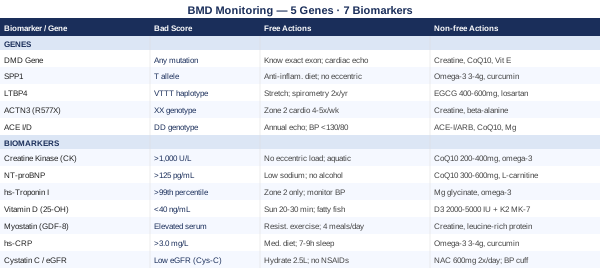
The 7 Biomarkers Worth Tracking in Becker Muscular Dystrophy
Biomarker monitoring in BMD is not a one-time blood draw at diagnosis. It is an ongoing practice that reveals how your body is responding over time and where intervention is most warranted. The seven markers below were selected for their direct relevance to the specific biological pathways involved in BMD — muscle membrane integrity, cardiac function, fibrosis-related inflammation, and metabolic compensation. Not all of them are ordered in standard clinical care. Knowing to ask for them specifically is the first step.
1. Creatine Kinase (CK)
Why it matters
Creatine kinase is the most established biochemical marker of skeletal muscle membrane damage. In BMD, insufficient or structurally abnormal dystrophin destabilizes the sarcolemma, causing persistent leakage of intracellular enzymes into the bloodstream. CK levels in BMD are typically 5 to 100 times the upper limit of normal, often ranging from 1,000 to 15,000 U/L compared to a normal adult range of roughly 38 to 174 U/L. As the disease progresses and muscle is replaced by fibrotic tissue, CK can paradoxically decrease — not because the condition is improving, but because there is less contractile tissue left to leak. Serial measurements over time carry more diagnostic weight than any single reading. According to the GeneReviews entry on Becker Muscular Dystrophy, markedly elevated CK is one of the earliest and most consistent biochemical findings across all dystrophinopathies.
How to measure it
CK is a standard blood test available at any clinical laboratory — Quest Diagnostics, LabCorp, or any hospital-affiliated lab. It can be ordered standalone or as part of a muscle enzyme panel. Out-of-pocket cost ranges from $15 to $75. Insurance routinely covers it with an appropriate neuromuscular diagnosis code. Annual measurement is a minimum for stable patients; more frequent testing is warranted after changes in physical activity, medication, or following cardiac events.
If the level is high: the plan without supplements
An acutely elevated CK significantly above your personal baseline warrants exercise modification before anything else. Eccentric muscle contractions — those where the muscle lengthens under load, such as walking downhill or lowering weights — cause disproportionately greater sarcolemmal stress in dystrophic muscle. Shifting activity toward concentric and isometric alternatives, prioritizing aquatic exercise where buoyancy reduces impact forces, and ensuring adequate rest between activity sessions can reduce unnecessary CK spikes without sacrificing cardiovascular conditioning. Prolonged bed rest is not the objective; strategic activity management is.
If the level is high: the plan with supplements or equipment
Coenzyme Q10 at 200 to 400 mg per day supports mitochondrial energy production in muscle cells and has membrane-stabilizing properties relevant to dystrophic muscle biology. Omega-3 fatty acids (EPA+DHA at 3 to 4 grams per day) reduce lipid peroxidation at the muscle membrane level. Creatine monohydrate at 3 to 5 grams per day is among the better-studied supplements in neuromuscular disease populations specifically — supporting phosphocreatine resynthesis in functional fibers with a strong safety record. None of these dramatically normalize CK, but they support the cellular environment that determines how much muscle is preserved over time. Recheck CK every 3 to 6 months when initiating these interventions.
2. NT-proBNP (N-Terminal Pro B-Type Natriuretic Peptide)
Why it matters
Dilated cardiomyopathy is the leading cause of death in adult BMD patients, and it frequently develops silently — before any breathlessness, edema, or chest symptoms appear. NT-proBNP is released by the ventricles under pressure and wall stress, making it one of the most sensitive early signals for emerging cardiac dysfunction, detectable before the left ventricular ejection fraction has visibly declined on echocardiography. Tracking this marker between echocardiograms closes a monitoring gap that standard care often leaves open.
How to measure it
NT-proBNP requires a specific blood test order — it is not included in standard panels. Cost ranges from $40 to $200 out of pocket, with insurance coverage generally available when ordered for cardiac monitoring in the context of a documented neuromuscular diagnosis. The clinical threshold for concern in adults under 75 is above 125 pg/mL, though values in BMD should always be interpreted alongside echocardiography findings by a cardiologist experienced with neuromuscular disease. Annual testing is appropriate in asymptomatic patients; every 6 months is reasonable when any cardiac abnormality has been detected.
If NT-proBNP is elevated: the plan without supplements
Any elevation above 125 pg/mL in a BMD patient without another clear explanation warrants cardiology referral and echocardiography. Lifestyle-level responses include a low-sodium diet (under 2,000 mg per day), which reduces cardiac preload meaningfully. Alcohol should be eliminated entirely — even moderate intake exerts direct toxic effects on cardiomyocytes, and the dystrophin-deficient cardiac cytoskeleton has reduced structural resilience to handle this. Daily weight monitoring (same time each morning) allows early detection of fluid retention, which precedes symptomatic heart failure.
If NT-proBNP is elevated: the plan with supplements or equipment
CoQ10 at 300 to 600 mg per day (divided into two doses) has the most robust human trial data for supporting cardiac energetics in dilated cardiomyopathy, including the Q-SYMBIO randomized trial demonstrating reduced mortality in heart failure with CoQ10 supplementation. L-carnitine at 1 to 2 grams per day supports fatty acid transport in cardiac mitochondria, which becomes impaired in failing heart tissue. Taurine at 2 to 4 grams per day has modest positive evidence in heart failure populations. All of these should be discussed with a cardiologist — they complement, not replace, pharmacological cardiac management (ACE inhibitors, beta-blockers, diuretics where indicated).
3. High-Sensitivity Cardiac Troponin I (hs-TnI)
Why it matters
High-sensitivity troponin I detects subclinical cardiac muscle injury at concentrations far below what standard troponin assays can identify. In BMD, the same dystrophin deficiency that damages skeletal muscle also affects cardiac muscle — meaning chronic low-level myocardial injury can occur years before ejection fraction declines. Serial hs-TnI measurement tracks the trend rather than a single snapshot, and a slow upward drift over years may indicate accelerating cardiac injury even when echocardiography appears reassuringly normal.
How to measure it
High-sensitivity troponin I is now standard at most hospital and large commercial laboratories. Cost ranges from $50 to $150 out of pocket. Elevations above the 99th percentile reference limit (which varies by assay but is often between 12 and 34 ng/L) indicate myocardial stress. In BMD patients, even low-level persistent elevations — values that would be dismissed as non-significant in a general cardiology context — are clinically meaningful and should prompt discussion.
If troponin I is elevated: the plan without supplements
Any elevation above the 99th percentile in a BMD patient without a clearly identifiable transient cause (intense exercise, dehydration) requires cardiac evaluation. Exercise programming should shift away from any maximal-intensity activities, as the dystrophin-deficient cardiac muscle does not have the structural resilience for high-demand outputs safely. Prioritizing zone 2 aerobic effort — sustained, conversational-pace cardio — supports cardiac adaptation without overloading the system. Close blood pressure monitoring is warranted, as even mild hypertension amplifies cardiac wall stress significantly.
If troponin I is elevated: the plan with supplements or equipment
Magnesium glycinate at 200 to 400 mg per day supports cardiac electrical stability and reduces ectopic firing risk in hearts under structural stress. Omega-3 fatty acids at 3 to 4 grams EPA+DHA per day have anti-arrhythmic and anti-inflammatory effects on cardiac tissue with multiple supporting trials. A wearable single-lead ECG monitor (such as the AliveCor KardiaMobile, approximately $79 to $149) enables home rhythm surveillance between clinical visits — particularly useful for detecting atrial arrhythmias, which are more prevalent in cardiomyopathic hearts and can be asymptomatic in their early stages.
4. 25-Hydroxyvitamin D
Why it matters
Vitamin D is not simply a bone mineral regulator. Vitamin D receptors are expressed in skeletal muscle, and active vitamin D (calcitriol) directly influences muscle protein synthesis pathways, mitochondrial function, and immune regulation. In people with reduced mobility or predominantly indoor activity — both common in progressive BMD — vitamin D deficiency is prevalent. Studies in neuromuscular disease populations consistently document low 25-OH vitamin D levels alongside associations with worse functional muscle scores. Vitamin D also modulates TGF-beta signaling, the same fibrotic pathway influenced by LTBP4 biology — making deficiency correction particularly relevant for patients with the higher-risk LTBP4 haplotype.
How to measure it
The 25-hydroxyvitamin D blood test is widely available and among the more affordable markers on this list. Out-of-pocket cost ranges from $30 to $100; direct-to-consumer lab services such as Marek Diagnostics often offer it for under $40. Optimal levels in the context of muscle health are generally considered to be in the range of 40 to 60 ng/mL. Testing twice yearly — once in winter and once in summer — captures the seasonal variation that significantly affects supplementation decisions.
If vitamin D is low: the plan without supplements
Direct sunlight exposure to large skin surface areas (arms and legs uncovered) for 20 to 30 minutes between 10 am and 2 pm can raise 25-OH vitamin D meaningfully in those who can safely access the outdoors. Dietary sources contribute modestly: fatty fish (salmon, mackerel, sardines), fortified dairy products, and egg yolks all provide vitamin D, but rarely enough to correct a true deficiency without supplementation. Short, consistent sun exposure is more effective than occasional prolonged sessions; sunburn confers no additional benefit.
If vitamin D is low: the plan with supplements or equipment
Vitamin D3 (cholecalciferol) at 2,000 to 5,000 IU per day is the standard correction dose for deficiency. Take it with the day's largest fat-containing meal to maximize absorption of this fat-soluble vitamin. Pair it with vitamin K2 in MK-7 form at 100 to 200 mcg per day — K2 ensures that calcium mobilized by vitamin D is directed to bone rather than soft tissue during correction. Recheck 25-OH vitamin D after 3 months. Doses above 5,000 IU per day should be guided by laboratory testing rather than maintained indefinitely, as toxicity (while uncommon) is entirely avoidable with periodic monitoring.
5. Myostatin (GDF-8)
Why it matters
Myostatin — growth differentiation factor 8 — acts as a biological brake on muscle growth. In healthy muscle, it limits hypertrophy. In BMD, chronic inflammation and muscle damage upregulate myostatin signaling, driving atrophy beyond what dystrophin deficiency alone would cause. Measuring serum myostatin reveals the anabolic-catabolic balance in remaining functional muscle — a ratio that determines whether those fibers are being preserved or progressively lost. Myostatin-targeting therapies are among the most actively studied strategies in dystrophinopathy research, and baseline measurement is becoming more clinically relevant as trials advance and eligibility criteria solidify.
How to measure it
Serum myostatin is not yet part of standard clinical care but is available through specialty and research-oriented laboratories. Cost ranges from $200 to $500. Academic medical centers with dedicated neuromuscular disease programs sometimes include it in research protocol panels. Patients enrolled in BMD natural history studies or clinical trials often have myostatin measured as a trial biomarker. Access is improving as this marker transitions from research to clinical relevance.
If myostatin is high: the plan without supplements
Resistance exercise, even at low intensity, is among the most potent natural suppressors of myostatin expression in skeletal muscle. In BMD, the key is muscle activation without causing significant membrane damage — which means avoiding eccentric-dominant loading and prioritizing concentric and isometric resistance work under the guidance of a physical therapist experienced with neuromuscular disease. Protein distribution across meals is equally important: consuming protein in 3 to 4 evenly distributed servings daily activates mTOR-mediated anabolic pathways more consistently than concentrating protein intake in a single meal.
If myostatin is high: the plan with supplements or equipment
Creatine monohydrate at 3 to 5 grams per day continuously supports phosphocreatine regeneration and has modest evidence for attenuating myostatin-related atrophy signals. Leucine-enriched protein at 30 to 40 grams per serving (containing at least 2.5 to 3 grams leucine) taken within 30 minutes of resistance activity maximally stimulates mTOR signaling, directly counteracting myostatin's catabolic effects at the cellular signaling level. Follistatin — a natural myostatin antagonist — is under active pharmaceutical investigation; dietary patterns that modestly upregulate follistatin include egg-rich diets and cruciferous vegetables, though clinical-grade delivery remains experimental.
6. High-Sensitivity C-Reactive Protein (hs-CRP)
Why it matters
In dystrophic muscle, inflammation is not merely a symptom — it is a driver. Activated macrophages and elevated cytokines, particularly TNF-alpha and IL-6, promote fibrotic remodeling that progressively replaces contractile muscle with collagen. High-sensitivity CRP is a reliable downstream marker of this systemic inflammatory activity. In cardiovascular medicine, physicians like Peter Attia highlight hs-CRP as one of the most actionable inflammatory markers precisely because it responds measurably to lifestyle changes and targeted interventions — making it useful for both initial assessment and tracking whether your anti-inflammatory efforts are producing any effect.
How to measure it
Hs-CRP must be specifically ordered as high-sensitivity CRP — standard CRP tests are less sensitive and miss the low-grade inflammation relevant here. Cost ranges from $15 to $60, and it is widely covered by insurance. For cardiovascular risk, values below 1.0 mg/L are optimal; below 2.0 mg/L is low risk; above 3.0 mg/L indicates elevated inflammatory activity warranting specific attention. In BMD, even values in the 1.5 to 3.0 mg/L range are worth addressing given the pro-fibrotic environment this creates. Test at baseline and every 3 to 6 months when actively modifying lifestyle or supplementation.
If hs-CRP is elevated: the plan without supplements
Diet is the most powerful lever. A consistent Mediterranean-style eating pattern — centered on extra-virgin olive oil, oily fish three or more times weekly, abundant vegetables, and legumes, with minimal ultra-processed foods — reduces hs-CRP in clinical trials more reliably than most supplements do. Sleep is the second lever: less than 6 hours per night substantially elevates CRP, and in the context of a condition already driving systemic inflammation, poor sleep compounds the damage. Modest reductions in visceral adiposity (even 5% body weight reduction in those who are overweight) produce measurable CRP decreases. Zone 2 aerobic activity 3 to 4 days per week is anti-inflammatory when performed below the threshold that triggers muscle damage.
If hs-CRP is elevated: the plan with supplements or equipment
Omega-3 fatty acids at 3 to 4 grams EPA+DHA per day are among the most consistently anti-inflammatory supplements available with a robust evidence base across multiple chronic disease populations. Curcumin with piperine (500 to 1,000 mg/day of a bioavailable formulation) reduces CRP and NF-kB-driven inflammation in human trials — a practical cycling protocol is 8 weeks on followed by 3 weeks off. Resveratrol at 500 mg/day with a fat-containing meal modestly enhances anti-inflammatory signaling via SIRT1 pathways. Recheck hs-CRP every 3 months during an active intervention period to assess response before adjusting strategy.
7. Cystatin C and eGFR
Why it matters
This is the biomarker most BMD monitoring plans miss entirely. Standard kidney function calculations depend on serum creatinine — which is a muscle metabolite. In patients with significant muscle loss, serum creatinine is low regardless of actual kidney function, creating a falsely reassuring eGFR. A BMD patient with substantial muscle wasting can show a creatinine level in the normal range while true renal function is meaningfully reduced. Cystatin C, by contrast, is produced by all nucleated cells at a constant rate independent of muscle mass, making cystatin C-based eGFR the standard recommended by Peter Attia and renal medicine specialists for any patient with abnormal muscle mass. In BMD, where medications including ACE inhibitors and diuretics affect the kidneys — and where actual renal reserve may be underestimated — accurate measurement matters for safe clinical management.
How to measure it
Standard metabolic panels include creatinine-based eGFR for $15 to $50 — but this is not sufficient for BMD patients. Cystatin C requires a specific order and costs $50 to $150 out of pocket. Request that your physician order cystatin C specifically and ask for the cystatin C-based eGFR interpretation using the CKD-EPI cystatin C equation. The combined creatinine + cystatin C formula (CKD-EPI 2021) offers the most precision when available. Annual testing is appropriate for stable patients; more frequent monitoring is warranted if creatinine trends are changing or medications are being adjusted.
If eGFR is low: the plan without supplements
Consistent hydration — 2 to 2.5 liters of fluid per day in temperate conditions — supports renal perfusion. NSAIDs (ibuprofen, naproxen) should be avoided entirely in any BMD patient with reduced renal reserve, as they are nephrotoxic and can precipitate acute kidney injury in the setting of chronic mild impairment. A complete medication review with a physician or clinical pharmacist to ensure all drug doses are appropriate for the actual GFR level is a fundamental safety measure. Discuss protein intake with a renal dietitian — adequate protein for muscle maintenance remains essential, but guidance on total load matters when GFR is below 45 mL/min.
If eGFR is low: the plan with supplements or equipment
N-acetylcysteine (NAC) at 600 mg twice daily has demonstrated renal-protective effects in high-risk patients exposed to nephrotoxic stressors and carries a favorable safety profile at this dose. Avoid high-dose antioxidant supplementation without medical guidance — some supplements (vitamin C above 1,000 mg/day, certain botanical extracts) can increase kidney stone risk or interact unpredictably with medications in renally-compromised patients. A validated home blood pressure cuff is an essential and inexpensive tool: hypertension both causes and accelerates GFR decline, and consistent home monitoring enables tighter blood pressure control than clinic-only measurements.
Biomarkers illuminate the current state of the disease. What they cannot fully explain is why that state looks the way it does — and why it may progress at a very different rate than someone else with a similar mutation. That requires looking at the genetics operating beneath the surface.
5 Gene Variants That Shape Your BMD Trajectory
The DMD gene determines that someone has BMD. It does not determine how severely they will experience it. Several well-documented modifier genes — working through inflammatory, fibrotic, and metabolic pathways — can produce disease courses ranging from surprisingly mild to rapidly progressive, even among patients with virtually identical dystrophin mutations. Modifier gene testing is increasingly accessible through standard clinical genetics panels, and knowing your profile can meaningfully shift which monitoring priorities and preventive measures deserve the most attention.
DMD Gene — Your Specific Mutation Defines Your Clinical Options
The DMD gene on chromosome Xp21.2 encodes dystrophin, the largest gene in the human genome at 2.4 million base pairs. In BMD, mutations preserve the reading frame, producing truncated but partially functional dystrophin — which is why BMD is milder than Duchenne MD, where near-complete dystrophin absence results from frame-disrupting mutations. Your specific exon deletion or duplication carries distinct prognostic implications and determines eligibility for specific therapeutic approaches. Deletions involving exons 45 to 55 are common in BMD and often associated with milder phenotypes; those spanning exon 51 are relevant because exon-skipping approaches designed for DMD intersect with BMD mutation profiles.
If the gene is bad: the plan without supplements
Know your exact exon deletion or duplication. If comprehensive mutation characterization has not been done, request full DMD gene panel sequencing (including deletion/duplication analysis) through a certified neuromuscular genetics program — this identifies your precise reading frame status and mutation-specific natural history data. Regular cardiac surveillance starting at or before age 10 (or at diagnosis in adults newly identified), pulmonary function testing at least annually, and physical therapy focused on contracture prevention in the specific muscle groups your mutation typically affects earliest are the non-negotiable care components to build around your genetic data.
If the gene is bad: the plan with supplements or equipment
Mutation-specific therapies are advancing rapidly. Clinical trials for exon-skipping, nonsense suppression, and AAV-delivered micro-dystrophin gene therapy are active and increasingly enrolling BMD participants with specific mutation profiles. The NIH clinical trials registry (clinicaltrials.gov) lists open studies searchable by mutation type and location — this is worth reviewing with a neuromuscular specialist at least annually given the pace of development. Supportively, creatine monohydrate (3 to 5 grams/day), CoQ10 (200 to 400 mg/day), and vitamin E (400 IU/day, not exceeding this dose) address downstream metabolic and oxidative consequences of dystrophin insufficiency regardless of specific exon deletion.
SPP1 — The Inflammatory Modifier
The SPP1 gene at chromosome 4q22.1 encodes osteopontin, a pro-inflammatory glycoprotein that drives macrophage polarization toward the M1 phenotype — the inflammatory subtype most associated with fibrotic muscle remodeling. A well-studied promoter polymorphism, the -66T>G variant (rs28357094), alters osteopontin expression levels and has been demonstrated in multiple human cohorts to modify disease severity in both BMD and DMD. Patients carrying the T allele (associated with higher osteopontin expression) show greater ambulatory loss and faster functional decline compared to those with the G allele. This finding has been replicated across independent European and North American cohorts, establishing SPP1 as one of the most robust genetic disease modifiers identified in dystrophinopathies.
Osteopontin promotes chronic macrophage activation in dystrophic muscle, contributing to the fibrotic environment that accelerates functional loss beyond what the dystrophin deficit alone would produce. This makes the inflammatory pathway an accessible therapeutic target for high-risk SPP1 genotypes.
If the gene is bad (T allele): the plan without supplements
Reduce all modifiable drivers of macrophage-driven inflammation. A consistently anti-inflammatory diet provides the most accessible daily lever: extra-virgin olive oil for its oleocanthal-mediated NF-kB inhibition, oily fish three or more times weekly for EPA/DHA, and systematic elimination of ultra-processed foods and refined seed oils. Adequate sleep — 7 to 9 hours with consistent timing — reduces overnight inflammatory cytokine production measurably. Avoid the specific activity patterns that trigger acute muscle membrane damage (high-eccentric loading), because each episode amplifies the SPP1-driven inflammatory cascade and compresses recovery time.
If the gene is bad: the plan with supplements or equipment
Corticosteroids (deflazacort or prednisone) are part of standard DMD management for their capacity to suppress the osteopontin-driven macrophage inflammatory response, and some neuromuscular clinicians consider them in specific BMD contexts — a discussion worth having with your neurologist in light of SPP1 status. For supplement-based approaches, omega-3 fatty acids at 3 to 4 grams EPA+DHA per day directly attenuate the macrophage polarization that osteopontin promotes. Curcumin with piperine (500 to 1,000 mg/day of a bioavailable form, cycling 8 weeks on/3 weeks off) targets NF-kB, the central transcription factor mediating osteopontin's pro-inflammatory and pro-fibrotic downstream effects.
LTBP4 — The Fibrosis Modifier
Latent TGF-beta binding protein 4 (LTBP4), on chromosome 19q13.2, regulates TGF-beta bioavailability — and TGF-beta is the primary molecular driver of fibrotic muscle remodeling. LTBP4 carries intronic variants that form two major haplotypes: IAAM and VTTT. Research published in Nature Genetics established these haplotypes as significant modifiers of disease progression in dystrophinopathies. Patients with the IAAM/IAAM genotype show significantly slower ambulation loss and delayed respiratory decline compared to those with the VTTT haplotype, which permits more TGF-beta to signal unchecked, accelerating fibrotic replacement of muscle tissue.
This modifier is clinically important because fibrosis — the replacement of contractile muscle with inextensible collagen — is irreversible. Earlier intervention in patients identified as VTTT haplotype carriers can preserve more functional muscle precisely by limiting the fibrotic progression before it consolidates.
If the gene is bad (VTTT haplotype): the plan without supplements
Physical therapy with a strong contracture-prevention focus becomes disproportionately important for VTTT haplotype patients. Fibrosis drives contracture formation faster than in IAAM patients with comparable dystrophin levels, making regular stretching and nighttime splinting protocols genuinely protective rather than merely precautionary. Serial spirometry monitoring (FVC and FEV1) at minimum twice yearly is warranted — respiratory muscle fibrosis can produce significant pulmonary restriction before dyspnea is noticed. Periodic overnight pulse oximetry helps detect nocturnal oxygen desaturation, which can precede daytime respiratory symptoms by years in this patient profile.
If the gene is bad: the plan with supplements or equipment
Losartan — an angiotensin receptor blocker that suppresses TGF-beta signaling — is under active clinical investigation in muscular dystrophy for its potential to slow fibrosis-related progression, with precedent from connective tissue disorders. EGCG (epigallocatechin gallate from standardized green tea extract at 400 to 600 mg/day) has demonstrated anti-fibrotic activity via TGF-beta pathway inhibition in cell and animal models of muscular dystrophy. Omega-3 fatty acids at 3 to 4 grams EPA+DHA modestly reduce TGF-beta signaling in human tissues. Vitamin E at 400 IU/day (not exceeding this dose) reduces lipid peroxidation in fibrosis-prone tissue environments. These are supportive measures — any decision about losartan specifically requires prescriber involvement.
ACTN3 (R577X) — The Muscle Fiber Composition Modifier
Alpha-actinin-3, encoded by ACTN3 on chromosome 11q13.2, is expressed exclusively in fast-twitch (type II) skeletal muscle fibers. The R577X polymorphism (rs1815739) generates a premature stop codon — meaning that individuals with the XX genotype produce no functional ACTN3 protein at all. This variant is common, affecting roughly 18% of people of European ancestry, and in the general population is associated with reduced sprint and explosive power capacity. In BMD, where fast-twitch fibers are particularly vulnerable to dystrophin-related membrane damage, the XX genotype reduces the already-limited reserve of high-force muscle fibers available for functional compensation.
This has direct implications for exercise programming and for setting realistic functional expectations at different disease stages. XX genotype patients with BMD may experience earlier plateau in maximal strength tasks than RR or RX genotype patients with comparable dystrophin levels.
If the gene is bad (XX genotype): the plan without supplements
Emphasize aerobic conditioning over power-based training. Zone 2 cardio — sustained effort at 60 to 70% maximum heart rate, the level where conversation is possible but effortful — builds mitochondrial density in remaining type I fibers without overloading a depleted type II reserve. Walking, cycling, swimming, and elliptical training at conversational pace for 30 to 45 minutes per session, 4 to 5 days per week, is appropriate for most ambulatory BMD patients in this category. Track perceived exertion (the Borg scale or talk test) rather than absolute pace, as exertion tolerance fluctuates with disease activity and temperature.
If the gene is bad: the plan with supplements or equipment
Creatine monohydrate at 3 to 5 grams per day supports phosphocreatine regeneration in both fiber types and is particularly relevant for the compromised fast-twitch system in ACTN3-null patients. Beta-alanine at 3.2 grams per day (in two divided doses of 1.6 grams to minimize the characteristic tingling paresthesia) increases intramuscular carnosine, which buffers muscle acidosis during efforts approaching anaerobic threshold — potentially extending functional capacity in moderate-intensity activities. Use beta-alanine in 8-week cycles followed by 4-week breaks. Leucine-enriched protein per meal (targeting 2.5 to 3 grams leucine per serving) maximizes the mTOR anabolic stimulus in remaining functional muscle across both fiber types.
ACE I/D — The Cardiac Risk Modifier
The ACE gene on chromosome 17q23.3 carries a well-characterized insertion/deletion polymorphism (I/D, rs4646994) that produces dramatically different levels of circulating and tissue ACE activity. The DD genotype generates roughly twice the ACE activity of the II genotype. In the context of BMD, elevated ACE activity means elevated angiotensin II — a peptide that promotes both cardiac hypertrophy and fibrotic remodeling of cardiomyocytes that are already structurally compromised by dystrophin deficiency. Cohort studies in dystrophinopathy patients have found that the D allele is associated with worse preserved cardiac function over time, particularly in terms of ejection fraction trajectory.
This is one of the molecular rationales for why ACE inhibitor and ARB therapy is specifically recommended in BMD cardiac management, and why DD genotype patients may benefit most from early prophylactic initiation even before measurable cardiac dysfunction appears.
If the gene is bad (DD genotype): the plan without supplements
Begin cardiac surveillance proactively. Echocardiography at or before age 10 (or at adult diagnosis), and at minimum annually thereafter, is the standard — but DD genotype patients arguably warrant twice-yearly echo once any baseline abnormality is detected. Home blood pressure monitoring with a validated automated cuff is inexpensive and immediately actionable: the target for BMD patients with cardiac involvement is generally below 130/80 mmHg. A consistently low-sodium diet (under 2,000 mg/day) and complete alcohol avoidance reduce the cardiac afterload that a dystrophin-deficient heart must manage under angiotensin II signaling pressure.
If the gene is bad: the plan with supplements or equipment
ACE inhibitors (lisinopril, enalapril, perindopril) or ARBs (losartan, irbesartan) are the most evidence-supported pharmaceutical interventions in BMD cardiac management — recommended by the American Academy of Neurology and American Heart Association for consideration even in asymptomatic BMD patients based on the structural vulnerability of the dystrophin-deficient heart. This is not a supplement-level intervention; it requires a prescriber, and DD genotype status strengthens the case for initiating it early in a cardiology conversation. Supportively, CoQ10 at 200 to 400 mg/day reduces oxidative stress in cardiomyocytes under angiotensin II-driven stress, and magnesium glycinate at 200 to 400 mg/day reduces ectopic cardiac firing and modestly supports vasodilation.
Knowing your modifier gene profile and tracking the right biomarkers tells you the shape of your disease. What exercise and muscle science have recently clarified is what you can actually do about it — including in ways that challenge what clinicians have historically recommended.
What Muscle Science Is Getting Right — And What Clinics Often Miss
For decades, the dominant clinical advice for muscular dystrophy patients leaned toward caution: avoid overexertion, minimize physical stress, rest when in doubt. That framework has been progressively challenged — and in some areas overturned — by research into muscle physiology, mitochondrial biology, and cardiorespiratory fitness. Much of this work has been synthesized for general audiences by researchers like Peter Attia in Outlive: The Science and Art of Longevity and by Andrew Huberman's podcast series on muscle biology and training science.
The central insight with direct relevance to BMD: the body adapts to the stress it receives, and controlled appropriate stress signals adaptation — while prolonged underloading accelerates the very atrophy and cardiac decline the caution was meant to prevent. The question is not whether to exercise, but how to calibrate exercise correctly within the constraints of BMD biology. The following principles represent the most evidence-relevant applications of current muscle science to BMD care.
Zone 2 Cardio Is Non-Negotiable for Cardiac and Mitochondrial Survival
Zone 2 training — sustained aerobic effort at roughly 60 to 70% maximum heart rate, where speech is possible but requires focus — is the most potent non-pharmacological stimulus for mitochondrial biogenesis in both skeletal and cardiac muscle. In BMD, where oxidative stress affects mitochondrial function in both tissues, 3 to 5 sessions per week of 30 to 45 minutes of zone 2 cardio meaningfully improves cellular energy production without triggering significant sarcolemmal damage. Swimming and cycling are the most appropriate modalities for most BMD patients because they minimize eccentric loading. Attia frames VO2 max — directly built by zone 2 and interval training — as the single most powerful modifiable predictor of long-term survival across chronic disease populations, an argument that applies with even greater urgency when cardiac and pulmonary compromise are the leading causes of death.
Eccentric Contractions Are Specifically Dangerous in Dystrophic Muscle
Eccentric contractions — where muscle lengthens under load, as in walking downstairs or lowering weights — cause disproportionately greater sarcolemmal microtrauma than concentric contractions in all muscle, but dramatically more so in dystrophin-deficient muscle. These microtraumas trigger the inflammatory cascade that drives both acute CK elevation and chronic fibrotic remodeling. Eliminating high-eccentric activities from BMD exercise programming is not conservative overcaution — it is evidence-based exercise engineering. This means preferring uphill walking over downhill, resistance machines that limit lowering phases, and aquatic exercise where buoyancy attenuates impact and eccentric stress throughout range of motion.
Protein Distribution Matters as Much as Total Intake
The muscle protein synthesis literature — synthesized across Attia's protocols and the academic work underlying them — consistently demonstrates that distributing protein evenly across 3 to 4 meals per day generates greater net muscle protein synthesis than eating the same total amount concentrated in one or two meals. For BMD patients where preserving every gram of functional fiber matters, this means targeting 0.4 to 0.5 grams of protein per kilogram of body weight per meal, with each meal anchored by a leucine-rich source (whey protein, eggs, salmon, chicken breast) to maximally stimulate mTOR-driven anabolic signaling. Do not save most protein for dinner.
Sleep Is Muscle Anabolism, Not Just Rest
During deep (slow-wave) sleep, growth hormone secretion peaks and the primary cellular repair pathways in muscle are most active. In BMD, inadequate sleep — below 7 hours — measurably elevates cortisol, amplifies inflammatory cytokine production, suppresses testosterone, and worsens the catabolic environment in already-stressed muscle. Sleep quality in BMD patients can be specifically compromised by nocturnal hypoventilation — another reason routine sleep assessment including periodic overnight pulse oximetry has dual value. Treating sleep as a core intervention rather than a lifestyle preference changes what is possible physiologically.
Grip Strength as a Monthly Home Biomarker
Grip strength is a validated functional biomarker correlating with total skeletal muscle mass, cardiac health, and all-cause mortality across multiple populations and decades of research. In BMD, monthly grip strength measurement with a hand dynamometer ($20 to $50 for reliable home devices) provides a low-cost snapshot of whether systemic muscular function is being preserved or declining between clinical visits. A consistent downward trend over 3 to 4 months is an actionable signal to intensify physical therapy referral, review nutrition, and reassess the full biomarker panel — before functional milestones are lost that are difficult to recover.
Creatine Is Not Just an Athletic Supplement
Creatine monohydrate has more than two decades of safety data and has been specifically studied in neuromuscular disease populations — not just athletic ones. In BMD and related conditions, creatine at 3 to 5 grams per day continuously supports phosphocreatine regeneration in functional fibers, modestly attenuates muscle damage markers, and improves submaximal exercise performance. It is one of the few supplements where the evidence base in dystrophic muscle contexts is sufficient to recommend without substantial qualification. The safety profile at this dose is excellent, including in long-term use.
Cold Exposure and Heat Therapy Have Complementary and Relevant Effects
Brief cold exposure — a 3 to 5 minute cold shower or cold water immersion 3 to 4 times per week — activates norepinephrine pathways that reduce systemic inflammatory tone and improve mood and alertness, relevant for BMD patients managing both chronic inflammatory load and the psychological burden of a progressive condition. Heat therapy through sauna use (15 to 20 minutes at 170°F, 3 to 4 times weekly) increases heat shock protein expression, which supports protein quality control in stressed cells, and provides cardiovascular conditioning through cardiac preconditioning mechanisms. Sauna should be approached conservatively in those with established cardiomyopathy — start with lower temperatures and shorter sessions, with physician awareness, before progressing.
Respiratory Muscle Training Deserves Proactive Attention
Inspiratory muscle training (IMT) with a threshold resistance device — commercially available for $40 to $80 — has demonstrated in randomized trials that progressive respiratory resistance training improves inspiratory muscle strength and can slow the decline of forced vital capacity in neuromuscular disease patients. Most patients and clinicians wait for respiratory function to decline before introducing respiratory support. Starting IMT when FVC is still well-preserved builds patient competence and meaningful reserve before the techniques are urgently needed. 15 to 20 minutes of IMT per day, 5 days per week, at 30% of maximum inspiratory pressure (increasing by 5% every 4 to 6 weeks under respiratory therapist guidance) is a reasonable protocol.
Mindset Is a Biological Input, Not Merely Psychological
Chronic depression and anxiety — which are significantly more prevalent in BMD patients and their caregivers than in the general population — produce measurable changes in inflammatory and cortisol profiles that directly worsen the biology of the disease. Elevated cortisol promotes muscle catabolism, suppresses immune function, elevates CRP, and worsens sleep architecture. Structured interventions including MBSR, regular outdoor exposure, and maintained social connection all have quantifiable anti-inflammatory effects in clinical populations. This is not a soft recommendation. In a disease where every anabolic advantage matters at the cellular level, psychological wellbeing is a physiological intervention.
The Minimum Effective Dose Principle Applies in BMD
The goal is not to train maximally but to train optimally — the minimum stimulus required to maintain cardiovascular adaptation, myostatin suppression, and neuromuscular signaling without causing cumulative membrane damage across the week. A 2015 Cochrane systematic review of exercise in slowly progressive neuromuscular disease found that moderate aerobic and mild resistance exercise, performed consistently, was associated with functional benefit without measurable acceleration of decline. This shifts the clinical frame from "how much can you do" to "how consistently can you do enough." Consistency over intensity is the correct optimization target in BMD.
Track Functional Milestones Systematically
The 6-minute walk test (6MWT), timed 4-stair climb, and hand grip dynamometry are three of the most clinically validated functional biomarkers in BMD, used in major clinical trials as primary endpoints. Measuring them at home monthly — the 6MWT requires only a hallway, a stopwatch, and a measured distance; grip dynamometry requires a hand device — creates a personal longitudinal dataset that no annual clinic visit can replicate. Trends over 6 to 12 months reveal trajectory more reliably than any single clinic assessment. This data, brought to appointments, fundamentally changes the quality of clinical conversations.
Complementary Approaches With Meaningful Evidence
Standard BMD care centers on pharmacology, cardiac monitoring, and physical therapy. Several complementary modalities have accumulated enough human clinical evidence to be worth considering as adjuncts — not replacements — for standard care in the specific context of neuromuscular disease. The following were selected based on clinical relevance to BMD biology and the quality of supporting evidence.
Yoga — Range of Motion Preservation and Respiratory Flexibility
Adapted yoga offers something standard physical therapy often underdelivers: a sustained, gentle, daily practice integrating breathing, posture awareness, and controlled movement that patients can largely self-manage. In BMD, contracture formation in the hip flexors, Achilles tendons, and thoracic spine is among the most modifiable contributors to functional decline — and regular sustained stretching is the primary tool for slowing it. The breathing dimension of yoga practice (pranayama) is particularly relevant, as controlled breathing exercises improve chest wall compliance and have been shown to modestly improve vital capacity in restricted populations.
A pilot study of adapted yoga in ambulant patients with slowly progressive neuromuscular disease demonstrated improvements in balance, respiratory muscle flexibility, and quality-of-life scores with no adverse events related to muscle overexertion over a 12-week intervention period. These findings are preliminary and limited by small sample size, but the risk profile of appropriately adapted yoga is low.
Practically, weekly sessions with a yoga therapist experienced in adaptive or chair yoga, combined with a daily 10 to 15 minute home practice targeting hip flexors, ankle plantar flexors, and thoracic extension, is a realistic starting protocol. Avoid any weight-bearing posture that involves significant eccentric loading or puts pressure directly on weakened muscle bellies. Bikram (hot yoga) is not appropriate for patients with established cardiomyopathy.
Breathing-Based Therapies — Protecting Pulmonary Function Proactively
Respiratory failure is a leading cause of morbidity in progressive BMD, and pulmonary function begins declining years before ventilatory support becomes necessary. Breathing-based therapeutic techniques — including lung volume recruitment (LVR), manually assisted cough techniques, and inspiratory muscle training (IMT) — are among the most evidence-supported complementary approaches in neuromuscular disease. The American Thoracic Society's clinical practice guidelines for chronic neuromuscular disease explicitly recommend LVR and assisted cough techniques when FVC declines below 70%.
IMT with a threshold resistance device has been evaluated in randomized controlled trials in mixed neuromuscular disease populations and found to improve inspiratory muscle strength and delay FVC decline. Training at 30% of maximum inspiratory pressure, with progressive increases of 5% every 4 to 6 weeks, represents a standard progressive protocol. Devices like the POWERbreathe threshold trainer are commercially available for $40 to $80.
Starting these techniques when FVC is still well-preserved — before they are urgently needed — builds patient competence and meaningful reserve capacity. Sessions of 15 to 20 minutes per day, 5 days per week, guided initially by a respiratory therapist or pulmonologist, integrate well alongside spirometry monitoring (minimum twice yearly for BMD patients). These approaches should complement rather than delay referral for nocturnal ventilatory support when FVC indicates the threshold for that intervention.
Massage Therapy — Soft Tissue Maintenance in Fibrosis-Prone Muscle
In muscles undergoing fibrotic remodeling — driven by the TGF-beta and macrophage-inflammatory pathways described in the genetics section — improved tissue circulation and mechanical stress at fascial interfaces may modestly slow collagen cross-linking and preserve remaining tissue extensibility. Myofascial release and lymphatic drainage massage techniques have been used clinically in neuromuscular disease populations to address progressive soft tissue restriction and reduce secondary contracture formation at key joints.
A study published in the Archives of Physical Medicine and Rehabilitation found that myofascial release techniques applied to patients with muscular dystrophy reduced contracture severity and improved passive range of motion at major joints over 12 weeks of treatment. The evidence base remains limited in volume, but the risk profile for massage from a trained therapist with neuromuscular experience is low when applied appropriately.
Biweekly sessions with a massage therapist trained in neuromuscular conditions or lymphatic drainage — focusing on the hip flexors, calf-Achilles complex, thoracic paraspinals, and shoulder girdle — is a reasonable addition to physical therapy. Communicate your BMD diagnosis explicitly before sessions begin: deep tissue high-pressure techniques applied directly over the belly of weakened dystrophic muscle are contraindicated and should be replaced by gentler myofascial and lymphatic approaches that work at fascial rather than contractile tissue level.
Mindfulness-Based Stress Reduction (MBSR) — Anti-Inflammatory Effect With Strong Evidence
The physiological loop between chronic psychological stress and worsening disease biology is particularly consequential in BMD. Sustained stress elevates cortisol, which promotes muscle catabolism and suppresses immune regulation; cortisol also amplifies TGF-beta-driven fibrosis signaling; and worsening physical function increases stress, completing the loop. Mindfulness-Based Stress Reduction (MBSR) — the structured 8-week protocol developed by Jon Kabat-Zinn — has the strongest evidence base of any mind-body intervention for chronic disease, with documented reductions in circulating CRP, IL-6, and cortisol across multiple clinical populations.
A meta-analysis published in Brain, Behavior, and Immunity found that MBSR and related mindfulness interventions significantly reduced CRP and IL-6 in individuals with chronic inflammatory conditions. In BMD, where these same inflammatory markers drive fibrosis and functional decline, the anti-inflammatory effect of sustained mindfulness practice carries direct biological relevance — not just quality-of-life benefit.
The standard MBSR protocol consists of 8 weeks of weekly 2.5-hour group sessions combined with approximately 45 minutes of daily home practice including body scan meditation, sitting practice, and mindful movement. Many hospital systems offer MBSR programs; online certified versions are available through institutions including UMass Memorial Medical Center. The time commitment is real and the program requires meaningful engagement to produce benefit — but so is the evidence supporting it.
Conclusion
Becker Muscular Dystrophy is not a single disease experience, and the range of severity between patients reflects real biological differences — differences that can now be partially understood and specifically addressed. The biomarkers covered in this article — from creatine kinase and NT-proBNP to cystatin C-based eGFR and myostatin — give you measurable windows into the processes most relevant to your particular disease trajectory. The genetic modifier profile, particularly SPP1, LTBP4, ACTN3, and ACE, explains much of the variability between patients and points toward the specific intervention priorities worth emphasizing in your case.
The most productive next step is not to implement everything at once, but to identify the biggest gaps in your current information. If comprehensive modifier gene testing has not been done, that is worth requesting from a neuromuscular genetics program. If NT-proBNP, high-sensitivity troponin I, or cystatin C have never been measured, those are specific, low-cost additions to any lab order. If hs-CRP and vitamin D are not already tracked regularly, they provide immediately actionable data. Bring the results of these tests to a conversation with a neuromuscular specialist or cardiologist with BMD experience. Better data does not replace clinical judgment — it gives that judgment something precise to work with.
Musculoskeletal: Muscle Conditions
Cardiovascular: Heart Conditions
Respiratory: Lung Conditions
Autoimmune: Inflammatory Conditions Connective Tissue Conditions
Urological: Kidney Conditions