This article contains AI generated content.
· UpdatedBethlem Myopathy Genes & Biomarkers: 3 Genes And 6 Biomarkers To Track
Introduction
Living with Bethlem myopathy means navigating a condition that moves slowly enough to feel manageable on most days, yet persistently enough to reframe what is possible over years and decades. Contractures develop. Proximal weakness gradually changes how you move. Respiratory muscles may become involved long before breathing problems become obvious. And through all of it, most patients receive a genetic diagnosis, a referral to physiotherapy, and advice that is genuinely well-meaning but rarely specific enough to be transformative. The standard guidance captures the surface of the problem without addressing the biology underneath.
The challenge is partly one of rarity. Bethlem myopathy affects roughly 1 in 200,000 people, and even experienced neurologists may have managed only a handful of cases. But it is also a challenge of communication — because the research has moved considerably beyond symptom management. The past decade has revealed that Bethlem myopathy is not just a structural problem in the extracellular matrix. It is also a condition of defective cellular housekeeping (autophagy), secondary mitochondrial dysfunction, ongoing low-grade inflammation, and progressive respiratory muscle vulnerability. Each of those mechanisms is partially addressable through specific, informed choices.
Generic advice — stretch regularly, stay active, see a physiotherapist — does not address why one person with a COL6A1 mutation maintains reasonable function into their sixties while another deteriorates faster. It does not explain which blood markers deserve regular monitoring, which secondary mechanisms should be prioritized, or what the evidence actually says about targeted interventions. The gap between what is now known and what most patients are told remains wide enough to matter.
This article bridges that gap through two main approaches. The first explores the three collagen VI genes responsible for Bethlem myopathy — what each mutation does, how it disrupts muscle biology, and what targeted strategies with or without supplementation may help compensate. The second covers six practical biomarkers worth tracking regularly, each chosen for its combination of accessibility, clinical relevance, and modifiability. Beyond those two core sections, additional strategies address a pivotal book on mitochondrial health, and four complementary approaches with meaningful evidence in neuromuscular conditions. The goal is not to promise a reversal — it is to ensure you are acting on the most complete and accurate information available.
Summary
Bethlem myopathy is caused by mutations in three collagen VI genes — COL6A1, COL6A2, and COL6A3 — that disrupt the structural scaffold surrounding muscle fibers and trigger a cascade of secondary damage including defective autophagy and mitochondrial dysfunction. The genetics section of this article explains what each gene does, why the type of mutation (dominant-negative versus haploinsufficiency) matters for prognosis, and what specific plans — with and without supplementation — can target the downstream biology. The biomarker section identifies six practical measures — from a basic CK blood test to grip strength dynamometry and spirometry — that reveal what the disease is actually doing, not just how you feel from day to day. Beyond those, you will find a summary of a groundbreaking book on mitochondrial health that reframes cellular strategy for muscle disease, and four complementary approaches — breathing-based training, yoga, massage, and mindfulness — that have genuine clinical evidence in neuromuscular conditions. Whether you were recently diagnosed or have been managing this condition for years, there are specific, testable steps here that most patients are never told about.
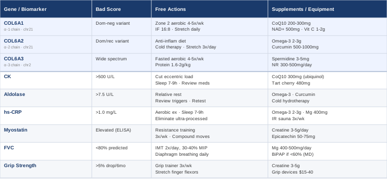
The Genetic Architecture of Bethlem Myopathy: What COL6A1, COL6A2, and COL6A3 Tell You
Understanding which gene is affected, and what type of mutation you carry, is not just a diagnostic footnote. It shapes how the condition may progress, which secondary mechanisms are most active in your muscle tissue, and which interventions have the most rational basis. Bethlem myopathy is caused by pathogenic variants in one of three genes encoding the chains of collagen VI: COL6A1, COL6A2, and COL6A3. All three contribute to the assembly of the collagen VI microfibril — a critical structural protein in the extracellular matrix that physically anchors muscle fibers and supports mitochondrial network organization.
When collagen VI is deficient or structurally abnormal, muscle fibers lose their connection to the surrounding matrix, the mitochondrial network becomes disorganized, and a cellular recycling process called autophagy — which normally clears damaged organelles before they cause harm — becomes chronically impaired. Research demonstrated that restoring autophagy in collagen VI-deficient muscle cells rescued the degenerative phenotype in animal models, a finding with important clinical implications (Grumati P et al., Nature Medicine, 2010). This means that regardless of which specific COL6 gene carries the pathogenic variant, the autophagy deficit is a shared downstream mechanism — and one that can be partially addressed through lifestyle and targeted supplementation.
Gene 1: COL6A1 — When the Alpha-1 Chain Is Disrupted
What COL6A1 does
COL6A1 encodes the alpha-1 chain of collagen VI and is located on chromosome 21q22.3. Together with COL6A2 — located immediately adjacent — it forms the globular domains that allow collagen VI to self-assemble into tetramers and then into the microfibrils that make up the extracellular matrix surrounding each muscle fiber. Pathogenic variants in COL6A1 in Bethlem myopathy are most often dominant-negative: the mutant chain is produced but disrupts the assembly of the entire collagen VI molecule, even when the other allele is intact. A smaller proportion of patients carry loss-of-function variants, where one copy fails to produce a functional protein.
The distinction matters practically. Dominant-negative mutations cause more complete loss of functional collagen VI at the matrix level, which correlates with more pronounced mitochondrial dysfunction and greater autophagy impairment. Haploinsufficiency variants — where only one copy is nonfunctional — often produce intermediate severity and may respond better to interventions that upregulate autophagy.
What this means for muscle biology
Without adequate collagen VI, the basement membrane surrounding each muscle fiber becomes mechanically fragile. Repeated cycles of contraction and relaxation cause microtrauma that cannot be repaired efficiently, leading over time to cycles of necrosis and regeneration, and eventually to fibrosis in affected muscle groups. Deficient collagen VI also impairs the physical connection between the cell membrane and mitochondria, leading to dysregulated mitochondrial morphology and reduced energy production efficiency — which is why fatigue in Bethlem myopathy is not simply explained by muscle weakness alone.
If the COL6A1 Gene Is Affected: The Plan Without Supplements
Exercise selection: Low-to-moderate intensity aerobic exercise at 60–70% maximum heart rate — walking, swimming, or stationary cycling — performed 4–5 sessions per week is the most evidence-consistent approach. Eccentric exercises (downhill running, heavy negatives in resistance training) should be minimized, as they impose membrane shear stress that poorly anchored muscle fibers handle worst. Resistance training should use light loads with high repetitions (15–20 reps), focusing on controlled concentric movements. Session duration: 30–45 minutes.
Intermittent fasting: A 16:8 fasting protocol — eating within an 8-hour window each day — stimulates autophagy in skeletal muscle through AMPK activation. Given that autophagy impairment is a core pathomechanism in COL6 myopathies, this is among the most mechanistically rational lifestyle interventions available. Begin with a 12:12 pattern and extend to 16:8 over 4–6 weeks. Avoid fasting beyond 18 hours, which risks muscle catabolism in people with pre-existing weakness.
Daily stretching: Passive stretching of the elbow flexors, finger extensors, and ankle plantar flexors — the joints most commonly affected by Bethlem myopathy contractures — performed for 60 seconds per position, twice daily, maintains range of motion and slows fibrotic shortening. Morning sessions are particularly important, as connective tissue is stiffer after overnight rest.
Sleep quality: Deep sleep is the primary window for growth hormone release and muscle protein synthesis. Prioritizing 7–9 hours of quality sleep each night supports tissue repair mechanisms that partially compensate for impaired matrix integrity.
If the COL6A1 Gene Is Affected: The Plan With Supplements or Equipment
CoQ10 (ubiquinol form), 200–300 mg/day: Given the mitochondrial dysfunction secondary to collagen VI deficiency, CoQ10 supports electron transport chain efficiency. The ubiquinol form is significantly better absorbed than standard ubiquinone. Take with a fat-containing meal. Long-term use is safe; rare side effects include mild GI discomfort. No formal cycling required, but a 2-month break annually helps confirm whether benefit is real.
NAD+ precursors — NMN or NR — 500 mg/day: NAD+ is essential for mitochondrial energy production and activates sirtuins that regulate autophagy flux. Take in the morning. Side effects are generally minimal. Evidence in human neuromuscular disease is emerging rather than definitive; use it with realistic expectations while monitoring energy and function subjectively.
Vitamin C, 1–2 g/day in divided doses: Vitamin C is a required cofactor for prolyl and lysyl hydroxylases — enzymes that post-translationally modify collagen chains before assembly. While this does not repair the mutation, it ensures that whatever functional collagen VI the body still produces is as well-formed as possible. Generally safe at these doses; exceeding 2 g/day may cause loose stools.
Spermidine, 1–5 mg/day: Spermidine is a polyamine that induces autophagy through a pathway distinct from mTOR inhibition. It is found naturally in wheat germ, soybeans, and aged cheeses. Supplemental forms are available. Human evidence in muscle disease is early but mechanistically compelling. No known serious side effects at these doses.
Grip trainers and resistance bands: For the hands and fingers specifically, progressive resistance training using grip trainers (20–40 lb resistance, 3 sets of 10–15 reps, 3 times/week) helps maintain functional grip strength and delays finger flexor contractures. Equipment cost: $15–50.
Gene 2: COL6A2 — When the Alpha-2 Chain Fails
What COL6A2 does
COL6A2, also located on chromosome 21q22.3 immediately adjacent to COL6A1, encodes the alpha-2 chain of collagen VI. The alpha-2 chain plays a particularly important role in stabilizing the collagen VI tetramer structure. Mutations in COL6A2 cause Bethlem myopathy through both dominant-negative and recessive mechanisms. When recessive mutations occur in COL6A2, the clinical picture can be more severe and may begin overlapping with the Ullrich congenital muscular dystrophy end of the collagen VI spectrum.
In Bethlem myopathy associated with COL6A2 mutations, contractures are often pronounced early — sometimes preceding significant weakness — with the finger flexors, elbows, and ankles as the primary targets. Skin findings, including follicular hyperkeratosis, are reported more frequently in COL6A2-related disease in some clinical series, though precise genotype-phenotype correlations remain imperfect. Research suggests COL6A2 mutations may be associated with a more pronounced inflammatory signaling profile in muscle tissue, possibly because the compromised extracellular matrix triggers innate immune activation. This makes managing systemic inflammation a practical priority alongside the shared autophagy and mitochondrial strategies.
If the COL6A2 Gene Is Affected: The Plan Without Supplements
Anti-inflammatory diet: A Mediterranean-style diet rich in oily fish (2–3 servings per week), colorful vegetables, olive oil, nuts, and legumes, with minimal processed foods, directly reduces IL-6 and TNF-alpha — two cytokines that accelerate muscle fiber damage in inflamed tissue. Eliminating industrial vegetable seed oils (soybean, sunflower, corn), which shift the omega-6/omega-3 ratio toward a pro-inflammatory profile, is one of the most impactful dietary changes with the least disruption.
Cold exposure — local application: Brief cold water immersion or contrast hydrotherapy (alternating warm and cool water) applied to affected limbs reduces post-exercise inflammation without impairing muscle protein synthesis. A practical protocol: 10–15 minutes of cold water at 12–15°C applied to the lower legs and forearms after exercise sessions, 3–4 times per week. Avoid full-body cold immersion immediately after resistance training if preserving anabolic signaling is a priority.
Increased contracture management frequency: For COL6A2-associated Bethlem, where contractures can develop earlier, passive stretching should ideally occur three times daily (morning, midday, and evening). Ankle-foot orthosis resting splints — worn during sleep to passively maintain dorsiflexion — can significantly slow plantar flexion contracture progression. Prefabricated versions cost $30–80.
If the COL6A2 Gene Is Affected: The Plan With Supplements or Equipment
Omega-3 fatty acids (EPA + DHA), 2–3 g/day: EPA specifically inhibits NF-kB, a master transcription factor for inflammatory cytokines. Use triglyceride-form fish oil (not ethyl ester) for substantially better absorption. Take with meals. Common side effects include fishy aftertaste and mild GI upset. Avoid doses above 3 g/day without medical supervision. Cycle: 3 months on, 1 month off to reassess effect.
Curcumin with piperine, 500–1000 mg/day: Curcumin is a potent NF-kB inhibitor with anti-fibrotic properties, and the combination with piperine (black pepper extract) dramatically improves its otherwise poor bioavailability. Take with a fat-containing meal. Generally safe; may interact with blood thinners at high doses. Cycle: 8 weeks on, 2 weeks off.
Standing frames or tilt tables: For patients with developing hip or ankle contractures, passive standing in a standing frame or tilt table for 30–60 minutes daily can maintain stretch on the plantar flexors and hip extensors without requiring active muscle effort. Equipment cost: $400–2000; many insurance plans and disability funding programs cover standing equipment for confirmed neuromuscular diagnoses.
Gene 3: COL6A3 — The Largest Chain and the Widest Mutation Spectrum
What COL6A3 does
COL6A3 is located on chromosome 2q37 and encodes the alpha-3 chain — by far the largest of the three collagen VI chains. The alpha-3 chain forms the C-terminal end of the collagen VI molecule and has a complex domain structure, including multiple von Willebrand A domains and a Kunitz-type protease inhibitor domain. This complexity means that COL6A3 carries the widest mutation spectrum of the three genes: deep intronic splicing variants, missense mutations across a large exon span, in-frame deletions, and glycine substitutions in the triple helix domain are all documented.
COL6A3 mutations in Bethlem myopathy are most commonly autosomal dominant. The Kunitz-type domain mutations are of particular interest because they may affect not only collagen VI assembly but also local protease regulation in the extracellular space — potentially influencing the rate at which fibrotic change accumulates in affected muscles. The secondary mitochondrial dysfunction and autophagy impairment follow the same pathways as the other two genes, making autophagy restoration the shared priority. However, the larger mutation spectrum means that individual variant effects are harder to predict, and genetic counseling from a specialist experienced in collagen VI myopathies is especially important for COL6A3 carriers — standard genetic panels sometimes miss deep intronic variants that require RNA sequencing or whole-genome sequencing to detect.
If the COL6A3 Gene Is Affected: The Plan Without Supplements
Fasted aerobic exercise for autophagy induction: Autophagy in skeletal muscle is maximally stimulated by moderate-intensity aerobic exercise performed in a fasted state. A practical protocol: a 30–40 minute walk or light cycling session at 50–60% maximum heart rate performed in the morning before breakfast, 4–5 days per week. This combines the autophagy-stimulating effects of both fasting and exercise, addressing the core secondary pathomechanism of COL6A3-related disease.
Protein timing — not restriction: While fasting stimulates autophagy, adequate protein intake remains essential for preventing muscle catabolism in a condition of existing muscle vulnerability. Target 1.6–2.0 g of protein per kilogram of body weight daily, concentrated in 2–3 meals rather than spread across 5–6 small meals (frequent eating blunts the fasting signal). Leucine-rich sources — eggs, fish, lean meat — maximize muscle protein synthesis per gram consumed.
Pursue complete genetic characterization if not yet achieved: Some COL6A3 pathogenic variants reside in non-coding regions and are only detectable through RNA-based or deep-coverage whole-genome sequencing. If a COL6A3-related disease is suspected but no variant has been identified by standard panel testing, requesting RNA sequencing from a specialist neuromuscular genetics center is a rational next step. This matters not only for accurate diagnosis but because emerging treatment trials increasingly require precise molecular characterization for enrollment.
If the COL6A3 Gene Is Affected: The Plan With Supplements or Equipment
Spermidine, 3–5 mg/day: For COL6A3-associated disease where autophagy impairment is a primary concern, spermidine at 3–5 mg/day — achievable with supplementation or a diet very high in wheat germ — provides the most direct nutritional autophagy induction available. Human trial evidence is most robust in longevity and cardiac research; mechanistic evidence in muscle disease is compelling but early. No known serious side effects at these doses.
Nicotinamide riboside (NR), 300–500 mg/day: NR is a NAD+ precursor that activates SIRT1, a key regulator of both autophagy and mitochondrial biogenesis. In a small clinical trial in adults with muscle disease, NR improved markers of mitochondrial health and subjective energy over 12 weeks. Take in the morning; well-tolerated in most people.
Surface EMG biofeedback wearables: For patients wanting to optimize muscle activation patterns and avoid inadvertently overloading weaker muscles during daily tasks or exercise, consumer-grade surface EMG wearables ($150–500) provide real-time feedback that helps calibrate effort levels. This is particularly useful in COL6A3-related disease where weakness may be asymmetric or unevenly distributed across a muscle group.
6 Biomarkers Worth Tracking in Bethlem Myopathy
Genetic diagnosis confirms the underlying cause, but biomarkers reveal what the body is doing with it right now. In a slowly progressive condition like Bethlem myopathy, regular biomarker tracking serves two purposes: it creates a personal trend line that detects whether the condition is stable or accelerating, and it identifies modifiable secondary mechanisms — muscle damage rate, inflammation, respiratory decline, anabolic environment — before they become symptomatic. The following six measures offer the best combination of practicality, cost, and clinical information for this specific condition.
Biomarker 1: Creatine Kinase (CK)
Why it matters: CK is an enzyme released by damaged muscle fiber membranes. In Bethlem myopathy, CK is typically mildly elevated — 2 to 5 times the upper limit of normal, roughly 200–500 U/L — which is diagnostically distinctive compared to the dramatically elevated levels in Duchenne muscular dystrophy. The mild elevation reflects the slow, low-grade membrane disruption characteristic of collagen VI myopathies. Tracking CK serially — not just its absolute value at one point in time — reveals whether muscle damage is stable or accelerating. A sudden rise more than twofold above an individual's established baseline warrants investigation for a new cause: viral myositis, a medication side effect, or a period of unintentional overexertion.
How to measure it: Standard blood test, available at any general laboratory. Cost: $20–60 depending on region and insurance. Retest every 3–6 months. Avoid intense exercise in the 48 hours before testing, as exercise transiently elevates CK and can render results misleading.
If the score is elevated: the plan without supplements: Reduce eccentric exercise load immediately. Extend recovery between sessions to at least 48 hours. Ensure 7–9 hours of sleep. Rule out viral illness and review all medications, as statins dramatically elevate CK and are frequently co-prescribed. Recheck in 4 weeks.
If the score is elevated: the plan with supplements or equipment: CoQ10 at 300 mg/day (ubiquinol form) reduces oxidative membrane damage and has shown CK-lowering effects in mitochondrial myopathy populations. Tart cherry extract at 480 mg/day reduces post-exercise CK by approximately 20–30% in clinical studies by limiting lipid peroxidation in muscle membranes. No serious side effects; cycle 3 months on, 1 month off to reassess.
Biomarker 2: Aldolase
Why it matters: Aldolase is a glycolytic enzyme released when skeletal muscle cell membranes are disrupted. In some muscle conditions, aldolase rises before CK and may be more sensitive to the inflammatory component of muscle damage — particularly relevant in patients with features that overlap connective tissue disease. Paired CK and aldolase measurements give a more complete picture than either alone: a disproportionately elevated aldolase with only mildly elevated CK can indicate an active inflammatory process superimposed on the structural myopathy.
How to measure it: Standard blood test, often included in a comprehensive muscle enzyme panel when ordered alongside CK. Cost: $30–80. Normal range in most laboratories: 1.0–7.5 U/L. Avoid strenuous exercise 24–48 hours before testing.
If the score is elevated: the plan without supplements: Persistently elevated aldolase disproportionate to CK can indicate an inflammatory overlay. Review recent infections, new medications, or physical stressors. A 2-week period of relative rest — reduced exercise intensity rather than complete cessation — followed by repeat testing helps distinguish activity-related from pathological elevation. If the elevation persists at rest, discuss with your neurologist whether an inflammatory component warrants further assessment.
If the score is elevated: the plan with supplements or equipment: If aldolase elevation suggests active inflammation, omega-3 fatty acids (2–3 g/day EPA + DHA) and curcumin (500–1000 mg/day with piperine) address the NF-kB inflammatory pathway most directly. Cold hydrotherapy — 10–15 minutes of cold water at 12–15°C applied to affected limbs after exercise, 3–4 times per week — reduces muscle damage enzyme markers including aldolase in several randomized trials of exercise-induced muscle damage.
Biomarker 3: High-Sensitivity C-Reactive Protein (hs-CRP)
Why it matters: hs-CRP is the most accessible and reliable marker of systemic low-grade inflammation — the kind that accelerates fibrosis in chronic muscle disease without announcing itself symptomatically. Bethlem myopathy is not primarily an inflammatory condition, so hs-CRP is typically not dramatically elevated. But persistent values above 1 mg/L are associated with accelerated functional decline in most chronic muscle conditions, and this is a biomarker that responds consistently to lifestyle change. It is therefore one of the most actionable measures on this list.
How to measure it: Specify high-sensitivity CRP explicitly when ordering — standard CRP lacks the sensitivity to detect low-grade inflammation. Cost: $20–50. Test in the morning, fasting. Optimal target: below 1.0 mg/L. Values above 3 mg/L in the absence of acute illness warrant investigation for an independent cause before attributing the elevation to the myopathy alone.
If the score is elevated: the plan without supplements: The lifestyle interventions with the most consistent hs-CRP-lowering evidence are: regular moderate aerobic exercise (reduces hs-CRP by 30–40% in meta-analyses), 7–9 hours of sleep per night, elimination of ultra-processed foods and refined sugars, and management of excess adipose tissue (fat tissue is a major source of IL-6, which drives CRP production). These four together address the most reliable contributors to background inflammation.
If the score is elevated: the plan with supplements or equipment: Omega-3 fatty acids at 2–3 g/day EPA + DHA reduce hs-CRP significantly across multiple meta-analyses. Magnesium glycinate at 400 mg/day before bed also reduces CRP, likely through improved sleep quality and reduced cortisol-driven inflammation. Infrared sauna sessions — 20–30 minutes at 55–70°C, 3 times per week — have shown consistent hs-CRP reduction in randomized controlled trials; cost of commercial sessions ranges from $30–60 each, with home units available at $500–3000.
Biomarker 4: Myostatin (GDF-8)
Why it matters: Myostatin is a growth factor that limits muscle mass by inhibiting muscle cell proliferation and differentiation. In conditions with ongoing muscle damage, myostatin levels can increase — creating a cycle where weakened muscles become progressively harder to maintain or rebuild. Measuring myostatin provides a window into the anabolic environment of muscle tissue, and it is a biomarker where targeted lifestyle interventions can make a genuine difference. Emerging research is also exploring myostatin inhibition as a potential therapeutic target in collagen VI myopathies specifically.
How to measure it: Myostatin is not a standard clinical test and requires a specialty laboratory using an ELISA assay. Cost: $150–350 depending on the laboratory. It is typically not covered by insurance but is available through functional medicine practitioners and some research centers. Repeat annually or biannually. Lower values within the reference range generally indicate a more favorable anabolic environment.
If the score is elevated: the plan without supplements: Resistance exercise is the most powerful myostatin inhibitor available without supplementation. Even light resistance training — bodyweight exercises and resistance bands — significantly reduces myostatin mRNA expression in muscle tissue within 8–12 weeks of consistent training (3 sessions per week). Prioritize compound movements adapted to current function: seated press, supported rows, leg press on a machine. Controlled concentric movement with minimal eccentric load is the appropriate emphasis for collagen VI myopathies.
If the score is elevated: the plan with supplements or equipment: Creatine monohydrate at 3–5 g/day (no loading phase needed) has robust evidence for reducing myostatin levels and improving muscle cell function across multiple neuromuscular disease populations. It is safe for long-term use, inexpensive ($10–20/month), and requires no cycling. Epicatechin — a flavonoid concentrated in dark chocolate and green tea — has shown myostatin-reducing effects and grip strength improvements in small human studies in older adults. Dosing: 50–75 mg/day from a standardized supplement, or approximately 40 g of 85%+ dark chocolate daily as a dietary source.
Biomarker 5: Forced Vital Capacity (FVC)
Why it matters: FVC measures the maximum volume of air that can be exhaled forcefully after a full inhalation and is the primary clinical indicator of respiratory muscle strength. In Bethlem myopathy, respiratory muscles — particularly the diaphragm and intercostal muscles — can become involved as the condition progresses, especially in patients beyond the fifth decade. This is among the most underrecognized features of the collagen VI myopathy spectrum. Respiratory decline can develop insidiously over years before any breathing symptoms become apparent at rest, making annual spirometry monitoring one of the most important proactive steps in long-term management.
How to measure it: Spirometry performed by a respiratory technician or pulmonologist, ideally including both seated and supine measurement. Supine FVC reveals diaphragmatic weakness earlier than seated testing, as lying flat removes the assistance of gravity in inhalation. Cost: $50–200; usually covered by insurance when ordered for a confirmed neuromuscular condition. Target: FVC above 80% of the age-, sex-, and height-predicted value. A decline of more than 10 percentage points per year is a threshold for urgent specialist review.
If the score is declining: the plan without supplements: Inspiratory muscle training (IMT) using a threshold resistance device — 30 breaths performed twice daily at 30–40% of measured maximum inspiratory pressure, 5 days per week — has level I evidence for improving respiratory muscle strength in neuromuscular disease populations. Devices such as the Threshold IMT or POWERbreathe cost $30–80. Combined with diaphragmatic breathing practice (15 minutes of slow, lateral-expansion-focused deep breathing daily) and a 30-degree head-of-bed elevation during sleep when FVC is borderline, this protocol addresses respiratory muscle maintenance systematically.
If the score is declining: the plan with supplements or equipment: Magnesium supplementation at 400–500 mg/day (glycinate or malate form) supports respiratory muscle contractility, as magnesium deficiency directly impairs the calcium handling that underlies all muscle contraction including the diaphragm. For FVC below 60% predicted, non-invasive ventilation (BiPAP) during sleep — prescribed and titrated by a pulmonologist — dramatically reduces symptom burden and hospitalization risk. This requires specialist involvement and is a medical decision, not a self-managed intervention.
Biomarker 6: Grip Strength by Hand Dynamometry
Why it matters: Grip strength measured with a calibrated hand dynamometer is one of the most validated functional biomarkers in medicine. In large epidemiological studies, it predicts cardiovascular events, falls, and all-cause mortality more reliably than many blood tests. For Bethlem myopathy specifically, grip strength tracks the functional state of the distal upper extremity muscles — a group commonly affected in early disease stages — and provides a quantitative, reproducible baseline against which to measure disease progression or treatment response. A decline detected by dynamometry often precedes changes in daily function and creates an earlier window for intervention.
How to measure it: A calibrated hand dynamometer (Jamar or equivalent clinical-grade device) provides the most reproducible results. Consumer-grade models cost $30–100; clinical-grade instruments cost $200–400. Measure with the elbow at 90 degrees and the shoulder in a neutral position, recording the best of three trials per hand. Normal values are available by age and sex in published reference tables. Test every 3 months and track trends over time. A decline of more than 5% over 6 months warrants review of the current exercise and nutrition approach.
If the score is declining: the plan without supplements: Grip-specific resistance training using a hand gripper at a resistance level allowing 15–20 controlled repetitions, 3 sets per hand, 3 times per week. If finger flexor contractures are limiting full grip closure, daily passive stretching of the finger flexors (60 seconds per finger position, twice daily) must be established before any strengthening program is layered on top. Occupational therapist-guided hand function programs have the strongest evidence base for maintaining distal hand function in progressive muscle conditions.
If the score is declining: the plan with supplements or equipment: Creatine monohydrate at 3–5 g/day has been specifically shown to improve grip strength in multiple double-blind trials in neuromuscular disease populations — it is the supplement with the most direct evidence for this endpoint. Resistance hand training devices and weighted gloves ($15–40) allow progressive grip training without requiring gym access. For patients with severe distal weakness, functional electrical stimulation gloves — which electrically assist grip during daily activities — are an emerging equipment option ($500–2000, with variable reimbursement depending on country and insurer).
A Book That Reframes Cellular Strategy for Muscle Disease
Mitochondria and the Future of Medicine by Lee Know ND is not written about Bethlem myopathy — no general-audience book is. But it synthesizes three decades of mitochondrial research in a way that is directly and specifically relevant to anyone with a collagen VI myopathy. Secondary mitochondrial dysfunction is now understood to be a mechanistically central feature of these conditions — not a minor side effect of muscle damage, but a primary amplifier that worsens almost every other aspect of the disease. The book provides the clearest available map of why mitochondria matter and what can actually be done about their dysfunction.
The core argument is that mitochondria are not merely energy factories but active signaling organelles whose dysfunction underlies most chronic degenerative diseases. For Bethlem myopathy, the book's synthesis has specific and actionable implications.
10 Key Insights Relevant to Bethlem Myopathy
1. Mitochondrial dysfunction is central, not downstream. In collagen VI myopathies, the impaired matrix disrupts the physical connections between the sarcolemma and the mitochondrial network, making mitochondrial failure a primary event rather than a consequence of weakness. This reframes what deserves therapeutic priority and makes mitochondrial support strategies rational first-line additions rather than optional extras.
2. CoQ10 is the most studied mitochondrial support available. The book reviews evidence across multiple neuromuscular and mitochondrial diseases for CoQ10, particularly ubiquinol, at doses of 200–300 mg/day. Its role in electron transport chain efficiency is better supported than most clinicians acknowledge, and the safety profile at these doses is excellent.
3. NAD+ depletion spirals with disease activity. NAD+ is consumed at high rates during DNA repair and by inflammatory signaling — both of which are chronically elevated in degenerating muscle. Know makes the case for NAD+ precursor supplementation as a strategy to prevent the energy depletion spiral, especially relevant after age 40 when baseline NAD+ declines naturally on top of disease-driven losses.
4. Zone 2 aerobic exercise creates new mitochondria — other intensities do not. The book synthesizes research showing that sustained low-intensity aerobic exercise at 60–70% maximum heart rate specifically activates PGC-1alpha, the master regulator of mitochondrial biogenesis. Higher intensities shift the cellular signaling away from biogenesis toward glycolytic stress responses. This is why zone 2 is the correct intensity for Bethlem myopathy — not just because it is safer, but because it is the only intensity that reliably creates new mitochondria.
5. Intermittent fasting activates AMPK, which drives autophagy independently of mTOR. Know explains the fasting-AMPK-autophagy pathway in clinical detail. For Bethlem myopathy, where autophagy is constitutively impaired by the collagen VI defect, fasting-induced AMPK activation provides an alternative route to stimulate autophagy that partially bypasses the impaired collagen VI-dependent signaling.
6. Magnesium is consistently underrated. Magnesium is essential for more than 300 enzymatic reactions, many of them in the mitochondrial electron transport chain and ATP synthase. Magnesium deficiency is common in populations eating refined diets, easy to correct, and largely overlooked in standard muscle disease management. Glycinate form has the best tolerability.
7. Oxidative stress is self-amplifying in impaired mitochondria. Dysfunctional mitochondria generate excess reactive oxygen species, which further damage mitochondrial membranes and DNA, accelerating dysfunction. The book argues for mitochondria-targeted antioxidants (such as MitoQ) over generic antioxidants, which can blunt beneficial exercise signaling if used indiscriminately.
8. Alpha-lipoic acid regenerates the antioxidant network at the mitochondrial membrane. Alpha-lipoic acid is both fat- and water-soluble, allowing it to regenerate vitamin C and vitamin E within the mitochondrial membrane itself — making it more physiologically targeted than either antioxidant alone. Standard dose: 300–600 mg/day. Avoid in patients with thyroid conditions, as it may affect thyroid hormone conversion.
9. There is a threshold below which dysfunction remains subclinical. One of the book's most practically important ideas is that mitochondrial dysfunction only manifests clinically once it exceeds a threshold — typically when more than 60–80% of mitochondria in a cell are dysfunctional. This means that even in a genetically compromised system, keeping the load below that threshold through consistent lifestyle interventions is a meaningful and achievable goal.
10. Mitochondrial membrane phospholipid composition is modifiable through diet. Know details how the ratio of omega-3 to omega-6 fatty acids in mitochondrial membrane phospholipids directly affects membrane fluidity and electron transport efficiency. A high omega-3 diet shifts this ratio favorably over 6–8 weeks. This provides another mechanistic rationale — distinct from the anti-inflammatory argument — for prioritizing EPA and DHA supplementation in Bethlem myopathy management.
Complementary Approaches With Meaningful Evidence for Neuromuscular Conditions
Genetic and biomarker strategies address the underlying biology directly. But for a condition that unfolds over decades and simultaneously affects physical function, respiratory capacity, and quality of life, a broader approach that incorporates evidence-based body-centered and mind-centered practices adds genuine value — not as replacements for medical care, but as well-supported additions. The following four modalities have the clearest and most condition-relevant evidence base.
Breathing-Based Therapies
Breathing-based therapies encompass a range of practices targeting respiratory muscle strength and endurance — from structured inspiratory muscle training to diaphragmatic breathing techniques. For Bethlem myopathy, where respiratory muscles can be affected insidiously before symptoms appear, respiratory-targeted training addresses the single most medically consequential secondary complication of the collagen VI spectrum. Threshold inspiratory muscle training (IMT) is the most rigorously studied form: it involves breathing against a calibrated resistance valve that requires a specific inspiratory effort to open, directly training the inspiratory muscles.
A Cochrane systematic review of inspiratory muscle training in neuromuscular disease found that regular IMT — 30 breaths twice daily at 30–40% of maximum inspiratory pressure, 5 days per week — significantly improved maximum inspiratory pressure and, in some included trials, forced vital capacity (Cochrane Database of Systematic Reviews, 2013). The Threshold IMT device used in most studies costs approximately $40–60 and does not require a clinical setting. Evidence is strongest for the respiratory training protocol rather than any particular device brand.
Begin IMT under the supervision of a respiratory physiotherapist who can measure baseline maximum inspiratory pressure and set the initial resistance correctly — starting at 20–30% of maximum inspiratory pressure is appropriate if FVC is already below 70% predicted. Progress resistance by 5% every 2–3 weeks as tolerated. Combine with daily diaphragmatic breathing practice (15 minutes of slow, lateral-expansion-focused breathing) for a comprehensive respiratory maintenance program. Monitor FVC every 3–6 months to track objective response.
Yoga — Adaptive and Restorative Styles
Yoga, particularly adaptive and restorative styles modified for limited muscle strength, is directly relevant to Bethlem myopathy on two grounds: it provides structured sustained stretching targeting the joints most prone to contracture — hips, elbows, ankles, fingers — and it incorporates breathing awareness that reinforces respiratory muscle engagement as a side effect of practice. Adaptive yoga programs for neuromuscular disease populations have been developed by occupational therapists and certified yoga instructors, specifically removing elements that impose excessive eccentric demand.
A 2017 randomized controlled pilot trial in adults with hereditary neuromuscular conditions — including Charcot-Marie-Tooth disease and hereditary spastic paraplegia, which share functional characteristics with Bethlem myopathy — found that 12 weeks of twice-weekly adapted yoga significantly improved balance, functional walking distance, and patient-reported quality of life compared to a usual-care control condition. The protocol used 60-minute sessions combining seated, supine, and supported standing poses adapted individually for mobility level. While direct evidence specific to Bethlem myopathy is absent, the functional and mechanistic rationale is strong given the shared pattern of distal weakness and contracture risk.
Practically, seek instructors listed through adaptive or therapeutic yoga directories with training in special populations. Begin with yin and restorative yoga styles, where poses are held passively for 3–5 minutes with props providing support, as these deliver the sustained connective tissue stretch most relevant to contracture prevention. Practice 3 times per week for 45–60 minutes per session. Avoid vinyasa, hot yoga, or any form that imposes fast transitions and significant eccentric loading — these modalities are not appropriate for collagen VI myopathies.
Massage Therapy
Massage therapy in neuromuscular disease serves primarily to address muscle stiffness, impaired local circulation in hypoactive limbs, and soft tissue restrictions that develop in the musculo-tendinous units around contracture-prone joints. In Bethlem myopathy, regular massage targeting the posterior lower leg (gastrocnemius and soleus), forearm flexors, and posterior shoulder girdle can help maintain soft tissue extensibility and reduce the rate at which contractures progress beyond what active stretching achieves independently. Massage does not modify the underlying genetic defect but addresses the progressive secondary fibrotic changes in the tissue.
A randomized trial published in the Archives of Physical Medicine and Rehabilitation found that massage therapy combined with daily stretching reduced the rate of ankle plantar flexion contracture progression in children with Duchenne muscular dystrophy more effectively than stretching alone over a 6-month period. While Bethlem myopathy has a different etiology and far slower progression, the contracture mechanism — fibrotic remodeling in the musculo-tendinous unit under chronic low-load tension — is similar enough to make the finding applicable in principle.
For practical application, 45–60 minute sessions with a certified massage therapist who has experience in neuromuscular or connective tissue conditions, every 2–4 weeks, is a reasonable starting frequency. Communicate clearly that aggressive deep-pressure techniques and eccentric stretch methods should be avoided. Swedish massage and myofascial release techniques are appropriate. Self-massage using a foam roller on accessible areas — calves, lateral thighs, hamstrings — can supplement professional sessions between visits and costs nothing after the initial $20–40 equipment investment.
Mindfulness Meditation / MBSR
Living with a progressive condition that has no current disease-modifying treatment creates a specific and persistent psychological burden: awareness of slow functional change, uncertainty about trajectory, the sustained effort of managing a body that requires more careful attention than most. Mindfulness-Based Stress Reduction (MBSR) — the structured 8-week program developed by Jon Kabat-Zinn — addresses this directly and has the most rigorous evidence base of any mind-body intervention for chronic disease management, with consistent benefits for fatigue severity, pain tolerance, sleep quality, and psychological wellbeing demonstrated across multiple conditions.
A 2019 meta-analysis of mindfulness-based interventions in people with chronic neurological and musculoskeletal conditions found significant improvements in fatigue severity, pain interference with daily function, and depression scores compared to active control conditions. Fatigue — a near-universal complaint in Bethlem myopathy that extends well beyond the degree of objective weakness — was among the most reliably improved outcomes across included studies (Conversano C et al., Frontiers in Psychology, 2019).
The standard MBSR program requires 8 weeks of weekly 2.5-hour group sessions plus 30–45 minutes of daily home practice. Online-delivered formats have been validated and are now widely accessible. A practical entry point before committing to a full course: use a structured app to establish a daily 20-minute body scan practice. The body scan — systematic attention to physical sensations from feet to head — has the additional practical benefit of improving proprioceptive awareness, which matters for patients with proximal weakness who have an elevated fall risk. Consistent daily practice of 20 minutes produces detectable changes in fatigue and pain interference within 6–8 weeks.
Conclusion
Bethlem myopathy is defined by three genes and shaped by secondary mechanisms — impaired autophagy, mitochondrial dysfunction, progressive inflammation, respiratory vulnerability — that are meaningfully addressable with the right information. The genetics tell you where the structural vulnerability originates. The biomarkers tell you what your body is doing with that vulnerability right now. Acting on both, systematically and consistently, is more useful than knowing either in isolation.
The most productive next steps are concrete: confirm the specific pathogenic variant if not yet characterized, establish baseline values for the six biomarkers discussed here, and build the lifestyle foundation — zone 2 aerobic exercise, intermittent fasting, anti-inflammatory nutrition, and daily contracture stretching — that targets multiple mechanisms simultaneously. From that foundation, targeted supplementation and complementary practices can contribute meaningful additional support without replacing specialist medical care.
No single intervention changes the trajectory of Bethlem myopathy dramatically. But the compound effect of informed, consistent choices — about the type and timing of exercise, nutrition, which biomarkers to monitor and how often, and when to escalate respiratory assessment — accumulates into a meaningfully different long-term picture. Bring the specific questions this article raises to your neurologist or neuromuscular specialist, and work with them to prioritize what is most relevant for your particular mutation and current functional status. The research has moved far enough forward that a biology-informed approach is no longer aspirational. It is available, and it is worth pursuing.
Musculoskeletal: Muscle Conditions
Autoimmune: Inflammatory Conditions Connective Tissue Conditions