This article contains AI generated content.
· UpdatedBipartite Patella – 5 Genes and 6 Biomarkers to Track
Introduction
If you have been told you have a bipartite patella, you've likely received one of two responses: "it's usually nothing" or "we'll manage the pain." Neither answer is particularly satisfying if your knee keeps flaring up during sport, after a long walk, or simply climbing stairs. The condition — where the kneecap develops as two (or sometimes three) distinct bone fragments instead of fully fusing — affects roughly 1 to 2% of the population. What rarely gets discussed is why some people with bipartite patella live entirely symptom-free while others experience chronic, recurring flares. The difference isn't always structural. Often, it's metabolic.
Generic advice tends to focus on rest, ice, and quadriceps strengthening. All reasonable starting points — but they don't address the underlying biology. Two people with identical imaging findings can have completely different outcomes. One is asymptomatic for life. The other deals with persistent pain tied to inflammation, poor bone quality, or connective tissue vulnerability. That gap in outcomes points to something measurable: individual differences in bone turnover, collagen integrity, inflammatory load, and genetic predisposition to skeletal development problems.
This is where targeted testing can genuinely change the conversation. Not because there is a cure hidden in a blood panel, but because these data points can help you and your clinician understand whether your condition is being amplified by low vitamin D, elevated bone resorption, systemic inflammation, or a structural vulnerability rooted in your collagen genes. Each of those has a different intervention strategy, and knowing which one applies to you matters considerably.
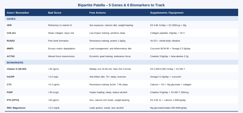
This article approaches bipartite patella from two complementary directions. The first — and most immediately actionable — identifies six key biomarkers that anyone managing this condition should consider tracking. The second explores five genes that research has linked to bone formation, collagen structure, and skeletal development, along with what you can do if your profile is unfavorable. Together they offer a much more specific framework for understanding your knee and for building a smarter long-term management plan.
6 Biomarkers to Track for Bipartite Patella
Biomarkers won't tell you that your patella is bipartite — imaging does that. What they reveal is the metabolic and inflammatory environment around the bone and connective tissue. Tracking these markers over time can identify patterns that explain why your condition is flaring, why healing seems slow, or why standard rehabilitation protocols aren't producing results. Most of these tests are accessible through a GP, sports medicine doctor, or functional medicine practitioner.
1. 25-Hydroxyvitamin D
Why it matters
Vitamin D is not just a supplement trend — it is a fat-soluble steroid hormone that directly regulates bone mineralization, calcium absorption, and the expression of genes involved in skeletal development. Low vitamin D is one of the most consistent findings in people with musculoskeletal pain, poor bone density, and impaired tissue healing. For bipartite patella — which represents a failure of ossification, the normal fusion of bone fragments during development — the role of vitamin D is particularly relevant. While low vitamin D in adulthood doesn't cause the condition retroactively, it can amplify pain, slow adaptation to load, and impair the structural integrity of the fibrocartilaginous tissue that holds the two fragments together. Several studies have linked vitamin D insufficiency to increased musculoskeletal pain sensitivity and slower soft-tissue repair, both of which directly complicate bipartite patella management.
How to measure it
A 25-hydroxyvitamin D blood test (also written as 25(OH)D) is the standard measure. It is inexpensive — typically $30 to $60 USD out of pocket, often covered by insurance. Most labs consider anything above 20 ng/mL as sufficient, but sports medicine and longevity-focused practitioners typically recommend targeting 40 to 60 ng/mL for optimal musculoskeletal function. Test twice per year: at the end of winter when levels are lowest and again mid-summer to understand your seasonal range.
If the score is low — the plan without supplements
Daily outdoor sun exposure is the most natural lever. Aim for 15 to 30 minutes of midday sun (arms and legs exposed) between April and October in northern latitudes. This can raise levels by 10 to 20 ng/mL over 8 to 12 weeks depending on skin tone, latitude, and starting level. Eating fatty fish (salmon, sardines, mackerel) three to four times per week, along with egg yolks and fortified foods, contributes meaningfully. This approach is slower but sustainable long-term.
If the score is low — the plan with supplements or equipment
Vitamin D3 supplementation at 2,000 to 5,000 IU per day is the most widely recommended approach when dietary and sun exposure is insufficient. Always pair it with vitamin K2 (MK-7, 100 to 200 mcg/day) to direct calcium into bone rather than soft tissue — this combination is strongly supported in the literature on bone health. Vitamin D does not need to be cycled; it can be taken year-round with dose adjustments based on retesting every 3 to 4 months. Toxicity is rare at physiological doses but can occur above 10,000 IU per day long-term without monitoring. A UVB lamp can help maintain levels through winter, though it requires proper protocol for safety.
2. High-Sensitivity C-Reactive Protein (hsCRP)
Why it matters
When bipartite patella becomes symptomatic — painful to the touch, inflamed with activity — inflammation is the immediate driver. High-sensitivity CRP is a protein produced by the liver in response to systemic inflammation. It doesn't localize to the knee specifically, but a chronically elevated hsCRP signals an inflammatory environment that makes every tissue — bone, tendon, cartilage — more vulnerable to irritation and slower to recover. Athletes with chronically elevated CRP consistently show longer soft-tissue healing times and greater pain sensitivity, two problems that directly complicate bipartite patella management. The good news is that hsCRP is highly modifiable through lifestyle changes.
How to measure it
The high-sensitivity CRP test is distinct from the standard CRP test — it detects lower concentrations and is more relevant for musculoskeletal and cardiovascular risk. Cost: $20 to $50. Optimal for musculoskeletal health is below 1.0 mg/L; values above 3.0 mg/L signal meaningful systemic inflammation. Test every 3 to 6 months if you're managing an active flare or tracking the effect of lifestyle changes.
If the score is high — the plan without supplements
Dietary intervention is the most powerful non-supplement lever. An anti-inflammatory eating pattern — built around vegetables, oily fish, olive oil, nuts, and whole foods while minimizing ultra-processed products — can reduce hsCRP by 30 to 50% within 8 to 12 weeks in most people. Sleep quality matters just as much: poor sleep (under six hours or highly fragmented) independently raises hsCRP within days. Moderate-intensity resistance training is consistently anti-inflammatory over time. Avoid high-impact loading during active flares, but don't remain sedentary — chronic inactivity independently raises systemic inflammation.
If the score is high — the plan with supplements or equipment
Omega-3 fatty acids (EPA + DHA, 2 to 4 grams per day) from fish oil have robust evidence for reducing hsCRP and systemic inflammatory markers. Take continuously, reassess at 3 months. Curcumin as a bioavailable formulation (BCM-95 or combined with piperine, 500 to 1,000 mg twice daily) shows consistent anti-inflammatory effects in musculoskeletal conditions. Cold water immersion (10 to 15 minutes at 10 to 15°C, three to four times per week) is increasingly used to modulate systemic inflammation, though direct evidence specific to knee conditions remains limited. Side effects: fish oil at high doses may slightly increase bleeding time; curcumin is generally well-tolerated but may interact with anticoagulants — discuss with your doctor if relevant.
3. Serum CTX (C-Telopeptide of Type I Collagen)
Why it matters
CTX (also called beta-CrossLaps or beta-CTX) is the gold-standard marker of bone resorption — the rate at which the body is breaking down old bone tissue. In a healthy remodeling cycle, resorption and formation stay balanced. When resorption outpaces formation, bone integrity degrades over time. For bipartite patella, high bone turnover is especially relevant because the fibrocartilaginous junction between the two bony fragments is a zone of mechanical stress and active remodeling. Elevated CTX suggests the bone environment is in a catabolic state, which can translate into greater pain at the fragment site, slower adaptation to loading, and potentially higher vulnerability to stress-related changes at the fragment border. Researchers tracking bone resorption markers in knee conditions have noted that elevated CTX correlates with worse pain outcomes in patellofemoral disorders.
How to measure it
Serum CTX should be drawn fasting in the morning between 7 and 10 AM — levels fluctuate significantly with food intake and time of day, making timing essential. Cost: $50 to $150. Optimal: below 0.3 ng/mL for adults under 65 (women post-menopause have higher reference ranges). Run alongside P1NP (see next section) to get the full bone turnover picture. Retest every 6 months when tracking interventions.
If the score is high — the plan without supplements
Resistance training is one of the most powerful non-pharmacological tools for reducing bone resorption. Mechanical loading signals osteoblasts (bone-forming cells) to become more active and suppresses excessive osteoclast-driven resorption. Focus on compound lower-body movements — leg press, split squats, hip hinge patterns — that load the femur and tibia without excessive shear force on the patellofemoral joint. Three sessions per week of progressive resistance training can meaningfully reduce CTX within 12 weeks. Sleep (7 to 9 hours per night) is also essential: growth hormone released during deep sleep is a major driver of anabolic bone signaling.
If the score is high — the plan with supplements or equipment
Calcium (total 1,000 to 1,200 mg/day from diet and supplementation combined, not primarily from supplements alone) and vitamin D3 together reliably reduce bone resorption markers. Magnesium (glycinate or malate form, 300 to 400 mg elemental magnesium in the evening) supports bone matrix formation and directly activates vitamin D. Collagen peptides (10 to 15 grams per day, type I/III, taken with vitamin C for maximal incorporation into tissue) may specifically support the fibrocartilaginous tissue at the bipartite junction. These are long-term interventions; reassess CTX at 6 months to confirm the trajectory is improving.
4. P1NP (Procollagen Type 1 N-Terminal Propeptide)
Why it matters
Where CTX measures bone breakdown, P1NP measures the rate of new bone formation. It is released as the body synthesizes new collagen during bone matrix production, making it the most sensitive and specific marker of osteoblast activity currently available. Tracking both CTX and P1NP together gives you the bone turnover ratio — a picture of whether your skeletal system is net-positive (building more than it breaks) or net-negative. For bipartite patella, the goal is a favorable ratio: sufficient bone-forming activity to maintain fragment density and support the connective tissue holding the two pieces together. The International Osteoporosis Foundation recommends P1NP as the reference standard for bone formation monitoring.
How to measure it
P1NP is measured from a standard fasting blood draw. Cost: $50 to $130. It is less commonly ordered than CTX but is increasingly available through functional medicine and sports medicine labs. Optimal levels vary by age and sex, but values below 35 mcg/L in adults suggest suppressed bone formation. Always request it alongside CTX to interpret the ratio meaningfully. Retest every 4 to 6 months.
If the score is low — the plan without supplements
Progressive resistance training — particularly forms that include impact loading such as jumping or brisk walking when the knee allows — is the most powerful physiological stimulus for raising P1NP. Even moderate resistance training shows measurable increases in bone formation markers within 8 to 12 weeks. The quality and duration of slow-wave sleep, during which growth hormone peaks, has a direct effect on P1NP levels. Reducing alcohol intake is meaningful: even moderate alcohol consumption (two or more drinks per day) suppresses osteoblast function detectably.
If the score is low — the plan with supplements or equipment
Creatine monohydrate (3 to 5 grams per day, taken continuously) has emerging evidence supporting bone formation in combination with resistance training — a systematic review published in the Journal of Musculoskeletal and Neuronal Interactions found positive effects on bone mineral density when creatine was paired with exercise. Vitamin K2 as MK-7 (100 to 200 mcg/day) directly activates osteocalcin, a bone matrix protein essential for mineralization, and has been shown in several trials to increase bone formation markers. Whole-body vibration platforms (15 to 20 minutes, three to five times per week at 25 to 30 Hz) have some evidence for stimulating bone formation markers, though results in younger, active populations are more preliminary.
5. Intact Parathyroid Hormone (iPTH)
Why it matters
Parathyroid hormone (PTH) regulates calcium and phosphate metabolism — specifically, it pulls calcium from bone when serum calcium drops too low. Chronically elevated PTH (secondary hyperparathyroidism) is most commonly driven by low vitamin D or insufficient dietary calcium, and it creates a persistently catabolic bone environment by overstimulating osteoclast activity. For someone managing bipartite patella who is trying to maintain fragment density and reduce pain, undetected secondary hyperparathyroidism is a significant and often overlooked obstacle. Many people with low vitamin D have elevated PTH without any obvious symptoms, and a standard blood panel won't catch it unless you specifically request this marker.
How to measure it
An intact PTH blood test costs $30 to $80 and ideally should be run alongside 25-OH vitamin D and serum calcium as a trio for full context. Optimal range: 10 to 55 pg/mL. Values consistently above 65 pg/mL in the presence of normal serum calcium warrant investigation for secondary hyperparathyroidism. Test once or twice a year when managing bone health long-term.
If the score is high — the plan without supplements
The most common driver of high PTH is low vitamin D — correcting sun exposure and dietary habits (as described in section 1) typically normalizes PTH over 8 to 12 weeks. Increasing dietary calcium from whole food sources (dairy, fortified plant milks, leafy greens, canned salmon or sardines with bones) reduces the calcium deficit that triggers sustained PTH release. Weight-bearing exercise plays a regulatory role by improving cellular calcium handling and reducing the demand on PTH-driven bone resorption.
If the score is high — the plan with supplements or equipment
Vitamin D3 at therapeutic doses (3,000 to 5,000 IU/day) combined with adequate total calcium intake (1,000 to 1,200 mg/day from food and supplements combined) is the primary intervention. PTH levels typically normalize within 3 months of consistent supplementation. Do not supplement calcium aggressively without vitamin D and K2 — the combination ensures calcium reaches bone rather than accumulating in soft tissue. Monitor PTH and vitamin D together at 3 months to confirm the expected response.
6. Red Blood Cell (RBC) Magnesium
Why it matters
Magnesium participates in over 300 enzymatic processes, including the activation of vitamin D, bone matrix formation, muscle relaxation, and pain signal modulation. Standard serum magnesium tests are notoriously unreliable for detecting deficiency because magnesium is primarily intracellular — serum levels remain artificially "normal" until total body stores are severely depleted. RBC magnesium is a far more accurate measure of true status. Low magnesium is associated with increased pain sensitization, heightened muscular tension, and impaired recovery — all of which amplify symptoms in a mechanically vulnerable knee. The muscles surrounding the patella (vastus medialis, quadriceps, iliotibial band) depend on adequate magnesium for proper tension regulation, making deficiency particularly relevant for bipartite patella patients.
How to measure it
RBC magnesium is available through most labs at $40 to $100. Optimal range is 5.2 to 6.5 mg/dL; many functional practitioners target the upper half of this range. Standard serum magnesium is not sufficient for detecting deficiency — specifically request RBC magnesium. Test every 3 to 6 months when supplementing or making dietary changes.
If the score is low — the plan without supplements
Dietary magnesium is abundant in dark leafy greens (spinach, Swiss chard), pumpkin seeds, almonds, black beans, avocado, and dark chocolate. A diet consistently built around these foods can raise RBC magnesium meaningfully within 6 to 8 weeks. Reducing alcohol and excessive coffee (both increase urinary magnesium excretion), and minimizing high-sugar diets (which deplete intracellular magnesium) matters equally. Epsom salt baths (two to three cups of magnesium sulfate per bath, 20 minutes, three times per week) are widely used for transdermal absorption and are beneficial for muscle relaxation around the knee regardless of the absorption debate.
If the score is low — the plan with supplements or equipment
Magnesium glycinate or magnesium malate (300 to 400 mg elemental magnesium per day, taken in the evening) are the most bioavailable and best-tolerated forms. Avoid magnesium oxide — it is poorly absorbed and largely ineffective for raising RBC levels. Supplementation is appropriate continuously for most people given the widespread prevalence of dietary deficiency. The main side effect is loose stools at higher doses — reduce by 100 mg if this occurs. Reassess RBC magnesium at 3 months to confirm repletion.
With the metabolic picture in view, the next layer worth exploring is genetic — because some of the variation in how bipartite patella manifests and responds to intervention is encoded before birth.
The Genetic Side: 5 Genes Linked to Bone Development and Connective Tissue
Genetic testing through panels like 23andMe, Genomind, or whole-genome sequencing services can reveal single nucleotide polymorphisms (SNPs) — common variants in your DNA that may subtly alter how proteins are produced. For bipartite patella, the most relevant genes relate to bone formation, collagen structure, and vitamin D metabolism. It's important to be clear: these variants don't cause bipartite patella on their own, and most have relatively modest effect sizes individually. But in combination with environmental factors — nutrition, training load, sleep — they can meaningfully shift outcomes.
VDR (Vitamin D Receptor)
What it affects
The VDR gene encodes the receptor through which vitamin D exerts its effects on bone cells, immune cells, and over 1,000 genes in total. Common SNPs — particularly BsmI (rs1544410), FokI (rs2228570), and TaqI (rs731236) — alter the efficiency with which cells respond to circulating vitamin D. Carriers of certain VDR variants have measurably lower bone mineral density and higher fracture risk even at the same serum vitamin D levels as non-carriers. For bipartite patella, VDR variants could partially explain why some people seem refractory to standard vitamin D supplementation.
If the gene is unfavorable — the plan without supplements
Maximize natural vitamin D production through consistent sun exposure (15 to 30 minutes midday, arms and legs exposed). Pair this with dietary sources of calcium and phosphorus to compensate for potentially reduced VDR-driven bone signaling. Weight-bearing exercise is especially important here, as mechanical load provides a VDR-independent stimulus for bone formation through mechanotransduction pathways.
If the gene is unfavorable — the plan with supplements or equipment
People with disadvantageous VDR variants often need to maintain serum 25(OH)D above 50 ng/mL — rather than the conventional 30 ng/mL — to achieve the same cellular response. This typically requires 4,000 to 6,000 IU D3 per day, monitored by blood testing every 3 months. Combine with vitamin K2 (MK-7, 200 mcg/day) and magnesium. Some practitioners use magnesium threonate specifically for people with VDR variants, given that magnesium is required for both D3 absorption and VDR expression.
COL1A1 (Collagen Type I Alpha 1)
What it affects
COL1A1 encodes the alpha-1 chain of type I collagen — the primary structural protein in bone, tendons, and ligaments. The Sp1 SNP (rs1800012) in the first intron of COL1A1 is one of the most studied genetic variants in musculoskeletal research. The "s" allele (versus the "S" allele) is associated with reduced collagen production, lower bone mineral density, and increased soft-tissue injury risk. This matters for bipartite patella because the fibrocartilaginous junction between the two bony fragments is a collagen-rich structure. If COL1A1 function is compromised, that junction is structurally weaker under mechanical stress, potentially explaining chronic pain and difficulty with rehabilitation.
If the gene is unfavorable — the plan without supplements
Reduce high-impact loading that places concentrated stress on the patellar fibrocartilaginous junction — particularly repetitive jumping or kneeling. Focus instead on low-impact strengthening (cycling, swimming, leg press at controlled range). Prioritize sleep, which is when collagen synthesis is most active. Avoid activities that chronically compress the anterior knee.
If the gene is unfavorable — the plan with supplements or equipment
Collagen peptides (15 grams per day, taken 30 to 60 minutes before exercise with vitamin C) are supported by research showing enhanced collagen synthesis in connective tissue around joints. A 2017 study published in the American Journal of Clinical Nutrition found that gelatin supplementation combined with vitamin C increased collagen synthesis markers in tendons. For someone with a COL1A1 variant, this may partially offset reduced baseline collagen production. Cycle: use continuously during periods of loading. No meaningful side effects reported.
RUNX2 (Runt-Related Transcription Factor 2)
What it affects
RUNX2 is the master transcription factor of osteoblast differentiation — it essentially controls the switch that turns progenitor cells into bone-forming cells. Variants in or near the RUNX2 gene have been associated with altered bone density, bone geometry, and skeletal development patterns in multiple population studies. While direct studies on RUNX2 and bipartite patella specifically are limited, the connection is mechanistically plausible: reduced RUNX2 activity during development could impair the ossification process responsible for patellar fusion. Early evidence suggests RUNX2 variants also influence how well bone responds to mechanical loading.
If the gene is unfavorable — the plan without supplements
Progressive mechanical loading is the primary stimulus that upregulates RUNX2 expression in osteoblasts — meaning consistent resistance training may partially compensate for reduced baseline RUNX2 activity. Prioritize compound lower-body strength work three to four times per week. Ensure adequate protein intake (1.6 to 2.0 grams per kilogram of body weight per day) to support osteoblast activity and bone matrix production.
If the gene is unfavorable — the plan with supplements or equipment
Vitamin D3 (through the VDR pathway) directly enhances RUNX2 expression in osteoblasts, making D3 supplementation particularly relevant for people with RUNX2-related variants. Strontium ranelate was historically used to support bone formation through pathways overlapping with RUNX2, but it is no longer widely available due to cardiovascular concerns. Whole-body vibration therapy may offer an alternative mechanical stimulus for osteoblast differentiation in people who cannot load heavily due to pain. Evidence remains in early stages for RUNX2-targeted interventions specifically.
MMP3 (Matrix Metalloproteinase-3)
What it affects
MMP3 encodes an enzyme that breaks down extracellular matrix components including collagen, proteoglycans, and fibronectin. The 5A/6A promoter polymorphism (rs3025058) affects MMP3 expression levels — carriers of the 5A allele produce more MMP3, leading to greater matrix degradation in connective tissues under stress. This is particularly relevant for soft-tissue structures around the knee: elevated MMP3 activity has been linked to higher rates of tendon and ligament injury and slower repair. For bipartite patella, elevated MMP3 expression around the fibrocartilaginous junction could make the connective tissue at the fragment interface more vulnerable to breakdown under mechanical load.
If the gene is unfavorable — the plan without supplements
Load management is the central strategy — avoiding activities that cause repeated acute stress to the patellar junction (deep squatting under heavy load, high-repetition kneeling) while maintaining strength through ranges of motion that don't compress the anterior knee. An anti-inflammatory diet is relevant here, as pro-inflammatory cytokines (particularly IL-1β and TNF-α) are among the primary activators of MMP3.
If the gene is unfavorable — the plan with supplements or equipment
Curcumin (BCM-95 formulation, 500 to 1,000 mg twice daily) has documented inhibitory effects on MMP3 expression through NF-κB pathway suppression. Omega-3 fatty acids (EPA + DHA, 2 to 3 grams per day) similarly reduce the inflammatory signals that upregulate MMP3. Collagen peptides (15 grams/day with vitamin C) may help offset matrix degradation by supporting synthesis of new connective tissue. These should be viewed as long-term interventions, not acute pain management — reassess symptom patterns every 3 months.
ACTN3 (Alpha-Actinin-3)
What it affects
ACTN3 encodes a structural protein found in fast-twitch muscle fibers. The R577X polymorphism (rs1815739) results in a complete absence of alpha-actinin-3 in XX homozygotes — roughly 18% of the general population. While this is primarily studied in athletic performance contexts, ACTN3 XX carriers show altered muscle mechanics, different injury recovery profiles, and in some studies, altered bone loading patterns due to differences in muscular force transmission. For athletes with bipartite patella, ACTN3 status may help explain variability in patellar stress responses to training and why some people experience flares at relatively low training volumes while others tolerate high loads asymptomatically.
If the gene is unfavorable — the plan without supplements
ACTN3 XX carriers may benefit from a greater emphasis on slow-twitch, endurance-type training patterns rather than explosive power work — not because power work is dangerous, but because the muscular force profiles may place different demands on the patellofemoral joint. Focus on controlled eccentric quad strengthening (e.g., slow decline squats, leg press with a 3-second lowering phase) which has specific evidence for patellofemoral pain management.
If the gene is unfavorable — the plan with supplements or equipment
Creatine monohydrate (3 to 5 grams per day) may be particularly beneficial for ACTN3 XX carriers seeking to maintain force output, as it partially compensates for reduced fast-twitch efficiency. Beta-alanine (3.2 grams per day, split into smaller doses to minimize tingling) supports muscular endurance during higher-repetition training. Both are widely safe and well-tolerated with continuous use.
Genetics and biomarkers give you the data layer. The next section offers a different kind of lens: a strategic framework for long-term musculoskeletal health that applies directly to how you manage this condition over years, not weeks.
What "Outlive" by Peter Attia Gets Right About Bone and Joint Health
Peter Attia's Outlive: The Science and Art of Longevity is not a knee-condition book, but several of its core arguments are directly applicable to anyone managing bipartite patella — particularly the idea that the standard medical response to joint and bone problems is reactive rather than preventive, and that the right biomarkers measured at the right time can change the entire trajectory.
1. Bone mineral density is a lagging indicator — track earlier markers
Attia argues that DEXA scans measure the outcome of bone loss, not its early drivers. CTX and P1NP are the leading indicators that tell you which direction the system is moving — years before a DEXA scan shows concern. For bipartite patella, monitoring these markers gives you a years-long window to intervene.
2. Muscle mass and bone are coupled — you cannot separate them
Every pound of muscle mass you maintain applies mechanical load to the skeleton that signals bone formation. Attia cites evidence that resistance training is one of the most bone-protective interventions available, with effects that persist into later decades. For bipartite patella, maintaining strong quadriceps and hip stabilizers is not just about reducing patellar stress — it is literally protective of the fragment structure over time.
3. Zone 2 cardio reduces systemic inflammation — and inflammation drives pain
Attia is particularly emphatic about low-intensity aerobic training (heart rate around 130 to 145 bpm, conversational pace) as a metabolic intervention. Consistent Zone 2 work reduces insulin resistance, lowers systemic inflammatory markers including hsCRP, and improves mitochondrial efficiency — all of which create a less inflammatory environment around injured or mechanically stressed tissue.
4. Protein intake is chronically underestimated for musculoskeletal repair
Attia recommends a minimum of 1.6 grams of protein per kilogram of body weight per day, and often higher for people managing tissue repair or bone loss. For bipartite patella — where the fibrocartilaginous junction is constantly being asked to tolerate mechanical load — adequate protein is the raw material for connective tissue maintenance. Most people eating standard Western diets fall short.
5. Sleep is the most underrated recovery intervention
Attia devotes significant attention to sleep architecture — specifically slow-wave and REM sleep stages — as critical windows for growth hormone secretion, bone remodeling, and tissue repair. Consistently poor sleep (under 7 hours, fragmented, or with disrupted slow-wave) raises cortisol, increases bone resorption markers, and slows collagen synthesis. For a condition where tissue adaptation to load is already compromised, chronic poor sleep is a compounding liability.
6. Stability and proprioception training is not optional for joint longevity
One of Attia's often-overlooked emphases is on single-leg stability, proprioceptive training, and fall prevention. For someone with bipartite patella, proprioceptive training around the knee — balance board work, single-leg stance progressions, controlled perturbation training — is directly relevant to reducing the risk of acute pain episodes caused by unexpected mechanical stress to the patellofemoral joint.
7. Consistent tracking over time beats one-time testing
Attia emphasizes that a single blood panel is a snapshot. The trend — what happens to your CTX over 6 months of intervention, or your hsCRP after changing your diet — is where the real information lives. For bipartite patella management, this means establishing a baseline on the six biomarkers above, making changes, and retesting systematically. One-time testing without follow-up is low-value.
8. VO2 max tracks overall resilience, including musculoskeletal recovery
Higher cardiorespiratory fitness (measured by VO2 max) is consistently associated with faster soft-tissue repair, lower inflammation, and better outcomes after musculoskeletal injury. While bipartite patella is structural, the pain it causes is mediated by the tissue environment — and a fitter, lower-inflammation system recovers faster. Attia treats VO2 max as the single most predictive biomarker of long-term physical function.
9. Biomarker interpretation requires context, not just normal ranges
Lab reference ranges are population averages, not optimal targets. A vitamin D level of 22 ng/mL is technically "normal" but functionally inadequate for bone health. Attia consistently advocates for targeting the optimal quartile, not simply escaping the flagged zone. The same logic applies to every biomarker discussed in this article.
10. The goal is function over decades, not symptom relief this month
Perhaps the most important reframe Attia offers: managing a structural knee condition is not about eliminating all pain immediately — it's about building the metabolic, muscular, and behavioral infrastructure that protects that joint for the next 20 to 40 years. That shift in time horizon changes which interventions you prioritize and how patient you are willing to be with the process.
Complementary Approaches Worth Considering
The following modalities have meaningful human evidence relevant to bipartite patella — specifically for managing patellofemoral pain, reducing local inflammation, supporting soft-tissue recovery, or improving neuromuscular control of the knee.
Low-Level Laser Therapy (Photobiomodulation)
Low-level laser therapy (LLLT), also called photobiomodulation, uses specific wavelengths of near-infrared or red light to penetrate tissue and stimulate mitochondrial activity, reduce inflammation, and promote tissue repair at the cellular level. For bipartite patella — particularly when the fibrocartilaginous junction is actively inflamed or slow to recover — LLLT offers a non-invasive tool to address the local tissue environment rather than just managing symptoms systemically.
A Cochrane review on LLLT for musculoskeletal conditions found consistent evidence for pain reduction in several soft-tissue and joint conditions, with patellofemoral pain specifically studied in multiple randomized controlled trials. A 2013 trial (Ferreira et al.) demonstrated significant pain reduction and functional improvement in patients with patellofemoral pain syndrome using 810 nm laser at the anterior knee.
For bipartite patella, seek a physiotherapist or sports medicine clinic with a Class IIIB or Class IV therapeutic laser (wavelength 810 to 1064 nm, power 50 to 500 mW). A typical protocol is 6 to 12 sessions over 3 to 6 weeks, applied directly over the anterior patella and medial border. Home devices (red light therapy panels at 660 to 850 nm) are a lower-cost option with weaker but reasonable evidence — use 10 to 15 minutes daily, 5 days per week, at the anterior knee. Side effects are minimal; avoid over the eyes and any active malignancy sites.
Massage Therapy
Massage therapy applied to the muscles surrounding the patellofemoral joint — particularly the quadriceps, iliotibial band, and vastus lateralis — addresses a key driver of bipartite patella flares: excessive lateral patellar traction. When the lateral structures are chronically tight, they create compressive and shear forces at the patellar junction that exacerbate pain. Manual soft-tissue therapy can reduce this tension, improve local circulation, and restore more balanced patellar tracking mechanics.
A 2015 systematic review in the Journal of Athletic Training found that soft-tissue mobilization combined with exercise produced superior outcomes for patellofemoral pain compared to exercise alone. Myofascial release targeting the lateral quadriceps and IT band was specifically effective at reducing anterior knee pain in this population.
Practically, aim for one to two sessions per week with a therapist experienced in sports medicine or orthopedic massage. Specifically request attention to the vastus lateralis, iliotibial band, and the lateral retinaculum around the patella. Between sessions, use a foam roller (30 to 60 seconds per area, slow passes) on the lateral quadriceps and IT band. Avoid direct deep pressure over the bifid patellar junction itself during an acute flare.
Tai Chi
Tai chi is a slow, flowing movement practice that simultaneously trains proprioception, single-leg balance, joint position sense, and lower-limb strength — all of which are relevant to patellofemoral stability. For bipartite patella, the risk of pain flares is often linked to unexpected mechanical stress or poor neuromuscular control during dynamic activities. Tai chi specifically targets the sensorimotor deficits that make joints vulnerable to these episodes.
A 2015 systematic review published in Arthritis Care & Research demonstrated that tai chi significantly reduced pain and improved physical function in people with knee osteoarthritis, with particular improvements in proprioception and balance. While bipartite patella differs from osteoarthritis, the neuromuscular deficits addressed are directly comparable, and the closed-chain, low-impact nature of tai chi is well-suited to an anterior knee condition.
Practically, begin with a beginner tai chi class (in person or video-based) two to three times per week. The Yang style short form is the most widely studied and accessible for beginners. Progress slowly over 6 to 8 weeks before adding more demanding postures. Focus on the single-leg weight transfer elements, which specifically challenge knee proprioception. Avoid deep knee flexion beyond 90 degrees during flares — most tai chi postures can be modified to a higher stance without losing the proprioceptive benefit.
Biofeedback
Biofeedback uses real-time monitoring — typically surface electromyography (sEMG) applied to the quadriceps — to help patients learn to selectively activate the vastus medialis oblique (VMO), the inner quadriceps muscle responsible for medial patellar tracking. Reduced VMO activation relative to the vastus lateralis is a well-documented contributor to patellofemoral pain, including in bipartite patella. Biofeedback training enables patients to correct this imbalance with precision that visual feedback alone cannot achieve.
A 2011 randomized controlled trial in the Journal of Orthopaedic & Sports Physical Therapy found that EMG biofeedback significantly improved VMO/VL activation ratios and pain outcomes in patellofemoral pain syndrome compared to exercise without biofeedback. The authors noted that biofeedback was particularly useful for patients who could not voluntarily isolate VMO activation through cuing alone.
Work with a sports physiotherapist who offers sEMG biofeedback as part of patellofemoral rehabilitation. A typical course is 8 to 12 sessions over 4 to 6 weeks, focusing on VMO activation during terminal knee extension, step-ups, and partial squats. Between sessions, practice the activation pattern learned in clinic during daily exercises. Home biofeedback devices (wearable sEMG monitors) are increasingly available and can extend training between sessions.
Breathing-Based Therapies
Chronic pain — including the low-level, persistent anterior knee pain that bipartite patella can produce — consistently dysregulates the autonomic nervous system, increasing sympathetic tone, pain sensitization, and systemic inflammation. Slow, diaphragmatic breathing techniques directly counter this: they activate the parasympathetic system, reduce cortisol, lower hsCRP, and dampen central pain amplification. For bipartite patella specifically, the relevance is indirect but real — a nervous system chronically in pain-alert mode heals more slowly and perceives pain at lower thresholds.
Research on slow-paced breathing (around 5 to 6 breath cycles per minute, also called resonance frequency breathing) has shown consistent reductions in pain perception and inflammatory markers across several musculoskeletal conditions. This breathing rate engages the baroreflex system and maximizes heart rate variability — a measurable indicator of autonomic balance and pain resilience.
Practically, use box breathing or resonance frequency breathing (inhale 4 to 5 seconds, exhale 4 to 5 seconds) for 10 to 15 minutes per day, ideally before sleep and after pain-aggravating activities. Apps such as Coherence Coach or HeartMath can guide the pace. A portable heart rate variability monitor (HRV4Training, Elite HRV) allows you to confirm you're breathing at your personal resonance frequency and track nervous system recovery over time. Side effects: essentially none, though some people experience brief lightheadedness when first slowing their breathing rate significantly — reduce the pace if this occurs.
Conclusion
Bipartite patella is a structural reality that won't be erased by a supplement or a blood panel. But the pain and dysfunction it causes are not inevitable — they are mediated by an inflammatory and metabolic environment that is genuinely measurable and meaningfully modifiable. Tracking the six biomarkers outlined here — vitamin D, hsCRP, CTX, P1NP, PTH, and RBC magnesium — gives you a concrete starting point for understanding what is driving your symptoms and what interventions are most likely to move the needle. Understanding your genetic profile in the five key areas adds another layer of personalization that can prevent years of trial-and-error with generic protocols.
The most practical next step is simple: start with a fasting blood draw that includes 25(OH)D, hsCRP, CTX (morning, fasting), and RBC magnesium. These four tests together cost well under $300 and will immediately tell you whether there are obvious metabolic targets to address. Discuss the results with a sports medicine physician or functional medicine practitioner who understands bone metabolism, and use the frameworks in this article to inform the conversation. Better information leads to better decisions — and for a condition this often misunderstood, that is where real progress begins.
Musculoskeletal: Bone Conditions Joint Conditions Tendon & Ligament Conditions Sports Injuries
Autoimmune: Inflammatory Conditions Connective Tissue Conditions