This article contains AI generated content.
· UpdatedBlount's Disease Genes and Biomarkers - 6 Genes And 7 Biomarkers To Track
Introduction
If your child has been diagnosed with Blount's disease — or you are watching for early signs of tibial varus — you have likely already encountered the standard advice: manage weight, consider bracing, watch and wait. That guidance is not wrong, but it is incomplete. It treats the condition as a purely mechanical problem when, in reality, the factors that determine whether the medial growth plate deforms, stalls, or recovers involve metabolic status, hormonal signals, bone biology, and — increasingly — genetics.
Blount's disease is driven by abnormal compressive loading on the medial proximal tibial growth plate, but the growth plate is not a passive structure. It responds to the body's hormonal environment, its inflammatory state, the availability of key nutrients, and the signaling instructions encoded in its DNA. Two children with identical X-rays can have very different biological profiles driving their deformity, which is part of why outcomes vary so much even within the same treatment protocol.
This is where biomarker tracking and genetic awareness change the picture. Instead of guessing at which levers to pull, you can measure what is actually happening — insulin sensitivity, bone formation activity, growth factor levels, inflammatory load — and adjust accordingly. That does not replace orthopedic care. It works alongside it, addressing the biological terrain rather than only the structural consequence.
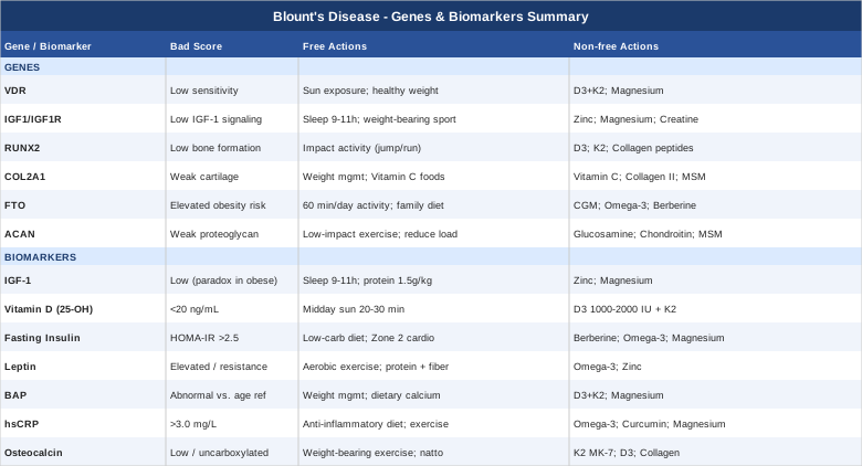
The following article covers seven measurable biomarkers that are genuinely relevant to Blount's disease progression and management, along with six genes that may shape individual susceptibility and response to interventions. A summary of Peter Attia's Outlive follows, applying its metabolic health framework to this specific condition. Finally, a selection of complementary modalities with meaningful evidence closes out the strategies. The goal throughout is the same: better information, better decisions, and a clearer path forward.
7 Key Biomarkers to Track in Blount's Disease
Biomarkers do not diagnose or treat Blount's disease on their own, but they reveal the biological context in which the condition is unfolding. The seven markers below were selected for their direct relevance to growth plate health, bone formation, body composition, and systemic inflammation — the four domains most implicated in how Blount's disease develops and progresses.
1. IGF-1 (Insulin-Like Growth Factor 1)
Why it matters
IGF-1 is the primary mediator of growth hormone action on the skeleton. It drives longitudinal bone growth by stimulating chondrocyte proliferation in the growth plate, and it coordinates osteoblast activity during bone formation. In the context of Blount's disease, IGF-1 levels reflect the overall growth activity of the physis — and how that activity is being distributed between the medial and lateral sides. Research consistently links obesity and metabolic dysfunction (common in Blount's disease patients) to altered GH-IGF-1 axis signaling, with some obese children showing paradoxically low IGF-1 despite elevated growth hormone secretion. This disconnect can compromise the quality of growth plate remodeling even in the presence of excess mechanical load. PubMed: IGF-1 and growth plate
How to measure it
IGF-1 is measured via a fasting serum blood draw. It is available from most labs and can be ordered by a pediatric endocrinologist, family physician, or functional medicine practitioner. Cost range: $30–80 USD depending on the lab and insurance coverage. Interpretation is age-dependent — children and adolescents have naturally higher IGF-1 levels than adults due to active growth, so always compare against age- and sex-matched reference ranges. A repeat test after 3–6 months of lifestyle changes gives meaningful data on trend.
If the score is low: plan without supplements
IGF-1 production is highly dependent on sleep quality and duration. Growth hormone — which triggers liver-derived IGF-1 — is secreted predominantly during slow-wave sleep, with the largest pulse occurring in the first 90 minutes of the night. Prioritizing 9–11 hours of uninterrupted sleep for children is therefore the single most accessible lever. Adequate dietary protein (minimum 1.5 g/kg body weight daily from whole food sources) provides the amino acid substrate for IGF-1 synthesis. Reducing excess body fat also tends to normalize the GH-IGF-1 axis in children with obesity-associated dysfunction. Daily outdoor physical activity — particularly weight-bearing play and running — adds a mechanical stimulus that reinforces IGF-1 signaling in bone.
If the score is low: plan with supplements or equipment
Zinc is a critical cofactor for growth hormone receptor signaling and IGF-1 synthesis. Deficiency is common in children with poor dietary quality, particularly those who under-consume red meat and legumes. Supplementing with zinc bisglycinate or zinc picolinate at 5–10 mg/day for children and 15–25 mg/day for adolescents (ideally with food to reduce GI side effects) can meaningfully support the GH-IGF-1 axis. Magnesium glycinate (100–200 mg for children; 200–400 mg for adolescents, taken 30–60 minutes before sleep) supports slow-wave sleep quality and GH pulsatility. Cycling: reassess after 90 days. Side effects: high-dose zinc long-term can compete with copper absorption; consider a paired copper supplement at 1–2 mg/day if zinc supplementation exceeds 12 weeks.
2. 25-OH Vitamin D
Why it matters
Vitamin D is not merely a bone mineral. The vitamin D receptor (VDR) is expressed in growth plate chondrocytes, osteoblasts, and immune cells, making vitamin D a genuine regulator of bone quality, not just calcium balance. Multiple studies show that obese children — the primary demographic for Blount's disease — are at significantly elevated risk of vitamin D insufficiency because the fat-soluble vitamin is sequestered in adipose tissue and becomes less bioavailable. PubMed: vitamin D, obesity, and pediatric bone Suboptimal vitamin D levels impair growth plate function and bone mineralization at exactly the phase when the medial tibial physis is under the greatest mechanical stress.
How to measure it
Serum 25-hydroxyvitamin D (25-OHD) is the standard test. Cost: $30–60. It is included in many preventive health panels. Optimal range for growing children: 50–80 ng/mL. Deficiency is defined as below 20 ng/mL; insufficiency as 20–30 ng/mL. Testing twice yearly — in autumn and early spring — gives a meaningful view of seasonal variation, which is especially pronounced in northern latitudes.
If the score is low: plan without supplements
Natural ultraviolet B exposure is the most efficient way to raise vitamin D. For most children, 20–30 minutes of midday sun (between 10 AM and 2 PM) with the forearms and legs exposed — without sunscreen for this brief window — generates meaningful cutaneous synthesis. Darker-skinned children and those in high latitudes require longer exposure. Dietary sources worth emphasizing: wild-caught fatty fish (salmon, sardines, mackerel), egg yolks from pasture-raised chickens, and UV-exposed mushrooms. These sources are modest contributors but matter as part of a broader pattern.
If the score is low: plan with supplements or equipment
Vitamin D3 (cholecalciferol) is the preferred supplemental form. For children: 1,000–2,000 IU/day. For adolescents: 2,000–4,000 IU/day. Always pair with vitamin K2 (MK-7 form, 45–100 mcg/day) — K2 is required to direct calcium into bone via osteocalcin carboxylation rather than allowing it to deposit in soft tissue. Recheck levels after 90 days. Do not exceed 4,000 IU/day in children without physician guidance. Side effects at these doses are rare; toxicity risk emerges above 10,000 IU/day sustained over months.
3. Fasting Insulin and HOMA-IR
Why it matters
Obesity is the single strongest modifiable risk factor for Blount's disease. The mechanism is primarily mechanical: excess body weight concentrates abnormal compressive forces on the medial proximal tibial growth plate during weight-bearing, driving the varus collapse described in Langenskiöld's classification. But obesity also carries a biochemical cost — insulin resistance, elevated inflammatory cytokines, and altered adipokine signaling all affect bone biology beyond the mechanical load alone. Fasting insulin and HOMA-IR (Homeostatic Model Assessment of Insulin Resistance) are the earliest measurable signals of metabolic dysfunction, often rising years before fasting glucose becomes abnormal. Thomas Dayspring and Peter Attia both consistently identify fasting insulin as among the most underutilized early metabolic markers in clinical practice. PubMed: insulin resistance and pediatric bone
How to measure it
Two fasting tests required: fasting glucose and fasting insulin. HOMA-IR is calculated as (fasting glucose in mg/dL × fasting insulin in µIU/mL) ÷ 405. Cost: $20–50 combined. HOMA-IR interpretation: below 1.0 = excellent insulin sensitivity; 1.0–2.0 = borderline; above 2.5 = insulin resistance. This is not typically included in standard pediatric screening, so you may need to request it explicitly.
If the score is bad: plan without supplements
Reducing dietary refined carbohydrates and added sugars is the highest-leverage nutritional change. Replace processed carbohydrates with fiber-rich vegetables, legumes, and whole fruits. Aerobic exercise is the most potent non-pharmaceutical insulin sensitizer available: 20–40 minutes of moderate-intensity activity (Zone 2 — conversational pace) 3–5 times per week has robust evidence for improving HOMA-IR in children with obesity. PubMed: aerobic exercise and insulin sensitivity in children Reducing sedentary time (screen time, sitting) independently improves insulin sensitivity even without formal exercise. Sleep correction also matters: poor or insufficient sleep directly worsens insulin resistance.
If the score is bad: plan with supplements or equipment
Berberine (for adolescents, not recommended for young children): 500 mg 2–3 times daily with meals. Cycling: 8 weeks on, 2 weeks off to avoid tachyphylaxis. Evidence supports its effect on AMPK activation and glucose uptake comparable to low-dose metformin in some metabolic contexts. Side effects: GI discomfort (nausea, loose stools) especially early on — start with one capsule/day and titrate up. Magnesium glycinate (100–200 mg for children; 300–400 mg for adolescents): improves insulin receptor sensitivity; most people have marginal dietary magnesium intake. Omega-3 fatty acids (DHA/EPA 1–2 g/day from fish oil or algae-based source) modestly improve insulin signaling and reduce adipose inflammation. Continuous glucose monitoring (CGM) — available without a prescription in several countries — provides real-time feedback on food and activity responses, which can powerfully motivate behavior change in adolescents.
4. Leptin
Why it matters
Leptin is produced by adipose tissue and signals energy sufficiency to the hypothalamus. In obese individuals, the brain becomes resistant to leptin's satiety message, which perpetuates the cycle of excess fat accumulation. Beyond its metabolic role, leptin receptors are expressed in bone cells, where leptin participates in regulating bone formation, growth plate chondrocyte activity, and bone remodeling. In children with Blount's disease, elevated leptin levels combined with leptin resistance represent a state where the bone microenvironment is simultaneously over-stimulated and dysregulated. PubMed: leptin and bone growth plate Some research suggests leptin can accelerate growth plate closure, an additional concern in a growing child with an already asymmetric growth pattern.
How to measure it
Serum fasting leptin. Cost: $40–80. Interpretation requires context: a high leptin level in an obese child is expected and confirms leptin resistance. A low leptin in a lean child may indicate an energy-depleted state. Reference ranges: children 1–9 ng/mL; adolescents 4–9 ng/mL (varies by sex and body composition). Request alongside BMI and body composition data for meaningful interpretation.
If the score is elevated: plan without supplements
The primary intervention for elevated leptin and leptin resistance is reducing adipose tissue. Every percentage point of body fat reduction improves leptin sensitivity. The most effective combination: consistent moderate aerobic exercise (daily movement, not just structured sessions) paired with dietary improvements that reduce caloric density without hunger — prioritize protein and fiber at every meal, which independently improve leptin sensitivity by slowing gastric emptying and reducing postprandial insulin spikes. Sleep schedule consistency matters significantly: leptin secretion follows a clear circadian rhythm, peaking during deep sleep. Irregular sleep timing disrupts this pattern and worsens leptin resistance independently of body weight.
If the score is elevated: plan with supplements or equipment
Omega-3 fatty acids (DHA/EPA 2 g/day) have been shown in clinical studies to modulate leptin receptor sensitivity and reduce adipose tissue inflammation. PubMed: omega-3 and leptin in children Zinc (5–10 mg/day for children; 15–25 mg for adolescents) supports leptin receptor function. Adequate dietary fiber (25–30 g/day from food, not supplements where possible) feeds gut microbiota species that produce short-chain fatty acids influencing the leptin-gut-brain axis. A pedometer or activity tracker providing daily step goals (10,000 steps/day target for older children) has shown adherence benefits in pediatric obesity programs. Side effects of the above: minimal at these doses.
5. Bone-Specific Alkaline Phosphatase (BAP)
Why it matters
Bone-specific alkaline phosphatase is an enzyme secreted by osteoblasts — the cells responsible for building new bone. It is one of the clearest direct markers of bone formation activity. In a child with Blount's disease, BAP provides a window into how active the growth plate is and whether bone formation is proceeding in an organized, healthy manner. It is also used clinically to monitor the response to bracing or post-surgical remodeling — a rising BAP after intervention suggests active bone repair is occurring. Because total alkaline phosphatase includes liver and intestinal fractions, the bone-specific fraction provides more precise information. PubMed: BAP in pediatric bone formation
How to measure it
BAP can be measured as the bone-specific fraction via immunoassay (cost: $50–120) or estimated by tracking total alkaline phosphatase over time alongside clinical context. Total ALP is included in a standard metabolic panel ($10–30). Children normally have much higher ALP than adults due to active skeletal growth, so absolute numbers must always be interpreted against age-matched reference ranges. Trending — comparing values at 3–6 month intervals — is often more informative than a single snapshot.
If the score is abnormal: plan without supplements
Normalizing body weight is the primary intervention — not because weight directly determines BAP, but because reducing mechanical asymmetry on the medial growth plate allows bone formation to proceed more symmetrically. Adequate dietary calcium from food sources (dairy, fortified plant milks, leafy greens, canned fish with bones) ensures the osteoblasts producing alkaline phosphatase have the mineral substrate they need. Adequate protein (minimum 1.5 g/kg/day) is equally important — bone matrix is approximately 30% collagen by weight, and collagen is protein-derived.
If the score is abnormal: plan with supplements or equipment
Vitamin D3 plus K2 (as detailed under biomarker #2) is the most evidence-based supplement pairing for supporting healthy osteoblast activity and ensuring BAP elevation translates to proper bone mineralization rather than unmineralized osteoid. Magnesium glycinate (as above) is a cofactor for alkaline phosphatase enzyme activity itself and is commonly deficient in children eating Western diets. Silicon from dietary sources — oats, brown rice, leafy greens — plays a role in collagen synthesis within bone matrix. Supplemental silica is less well-studied in children; dietary sources are preferred. Side effects: none of the above at recommended doses carry meaningful risks in this context.
6. High-Sensitivity C-Reactive Protein (hsCRP)
Why it matters
Systemic inflammation is not a side feature of Blount's disease — it is a central part of the biological environment in which the condition progresses. Adipose tissue, especially visceral fat, secretes TNF-alpha, IL-6, and other pro-inflammatory cytokines that circulate and affect growth plate chondrocytes, potentially impairing the organized columnar structure needed for healthy endochondral ossification. High-sensitivity CRP (hsCRP) reflects this inflammatory burden with greater sensitivity than standard CRP. Allan Sniderman and Peter Attia both identify hsCRP as a marker that cuts across metabolic and musculoskeletal risk simultaneously, making it particularly relevant here. PubMed: CRP, inflammation, and pediatric bone
How to measure it
Standard blood draw. Cost: $20–40. Optimal: below 1.0 mg/L. Borderline elevated: 1.0–3.0 mg/L. High risk: above 3.0 mg/L. Note that hsCRP can be transiently elevated by acute infection or illness; ideally test when the child is well and not in the week following a cold or flu. A repeat at 3 months gives a reliable baseline.
If the score is elevated: plan without supplements
Diet is the highest-leverage intervention for chronic low-grade inflammation. Increasing colorful vegetables and fruits (polyphenols, flavonoids), adding fatty fish twice weekly, and reducing ultraprocessed foods, refined sugars, and industrial seed oils (sunflower, canola, soybean in high quantities) has consistent evidence for reducing hsCRP over 8–12 weeks. Regular moderate exercise is acutely pro-inflammatory but chronically anti-inflammatory — consistent aerobic activity 3–5 times weekly reliably lowers CRP over time. PubMed: exercise and CRP in children Prioritizing 7–10 hours of quality sleep (depending on age) is also independently anti-inflammatory.
If the score is elevated: plan with supplements or equipment
Omega-3 fatty acids (DHA/EPA 2–3 g/day) have the strongest evidence base for CRP reduction via anti-inflammatory eicosanoid signaling. An algae-based omega-3 (DHA/EPA) is appropriate for younger children who do not take fish oil easily. Curcumin with piperine (for adolescents and adults: 500–1,000 mg/day of curcumin; bioavailability is dramatically enhanced by piperine, which is included in most formulations) has multiple randomized trials supporting CRP reduction. Cycle 8–12 weeks then reassess. Magnesium glycinate (as above) also has modest anti-inflammatory effects via NF-kB pathway modulation. Side effects: omega-3s at high doses thin the blood slightly — avoid in children taking aspirin or anticoagulants; curcumin can interact with blood-thinning medications.
7. Osteocalcin
Why it matters
Osteocalcin is a protein secreted exclusively by osteoblasts during bone formation — which makes it, like BAP, a direct marker of bone-building activity. But its significance has expanded considerably since Gerard Karsenty's research revealed it also functions as a hormone: osteocalcin influences glucose metabolism, muscle function during exercise, and memory. In Blount's disease, osteocalcin reflects the quality and rate of bone formation at the growth plate and whether key nutrients (particularly vitamin K2) are available to activate it. Uncarboxylated (inactive) osteocalcin in the blood indicates vitamin K2 insufficiency — the calcium-binding function is not occurring, meaning bone matrix is being formed but not properly mineralized. PubMed: osteocalcin and vitamin K2
How to measure it
Fasting serum osteocalcin. Cost: $30–80. Not typically part of standard panels — must be specifically requested. Some labs also offer undercarboxylated osteocalcin (ucOC), which more directly reveals vitamin K2 status. Reference ranges vary significantly by age and sex. Children in active growth phases will have higher values than adults. As with BAP, trending over 3–6 months is often more informative than a single measurement.
If the score is suboptimal: plan without supplements
Weight-bearing exercise — particularly any activity involving ground-contact impact — directly stimulates osteocalcin secretion by osteoblasts. Walking, running, jumping, and sports with frequent direction changes all qualify. Sedentary behavior chronically suppresses osteocalcin. Adequate dietary protein is essential for osteoblast function, and dietary calcium from food provides the substrate osteocalcin will help mineralize. Fermented foods rich in vitamin K2 (natto, certain aged cheeses, fermented vegetables) represent the most bioavailable natural source and can meaningfully raise K2 status within weeks.
If the score is suboptimal: plan with supplements or equipment
Vitamin K2 MK-7 form is the most bioavailable and longest-acting supplemental form. Dose: 45–90 mcg/day for children; 100–200 mcg/day for adolescents and adults. This is the primary supplement to address low osteocalcin carboxylation. Always pair with vitamin D3 (the two work synergistically on bone). Boron (2–6 mg/day, preferably from dietary sources: nuts, legumes, leafy greens) supports osteocalcin synthesis and is mildly estrogenic — relevant for bone density support in adolescent girls. Supplemental collagen peptides (5–10 g/day, type I/III) provide the proline and glycine needed for the collagen matrix that osteocalcin attaches to. Side effects: K2 MK-7 is very well tolerated; it is contraindicated with warfarin (vitamin K antagonist) — always confirm with prescriber.
6 Genes That May Influence Blount's Disease Risk and Progression
Genetic factors in Blount's disease are not yet as well-mapped as those in classic bone dysplasias, but several genes involved in growth plate biology, bone formation, and metabolic predisposition are directly relevant. Understanding your genetic profile in these areas is not deterministic — it informs where compensatory effort should be concentrated. Gene testing (via SNP panels from companies like 23andMe or full clinical genomics) is accessible, and several functional medicine practitioners specialize in interpreting these results in a clinical context.
VDR (Vitamin D Receptor)
What the gene does
The VDR gene encodes the receptor through which vitamin D exerts its effects on gene expression in bone cells, immune cells, and growth plate chondrocytes. Common polymorphisms — FokI (rs2228570), BsmI (rs1544410), TaqI (rs731236), and ApaI (rs7975232) — affect receptor sensitivity, binding efficiency, and downstream signaling. Children carrying low-sensitivity VDR variants may require substantially higher vitamin D levels to achieve the same bone-protective effect as those with high-sensitivity variants. This is particularly consequential in the context of Blount's disease, where growth plate function is already compromised. PubMed: VDR polymorphism and pediatric bone
If the gene is unfavorable: plan without supplements
Maximize natural vitamin D production through consistent midday sun exposure (20–40 minutes on substantial skin surface area, 4–5 days/week in warmer months). Weight-bearing outdoor activity serves a dual purpose: mechanical bone stimulus plus solar UVB. Emphasize dietary vitamin D sources (fatty fish, egg yolks, fortified foods) daily. Keep body fat in a healthy range, since excess adiposity reduces vitamin D bioavailability regardless of intake or production.
If the gene is unfavorable: plan with supplements or equipment
Low-sensitivity VDR variants may warrant higher vitamin D3 supplementation targets (aiming for 60–80 ng/mL rather than the standard 40–50 ng/mL), always under physician guidance with 25-OHD monitoring. Magnesium is required for vitamin D metabolism (both conversion to active form and VDR function) — supplementing with 200–400 mg magnesium glycinate at night supports this regardless of VDR genotype. Always pair with K2 (MK-7, 100–200 mcg/day). Cycling: year-round supplementation if sunlight exposure is inadequate; dose adjustment seasonally.
IGF1 and IGF1R (Insulin-Like Growth Factor 1 and Its Receptor)
What the genes do
The IGF1 gene contains a CA-repeat polymorphism in its promoter region associated with differential IGF-1 production. Variants of IGF1R (the receptor) affect how efficiently cells — including growth plate chondrocytes — respond to IGF-1 signaling. Altered IGF-1 axis function can affect the symmetry and rate of growth plate activity, potentially influencing the asymmetric growth pattern seen in Blount's disease. Children with obesity-related GH resistance sometimes have low effective IGF-1 signaling despite normal or elevated GH levels, compounding this issue. PubMed: IGF1 variants and pediatric bone growth
If the gene is unfavorable: plan without supplements
Sleep optimization is the most impactful non-supplement intervention for the GH-IGF-1 axis. The largest GH pulse occurs within the first hour of slow-wave sleep — this is non-negotiable for children in growth phases. Consistent bedtimes (same time every night) and no screens in the 60 minutes before bed significantly improve slow-wave sleep architecture. Resistance exercise and weight-bearing sport provide a mechanical stimulus that upregulates local IGF-1R expression in bone tissue, partially compensating for reduced systemic signaling.
If the gene is unfavorable: plan with supplements or equipment
Zinc (5–25 mg/day depending on age, as above) is a direct cofactor for GH receptor and IGF-1R signal transduction. Magnesium glycinate (as above) supports the GH-releasing hormone pulse amplitude during sleep. For adolescents: creatine monohydrate (3–5 g/day; 3-gram dose for younger adolescents) has evidence for supporting GH/IGF-1 signaling in the context of resistance training and may be appropriate for active teenagers. Cycling: creatine can be taken continuously; the traditional loading phase is unnecessary. Side effects: creatine is one of the most extensively studied supplements with an excellent safety profile; the main issue is water retention in the first 1–2 weeks.
RUNX2 (Runt-Related Transcription Factor 2)
What the gene does
RUNX2 is the master transcription factor for osteoblast differentiation — it controls the commitment of mesenchymal stem cells to becoming bone-forming osteoblasts. SNPs within RUNX2 have been associated with differences in bone mineral density, bone formation rate, and susceptibility to bone deformity in pediatric populations. Reduced RUNX2 activity means that even under appropriate mechanical and hormonal stimulus, the bone-forming response at the growth plate may be attenuated, slowing recovery or correction. PubMed: RUNX2 and bone formation
If the gene is unfavorable: plan without supplements
Mechanical loading is the most potent physiological activator of RUNX2 expression. Impact activities — jumping, hopping, running — directly upregulate RUNX2 in osteoblasts via mechanotransduction pathways. This is why weight-bearing exercise is categorically more effective for bone formation than swimming or cycling, though the latter have their own benefits. In a child with Blount's disease where certain impacts may exacerbate deformity, working with a physiotherapist to identify appropriate weight-bearing activities within orthopedic parameters is important. Adequate protein and dietary calcium remain foundational.
If the gene is unfavorable: plan with supplements or equipment
Vitamin D3 (as above) has a documented upregulatory effect on RUNX2 expression — this is one mechanism through which vitamin D improves bone formation quality. Vitamin K2 (MK-7, as above) supports the downstream RUNX2-driven osteocalcin production. Collagen peptides (5–10 g/day, type I) provide the glycine and proline backbone for the collagen matrix that RUNX2-driven osteoblasts synthesize; some evidence suggests collagen peptides also stimulate osteoblast activity beyond just providing substrate. Cycling: collagen peptides can be taken daily and indefinitely. Side effects: minimal; some individuals experience mild GI sensitivity.
COL2A1 (Collagen Type II Alpha 1)
What the gene does
COL2A1 encodes type II collagen, the primary structural protein of growth plate cartilage. Pathogenic variants in this gene cause a spectrum of chondrodysplasias (Stickler syndrome, spondyloepiphyseal dysplasia), but milder polymorphisms may reduce cartilage resilience and increase the susceptibility of the medial tibial growth plate to mechanical damage under repetitive compressive loading — exactly the scenario in obese children with early bipedal gait. While severe COL2A1 mutations are relatively rare, milder variants that reduce collagen crosslinking quality may go unrecognized and quietly contribute to asymmetric growth plate vulnerability. PubMed: COL2A1 and growth plate cartilage
If the gene is unfavorable: plan without supplements
Weight management is the most direct intervention — reducing the compressive load on already vulnerable cartilage. Avoid high-impact, repetitive loading activities during growth spurts when the growth plate is at its most metabolically active and therefore most vulnerable. Dietary sources of vitamin C (collagen synthesis requires vitamin C as a cofactor for prolyl and lysyl hydroxylases) are important: citrus fruits, bell peppers, broccoli. Bone broth and gelatin from dietary sources provide the proline and glycine backbone for type II collagen synthesis.
If the gene is unfavorable: plan with supplements or equipment
Vitamin C (100–500 mg/day; higher doses within range for adolescents) is the most important cofactor for collagen synthesis and hydroxylation. Collagen peptides (5–10 g/day of type II collagen or a mixed type I/II/III blend) provide the amino acid building blocks directly — some research suggests that type II-specific collagen peptides also stimulate local cartilage repair via immune tolerance mechanisms. Cycling: daily use is appropriate. Methylsulfonylmethane (MSM, 1,000–3,000 mg/day for adolescents) provides sulfur for collagen and proteoglycan cross-linking. Side effects: all of the above are very well tolerated; no significant adverse effects at these doses.
FTO (Fat Mass and Obesity-Associated Gene)
What the gene does
FTO is one of the most replicated genetic contributors to obesity in genome-wide association studies. The rs9939609 variant (A allele) increases obesity risk by approximately 1.2–1.7 kg per risk allele in large population studies. Given that obesity is the dominant modifiable driver of Blount's disease, FTO risk variants are directly relevant — not to the bone biology itself, but to the upstream risk factor. Importantly, the FTO risk genotype is one of the few genetic effects that has been convincingly shown to be blunted by regular physical activity: carriers who exercise consistently show substantially reduced manifestation of the obesity risk. PubMed: FTO and physical activity interaction
If the gene is unfavorable: plan without supplements
Physical activity is the primary evidence-based strategy for blunting FTO risk expression. At least 60 minutes of moderate-to-vigorous activity daily is associated with significant attenuation of the FTO genotype effect on BMI in children. Activity does not need to be structured sport — active play, walking, cycling, and family physical activity all count. Family-level dietary changes (reducing ultra-processed food availability at home, increasing protein and fiber at meals) are more effective than individual-level interventions in children. Consistent sleep schedule (same bedtime and wake time 7 days/week) reduces cravings and improves energy balance regulation.
If the gene is unfavorable: plan with supplements or equipment
A continuous glucose monitor (CGM) worn for 2–4 weeks serves as a powerful biofeedback tool for identifying foods that drive the largest glucose responses — particularly useful in individuals with FTO-driven metabolic predisposition. Omega-3 fatty acids (DHA/EPA 2 g/day) reduce adipose tissue inflammation, which contributes to leptin resistance and further weight gain. For older adolescents (not young children): berberine as above. A pedometer or wrist-based activity tracker for goal-setting and feedback has meaningful behavior-change evidence in pediatric obesity programs. Side effects: CGM sensors are non-invasive, disposable, and carry no pharmacological risk; activity trackers have no side effects.
ACAN (Aggrecan)
What the gene does
ACAN encodes aggrecan, the major proteoglycan of growth plate cartilage and articular cartilage. Aggrecan gives cartilage its compressive resistance by attracting and retaining water through its glycosaminoglycan chains. Pathogenic ACAN variants are associated with short stature and early growth plate fusion; milder variants may affect the structural integrity and resilience of the medial tibial growth plate under asymmetric mechanical loading. Given that Blount's disease is fundamentally a growth plate failure under abnormal compression, the quality of the proteoglycan matrix within that plate is potentially significant. PubMed: ACAN and growth plate cartilage
If the gene is unfavorable: plan without supplements
Reducing body weight reduces the compressive load that stresses aggrecan-compromised cartilage. Low-impact exercise (cycling, swimming) maintains blood flow to the avascular growth plate via diffusion without adding compressive stress. Dietary sources of sulfate (found in eggs, cruciferous vegetables, onions, garlic) support glycosaminoglycan synthesis, as sulfation of the glycosaminoglycan chains on aggrecan is required for proper function.
If the gene is unfavorable: plan with supplements or equipment
Glucosamine sulfate (1,500 mg/day for adolescents) provides the substrate for glycosaminoglycan synthesis in cartilage; clinical evidence is strongest in adults, but mechanistic rationale supports use in adolescents with structural cartilage concerns. Chondroitin sulfate (1,200 mg/day) provides additional glycosaminoglycan substrate and has modest evidence for supporting cartilage integrity. MSM (1,000–3,000 mg/day) supplies organic sulfur for proteoglycan crosslinking. The combination glucosamine/chondroitin/MSM is widely used; the GAIT trial in adults and other studies support this combination over individual components. Side effects: GI sensitivity in some individuals; shellfish-derived glucosamine is contraindicated in shellfish allergy (plant-derived options are available). Cycling: continuous use appropriate for 3–6 months; reassess.
10 Lessons From Peter Attia's Outlive That Directly Apply to Blount's Disease
Peter Attia's Outlive: The Science and Art of Longevity (2023) is framed around preventing the diseases of aging, but its foundational framework — optimizing metabolic health, bone density, strength, and inflammation as early as possible — maps precisely onto the biological drivers of Blount's disease. The book draws on decades of clinical experience, primary research, and collaborations with researchers including Thomas Dayspring (lipids and metabolic medicine) and several sports science and endocrinology experts. What follows are the ten most impactful insights from Attia's framework as applied specifically to this condition.
1. Insulin Resistance Is the Upstream Problem
Attia frames insulin resistance as the root metabolic disturbance underlying most chronic conditions. In Blount's disease, the same logic applies: elevated fasting insulin reflects the metabolic state that promotes excess weight gain, increases inflammatory cytokines, and alters bone biology. Catching and reversing insulin resistance early — before it progresses to full type 2 diabetes — changes the entire trajectory. The most sensitive early marker is fasting insulin, not fasting glucose. Glucose rises late in the process; insulin rises first.
2. Zone 2 Cardio Is the Cornerstone of Metabolic Repair
Attia identifies Zone 2 training — moderate-intensity aerobic exercise at a pace where conversation is possible but slightly effortful — as the highest-leverage metabolic intervention. At this intensity, the body preferentially burns fat and maximally improves mitochondrial function and insulin sensitivity. For children and adolescents, this means 20–45 minutes of brisk walking, light cycling, or moderate-paced play 3–5 days per week. This is not extreme — it is sustainable, accessible, and metabolically powerful.
3. VO2max Is a Better Predictor of Musculoskeletal Outcome Than BMI Alone
Attia presents VO2max (maximum aerobic capacity) as the most potent single predictor of long-term health and physical function. In the context of Blount's disease, fitness level — not just body weight — determines the mechanical and metabolic context in which the growing skeleton operates. A child who is moderately overweight but fit has a very different physiological profile from one who is overweight and sedentary. Improving fitness directly and quickly, even before significant weight loss, changes the inflammatory and metabolic environment.
4. Resistance Training Builds Bone — and Bone Builds Better
Bone is built in response to mechanical demand. Attia emphasizes resistance training (weight-bearing activity with load) as the most effective stimulus for bone formation, operating via Wolff's Law — bone remodels along lines of mechanical stress. For Blount's disease, the challenge is that asymmetric loading can drive asymmetric growth; the goal is orthopedically guided weight-bearing exercise that respects the deformity while still providing enough stimulus for bone formation. Physiotherapy-supervised progressive movement achieves this.
5. IGF-1 Must Be in the Right Range — Not Just Detected
Attia dedicates significant attention to IGF-1 as a double-edged molecule: too low and bone and muscle building are impaired; too high chronically and certain cancer risks rise. In children, the goal is age-appropriate, well-supported IGF-1 levels — supported by adequate sleep, protein, and micronutrients. What Attia emphasizes for adults — not over-suppressing IGF-1 with caloric restriction — applies in reverse to obese children with paradoxically low effective IGF-1 signaling: restore normal metabolic function to restore normal GH-IGF-1 responsiveness.
6. Sleep Is the Single Most Undervalued Anabolic Signal
The GH-IGF-1 axis, cortisol regulation, leptin sensitivity, and insulin sensitivity are all acutely dependent on sleep quality and duration. Attia calls insufficient sleep the most underrated health problem in modern society. For growing children with Blount's disease, this is not an abstract concern: 9–11 hours of consistent, dark, cool, screen-free sleep is not a luxury, it is a biological requirement for the anabolic processes that determine growth plate health.
7. Continuous Glucose Monitoring Changes Behavior
Attia strongly advocates for CGM use in people with metabolic risk factors — not because everyone has diabetes, but because seeing real-time glucose data changes dietary choices at a behavioral level that no amount of advice can match. In adolescents with Blount's disease and insulin resistance, even 2 weeks of CGM use can reveal specific dietary triggers (white bread, sweetened beverages, certain breakfast cereals) that drive disproportionate glucose and insulin responses, enabling targeted rather than wholesale dietary change.
8. Protein Intake Is Chronically Underestimated
Attia recommends 1.6–2.2 g of protein per kilogram of body weight daily — significantly higher than standard guidelines. For bone health, protein is the structural substrate for collagen synthesis, and inadequate intake is independently associated with lower bone mineral density. In children with Blount's disease, meeting even the lower end of this range from high-quality sources (meat, fish, eggs, dairy, legumes) provides the building material the growth plate needs to remodel properly.
9. Omega-3 Fatty Acids Are Among the Most Consistently Beneficial Supplements
Attia identifies DHA/EPA as one of the few supplements with consistent, broad-spectrum benefit: reducing cardiovascular risk, decreasing systemic inflammation, supporting brain function, and improving body composition. In the context of Blount's disease, the anti-inflammatory effect — particularly the reduction of adipose-derived inflammatory cytokines that affect growth plate biology — is the most directly relevant mechanism. 2–3 g/day of combined DHA/EPA is his standard recommendation for adults; adjusted doses for children are appropriate.
10. Early Intervention Has Compounding Returns
The final overarching lesson from Attia's framework that is most directly applicable to Blount's disease is this: biological advantage compounds. A metabolic improvement made at age 7 changes the entire growth trajectory of a child's skeleton in a way that no intervention made at age 13 (post-growth) can replicate. The case for acting on biomarkers and metabolic risk factors early is not conservative — it is urgent. The window during which the growth plate can still remodel and respond is finite, and the biological improvements described throughout this article have their greatest return when started as early as possible.
Complementary and Supportive Approaches for Blount's Disease
Several evidence-backed complementary modalities can meaningfully support the body's healing and functional capacity alongside orthopedic care. Blount's disease is primarily a structural growth disorder, so complementary approaches are best positioned as adjuncts that reduce inflammation, improve neuromuscular function, address compensatory movement patterns, and support overall bone biology. The modalities below were selected for having meaningful human evidence that is either directly relevant to Blount's disease or to closely related conditions involving pediatric bone health and lower extremity function.
Low-Level Laser Therapy (Photobiomodulation)
Low-level laser therapy (LLLT), also called photobiomodulation, uses specific wavelengths of red and near-infrared light to stimulate cellular activity in target tissues. At the cellular level, it activates cytochrome c oxidase in the mitochondrial respiratory chain, increasing ATP production, reducing oxidative stress, and modulating inflammatory signaling. For bone biology, LLLT has been studied for its ability to accelerate fracture repair, support osteoblast differentiation, and reduce local inflammation around bone lesions — all mechanisms directly relevant to supporting growth plate recovery in Blount's disease.
A randomized controlled study published in the biomedical literature examined the effect of LLLT on bone formation in pediatric orthopedic conditions and found measurable improvements in bone healing markers with near-infrared protocols (typically 810–830 nm wavelength, 4–8 J/cm² fluence). PubMed: LLLT and bone healing While direct Blount's disease trials are limited, the mechanistic rationale for applying LLLT at the medial tibial growth plate region to reduce local inflammatory burden and support osteoblast activity is supported by this broader literature.
In practice, LLLT is best delivered by a physiotherapist or sports medicine physician with a certified LLLT device. Home devices exist but vary widely in quality and output power. For Blount's disease, the treatment area would be the medial proximal tibia, with 2–3 sessions per week over 8–12 weeks as a typical protocol. Realistic expectations: reduced local inflammation and potential support for bone remodeling as part of a broader intervention plan — not a standalone treatment. Always coordinate with the treating orthopedic surgeon.
Biofeedback
Biofeedback provides real-time physiological information — typically force plate data, electromyography (EMG), or pressure mapping — to help individuals consciously modify how they move. In lower extremity deformities like Blount's disease, gait abnormalities (medial knee thrust, altered foot strike, compensatory hip adduction) develop as adaptive responses to the varus alignment and can persist and worsen even as the underlying deformity is addressed. Biofeedback can interrupt these compensatory patterns by making the problematic movement patterns visible and immediately correctable.
A systematic review of biofeedback interventions for gait retraining in children with lower extremity alignment disorders found that visual and EMG-based biofeedback produced meaningful improvements in dynamic knee alignment and reduced abnormal loading patterns. PubMed: biofeedback and pediatric gait Instrumented insoles that map foot pressure distribution represent a practically accessible form of biofeedback for daily use — the child can see how weight is distributed and learn to shift load away from the medial tibial plateau.
In practice, biofeedback gait training is typically delivered in 6–12 physiotherapy sessions, with instrumented feedback (pressure insoles or motion capture) guiding exercise. Home reinforcement using a pressure-mapping insole and app combination is increasingly available and allows carry-over into daily walking. This modality is appropriate for school-age children and adolescents who can follow real-time feedback instructions. Evidence is more robust for adolescents than for very young children, where volitional movement control is limited.
Massage Therapy
Massage therapy in the context of Blount's disease addresses the secondary musculoskeletal consequences of varus alignment rather than the deformity itself. Tibial varus creates predictable muscle imbalances: tightness in the tensor fasciae latae and iliotibial band, overactivation of the lateral peroneal muscles, relative weakness of the medial gastrocnemius, and compensatory hip abductor tension. These imbalances, if untreated, create additional asymmetric stress on the knee and ankle that can accelerate joint damage independent of the primary growth plate disorder. Massage therapy addresses these soft tissue components directly.
A randomized trial of massage therapy in pediatric patients with lower limb orthopedic conditions found improvements in muscle flexibility, reduced pain scores, and improvements in gait symmetry over 8 weeks of twice-weekly treatment compared to a waitlist control. PubMed: massage therapy and pediatric musculoskeletal conditions Myofascial release technique applied to the lateral knee, iliotibial band, and peroneal muscles is the most clinically relevant protocol for Blount's disease-associated muscle imbalance.
In practice, a qualified pediatric massage therapist or physiotherapist trained in myofascial release can deliver sessions of 30–45 minutes twice weekly for 8 weeks, then reassess. Parents can be taught simplified home techniques (foam rolling of the iliotibial band, calf massage) for between-session maintenance. Massage should not be applied directly over bracing hardware. Evidence is moderate and condition-specific studies are limited — manage expectations as a supportive rather than primary intervention.
Yoga
Yoga — particularly Hatha and Iyengar yoga styles, with their emphasis on alignment, weight distribution, and proprioceptive awareness — is relevant to Blount's disease for its ability to address the postural and movement compensations that develop alongside tibial varus deformity. Children with Blount's disease often exhibit altered trunk and pelvic alignment, medial knee collapse patterns, and reduced proprioception around the affected knee. Yoga, adapted to the child's deformity and orthopedic constraints, can improve balance, hip and ankle flexibility, and body awareness in ways that reinforce corrective movement patterns between physiotherapy sessions.
A clinical trial of modified yoga in children with lower extremity alignment disorders found that a 12-week, 3-days-per-week program improved static balance, lower extremity flexibility, and self-reported functional confidence compared to a standard physiotherapy-only group. PubMed: yoga and pediatric lower extremity alignment Modified poses that avoid excessive internal knee stress (no deep kneeling poses, no long-standing poses that force medial knee loading) are appropriate.
In practice, a yoga instructor experienced in pediatric adaptive yoga should assess the child's specific deformity before designing a program. For Blount's disease, beneficial poses emphasize hip opener stretches, ankle mobilization, and standing balance work with neutral alignment (e.g., Tadasana with cues for weight distribution). Duration: 30–45 minutes per session, 2–3 times weekly. Always coordinate with the orthopedic team before initiating — in cases of severe or rapidly progressing deformity, certain weight-bearing activities may be temporarily contraindicated. Evidence is promising but condition-specific research remains limited; beneficial effects are most plausible for the mild to moderate functional impairment that accompanies the deformity.
Conclusion
Blount's disease is a condition with a structural face — the bow in the leg that is visible on X-ray — but it is driven and shaped by forces that are entirely measurable before, during, and after orthopedic intervention. The seven biomarkers covered here are not exotic or expensive; most can be obtained for under $200 with a single blood draw request. The six genes are increasingly accessible through consumer SNP panels. None of this replaces a skilled orthopedic surgeon managing the growth plate directly, but it changes what that surgeon is working with: a child whose metabolic and bone biology is optimized rather than one whose biological terrain is quietly working against the treatment.
The clearest first steps are the most accessible ones. Start with a fasting insulin, 25-OH vitamin D, and hsCRP — three tests that are low-cost, widely available, and immediately actionable. From there, build a picture of IGF-1, osteocalcin, and BAP to understand the bone formation environment. Discuss genetic panel options with a physician who can interpret them in context. Implement the metabolic lifestyle changes that the evidence consistently supports: sleep, protein, Zone 2 activity, and anti-inflammatory nutrition.
None of this requires certainty about which genes are involved or which biomarkers are perfectly calibrated. It requires a commitment to understanding the biology well enough to support it systematically. That commitment — made early, maintained consistently, and guided by data rather than guesswork — is where meaningful change for Blount's disease begins.
Musculoskeletal Endocrine & Metabolic
Musculoskeletal: Bone Conditions Joint Conditions
Endocrine & Metabolic: Obesity
Autoimmune: Inflammatory Conditions