This article contains AI generated content.
· UpdatedBullous Pemphigoid Genes Biomarkers — 5 Genes and 6 Biomarkers to Track
Introduction
Living with bullous pemphigoid carries a particular kind of exhaustion. The large, tense blisters, the relentless itching that often signals a flare before any blister appears, the weight of long-term corticosteroids and their cascading side effects — none of this is trivial, and most of it remains under-explained in a standard clinical encounter. You receive a diagnosis confirmed by immunofluorescence, a prescription, and a broad prognosis. Rarely do you receive a personalized picture of what is driving your disease at the molecular level today.
Generic dermatology protocols exist because they work reasonably well for most people, most of the time. But bullous pemphigoid varies enormously between individuals: in which antibodies dominate, in how inflammatory markers shift over time, in whether the Th2 allergic axis or the tissue-damaging effector arm is most engaged. Two people with confirmed BP can be biochemically quite different, and those differences matter when deciding how aggressively to treat, which secondary approaches to explore, and which lifestyle factors may be amplifying the immune response in your specific case.
This article approaches the condition through two complementary lenses. The primary section covers the six most clinically useful biomarkers for BP — not just to confirm the diagnosis, but to monitor disease activity, detect flares early, and identify where intervention may be most useful. The second section examines five gene variants linked to BP susceptibility and immune behavior, with evidence-based strategies for each one. Following those, you will find a distillation of a paradigm-challenging approach from the autoimmune medicine literature, and a curated set of complementary modalities that have genuine clinical evidence behind them.
Better information does not guarantee a cure. It does, however, change the quality of decisions you and your care team can make — and in a condition where both over-treatment and under-treatment carry real costs, that precision is worth pursuing.
Summary
This article maps bullous pemphigoid through two practical tools: measurable biomarkers and genetic factors. The six biomarkers covered — anti-BP180 antibody titer, anti-BP230 titer, total IgE, eosinophil count, interleukin-31, and high-sensitivity CRP — each capture a different dimension of the disease, from autoantibody burden to the cytokine behind relentless itch. For each, you will find out how to get tested, what an abnormal result reveals about your immune state, and what to do about it with and without supplements. The genetics section covers five key genes — including the major HLA risk allele and the autoantigen-encoding COL17A1 — with targeted intervention plans for each. Beyond biomarkers and genes, the article includes ten research-backed insights from The Autoimmune Solution that challenge the purely immunosuppressive standard of care, and five complementary approaches — including the Autoimmune Protocol, mindfulness-based stress reduction, microbiome-directed therapy, photobiomodulation, and breathing practice — each with specific protocols. Every section gives you something concrete to act on.
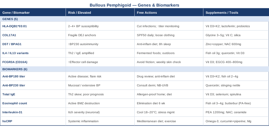
The 6 Biomarkers That Matter Most in Bullous Pemphigoid
Once a diagnosis is confirmed, the clinical challenge shifts from detection to monitoring. Biomarkers in bullous pemphigoid are not static — they fluctuate with disease activity, treatment response, and environmental triggers. The six markers below answer a different question than a skin biopsy: not whether you have BP, but how active it is right now, which immune pathways are most engaged, and where there may be room to intervene. Some require specialty labs; others appear on a standard blood panel. Together they provide a considerably sharper picture than clinical examination alone.
Biomarker 1: Anti-BP180 (NC16A Domain) ELISA Titer — The Central Indicator
Why it matters and what it reveals. Anti-BP180 antibodies targeting the NC16A extracellular domain of type XVII collagen are the defining laboratory feature of BP. They are detectable in approximately 80–90% of patients and correlate more reliably with disease activity than virtually any other measurable parameter. When titers fall, disease typically responds to treatment. When they rise — often before visible blisters form — a flare is frequently imminent. Crucially, the titer magnitude tracks with clinical severity: very high titers generally correspond to more extensive blistering, faster disease progression, and higher relapse risk after immunosuppressive tapering. Several published cohort studies have established anti-BP180 NC16A levels as a reliable surrogate for clinical severity and treatment response, making this the single most important blood test to track in active BP.
How to measure it. Anti-BP180 ELISA is available through specialty dermatology reference labs and academic medical centers. It is not included in a standard autoimmune panel and must be ordered specifically by name. Cost: approximately $80–180 depending on the laboratory. Physician referral is typically required. Testing every 3–6 months during active disease — or promptly at any suspected flare — is clinically reasonable. Request the NC16A-specific ELISA, not the older indirect immunofluorescence method, for quantitative monitoring.
If the score is high — the plan without supplements. High anti-BP180 titers are a signal to prioritize disease management in partnership with a dermatologist, not a situation to manage independently. Practically, this means conducting a thorough medication review: furosemide, ACE inhibitors, nonsteroidal anti-inflammatory drugs, penicillamine, and DPP-4 inhibitors (widely used for type 2 diabetes) are all well-documented triggers for drug-induced or drug-exacerbated BP. Discontinuing or substituting an offending drug — done with physician supervision — has in some cases produced substantial antibody titer reduction and clinical improvement without immunosuppressive escalation. Beyond medication review, a structured anti-inflammatory dietary pattern (removing ultra-processed foods, refined sugar, and common allergens), consistent sleep of 7–9 hours nightly, and regular moderate movement all reduce systemic inflammatory load and support the immune environment in which treatment works.
If the score is high — the plan with supplements or equipment. Vitamin D insufficiency is disproportionately common in BP patients; published cohort data show that serum 25(OH)D levels are significantly lower in BP patients than in matched healthy controls. Vitamin D3 at 2,000–5,000 IU per day — adjusted to target serum 25(OH)D levels of 40–60 ng/mL — modulates regulatory T-cell activity and can reduce the imbalance between autoreactive and suppressive immune populations. Combine with K2 (MK-7 form, 100–200 mcg/day) to direct calcium appropriately; both are fat-soluble and best taken with a fat-containing meal. Test serum 25(OH)D every 3 months during dose adjustment. No cycling required at standard doses. Fish oil (EPA+DHA, 2–4 g/day) reduces prostaglandin E2 and leukotriene production, modulating the inflammatory milieu that amplifies autoantibody-mediated damage. Take with food to reduce GI side effects. At doses above 3 g/day, discuss with your doctor if you take anticoagulants, as omega-3s have mild blood-thinning effects.
Biomarker 2: Anti-BP230 ELISA Titer — The Second Autoantibody
Why it matters and what it reveals. BP230, encoded by the DST gene, is the second major autoantigen in bullous pemphigoid. Anti-BP230 antibodies are detected in approximately 50–60% of patients and are less tightly correlated with disease activity than anti-BP180 — they do not predict flares as reliably. However, they add meaningful diagnostic confidence in the roughly 10–15% of BP patients who are anti-BP180 negative, and some research suggests that anti-BP230 positivity may associate with more extensive skin involvement and mucosal disease. In patients with both antibodies elevated, the combined autoantibody burden may correlate with greater severity than either marker alone.
How to measure it. Anti-BP230 ELISA is typically ordered alongside anti-BP180 from the same specialty laboratories. Cost: $80–160 as a standalone test, often lower when ordered as part of a bullous disease antibody panel. Testing frequency follows the same logic as BP180 — every 3–6 months during active disease, or at any suspected flare.
If the score is high — the plan without supplements. Because anti-BP230 correlates less directly with activity than anti-BP180, an elevated titer primarily calls for a conversation with your dermatologist rather than self-directed action. From a lifestyle standpoint, the same anti-inflammatory strategies apply: eliminate documented drug triggers, optimize sleep architecture, reduce dietary refined carbohydrates, and address known allergen exposures. UV-B narrowband phototherapy, used under medical supervision, has shown some ability to modulate the Th2-dominant immune response that drives IgG autoantibody production in autoimmune blistering diseases — worth discussing with a specialist experienced in BP.
If the score is high — the plan with supplements or equipment. Quercetin (500–1,000 mg/day, taken with food) is a polyphenol flavonoid that stabilizes mast cells and reduces Th2-driven cytokine secretion including IL-4 and IL-13 — the upstream drivers of IgG class switching toward autoantibody production. It has a well-characterized safety profile and is widely studied in allergic and inflammatory conditions. A conservative cycling approach — 10 weeks on, 2 weeks off — minimizes any theoretical accumulation; side effects at standard doses are rare and mild. Stinging nettle extract (Urtica dioica, 300–600 mg standardized extract/day) has modest evidence as a natural antihistamine with anti-Th2 inflammatory properties, potentially complementary in Th2-dominant autoimmune states. Use in 8-week courses; occasional mild GI discomfort is the most common side effect.
Biomarker 3: Total IgE — The Allergic Immune Signature
Why it matters and what it reveals. Bullous pemphigoid is not a classical autoimmune disease in the Th1-dominant sense — it has a pronounced Th2, allergic character. Total serum IgE is elevated in a substantial proportion of BP patients, and multiple published studies have found correlations between high total IgE and greater disease severity, more intense pruritus, higher rates of recurrence, and reduced treatment response to conventional corticosteroids. This Th2 skew has become therapeutically significant: it directly explains the efficacy of omalizumab (anti-IgE) and dupilumab (anti-IL-4Rα) in refractory BP — both biologics target the same Th2 pathways that elevate IgE. A high total IgE result, in this context, is not just a number — it identifies the immune polarization driving your disease and points toward specific targets.
How to measure it. Total IgE is available on any standard blood panel. Cost: $20–60. It requires no specialty lab and can be ordered at virtually any clinical visit. For adults, values above 150 IU/mL are generally considered elevated; many active BP patients present with values of 500–2,000+ IU/mL. Test every 3–6 months during active disease; it serves as a rough proxy for Th2 activity between more expensive antibody assays.
If the score is high — the plan without supplements. Elevated total IgE signals an overactive Th2/allergic immune pathway. The actionable response is a systematic allergen audit: identify and reduce exposure to common inhalant allergens (house dust mites, mold spores, pet dander, cockroach antigens), which independently amplify Th2 tone even in the absence of overt allergy symptoms. Physical steps with strong evidence: encasing mattresses and pillows in allergen-proof covers, washing bedding weekly at 60°C, reducing indoor humidity below 50% to limit dust mite and mold growth. Dietary pattern review is also relevant — high omega-6 vegetable oil consumption, frequent dairy, and excess refined sugar all tend to amplify Th2-dominant immune responses in susceptible individuals.
If the score is high — the plan with supplements or equipment. Vitamin D3 (2,000–5,000 IU/day with K2-MK7 100–200 mcg) has documented ability to shift immune balance from Th2 toward Th1 and Treg, directly addressing the IgE-producing arm of the immune response. This is one of the better-supported immunomodulatory applications of vitamin D in the published literature. Selenium (100–200 mcg/day as selenomethionine) supports regulatory T-cell function and modulates the Th1/Th2 balance at the level of cytokine signaling. Use for 12-week courses, then test serum selenium before continuing; the therapeutic window is narrow — do not exceed 400 mcg/day, and toxicity above that threshold is real. Spirulina (2–3 g/day, in capsule or powder form) has shown some evidence for IgE modulation in small human trials; the evidence base is limited but mechanistically coherent. Take daily with food; side effects are rare at standard doses.
Biomarker 4: Blood Eosinophil Count — Tracking the Blister-Driving White Cell
Why it matters and what it reveals. Eosinophils are among the most damaging immune cells in BP. They release proteases, elastase, and reactive oxygen species that directly degrade the basement membrane zone proteins — including BP180 — accelerating blister formation once autoantibodies have targeted the dermal-epidermal junction. Peripheral eosinophilia (elevated blood eosinophil count) is present in up to 50% of BP patients and tends to correlate with disease activity; declining counts frequently accompany clinical improvement. Tissue eosinophilia on skin biopsy is even more closely linked to blister severity and is one of the histological hallmarks of the condition. Tracking peripheral eosinophil count gives a cheap, rapidly accessible proxy for tissue inflammation.
How to measure it. Eosinophil count is part of a complete blood count with differential (CBC-diff) — one of the most accessible tests in medicine. Cost: $15–40, available at any laboratory, requires no specialty referral. A normal eosinophil count is 0.1–0.5 × 10⁹/L; values above this threshold are considered elevated. Some active BP patients present with counts three to five times the upper limit. Test every 4–8 weeks during active disease and monthly when initiating or tapering treatment — it responds faster than antibody titers to immune changes.
If the score is high — the plan without supplements. Elevated eosinophils in BP typically reflect active disease, but food sensitivities can independently drive eosinophilia and amplify the underlying baseline. A structured 6-week elimination of the most commonly implicated foods — wheat, dairy, soy, eggs, tree nuts, and shellfish — followed by systematic reintroduction (one group every 5–7 days) can reveal whether dietary patterns are compounding eosinophilic inflammation on top of the BP-driven baseline. Allergen reduction strategies described above also apply, since inhalant allergens recruit eosinophils via the same IL-5/eotaxin pathway.
If the score is high — the plan with supplements or equipment. Fish oil (EPA+DHA, 3–4 g/day) has consistent evidence for reducing peripheral eosinophil counts across allergic and inflammatory conditions. Its mechanism is competitive inhibition of arachidonic acid in prostaglandin and leukotriene pathways, reducing leukotriene B4 — a potent eosinophil chemoattractant. Take with food; allow 8–12 weeks before reassessing counts. Butterbur extract (Petasites hybridus, standardized to 7.5 mg petasin per capsule, 50 mg twice daily) has anti-eosinophilic and anti-leukotriene activity with clinical evidence primarily from allergic rhinitis; the mechanism is relevant to BP's eosinophilic inflammation. Critically: use only PA-free (pyrrolizidine alkaloid-free) certified extracts to avoid hepatotoxicity. Cycle: 8 weeks on, 4 weeks off. Discuss with your doctor if you are on blood pressure medications.
Biomarker 5: Interleukin-31 (IL-31) — The Itch Cytokine
Why it matters and what it reveals. If anti-BP180 explains the blistering, IL-31 largely explains the itch. This cytokine, produced by activated Th2 cells and mast cells, binds directly to receptors on sensory neurons and triggers the neural signaling cascade of pruritus. Research published in dermatological immunology journals has found serum IL-31 significantly elevated in BP patients compared to healthy controls, and — crucially — the elevation correlates with itch severity rather than with blister count. This means the two dominant symptoms of BP can be driven by partially independent mechanisms, and tracking IL-31 separately from antibody titers provides a more complete picture of what is causing the most suffering. The clinical importance of this distinction is growing: nemolizumab, a monoclonal antibody targeting the IL-31 receptor, has demonstrated efficacy in BP-related pruritus in Phase II clinical trials, validating IL-31 as both a biomarker and a therapeutic target.
How to measure it. Serum IL-31 requires a specialty cytokine or immunology laboratory and is not part of routine clinical panels. It is increasingly available through academic dermatology centers and large reference labs. Cost: $100–300 depending on the laboratory. Testing every 2–3 months during active disease, or specifically before and after anti-itch interventions, makes clinical sense. The absence of a universal reference range is a current limitation — results are most useful as relative comparisons over time in the same individual rather than against population norms.
If the score is high — the plan without supplements. IL-31 is dramatically amplified by heat, making temperature management one of the most accessible evidence-consistent tools for managing BP itch. Keeping sleeping environments at 18–20°C, using cooling gel pads or cooling clothing during flares, and replacing hot showers with lukewarm water consistently reduces IL-31-mediated pruritus signaling. Psychological stress is a potent mast cell activator — and mast cells are a primary source of IL-31 — making structured stress reduction a genuine clinical priority rather than a lifestyle recommendation. Minimizing skin microtrauma from scratching (which triggers further mast cell degranulation and more IL-31 release) through distraction techniques, cold compresses, and trimmed fingernails interrupts the itch-scratch-amplification cycle.
If the score is high — the plan with supplements or equipment. Palmitoylethanolamide (PEA, 600 mg twice daily) is an endocannabinoid precursor compound with documented anti-inflammatory and anti-pruritic properties; it reduces mast cell activation and has been studied in neuropathic itch conditions with a favorable safety profile. No cycling is required; side effects are rare. N-acetylcysteine (NAC, 600 mg twice daily) reduces oxidative stress in activated mast cells and has shown some ability to modulate cytokine profiles including IL-31 in inflammatory conditions. Take with food; 12-week cycles with 4-week breaks is a conservative protocol. Topical barrier creams with ceramides do not reduce serum IL-31 directly, but they interrupt the skin disruption that amplifies mast cell activation at the surface — particularly important during flares.
Biomarker 6: High-Sensitivity C-Reactive Protein (hsCRP) — The Systemic Inflammation Barometer
Why it matters and what it reveals. CRP is a non-specific inflammatory marker, but in bullous pemphigoid it captures something the disease-specific antibody tests cannot: the overall systemic inflammatory state in which BP is occurring. BP does not exist in metabolic isolation — it clusters with obesity, cardiovascular disease, type 2 diabetes, and in some cases internal malignancy, all of which elevate baseline inflammation. Elevated hsCRP in a BP patient signals that the inflammatory burden extends beyond the skin, which has implications for cardiovascular risk, the body's response to corticosteroids, and overall prognosis. Elevated baseline hsCRP has been associated with longer time to remission in autoimmune blistering disease cohorts, making it a clinically relevant indicator of how difficult achieving control may be.
How to measure it. High-sensitivity CRP (hsCRP — not standard CRP) is a more sensitive assay capable of detecting low-grade inflammation below the threshold of standard CRP. Cost: $15–40, universally available at any laboratory. Target values for a BP patient aiming for optimal immune context: below 1 mg/L. Values between 1–3 mg/L indicate moderate background inflammation; above 3 mg/L suggests significant systemic inflammatory burden. Test at baseline and every 3–4 months. Acute infections transiently spike CRP — repeat the test 4 weeks after any illness to distinguish sustained from transient elevation.
If the score is high — the plan without supplements. The most consistent lifestyle drivers of elevated hsCRP are excess visceral adipose tissue, chronic sleep debt, physical inactivity, and high-glycemic dietary patterns — none of which are unique to BP but all of which amplify its inflammatory milieu. A Mediterranean-style dietary pattern (high in fatty fish, extra-virgin olive oil, polyphenol-rich vegetables and fruits, low in refined carbohydrates and industrial seed oils) has the strongest evidence base for CRP reduction of any dietary approach, with effects appearing in 8–12 weeks. Resistance training two to three times per week has anti-inflammatory effects comparable to aerobic exercise and additionally improves insulin sensitivity — reducing the glycemic component of inflammation simultaneously.
If the score is high — the plan with supplements or equipment. Omega-3 fatty acids (EPA+DHA, 2–4 g/day) reduce hsCRP in multiple meta-analyses — this is one of their most robustly supported applications. Curcumin combined with piperine (500 mg curcumin + 5 mg piperine, twice daily with food) is among the most evidence-backed anti-inflammatory supplements available; piperine increases curcumin bioavailability approximately 20-fold. Cycle: 12 weeks on, 4 weeks off to prevent GI habituation; occasional GI upset at higher doses. Magnesium glycinate (300–400 mg taken at night) reduces hsCRP in magnesium-deficient individuals — a common finding given that most people consume inadequate dietary magnesium — and concurrently improves sleep quality, addressing two CRP drivers simultaneously. No cycling required; the most common side effect at higher doses is softened stools.
With these six markers tracked regularly, you shift from reactive to proactive management — catching escalating disease before visible blistering, identifying which immune pathways are most active, and being able to measure whether an intervention is actually working. The next layer of understanding comes from genetics, which explains something about why your immune system is wired toward this response in the first place.
What Your Genes Can Reveal About Bullous Pemphigoid
Genetic factors in bullous pemphigoid do not operate deterministically — having a risk variant does not mean the disease was inevitable, and it does not mean progression is fixed. What genes do provide is a map of your biological predispositions: which immune responses your system runs more efficiently, where it may be overactive, and which nutritional inputs or lifestyle practices may have outsized impact on your specific biology. The framework drawing on genomic medicine — advanced by researchers like Ali Torkamani at Scripps Research Institute and applied clinically by practitioners like Gary Brecka — treats gene variants not as sentences but as actionable information: knowing you carry a variant that upregulates IL-13 production, for instance, tells you exactly which anti-inflammatory inputs are most mechanistically justified for your case.
The five genes below have the strongest evidence base in BP genetics research. These variants influence susceptibility, severity, and the quality of the immune response — and for each one, there are specific compensating strategies.
Gene 1: HLA-DQB1*03:01 — The Major Risk Allele
HLA-DQB1 encodes a key component of the MHC class II complex — the molecular machinery that presents peptide antigens to CD4+ T-helper cells, determining which foreign (and self) proteins trigger an immune response. The specific allele HLA-DQB1*03:01 has been identified in genome-wide association studies of BP as the strongest common genetic risk factor, conferring approximately two to four times greater susceptibility relative to non-carriers. The prevailing hypothesis is that this allele presents BP180-derived peptides with particular efficiency to autoreactive T cells, creating the T-cell help that drives autoantibody production. This allele is more common in European ancestry populations and accounts for a meaningful proportion of the heritability of BP.
HLA typing is available through immunology laboratories (cost: $100–400) or via consumer genomics platforms with third-party interpretation tools such as Promethease or SelfDecode.
If the gene is bad — the plan without supplements. HLA alleles are not modifiable — but the environmental inputs that activate the autoimmune pathway through them are. The most evidence-consistent approach is systematically reducing molecular mimicry triggers: chronic infections (H. pylori, Epstein-Barr virus, oral pathogens) that present peptides structurally similar to BP180, and gut barrier disruption that allows dietary and bacterial peptides to enter systemic circulation where they can activate HLA-DQB1-presented autoreactivity. Surveillance frequency is also actionable: carriers of this allele who are in remission should consider more frequent anti-BP180 titer monitoring — every 3–4 months rather than every 6 — given their elevated biological baseline risk of re-activation.
If the gene is bad — the plan with supplements or equipment. Vitamin D3 (2,000–5,000 IU/day with K2-MK7 100–200 mcg) directly modulates T-regulatory cell induction — the immune population most capable of suppressing the autoreactive T-cell activity linked to this HLA allele. This is among the most mechanistically justified uses of vitamin D in this context. Test serum 25(OH)D every 3 months. Lactoferrin (300–600 mg/day, taken between meals) is an iron-binding glycoprotein with emerging immunological evidence for modulating immune tolerance, particularly in combination with gut dysbiosis and HLA-linked autoreactivity. Cycle: 8 weeks on, 4 weeks off; well-tolerated in human studies. A probiotic combination of Lactobacillus rhamnosus GG and Bifidobacterium longum supports gut barrier integrity, reducing the antigenic load that reaches HLA-linked T cells in the gut-associated lymphoid tissue.
Gene 2: COL17A1 — The Autoantigen Gene
COL17A1 encodes type XVII collagen — the same protein that anti-BP180 antibodies target. This is significant: variants in the gene encoding the autoantigen itself may alter the three-dimensional conformation of the protein, potentially affecting how recognizable it is to the immune system, and may also affect the structural stability of the hemidesmosomal complex at the dermal-epidermal junction. Some COL17A1 variants have been linked to reduced protein stability under mechanical or oxidative stress, meaning the anchor points holding the epidermis to the dermis may be inherently more vulnerable to disruption once autoimmune targeting begins.
COL17A1 variant analysis requires whole-exome or whole-genome sequencing, or a targeted connective tissue/skin fragility gene panel. Cost: $200–500 through clinical genetics services or $200–400 via consumer platforms with exome add-on services.
If the gene is bad — the plan without supplements. The immediate priority is structural protection of the skin. Since COL17A1 variants may compromise the hemidesmosomal anchor points, mechanical friction and UV-induced oxidative damage become more consequential than in the average person. Wearing loose-fitting, non-abrasive, seamless clothing; applying broad-spectrum SPF 30–50 daily even in low-sun conditions; avoiding adhesive tape directly on skin; and using gentle, fragrance-free, ceramide-rich skincare products all reduce the physical triggers that can initiate localized blister formation in structurally vulnerable skin.
If the gene is bad — the plan with supplements or equipment. Type XVII collagen is assembled from glycine, proline, and hydroxyproline. Glycine supplementation (3–5 g/day, preferably in the morning dissolved in water) provides the amino acid that constitutes approximately one-third of the collagen primary sequence and is the rate-limiting precursor in endogenous collagen synthesis; it is inexpensive and well-tolerated. Vitamin C (500–1,000 mg/day as ascorbic acid or buffered calcium ascorbate) is essential for hydroxylation of proline residues — without it, collagen triple-helix formation is impaired regardless of amino acid availability. Silica from standardized horsetail or bamboo extract (delivering 10–20 mg bioavailable silica/day) has published evidence supporting collagen cross-linking and tensile strength in skin and connective tissue. All three are well-tolerated long-term; take with food.
Gene 3: DST (BPAG1) — The Second Autoantigen
The DST gene encodes BP230, a plakin-family cytoskeletal linker protein that anchors keratin intermediate filaments to hemidesmosomes. Like COL17A1, variants in DST that affect protein conformation or stability may alter both the immunogenicity of BP230 and the mechanical resilience of the intracellular anchoring system. A clinically important subset of BP patients is predominantly or exclusively anti-BP230 positive — and anti-BP180 negative — suggesting that DST variants may define a distinct pathological subtype with different trigger patterns, antibody dynamics, and potentially different treatment responses. Research in this area is less advanced than for COL17A1 but is growing.
If the gene is bad — the plan without supplements. BP230 is an intracellular protein whose immunogenic exposure depends partly on cellular stress and keratinocyte apoptosis — processes that are significantly amplified by oxidative stress, UV damage, and chronic sleep deprivation. Reducing these exposures is directly relevant. An anti-inflammatory diet (eliminating refined sugar, processed seed oils high in omega-6, and alcohol) reduces the oxidative stress that accelerates protein oxidation and misfolding, potentially reducing the immunogenic stimulus. Sleep of 7–9 hours nightly, during which the bulk of skin cell repair and apoptotic clearance occurs, is particularly relevant to the DST subtype.
If the gene is bad — the plan with supplements or equipment. Zinc (15–25 mg/day as zinc glycinate or zinc citrate) supports keratinocyte differentiation, DNA repair, antioxidant enzyme synthesis (zinc-SOD), and wound healing — all directly relevant when skin integrity is compromised by BP230 pathway dysfunction. Always balance with copper: 1–2 mg/day of copper per 15 mg zinc supplemented, since high-dose zinc chronically displaces copper absorption. Cycle: 12 weeks on, 4 weeks off; monitor serum zinc and copper at baseline and at 12 weeks. NAC (600 mg twice daily, 12-week cycles) reduces keratinocyte oxidative stress and supports glutathione recycling, offering a layer of protection for the intermediate filament network in DST-variant carriers.
Gene 4: IL4 and IL13 Pathway Variants — The Th2 Amplifiers
Interleukins 4 and 13 are the master cytokines of Th2 immunity — the immune polarization pattern that defines BP, atopic dermatitis, and allergic disease. Two well-studied functional polymorphisms are particularly relevant: the IL4 -590C/T promoter variant, which increases IL-4 transcription, and the IL13 R130Q variant (rs20541), which produces a hypersecretory IL-13 with reduced feedback inhibition. Carriers of these variants mount stronger Th2 responses to allergens and autoantigens, which in BP translates to more intense eosinophilic inflammation, higher total IgE production, more robust IgG4 autoantibody class switching toward BP180, and potentially greater itch severity via secondary IL-31 upregulation. Gary Brecka's framework emphasizes that cytokine pathway variants like these are among the most actionable in genetics, because their downstream products can be specifically targeted by lifestyle interventions, nutrients, and increasingly by precision biologics.
If the gene is bad — the plan without supplements. The Th2-amplifying effect of IL4 and IL13 variants is most powerfully activated by allergen exposure, gut dysbiosis, and the absence of Th1/Treg-stimulating microbial inputs — a situation exacerbated by modern hyper-hygienic environments and antibiotic overuse. Paradoxically, microbe-rich environments (outdoor activity, contact with soil, consumption of fermented foods) provide Th1-stimulating signals that can partially offset the genetic Th2 bias. Daily consumption of live-culture fermented foods (kefir, kimchi, sauerkraut, live yogurt if tolerated), time outdoors, and reduction of antimicrobial household products all provide low-level Th1 stimulation that counterbalances the IL4/IL13 variant tendency.
If the gene is bad — the plan with supplements or equipment. Fish oil (EPA+DHA, 3 g/day) shifts the eicosanoid profile from pro-Th2 (PGE2, via arachidonic acid) toward a more balanced state (series-3 prostaglandins), directly counteracting the eicosanoid milieu that IL-4 and IL-13 overexpression creates. Quercetin (500–1,000 mg/day) inhibits IL-4 and IL-13 secretion from mast cells and basophils at the cellular level, acting as a natural Th2 brake independent of the upstream genetic variant. Vitamin D3 (3,000–5,000 IU/day with K2) induces Treg cells that suppress both Th1 and Th2 excess; in IL4/IL13 variant carriers, Treg induction is a priority. Monitor serum 25(OH)D quarterly when dosing at the higher end of this range. All three supplements can be taken continuously without cycling.
Gene 5: FCGR3A (CD16A) — The Antibody Amplifier
FCGR3A encodes FcγRIIIA (CD16A), the Fc receptor on natural killer cells and macrophages that binds the Fc portion of IgG antibodies. The Val158Phe (V158F) polymorphism creates two functionally distinct variants: the high-affinity V/V genotype binds IgG antibodies significantly more strongly than the F/F variant, activating NK cells and macrophages more efficiently when they encounter IgG-coated targets. In bullous pemphigoid, anti-BP180 IgG deposits coat the basement membrane zone — meaning that V/V FCGR3A carriers may mount a more vigorous effector response to any given antibody titer, translating to more severe blistering relative to their anti-BP180 level. This may explain some of the clinical variability between patients with similar antibody titers but very different disease severity.
If the gene is bad — the plan without supplements. The practical implication of the V/V variant is that anti-BP180 titer monitoring takes on added importance — even moderately elevated titers may produce disproportionate tissue damage in these individuals. This is also a reason to be particularly conservative about skin trauma triggers (friction, UV exposure, mechanical pressure), which recruit Fc-receptor-bearing NK cells and macrophages to the skin surface where they amplify complement-mediated and antibody-dependent damage. More frequent clinical skin self-examinations — weekly during remission — allow earlier detection of subclinical blister formation and earlier treatment intervention.
If the gene is bad — the plan with supplements or equipment. Vitamin D3 has documented effects on Fc receptor expression and function in macrophages, with some evidence suggesting it reduces the efficiency of IgG-triggered inflammatory amplification — a particularly relevant mechanism for V/V FCGR3A carriers. This provides an additional layer of mechanistic justification for the vitamin D priority already established in the biomarker section. Green tea extract (EGCG, 400–800 mg/day of standardized extract, decaffeinated form) reduces Fc-receptor-mediated cytokine release from activated macrophages in vitro and has some human evidence in autoimmune and inflammatory conditions. Use for 12-week cycles; at doses above 800 mg/day EGCG there is theoretical risk of liver stress — stay within this ceiling, avoid on an empty stomach, and do not combine with other hepatically processed compounds without physician awareness.
Moving from what is inherited to what is restorable: the next section draws on a body of research that challenges the standard immunosuppressant-only model of autoimmune disease management and proposes a set of upstream interventions with genuine scientific backing.
Ten Insights From "The Autoimmune Solution" That May Change Your Approach
Amy Myers is a physician who developed her own autoimmune thyroid disease while in medical school and rebuilt her health through approaches that fell well outside her conventional training. Her 2015 book The Autoimmune Solution synthesizes evidence from gastroenterology, functional immunology, and environmental medicine to argue that autoimmune diseases are not simply genetic bad luck requiring lifelong immunosuppression — they are dynamic processes shaped by environmental inputs that can, in many cases, be meaningfully modified. The book has limitations: some claims extend beyond current clinical trial evidence, and it is not a replacement for medical care. But its core insights draw on real, published research, and its framework is more challenging to prevailing dermatology practice than it might initially appear.
1. Autoimmune Disease Develops on a Spectrum Over Years
Drawing on the "autoimmune spectrum" model established by immunologist Noel Rose and the mechanistic framework of gastroenterologist Alessio Fasano, Myers argues that autoimmune diseases do not appear suddenly — they develop over years of gradual immune dysregulation, passing through a pre-clinical phase where autoantibodies are detectable but symptoms are absent. For BP, this means that anti-BP180 antibodies can be circulating — and presumably causing low-level hemidesmosomal damage — long before a first visible blister. This reframes monitoring: testing biomarkers in at-risk individuals (such as confirmed HLA-DQB1*03:01 carriers with a family history of autoimmune disease) before symptoms appear is biologically logical, not merely academic.
2. Intestinal Permeability Is a Required Gateway
Fasano's research on zonulin — a protein that regulates the opening of tight junctions between intestinal epithelial cells — provides the mechanistic basis for Myers' central claim. A compromised gut barrier allows bacterial lipopolysaccharides, undigested food protein fragments, and microbial peptides to enter systemic circulation, where they can trigger and sustain immune reactions via molecular mimicry. For BP patients, the gut-skin connection is not metaphorical — the immune cells that recognize the gut lining are closely related to those that patrol the dermal-epidermal junction. Repairing intestinal permeability is, in Myers' view, foundational to any autoimmune intervention. Fasano's 2012 review on leaky gut and autoimmune diseases (available in the published literature via PubMed) provides the foundational evidence base for this mechanism.
3. Gluten Triggers Gut Permeability Regardless of Celiac Status
Myers recommends eliminating gluten for all autoimmune patients, not exclusively those with celiac disease. The evidence for this is stronger in some conditions (Hashimoto's thyroiditis, dermatitis herpetiformis) than in BP specifically — no BP-specific gluten elimination trial has been published. However, gliadin (the immunologically active component of gluten) triggers zonulin release and gut junction opening even in the absence of celiac genes, which means the permeability effect is not restricted to celiac patients. A 60-day strict gluten elimination — removing all wheat, rye, barley, and contaminated oats — is a reasonable diagnostic experiment for any BP patient who has not yet tried it, with anti-BP180 titers and eosinophil count as objective comparison points before and after.
4. Chronic Infections Sustain Molecular Mimicry
Low-grade persistent infections — Helicobacter pylori, Epstein-Barr virus, cytomegalovirus, and oral periodontopathic bacteria — can sustain autoimmunity through molecular mimicry: their surface proteins share structural sequences with host proteins (including, in some cases, basement membrane zone proteins), and the immune response trained to fight the infection cross-reacts with self-tissue. For BP specifically, published case reports have documented disease improvement or remission following H. pylori eradication in infected patients, though robust randomized trial evidence is lacking. Myers recommends systematic testing for these infections in any autoimmune patient not responding as expected to standard treatment — a low-cost investigative step with meaningful upside.
5. Toxic Burden Modifies Immune Identity
Heavy metals (mercury, lead, arsenic) and organic chemicals (pesticides, solvents, plasticizers) can modify immune function by altering protein conformation, generating reactive oxygen species that create neoantigens, and directly disrupting regulatory T-cell populations. Myers advocates a thorough toxin exposure audit for autoimmune patients: occupational chemical exposure, dental amalgam fillings, consumption of high-mercury fish (tuna, swordfish, king mackerel), and water quality. Supporting hepatic detoxification through adequate cruciferous vegetable intake, sufficient protein, and glutathione precursors (NAC, whey, glycine) is the practical correlate. For BP patients with unexplained disease persistence despite adequate treatment, heavy metal testing (red blood cell mercury, whole blood lead, plasma arsenic) is worth considering.
6. Stress Produces Glucocorticoid Resistance in Immune Cells
The relationship between stress and autoimmunity is not vague. Chronic psychological stress elevates cortisol, which initially suppresses inflammation — but over months, prolonged cortisol exposure induces glucocorticoid receptor downregulation in immune cells, producing a state where immune cells no longer respond to cortisol's anti-inflammatory signals. This is the mechanism behind stress-driven autoimmune flares: the very hormone that should contain the immune response has become ineffective. For BP patients on systemic corticosteroids, chronic stress may be partially explaining suboptimal treatment response. Structured daily stress management — MBSR, vagal breathing, progressive muscle relaxation — is, in this framing, not optional self-care but a direct intervention in immune pharmacology.
7. Specific Nutrient Deficiencies Drive Immune Dysregulation
Myers identifies a core set of nutrients whose deficiency is simultaneously common in autoimmune patients and mechanistically important for immune regulation: vitamin D (impairs Treg induction), zinc (impairs regulatory immune cell development and skin repair), selenium (impairs glutathione peroxidase and thyroid-adjacent immune regulation), omega-3 fatty acids (impairs resolution of inflammation), magnesium (impairs over 300 enzymatic reactions including many immune-relevant ones), and vitamin B12 (impairs methylation and therefore gene expression). The clinical recommendation is not to supplement blindly but to test for actual deficiencies — 25(OH)D, serum zinc, RBC magnesium, plasma selenium, homocysteine as a B12 proxy — and correct them specifically.
8. Microbiome Diversity Is Itself an Immune Regulator
Butyrate-producing colonic bacteria — particularly Faecalibacterium prausnitzii, Roseburia intestinalis, and Eubacterium hallii — produce short-chain fatty acids that directly induce the differentiation of regulatory T cells in the gut-associated lymphoid tissue. These Tregs are not sequestered in the gut; they migrate systemically and are among the primary immune populations suppressing the autoantibody-producing B-cell response in conditions like BP. Repeated antibiotic courses, low-fiber diets, and chronic proton pump inhibitor use all deplete these bacteria. Myers' protocol prioritizes microbiome repair as a core immune-regulatory intervention — not a wellness ancillary.
9. Medications Deserve a Systematic Audit
This is among Myers' most clinically actionable — and underappreciated — recommendations in the BP context. Drug-induced bullous pemphigoid is a well-established clinical entity, with furosemide, DPP-4 inhibitors, ACE inhibitors, penicillamine, checkpoint immunotherapy agents, and several other widely used drugs as established triggers. Beyond drug-induced cases, many medications with broad immune effects (long-term PPIs altering the gut microbiome, extended antibiotics, NSAID-related gut permeability changes) may be contributing to autoimmune disease activity without being recognized as factors. A systematic, physician-guided review of all ongoing medications — asking whether any might be contributing to immune dysregulation — is a step few clinicians routinely take.
10. Remission Is a Goal, Not a Coincidence
Myers frames autoimmune remission not as a lucky accident that happens to some patients but as the expected result when all the upstream drivers of immune dysregulation are systematically addressed. For BP, where spontaneous remission does occur — particularly in younger, healthier patients — this framework suggests that the proportion of patients achieving lasting remission may be meaningfully improved by removing triggers, repairing the gut, correcting nutrient deficiencies, managing stress, and reducing toxic exposures alongside medical treatment. It does not mean abandoning corticosteroids or immunosuppressants — it means giving them the best possible biological environment in which to work, and systematically pursuing the conditions under which the immune system might regain tolerance.
Complementary Approaches With Real Evidence
The following five modalities address the biological context in which bullous pemphigoid operates — immune regulation, gut ecology, stress physiology, and wound healing — rather than the disease directly. Each has meaningful published human evidence, either for BP specifically or for conditions sharing its core pathophysiology. None replace dermatological care; all are intended as adjuncts to it.
The Autoimmune Protocol (AIP)
The Autoimmune Protocol, developed by Dr. Sarah Ballantyne and described in detail in The Paleo Approach, is a comprehensive elimination-and-reintroduction dietary strategy designed specifically for autoimmune conditions. The elimination phase removes foods theorized to contribute to gut barrier disruption or immune activation — including all grains, dairy, legumes, eggs, nightshades, nuts, seeds, vegetable oils, and alcohol — while emphasizing nutrient density through organ meats, fatty fish, fermented vegetables, and a wide variety of colorful produce. After 30–90 days of strict elimination, foods are reintroduced systematically, one category at a time, to identify individual immune triggers. The protocol also includes a lifestyle component covering sleep, stress, and movement.
Human clinical evidence for AIP exists in inflammatory bowel disease: a pilot study by Konijeti and colleagues (2017, published in Inflammatory Bowel Diseases) found significant reduction in inflammatory markers and clinical symptom scores in Crohn's disease and ulcerative colitis patients after 6 weeks on AIP, with endoscopic remission in a meaningful proportion. For autoimmune blistering diseases specifically, dedicated trial evidence does not yet exist — but the mechanistic rationale is directly applicable to BP's Th2-dominant, gut-influenced pathology, and the protocol is the most systematically constructed dietary approach to autoimmunity currently available.
For BP patients, a realistic entry point is a structured 60-day elimination phase, ideally coordinated with a nutritional therapist to ensure adequate caloric and micronutrient intake throughout. Reintroduction should follow a deliberate schedule — one food group every 5–7 days — with anti-BP180 titers and eosinophil counts measured before the protocol begins and again after 60 days for objective comparison. Given BP's strong IgE and Th2 character, dairy and egg elimination is particularly worth evaluating carefully; both are known to amplify IgE-mediated immune responses in genetically susceptible individuals. Given the dietary restriction involved, this is not a protocol to undertake casually — medical supervision and laboratory monitoring are advisable.
Mindfulness-Based Stress Reduction (MBSR)
MBSR is a structured 8-week program developed by Jon Kabat-Zinn at the University of Massachusetts Center for Mindfulness, combining body scan practice, seated meditation, mindful yoga, and group dialogue into a protocol with measurable physiological effects. Its relevance to BP is mechanistic: documented MBSR effects include reduction of salivary and plasma cortisol, reduction of circulating IL-6 and TNF-alpha, and in some studies an increase in regulatory T-cell populations — all directly relevant to the autoimmune-inflammatory pathophysiology of BP. The chronic stress → glucocorticoid resistance → immune dysregulation cascade described in the previous section is precisely the mechanism MBSR interrupts.
A landmark randomized controlled trial by Kabat-Zinn and colleagues, published in Psychosomatic Medicine, found that mindfulness practice during UV phototherapy for psoriasis — another immune-mediated skin condition — significantly accelerated skin clearing compared to phototherapy alone. While no equivalent BP-specific trial exists, a meta-analysis in Brain, Behavior, and Immunity confirmed that MBSR produces statistically significant reductions in circulating inflammatory markers including CRP across multiple immune-mediated and inflammatory conditions. The biological plausibility is strong; BP-specific evidence awaits.
The standard MBSR protocol requires 2.5 hours per week for 8 weeks, plus a single all-day retreat, with 40–45 minutes of daily home practice. For BP patients, initiation during stable or remission phases allows learning the technique without the cognitive difficulty of active disease suffering. Accessible entry points include the University of Massachusetts Center for Mindfulness online program, the Palouse Mindfulness free online MBSR course, or structured MBSR apps. For patients with stress-correlated flares — which many BP patients anecdotally report — this is addressing a real, measurable trigger, not a placebo-level intervention.
Microbiome-Directed Therapies
The gut-skin axis — the bidirectional relationship between gut microbiome composition and skin immune status — is one of the more rigorously studied emerging frameworks in immunological dermatology. Gut microbiome dysbiosis (reduced diversity, depletion of butyrate-producing species, overgrowth of pro-inflammatory bacteria) has been documented in psoriasis, atopic dermatitis, rosacea, and acne. While BP-specific microbiome studies are limited in number, the mechanistic connection is direct: the butyrate produced by Faecalibacterium prausnitzii and Roseburia intestinalis inhibits histone deacetylases in colonic regulatory T-cell precursors, stimulating their differentiation into the Tregs that are exported systemically to suppress autoantibody-producing B cells. Depleted microbiome diversity removes this Treg generation at its source.
A randomized clinical trial of probiotic intervention in atopic dermatitis — a condition sharing BP's Th2/IgE immune architecture — demonstrated that a 16S rRNA-informed probiotic selection reduced serum IL-4 and IL-13 levels and improved skin barrier scores compared to placebo. The mechanistic overlap with BP's Th2 pathology is direct. Additionally, a meta-analysis of probiotic supplementation in autoimmune conditions published in Nutrients (2019) found significant reductions in circulating inflammatory cytokines across multiple autoimmune diseases with consistent probiotic supplementation over 8 or more weeks.
For BP patients, a practical microbiome-directed approach has four components: a prebiotic-rich dietary baseline (minimum 25–30g varied fiber per day from vegetables, legumes if tolerated post-AIP reintroduction, and resistant starches); daily fermented food consumption (kefir, live-culture yogurt if dairy is tolerated, kimchi, sauerkraut, miso); a probiotic supplement containing Lactobacillus rhamnosus GG, Bifidobacterium longum, and Bifidobacterium infantis taken before the largest meal; and optional baseline microbiome profiling (through services such as Biomesight or Genova's GI Effects panel) to identify specific depletions before selecting probiotics. This is a supportive strategy targeting immune dysregulation at its intestinal root — not a replacement for medical management.
Low-Level Laser Therapy / Photobiomodulation
Low-level laser therapy (LLLT), also called photobiomodulation (PBM), uses low-intensity red and near-infrared light at therapeutic wavelengths (typically 630–850 nm) to stimulate mitochondrial cytochrome c oxidase activity in cells, increasing ATP production, reducing local oxidative stress, modulating inflammatory cytokine expression, and accelerating wound healing. It generates no significant heat and has an excellent safety profile at therapeutic doses. In the context of BP, its most immediate clinical relevance is in supporting healing of the erosions left by ruptured blisters — wounds that are slow to close in immunocompromised or corticosteroid-treated patients and carry significant secondary infection risk.
Multiple randomized controlled trials and meta-analyses have demonstrated LLLT's efficacy in chronic wound healing acceleration. A meta-analysis in Photomedicine and Laser Surgery covering 12 RCTs of various chronic wound types found statistically significant improvement in wound closure rates with photobiomodulation compared to sham. Studies in immunocompromised wound populations (including post-transplant recipients receiving immunosuppression) have shown wound healing time reductions of 30–40% with photobiomodulation at 660 nm — particularly relevant to the BP patient population, which is frequently elderly, corticosteroid-treated, and at elevated risk of slow wound healing and wound infection.
For home use, photobiomodulation panels (from manufacturers such as Joovv, RedRush, or portable handheld devices) are widely available. For wound healing in BP erosions, red light at 630–660 nm is most relevant. Apply to healed or healing erosions (not to active tense blisters or open wounds without medical clearance) for 5–10 minutes per site, once or twice daily. Do not apply over the eyes or to any suspicious skin lesion. Patients on photosensitizing medications (tetracyclines, certain diuretics) should consult their dermatologist before beginning. This is a supportive adjunct to wound dressings and infection prevention, not a primary treatment for active BP.
Breathing-Based Therapies
Slow, diaphragmatic breathing at approximately 6 breaths per minute — sometimes called coherent breathing or resonance frequency breathing — produces measurable effects on the autonomic nervous system and, through it, on immune function. At this breathing rate, heart rate variability reaches its maximum (a marker of robust vagal tone), and the cholinergic anti-inflammatory pathway — the mechanism by which vagal nerve activation suppresses macrophage cytokine production, particularly TNF-alpha and IL-1β — is strongly activated. This pathway is directly relevant to the tissue inflammation that amplifies BP disease activity following autoantibody deposition.
A randomized controlled trial published in Brain, Behavior, and Immunity demonstrated that a structured slow pranayama breathing practice (6 breaths per minute, 20 minutes daily, 8 weeks) produced significant reductions in serum IL-6 and TNF-alpha in participants with elevated inflammatory markers. A study in Frontiers in Human Neuroscience found that heart rate variability biofeedback — which achieves the same 6-breaths-per-minute target using real-time feedback — increased regulatory immune markers and reduced cortisol reactivity over 8 weeks of consistent practice. These are not marginal effects; the cytokine reductions are in the same range as those produced by several anti-inflammatory supplements.
The practical protocol is straightforward: inhale for 5 seconds, exhale for 5 seconds, through the nose, 20 minutes per day. A longer exhale (inhale 4 seconds, exhale 6 seconds) further amplifies parasympathetic and anti-inflammatory activation. The Resperate device provides biofeedback guidance; apps such as Othership, Insight Timer, or HRV4Training offer accessible alternatives. An 8-week consistent practice period is needed before measurable autonomic and inflammatory changes consolidate. For BP patients with documented stress-correlated flares, this is among the highest-return, lowest-cost interventions available — and it is entirely compatible with all medical treatment.
Conclusion
Bullous pemphigoid is manageable — but managing it well requires more than a diagnosis and a prescription. The six biomarkers covered here can tell you where your immune activity stands right now: which antibodies are driving disease, how intensively the Th2 pathway is engaged, whether systemic inflammation is elevated beyond the skin, and what is generating the itch that may be more disabling than the blisters. The five genetic factors explain something meaningful about why your immune system is prone to this response and which specific nutritional and lifestyle compensations are most mechanistically justified for your biology. The upstream approaches — from AIP to microbiome repair to breathing practice — address the biological environment in which both BP and its treatment operate.
The most useful next step is specific: identify which of these biomarkers you have not yet measured, and bring that conversation to your dermatologist or a functional medicine physician. If anti-BP180 is already being tracked, adding total IgE and hsCRP to the next panel requires no specialist order and costs under $100 combined. If stress is a consistent pattern before your flares, beginning a 20-minute daily breathing practice costs nothing and has real mechanistic support. Precision compounds. Choose one area, start there, and measure what changes.