This article contains AI generated content.
· UpdatedCalcific Bursitis: 5 Genes And 6 Biomarkers To Track
Introduction
Calcific bursitis tends to arrive without a clear explanation and stay far longer than expected. You may have been told it is wear and tear, that you need more rest, or that anti-inflammatories should manage it. Those approaches help some people some of the time. What they rarely address is the underlying biology that made your bursae a target for calcium deposition in the first place — and why some people heal quickly while others cycle through flare-ups for months or years with no real answers.
The science of soft tissue calcification has become considerably more specific over the past decade. Researchers have identified proteins that act as natural inhibitors of calcium crystal formation, enzymes that destroy those inhibitors, and genetic variants that shift the threshold in predictable ways. Much of this comes from cardiology research on vascular calcification, which uses the same molecular mechanisms now being mapped onto musculoskeletal tissues with growing precision.
What most patients receive is advice calibrated to average outcomes. This article does not contradict clinical care — physiotherapy, corticosteroid injection, and shockwave therapy all have legitimate roles. What it adds is a layer of specificity: which biomarkers you can measure to understand your individual calcification profile, which genetic variants may be raising your risk, and what practical steps follow from each finding.
Better information does not guarantee faster recovery, but it allows you to aim more precisely. This article walks through six key biomarkers, five relevant genes, a reframing of the calcium-magnesium story through a landmark book, and three complementary approaches with genuine clinical evidence for this condition specifically.
Summary
This article looks at what drives calcific bursitis at the biological level, going considerably beyond standard advice. The biomarker section covers six measurable factors — including fetuin-A, RBC magnesium, and alkaline phosphatase — that reveal why calcium is accumulating in your bursae and what each abnormal result calls for in terms of diet, lifestyle, and targeted supplementation. The genetics section explains five genes (ANKH, ENPP1, ALPL, BMP2, and IL1B) that control how well your body prevents soft tissue calcification, with specific action plans for each variant. Beyond those two core sections, the article covers a key book on magnesium and mineral imbalance that directly applies to this condition, and three evidence-based complementary approaches — including low-level laser therapy — that have been tested in clinical trials specifically for calcific deposits in joint tissue. The goal throughout is precision over generality: a reading that maps your specific profile and what to do about it.
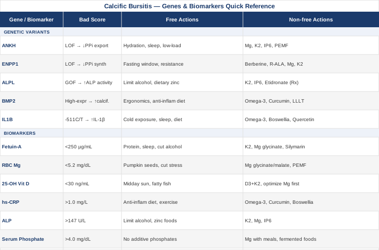
6 Biomarkers to Track if You Have Calcific Bursitis
Understanding why your bursae are accumulating calcium starts with measuring the biology that either prevents or promotes that accumulation. These six biomarkers are not exotic — most can be ordered through a standard lab, sometimes as part of a basic metabolic panel — but they are rarely ordered together with calcific bursitis in mind. That shift in framing is what this section asks you to make.
Biomarker 1: Fetuin-A — Your Body's Most Important Calcification Brake
Fetuin-A is a glycoprotein produced primarily by the liver and secreted into the bloodstream. Its primary function is to inhibit the spontaneous precipitation of calcium phosphate into crystals in soft tissue. Think of it as a chaperone protein: it binds calcium and phosphate in the blood and prevents them from crystallizing where they should not. Without adequate fetuin-A circulating in your blood, even normal calcium and phosphate concentrations can begin to crystallize in mechanically stressed or inflamed tissue — exactly the environment present in an irritated bursa.
Low fetuin-A levels have been consistently associated with vascular calcification, soft tissue calcification, and accelerated mineral deposition in several clinical populations. Research on fetuin-A and ectopic calcification has demonstrated that fetuin-A-deficient animal models develop extensive soft tissue calcification even when fed diets with normal calcium content — highlighting that it is not simply about how much calcium you consume, but whether your body can keep it in solution.
Why it matters for calcific bursitis: If your fetuin-A is low, your blood is less equipped to keep calcium in solution under biological stress. Even a modest local inflammatory trigger — repetitive micro-trauma at a bursa, for instance — can tip the balance toward crystal deposition when the circulating inhibitor is already depleted. Fetuin-A also has anti-inflammatory properties in its own right, so low levels may amplify the local inflammatory response that drives the condition.
How to Measure It
Fetuin-A is measured via a specialty blood test. It is not part of standard panels but is available through larger commercial labs and functional medicine providers. Cost ranges from $80 to $200 depending on the lab and whether a physician order is required. Optimal levels in healthy adults generally fall above 250 µg/mL, though reference ranges vary by lab. Some functional labs report this as AHSG (the gene that encodes it).
If the Score Is Low: The Plan Without Supplements
Fetuin-A is a liver-synthesized protein, so liver health and adequate protein intake are foundational. Prioritize adequate dietary protein — aim for 1.2 to 1.6 g of protein per kilogram of body weight daily from whole food sources. Insufficient protein intake is a known driver of reduced hepatic protein synthesis, including fetuin-A. Reduce the inflammatory load: a diet low in ultra-processed foods, refined seed oils, and added sugars reduces the systemic inflammation that suppresses fetuin-A production. Address sleep quality — hepatic protein synthesis peaks during deep sleep, and chronic sleep deprivation measurably reduces circulating plasma proteins. Alcohol, even in moderate amounts, impairs hepatic function and lowers protein secretion; reducing or eliminating it during a flare is a direct lever.
If the Score Is Low: The Plan With Supplements or Equipment
Vitamin K2 (MK-7): 100–200 mcg daily. K2 activates matrix Gla protein (MGP), a key inhibitor of soft tissue calcification that works synergistically with fetuin-A. Both are part of the same anti-calcification network. No cycling required for MK-7. Side effects: Rare; caution in patients on warfarin — K2 counteracts anticoagulant effects.
Magnesium glycinate: 300–400 mg daily. Supports liver function and works upstream of fetuin-A by directly inhibiting hydroxyapatite crystal nucleation. Take with dinner. No cycling required. Side effects: Loose stools at doses above 500 mg; titrate up from 150–200 mg.
Silymarin (milk thistle extract): 140–300 mg daily standardized to 70–80% silymarin content. Supports hepatic protein synthesis and reduces liver inflammatory load. No cycling required. Side effects: Rare gastrointestinal discomfort.
Low-dose melatonin at night (0.3–1 mg): Melatonin supports liver regeneration and protein synthesis during nighttime, with emerging evidence it supports fetuin-A levels in metabolic contexts. Cycle to avoid habituation: 4–6 weeks on, 1–2 weeks off. Side effects: Morning grogginess at higher doses; low-dose forms are preferred.
Biomarker 2: Red Blood Cell (RBC) Magnesium — The Crystallization Blocker Hidden in Plain Sight
This is the most underappreciated test in the context of calcific bursitis. Standard serum magnesium is nearly useless for detecting functional deficiency — the body will sacrifice intracellular and tissue magnesium to keep serum levels in the normal range until deficiency becomes severe. The RBC magnesium test measures magnesium inside red blood cells and is a far more reliable indicator of true intracellular magnesium status. Most people with calcific conditions who check only serum magnesium are flying blind.
Magnesium has a direct, well-documented role in preventing calcium phosphate crystallization. At the molecular level, magnesium ions compete with calcium for binding sites on hydroxyapatite crystal surfaces, effectively blocking crystal growth before it gains momentum. Epidemiological and mechanistic evidence supports this: multiple studies have found that higher magnesium tissue levels are associated with lower rates of soft tissue and joint calcification. Magnesium also modulates local inflammation by suppressing NF-κB activation — a key transcription factor in the inflammatory cascade that drives bursal irritation and osteogenic cell transformation.
How to Measure It
Serum magnesium: Usually part of a standard metabolic panel, approximately $10–30. Useful to rule out overt deficiency but misses subclinical states reliably.
RBC magnesium: Specialty test available through most major labs, approximately $40–100. Optimal range as referenced by most functional medicine practitioners is 5.2–6.5 mg/dL. Below 5.2 mg/dL is suboptimal regardless of serum levels. Retest every 3–4 months while supplementing to track response.
If the Score Is Low: The Plan Without Supplements
High-magnesium dietary sources: pumpkin seeds (approximately 150 mg per ounce), dark chocolate above 85% cacao, almonds, boiled spinach, black beans, and avocado. Include two to three high-magnesium foods daily as a baseline habit. Reduce magnesium wasters: alcohol, excess refined sugar, chronic psychological stress (cortisol drives urinary magnesium excretion), and high supplemental calcium without cofactors all deplete intracellular magnesium. Improve gut absorption: gut inflammation reduces magnesium absorption capacity. Removing processed food, addressing dysbiosis if present, and prioritizing gut-supportive foods (fermented, fibrous) raises the absorption floor over 8–16 weeks.
If the Score Is Low: The Plan With Supplements or Equipment
Magnesium glycinate: 300–400 mg elemental magnesium daily. Best absorbed form for raising intracellular levels without causing GI issues. Take with dinner. No cycling required. Side effects: Loose stools at doses above 500 mg; start lower and titrate.
Magnesium malate: 300–400 mg daily. Highly bioavailable; may be preferred if fatigue accompanies the condition since malate supports the citric acid cycle. Take morning or afternoon. Side effects: Similar to glycinate.
Topical magnesium chloride oil: Applied directly over the affected joint area, 5–10 sprays twice daily. Bypasses gut absorption. Anecdotal evidence is stronger than trial evidence for this route, but it functions well as an adjunct, particularly for local tissue delivery. Side effects: Mild skin tingling or temporary redness, which typically resolves within weeks of regular use.
PEMF (Pulsed Electromagnetic Field) devices: Emerging evidence suggests PEMF improves intracellular magnesium dynamics and reduces calcium crystal accumulation in connective tissue. Devices from established brands are used 20–30 minutes daily to the affected joint. Side effects: Rare; contraindicated with implanted electrical devices such as pacemakers.
Biomarker 3: 25-OH Vitamin D — The Calcium Traffic Controller
Vitamin D's relationship with calcification is often misunderstood, and the misunderstanding works in both directions. Some people avoid vitamin D supplementation fearing it will worsen calcium deposition. Others supplement aggressively without tracking levels. Neither approach is well-reasoned.
Vitamin D deficiency impairs calcium homeostasis — it raises parathyroid hormone (PTH), disrupts normal bone mineral exchange, and contributes to the type of local tissue stress and immune dysregulation that predisposes tendons and bursae to pathological mineralization. The calcium that would normally be directed into bone becomes destabilized and available for soft tissue deposition under these conditions. Conversely, very high vitamin D (above 100 ng/mL, particularly with simultaneous calcium supplementation) can cause hypercalcemia and increase ectopic calcification risk. Research on vitamin D and calcific musculoskeletal conditions is still building but mechanistically the therapeutic window is clear. The clinical target for most functional medicine practitioners — including Peter Attia — sits at 40–60 ng/mL.
How to Measure It
A standard 25-OH vitamin D blood test costs $30–80 and is widely available. The conventional normal range is 20–100 ng/mL, but 40–60 ng/mL represents the functional sweet spot for immune regulation, inflammation control, and calcium metabolism without excess. Test in late winter to capture your true low-point level; retest after 3 months when adjusting dosing.
If the Score Is Below 30 ng/mL: The Plan Without Supplements
Midday sun exposure of 15–30 minutes on arms and legs, three to five times per week depending on latitude and skin tone, produces meaningful vitamin D in summer months. At higher latitudes in winter, sun exposure alone is insufficient and supplementation becomes necessary regardless of diet. Dietary sources including fatty fish (salmon, sardines, mackerel), egg yolks, and beef liver contribute meaningfully but rarely bring levels to the 40–60 ng/mL range without supplementation.
If the Score Is Below 30 ng/mL: The Plan With Supplements
Vitamin D3 combined with K2 (MK-7): D3 at 2,000–5,000 IU daily, paired with vitamin K2 MK-7 at 100–200 mcg daily. K2 activates the proteins (MGP, osteocalcin) that direct calcium toward bone and away from soft tissue — making it an essential partner when supplementing D3. Never supplement D3 without K2 if soft tissue calcification is a concern. Retest every 3 months when dose-adjusting. Side effects: At doses above 5,000 IU, retest more frequently; avoid concurrent high-dose calcium supplementation.
Vitamin D cofactors — magnesium first: Magnesium is required to convert vitamin D to its active form (calcitriol) via hepatic and renal hydroxylase enzymes. If magnesium is depleted, D3 supplementation is partially ineffective and may even increase magnesium depletion further. Ensure RBC magnesium is optimized before or simultaneously with D3 supplementation.
Biomarker 4: hs-CRP — The Inflammation Alarm That Feeds Calcium Deposition
High-sensitivity C-reactive protein (hs-CRP) is a standard measure of systemic inflammation produced by the liver in response to circulating IL-6. Elevated hs-CRP reflects an inflammatory environment that, sustained over months and years, damages the microstructure of soft tissues — tendons, bursal linings, connective tissue matrices — creating conditions that favor calcium deposition in the local environment.
The link between chronic low-grade inflammation and calcific bursitis is documented across multiple lines of evidence. Inflammatory cytokines such as IL-1β and TNF-α have been shown to upregulate the expression of BMP-2 (bone morphogenetic protein 2), which in turn promotes osteoblast-like transformation of local mesenchymal cells — essentially driving soft tissue to behave like bone-forming tissue. Multiple studies have confirmed elevated inflammatory markers in patients with active calcific deposits compared to healthy controls matched for age and lifestyle. Tracking hs-CRP gives you a proxy for the ongoing inflammatory drive and a concrete target for intervention — not just a description of the problem.
How to Measure It
hs-CRP is available at most standard labs, costing $20–50 and often included in cardiovascular screening panels. Optimal levels: Below 1.0 mg/L. Between 1.0 and 3.0 mg/L represents intermediate systemic inflammation. Above 3.0 mg/L is elevated and warrants attention. Rule out acute infection or injury as a confound before interpreting a single elevated result — hs-CRP spikes dramatically with even a minor illness and requires a baseline measurement after recovery.
If the Score Is Elevated: The Plan Without Supplements
Anti-inflammatory dietary pattern: Emphasize fatty fish three to four times per week, extra virgin olive oil as a primary fat, leafy greens, berries, and cruciferous vegetables. Reduce ultra-processed foods, refined carbohydrates, and seed oils used at high cooking temperatures. This is not about perfection — it is about changing the ratio of pro-inflammatory to anti-inflammatory inputs in your daily diet.
Sleep: hs-CRP rises measurably with chronic sleep under six hours. Prioritize seven to nine hours with consistent timing. Two weeks of improved sleep shows measurable hs-CRP reduction in most studies.
Exercise: Regular moderate aerobic exercise — 150 or more minutes per week at zone 2 intensity — reduces circulating inflammatory markers. Resistance training three to four times per week adds anti-inflammatory myokine signaling. Sedentary behavior independently elevates hs-CRP regardless of diet.
Cold exposure: Two to four minutes of cold water immersion three to five times per week has shown measurable reductions in IL-6 and CRP in controlled studies. Start at cool (not ice-cold) water and progress over several weeks.
If the Score Is Elevated: The Plan With Supplements or Equipment
Omega-3 (EPA + DHA): 2–4 g of combined EPA/DHA daily from fish oil or algae oil. One of the most consistently proven anti-inflammatory nutritional interventions across dozens of randomized trials. Take with food. No cycling needed. Side effects: Fish-flavored burp at higher doses (use enteric-coated or refrigerate capsules); mild blood-thinning effect at doses above 3 g — relevant if taking anticoagulants.
Curcumin (bioavailable form): 500–1,000 mg daily using a high-absorption form (Meriva, Theracurmin, or BCM-95). Standard curcumin powder has poor bioavailability. Multiple RCTs show hs-CRP reduction with bioavailable curcumin. Cycle: 8 weeks on, 2–4 weeks off. Side effects: Rare GI irritation; enhances blood-thinning effects of certain medications.
Boswellia serrata (AKBA-standardized): 100–200 mg of AKBA-standardized extract daily. Inhibits 5-LOX, an inflammatory enzyme upstream of joint-specific inflammation. Cycle: 8–12 weeks on, 4 weeks off. Side effects: Occasional nausea.
Red light therapy (photobiomodulation): 10–20 minutes daily to the affected joint area using 630–850 nm wavelengths. Reduces local inflammatory cytokines through mitochondrial signaling cascades. See the Complementary Approaches section for full detail. Cost: $300–2,000 for quality panels.
Biomarker 5: Alkaline Phosphatase (ALP) — The Enzyme That Dismantles Your Natural Crystal Inhibitor
Alkaline phosphatase is almost never discussed in the context of calcific bursitis, yet it sits at the center of the biochemical story. ALP — specifically tissue-nonspecific alkaline phosphatase (TNAP), encoded by the ALPL gene — is an enzyme whose function is to hydrolyze inorganic pyrophosphate (PPi). And PPi is one of the body's most potent natural inhibitors of hydroxyapatite crystal formation.
Healthy tissue maintains a balance between PPi (which inhibits crystal growth) and inorganic phosphate (which promotes it). When ALP activity is chronically elevated, it tips this balance by destroying the PPi brake. The result is a local tissue environment where calcium phosphate crystals can nucleate and grow with less resistance — exactly the pathophysiology underlying calcific bursitis. Research on this mechanism has expanded significantly in vascular calcification literature, where elevated ALP is now recognized as an independent risk factor for arterial mineral deposits. The same pathway operates in periarticular soft tissue.
How to Measure It
ALP is part of the standard comprehensive metabolic panel (CMP), costing $10–30 when run alongside other routine tests. Normal range: 44–147 U/L in most labs. Elevated ALP can reflect liver disease, bone turnover, or TNAP overactivity. If liver function tests (ALT, AST) are normal, elevated ALP is more likely to reflect bone or soft tissue TNAP activity. Consider requesting bone-specific ALP (bALP) to isolate that contribution, approximately $60–120.
If the Score Is Elevated: The Plan Without Supplements
First rule out liver pathology: if ALT and AST are also elevated, liver disease is the likely driver and must be addressed separately through medical evaluation. If liver enzymes are normal, focus on: reducing alcohol intake (alcohol directly raises hepatic ALP), increasing zinc-rich whole foods (oysters, red meat, pumpkin seeds — ALP is a zinc-metalloenzyme and mild zinc dysregulation contributes to abnormal enzyme kinetics), and introducing fermented foods rich in natural menaquinones (natto, aged cheeses) as a dietary K2 source.
If the Score Is Elevated: The Plan With Supplements or Equipment
Vitamin K2 (MK-7): 150–200 mcg daily. Evidence from vascular calcification research suggests K2 modulates TNAP expression and supports normal mineralization pathways. Priority supplement for elevated ALP. No cycling required. Side effects: Caution with anticoagulants.
Magnesium: 300–400 mg daily as glycinate or malate. Magnesium competes with calcium for enzyme active sites and supports PPi stability in the extracellular matrix.
Inositol hexaphosphate (IP6): 1–2 g daily between meals. A naturally occurring compound that directly inhibits calcium phosphate crystal formation through chelation of mineral deposits. Emerging research only; use as an adjunct rather than a primary strategy. Cycle: 8 weeks on, 4 off. Side effects: May reduce absorption of zinc and iron if taken with meals — always take on an empty stomach.
Biomarker 6: Serum Phosphate — The Overlooked Calcification Fuel
Calcium phosphate crystals — the form that accumulates in calcific bursitis, specifically hydroxyapatite — require both calcium and phosphate. While almost all attention in clinical discussions goes to calcium, phosphate is the overlooked partner that drives the reaction forward. You cannot form a calcium phosphate crystal without phosphate, and modern diets provide it in abundance.
Processed foods routinely contain inorganic phosphate additives used as preservatives, emulsifiers, and flavor enhancers. These additives are absorbed at 90% or more bioavailability — far higher than the organic phosphate in whole foods, which is absorbed at roughly 40–60%. Carbonated soft drinks contain phosphoric acid. The combined effect in many people is a chronically elevated serum phosphate that creates a chemical environment favorable to ectopic crystal formation whenever local tissue conditions are strained. Studies in nephrology and cardiovascular medicine have demonstrated this clearly in high-phosphate populations; the musculoskeletal implications are underresearched but mechanistically direct.
How to Measure It
Serum phosphate is part of the CMP, approximately $10–30 added to standard blood work. Optimal range: 2.5–4.0 mg/dL. Above 4.0 mg/dL in someone without renal disease is worth addressing. Phosphate has a diurnal variation — fasting morning measurements are the most consistent. Retest after 8–12 weeks of dietary intervention.
If the Score Is Elevated: The Plan Without Supplements
Eliminate phosphate additives from the diet by reading ingredient labels. Avoid any packaged food listing "phosphate," "phosphoric acid," "disodium phosphate," or "polyphosphate" — this primarily means eliminating ultra-processed foods and carbonated soft drinks. Switch to whole food protein sources: organic phosphate in meat, legumes, and whole grains is absorbed at roughly half the efficiency of additive phosphate. This single dietary change can lower serum phosphate meaningfully over 6–8 weeks. Limit very high-protein intake from processed protein powders or bars with phosphate additives; aim for 1.2–1.8 g/kg/day from whole food sources.
If the Score Is Elevated: The Plan With Supplements or Equipment
Magnesium with meals: 200–300 mg of magnesium glycinate or citrate taken with meals. Magnesium competes with phosphate for shared intestinal transporters and modestly reduces phosphate absorption. This is one practical reason to take magnesium with food rather than away from it in this context.
Diverse fermented foods daily: Emerging evidence suggests that gut microbiome composition affects phosphate bioavailability and renal phosphate handling. Including kefir, kimchi, sauerkraut, or yogurt daily supports a microbiome profile associated with lower phosphate burden. Daily, indefinitely. Side effects: Bloating during the first one to two weeks of introduction.
Calcium carbonate with meals (physician-supervised only): Used clinically in kidney disease to bind dietary phosphate in the gut before absorption. Not appropriate without medical supervision in people with normal renal function, as it raises calcium independently. Mention this option to your physician if serum phosphate remains elevated after dietary changes.
The six biomarkers above form an interconnected picture: low fetuin-A, depleted RBC magnesium, elevated ALP, and high serum phosphate collectively create a body chemistry where calcium phosphate crystals form readily in stressed soft tissue like bursae. Understanding your own profile within this network allows targeted intervention rather than guesswork. The genetics section below explains why these biomarker patterns can be more persistent in some people than others.
What Your Genes May Reveal About Calcific Bursitis
The genetic underpinning of calcific bursitis and related crystalline deposition disorders centers largely on one biochemical axis: the regulation of extracellular pyrophosphate (PPi). PPi is the body's natural crystallization inhibitor in periarticular tissue, and several genes control how much of it gets produced, transported, and destroyed. Variants in these genes do not guarantee disease — environment, diet, and lifestyle remain essential — but they shift the threshold significantly. Understanding which variants you carry allows targeted preventive action rather than waiting for symptoms to escalate.
Gene 1: ANKH — The Pyrophosphate Gatekeeper
What it does: The ANKH protein is a transmembrane channel that transports inorganic pyrophosphate (PPi) from inside cells to the extracellular matrix. More extracellular PPi means greater inhibition of hydroxyapatite crystal formation in the surrounding tissue.
What a loss-of-function variant does: Reduced-activity ANKH variants lower the available pool of extracellular PPi, shifting local tissue chemistry toward crystal-permissive conditions. Research on the ANKH gene has established it as a causal factor in calcium pyrophosphate deposition (CPPD) disease — a crystalline arthropathy that shares pathophysiology with calcific bursitis. ANKH variants have been identified in both familial and sporadic cases of periarticular calcification.
If the Gene Is Affected: The Plan Without Supplements
The compensatory focus without supplements is on protecting the remaining PPi pool. Prioritize high dietary magnesium (see Biomarker 2) since magnesium works as a direct crystal inhibitor independent of the PPi system. Maintain optimal hydration — even mild chronic dehydration concentrates bursal and synovial fluids, raising the local calcium/phosphate product above the crystallization threshold. Reduce joint-loading activities during active flares to limit cell stress and the associated ATP hydrolysis that generates both useful and harmful by-products. Sleep quality is directly relevant: cellular processes that support PPi generation are most active during deep sleep; prioritizing 7–9 hours is not generic advice but a biochemical imperative in this context.
If the Gene Is Affected: The Plan With Supplements or Equipment
Magnesium glycinate: 300–400 mg daily. Compensates partially for reduced PPi by directly inhibiting crystal growth at nucleation sites. No cycling required. Side effects: Loose stools at excess doses.
Vitamin K2 (MK-7): 150–200 mcg daily. Activates matrix Gla protein (MGP), a complementary inhibitor of soft tissue mineralization that operates through a PPi-independent mechanism — providing a parallel brake.
Inositol hexaphosphate (IP6): 1–2 g daily between meals. Provides a further crystallization-inhibiting pathway. Cycle: 8 weeks on, 4 off. Side effects: Take away from meals to avoid mineral chelation.
PEMF therapy: 20–30 minutes daily over the affected joint. Emerging data suggests PEMF influences crystal formation kinetics in periarticular tissue and may improve PPi bioavailability at the cellular level. Side effects: Contraindicated with implanted electrical devices.
Gene 2: ENPP1 — The Pyrophosphate Factory
What it does: ENPP1 (ectonucleotide pyrophosphatase/phosphodiesterase 1) generates extracellular PPi from extracellular ATP. Where ANKH transports PPi out of cells, ENPP1 creates it in the extracellular space. Together they are the two primary contributors to the periarticular PPi pool.
What a loss-of-function variant does: ENPP1 variants that reduce enzyme activity lower PPi generation, creating the same crystal-permissive environment as ANKH variants but through a different upstream mechanism. Importantly, ENPP1 variants are also linked to insulin resistance — an overlap that matters because metabolic dysfunction independently raises soft tissue calcification risk through shared inflammatory pathways. Research on ENPP1 demonstrates reduced extracellular PPi in affected individuals and confirms the dual metabolic-calcification risk.
If the Gene Is Affected: The Plan Without Supplements
Address insulin sensitivity directly: time-restricted eating (14–16 hour fasting windows), resistance training three to four times per week, and reducing refined carbohydrate load all improve insulin sensitivity through AMPK activation — which also modulates ENPP1 activity. ENPP1 variants raise your metabolic risk alongside your calcification risk; the lifestyle factors that improve one meaningfully help the other. Monitor fasting glucose and HbA1c alongside your calcification biomarkers and track them together as connected indicators.
If the Gene Is Affected: The Plan With Supplements or Equipment
Berberine: 500 mg twice daily with meals. Improves insulin sensitivity via AMPK activation (similar mechanism to metformin). Addresses the ENPP1-metabolic axis directly. Cycle: 8–12 weeks on, 4 weeks off. Side effects: GI discomfort at initiation; do not combine with blood sugar-lowering medications without monitoring.
Alpha lipoic acid (R-ALA form): 300–600 mg daily. Antioxidant with insulin-sensitizing properties and direct effects on cellular energy metabolism. Side effects: Rare nausea; mild blood sugar lowering effect at higher doses.
Magnesium, K2, and PEMF as described in the ANKH section apply equally here — the mineralization-inhibiting strategy is consistent regardless of which PPi gene is affected.
Gene 3: ALPL — The Enzyme That Degrades Your Crystal Inhibitor
What it does: ALPL encodes tissue-nonspecific alkaline phosphatase (TNAP), which hydrolyzes PPi into inorganic phosphate. TNAP is the enzymatic counterweight to ANKH and ENPP1 — it removes the very inhibitor they produce. In healthy bone, TNAP activity is essential for normal mineralization. In soft tissue, chronically elevated TNAP tips the balance toward pathological crystal formation.
What a high-activity variant does: Gain-of-function or upregulated ALPL variants increase TNAP activity, causing faster PPi degradation in periarticular tissue. This is the genetic basis for what Biomarker 5 (serum ALP) measures in practice — elevated serum ALP may be the functional readout of this gene's activity. ALPL variants have been extensively studied in vascular and soft tissue calcification contexts.
If the Gene Is Affected: The Plan Without Supplements
Limit factors that upregulate TNAP activity: high alcohol intake, severe caloric restriction (which paradoxically elevates bone ALP as the body responds to nutritional stress), and very low protein diets. Ensure dietary zinc from whole food sources rather than high-dose supplementation — TNAP requires zinc as a cofactor, and both deficiency and excess disrupt enzyme kinetics.
If the Gene Is Affected: The Plan With Supplements or Equipment
Vitamin K2 (MK-7): 200 mcg daily. Evidence from the vascular calcification literature suggests K2 can modulate TNAP expression. Priority intervention here. No cycling required.
IP6 (inositol hexaphosphate): 1–2 g daily between meals. Works downstream of TNAP to inhibit crystallization even when PPi is being degraded at an elevated rate. Cycle: 8 weeks on, 4 off.
Etidronate (bisphosphonate, prescription only): A bisphosphonate that directly inhibits TNAP activity and has been studied specifically in calcific tendinitis. This is a physician conversation — not a self-managed supplement — but worth raising explicitly if ALPL variants are confirmed on genetic testing, as standard rheumatology protocols may not include this approach without patient advocacy.
Gene 4: BMP2 — The Tissue Calcification Promoter
What it does: Bone morphogenetic protein 2 (BMP2) is a growth factor that drives cells toward an osteogenic (bone-forming) fate. In healthy bone remodeling, this is necessary. In soft tissue — tendons, bursae, ligaments — BMP2 expression drives pathological ossification by transforming local mesenchymal stem cells into osteoblast-like cells that deposit mineral.
What a high-expression variant does: Variants that increase BMP2 expression or heighten local tissue sensitivity to BMP2 signaling raise the likelihood that bursal cells undergo osteogenic transformation in response to inflammatory stimuli. Studies on BMP2 in calcific tendinopathy have directly shown elevated BMP2 expression in calcific versus non-calcific tendon tissue, confirming this as an active driver rather than a passive association.
If the Gene Is Affected: The Plan Without Supplements
Chronic low-grade inflammation is the environmental trigger that activates BMP2 in soft tissue — the NF-κB and IL-1β signaling that you can reduce through diet, sleep, and exercise (see Biomarker 4) is precisely what turns a high-BMP2 genotype into an active problem. Ergonomic adjustments to reduce repetitive micro-trauma at the affected joint are also a direct BMP2 mitigation strategy: mechanical stress activates BMP2 transcription, and reducing that input lowers the activation signal.
If the Gene Is Affected: The Plan With Supplements or Equipment
Omega-3 (EPA/DHA): 3–4 g daily. Downregulates NF-κB and the BMP2-associated inflammatory cascade. No cycling needed. Long-term use.
Curcumin (BCM-95 or Meriva form): 500–1,000 mg daily. Inhibits NF-κB directly — a key upstream driver of BMP2 induction in soft tissue. Cycle: 8 weeks on, 2–4 off.
Low-level laser therapy (LLLT): Photobiomodulation has been shown in studies to modulate BMP2 expression in tendon and bursal tissue and reduce the osteogenic transformation of local mesenchymal cells. See the Complementary Approaches section below for full protocol detail.
Gene 5: IL1B — The Inflammation Amplifier That Fuels Crystal Formation
What it does: The IL1B gene encodes interleukin-1 beta (IL-1β), a master pro-inflammatory cytokine. IL-1β drives NF-κB activation, upregulates BMP2 expression, recruits immune cells to inflamed tissue, and sustains the inflammatory microenvironment that characterizes active calcific bursitis. It is one of the most studied cytokines in inflammatory joint disease.
What a high-activity variant does: High-expression IL1B promoter polymorphisms (such as -511C/T) produce more IL-1β in response to the same inflammatory stimulus — creating a more intense and sustained local inflammatory response that more aggressively promotes osteoblast-like transformation of bursal cells and prolongs the phase in which calcification is actively progressing. Research on IL1B variants has demonstrated associations across a range of inflammatory and degenerative musculoskeletal conditions.
If the Gene Is Affected: The Plan Without Supplements
All anti-inflammatory lifestyle factors from the hs-CRP section apply with heightened importance here. If you carry an active IL1B variant, sleep deprivation, chronic stress, sedentary behavior, and inflammatory diet patterns translate more directly into elevated local IL-1β and a more aggressive calcification cycle. Cold exposure — two to four minutes of cold water immersion three to five times per week — has measurably reduced circulating IL-1β in controlled studies and is one of the most accessible non-supplement interventions.
If the Gene Is Affected: The Plan With Supplements or Equipment
Omega-3 (EPA/DHA): 3–4 g daily. One of the strongest nutritional IL-1β inhibitors with a robust evidence base. No cycling required.
Boswellia serrata (AKBA-standardized): 100–200 mg daily. Specifically inhibits the 5-LOX pathway upstream of IL-1β inflammatory cascades. Cycle: 8–12 weeks on, 4 off.
Quercetin with bromelain: 500–1,000 mg daily. Natural IL-1β downregulator; bromelain improves quercetin absorption. Cycle: 8 weeks on, 2–4 off. Side effects: Rare GI effects at high doses.
Red light therapy (photobiomodulation): 10–20 minutes daily at 630–850 nm over the affected joint. Reduces local IL-1β expression in joint tissue through mitochondrial signaling. Increasingly accessible through consumer-grade devices.
The genetics picture and the biomarker picture converge on the same set of practical interventions — which is a useful reassurance. Whether you start with testing or with targeted lifestyle changes, the direction is consistent: support PPi production, reduce ALP activity, lower inflammation, and optimize magnesium and K2 status. The book below helps explain why the magnesium-calcium balance is often the leverage point that practitioners miss.
The Book That May Change Your Understanding of Calcific Bursitis
The Magnesium Miracle by Dr. Carolyn Dean — updated most recently in 2017 — synthesizes several decades of research on magnesium's role in over 65 conditions, with soft tissue calcification receiving direct and detailed treatment. What makes it particularly useful for someone navigating calcific bursitis is that it challenges one of the most pervasive assumptions in conventional care: that calcium supplementation is broadly beneficial and that calcification disorders are primarily about too much calcium rather than a failure of the regulatory system that keeps calcium in the right places.
10 Key Insights From This Book That Apply Directly to Calcific Bursitis
1. Magnesium deficiency is far more common than serum testing reveals. Serum magnesium represents less than 1% of total body magnesium. The 99% inside cells and bones is depleted silently for years before serum levels drop. Most people with soft tissue calcification conditions have never had intracellular magnesium properly measured.
2. Calcium and magnesium must be balanced — not just supplemented. The standard advice to supplement calcium without equivalent attention to magnesium is a practical driver of soft tissue calcification. Excess unbalanced calcium, with insufficient magnesium to regulate it, predisposes tissues to mineral deposition in joints and bursae.
3. Magnesium directly prevents calcium from entering cells inappropriately. At the cellular level, magnesium functions as a natural calcium channel blocker. When intracellular magnesium is low, calcium flows more freely into cells — including cells lining the bursae — creating the intracellular environment in which calcification begins.
4. Refined foods strip magnesium from the diet at a population scale. Modern agricultural and food processing removes 80–90% of the magnesium content from whole grains and vegetables. The typical Western diet provides roughly 50–70% of the daily magnesium requirement. This chronic deficit accumulates over decades.
5. Stress is a major magnesium depleter. The cortisol response to chronic psychological stress directly drives urinary magnesium excretion. People under prolonged work or life stress are significantly more likely to be magnesium-depleted — and they are also more likely to have elevated inflammatory markers that promote calcific bursitis.
6. Vitamin D supplementation without magnesium can worsen calcification risk. Dean's work aligns with the biochemistry: D3 requires magnesium for conversion to its active form. Supplementing D3 in a magnesium-depleted person can increase apparent D3 demand, worsen magnesium depletion, and paradoxically increase ectopic calcium deposition risk. The two must be co-optimized.
7. The calcium-to-magnesium ratio in soft tissue may matter more than absolute levels. The calcification problem is not always about total calcium being high — it is about the ratio of calcium to magnesium in local tissue being skewed. Raising magnesium rebalances this ratio without requiring calcium restriction.
8. Magnesium deficiency impairs ATP production, and ATP is required for PPi synthesis. This is the often-missed mechanical link: PPi (the crystal inhibitor we discussed in the genetics section) is generated from ATP. If magnesium is depleted — and magnesium is an essential cofactor in the ATP-generating processes of cellular respiration — PPi generation is itself compromised. Low magnesium thus reduces the body's ability to maintain the PPi-based crystal brake. This is the precise mechanism connecting the magnesium biomarker to the ANKH and ENPP1 genetic story.
9. Forms of magnesium matter significantly for absorption and clinical effect. Dean covers in detail why magnesium oxide (the most common form in cheap supplements) is poorly absorbed. Glycinate, malate, threonate, and citrate forms absorb meaningfully better. Liquid ionic magnesium is the best-absorbed option for those with gut absorption issues.
10. The threshold for clinical response is higher than most doctors assume. Achieving meaningful intracellular magnesium repletion typically requires 300–600 mg of well-absorbed elemental magnesium daily for 3–6 months before intracellular levels normalize. Short-term supplementation trials that show no effect are often simply underdosed or too brief to assess the full biological response.
Complementary Approaches With Clinical Evidence for Calcific Bursitis
The approaches below are included because they have human clinical evidence — not just plausibility — for calcific bursitis or closely related periarticular calcification conditions. They are not cures and do not replace medical management, but they can meaningfully contribute to symptom resolution and tissue recovery when applied consistently alongside the biomarker and genetic strategies above.
Low-Level Laser Therapy (Photobiomodulation)
Low-level laser therapy (LLLT), also known as photobiomodulation, uses specific wavelengths of red and near-infrared light (typically 630–905 nm) to stimulate mitochondrial function in tissue, reduce local inflammatory cytokine production, and support cellular repair processes. In the context of calcific bursitis, it is relevant for two reasons: it has direct evidence for reducing calcium deposit size in calcific shoulder pathology, and it modulates the BMP2 and IL-1β pathways that drive calcium deposition at the cellular level.
A randomized controlled trial published in a peer-reviewed journal and indexed at PubMed demonstrated significant improvements in pain and function in patients with calcific bursitis treated with LLLT compared to placebo, with evidence of reduced calcium deposit size on imaging in a proportion of treated patients. The evidence for LLLT in calcific shoulder pathology is stronger than for most soft tissue conditions, with multiple trials published over the past two decades.
For practical application, LLLT can be delivered through clinic-based class III or IV laser devices (typically 2–3 sessions per week for 6–8 weeks under physiotherapy supervision) or through consumer-grade red light panels at 630–850 nm used at home for 10–20 minutes daily directly over the affected joint. Professional devices deliver higher irradiance and are more validated in trials; consumer devices are a reasonable adjunct between professional sessions. Allow 8–12 weeks for full assessment of benefit. Side effects are minimal when used as directed; avoid looking directly into laser sources without appropriate eye protection.
Mindfulness Meditation and MBSR
Mindfulness-based stress reduction (MBSR) is an 8-week structured program developed at the University of Massachusetts Medical School combining meditation, body scan practice, and gentle movement. Its relevance to calcific bursitis is primarily through two mechanisms: reduction of systemic inflammation (chronic psychological stress is a documented driver of elevated IL-6, CRP, and IL-1β — the same inflammatory markers that promote calcification) and improvement of pain catastrophizing and pain-related disability, which independently affect recovery trajectory in musculoskeletal conditions.
Multiple controlled trials have demonstrated that MBSR reduces circulating inflammatory markers including hs-CRP and IL-6, with effect sizes comparable in some studies to low-dose anti-inflammatory supplementation. For chronic pain associated with calcific bursitis — which can persist even as calcium deposits begin resolving — MBSR specifically reduces pain catastrophizing, a psychological factor that independently predicts disability and delayed recovery regardless of the structural findings on imaging.
The standard MBSR protocol involves eight weekly sessions of approximately 2.5 hours plus daily home practice of 30–45 minutes. Shorter programs (4-week versions, app-based programs such as Insight Timer's structured MBSR courses) deliver partial benefit and are more realistic for most people managing an active condition. Even 10–15 minutes of daily body scan or focused breathing practice shows measurable stress-biomarker effects within 4–6 weeks. The goal is not relaxation per se — it is a measurable reduction in the stress-driven inflammatory load that sustains the calcification cycle.
Massage Therapy
Manual massage therapy applied to the musculature surrounding the affected bursa — the rotator cuff muscles and periscapular muscles in shoulder bursitis, for example — does not directly dissolve calcium deposits, but it addresses two important secondary factors: circulatory restriction and compensatory muscle tension. Both are well-documented accompaniments to calcific bursitis that worsen symptoms and delay tissue remodeling.
Evidence from clinical studies on massage therapy in shoulder pathology — including bursitis and rotator cuff conditions — supports meaningful improvements in pain, range of motion, and functional scores. The mechanisms include improved local circulation (which supports the delivery of anti-inflammatory mediators and the clearance of inflammatory by-products), reduction of hypertonic muscle bands that increase mechanical loading on the affected bursa, and parasympathetic nervous system activation that reduces systemic cortisol and its downstream inflammatory effects.
In practice, deep tissue massage or myofascial release focused on the muscles directly overlying and surrounding the affected joint is the most appropriate technique. Sessions of 45–60 minutes, one to two times per week during active flare phases, transitioning to monthly maintenance once symptoms stabilize. Self-massage tools (foam rollers, massage guns used at low intensity away from the bursa itself — not directly over an inflamed bursa) can extend the benefit between sessions. Avoid direct pressure on the bursa during active inflammation; work the surrounding musculature and address the affected area only as pain allows. A licensed massage therapist familiar with musculoskeletal pathology should be the first point of contact.
Conclusion
Calcific bursitis is not simply a mechanical problem or a calcium excess problem — it is a failure of the regulatory biology that normally prevents calcium from crystallizing in soft tissue. The biomarkers covered in this article give you measurable entry points: fetuin-A, RBC magnesium, 25-OH vitamin D, hs-CRP, alkaline phosphatase, and serum phosphate together paint a picture of your individual calcification profile that is far more actionable than a diagnosis alone. The genetic variants in ANKH, ENPP1, ALPL, BMP2, and IL1B explain why some people are more susceptible than others — and more importantly, they point toward the specific compensatory strategies that raise the threshold back up.
The next smart step is to start where you have the most information. If you have never had RBC magnesium, fetuin-A, hs-CRP, and a full metabolic panel ordered together, that is the foundation. If you have access to genetic testing (through providers such as 23andMe with third-party interpretation tools, or a functional medicine physician), the five genes discussed here give you a meaningful layer of precision on top of the biomarker data. Take that information into a conversation with a physician, physiotherapist, or functional medicine practitioner who is open to individualized approaches — and use this article as the framework for that conversation.
Musculoskeletal: Bone Conditions Joint Conditions Tendon & Ligament Conditions
Autoimmune: Inflammatory Conditions Connective Tissue Conditions