This article contains AI generated content.
· UpdatedCalcific Tendinitis — Genes And Biomarkers: 5 Genes And 7 Biomarkers To Track
Introduction
If you've been diagnosed with calcific tendinitis, you already know the pattern: calcium crystals form inside a tendon — most commonly the supraspinatus in the shoulder — and cause pain that ranges from a dull background ache to sudden, acute attacks that make simple movements nearly impossible. You may have seen the calcium deposit on an ultrasound or X-ray, been told to rest and take anti-inflammatories, and returned months later to find it still there. The frustration isn't the pain alone — it's the sense that nothing in the explanation quite fits your situation.
What standard advice misses is that calcific tendinitis isn't simply a mechanical problem caused by overuse. It's a biological process driven by how your body manages calcium, handles inflammation, and maintains tendon cell integrity. These processes vary significantly between individuals based on measurable biochemical signals and, to a real extent, on genetic variants that affect the underlying pathways. Two people with the same job, the same posture, and the same training load can have entirely different outcomes because their internal biology is different in ways that standard care doesn't account for.
This is where biomarkers and genetic variants become practically useful. Biomarkers — measured through routine or functional blood panels — give you a real-time view of your inflammatory load, vitamin and mineral status, and calcification-regulating proteins. Genetic variants, accessed through consumer testing services and interpreted with the right tools, tell you where your baseline predispositions lie. Neither tells the whole story alone, but together they create a map that generic advice cannot.
This article covers both. The primary section examines seven biomarkers with direct relevance to calcific tendinitis — what they measure, what a bad result means, and what to do about it. A second section walks through five genetic variants implicated in tendon calcification biology, with practical compensation strategies for each. A third section summarizes a key book that reframes how calcium behaves in the body. The goal isn't a cure — it's the kind of specific, grounded information that allows you to make better decisions about your care.
7 Biomarkers That Matter More Than Most Doctors Test
Biomarkers are measurable signals in your blood that reflect what's happening in your body's biochemistry right now. For calcific tendinitis, the most informative markers aren't simply serum calcium — they cover inflammation levels, calcium metabolism regulators, and proteins that directly inhibit or enable crystal growth in soft tissue. Getting these tested through a functional medicine panel or by requesting them individually from your GP gives you a concrete, actionable picture.
1. High-Sensitivity C-Reactive Protein (hs-CRP)
Why it matters: hs-CRP is produced by the liver in response to IL-6 — a key pro-inflammatory cytokine. In calcific tendinitis, chronic low-grade inflammation is not just a symptom; it's part of the mechanism. Inflammatory signaling promotes the transformation of tenocytes (tendon cells) into chondrocyte-like cells, which then produce matrix vesicles that seed hydroxyapatite crystal growth. Elevated hs-CRP tells you this background inflammatory environment is active. Research published in Calcified Tissue International has noted correlations between elevated systemic inflammation markers and worse calcific tendinitis presentations.
Optimal range: Below 0.5 mg/L (per Peter Attia's protocol); below 1.0 mg/L is acceptable. Above 3 mg/L is high-risk territory.
How to Measure It
hs-CRP is a standard blood draw available at most labs. It's often included in inflammatory panels or can be ordered alone. Cost: $10–30 USD. It should be interpreted fasted, not during an acute infection or injury, which would give artificially elevated readings. Aim to test every 6–12 months while monitoring interventions.
If the Score Is Bad, the Plan Without Supplements
An elevated hs-CRP first requires an honest audit of lifestyle. The Mediterranean dietary pattern consistently reduces hs-CRP by 20–30% within 12 weeks — this means olive oil, fatty fish, legumes, vegetables, and a significant reduction in seed oils and ultra-processed foods. Sleep quality is a surprisingly large lever: poor sleep (under 7 hours or fragmented sleep) raises CRP by 40% or more in research settings. Address sleep architecture before anything else. Zone 2 aerobic exercise (150–180 minutes per week at a pace where you can hold a conversation) has robust anti-inflammatory effects over time. Treating gum disease and dental infections is often overlooked but is one of the largest drivers of chronically elevated CRP. Commit to dietary and sleep changes for at least 12 weeks before reassessing.
If the Score Is Bad, the Plan With Supplements or Equipment
Omega-3 fatty acids (EPA + DHA): 2–4g/day from quality fish oil or algae-based omega-3 has been shown in multiple trials to reduce hs-CRP by 15–25%. Use a triglyceride-form product for best absorption. Cycling: safe year-round. Side effects: mild blood thinning at higher doses; disclose to any prescribing physician. Curcumin with piperine: 500–1000mg/day; reduces NF-κB inflammatory signaling. Cycle 8 weeks on, 2–4 weeks off. Side effects: may interact with anticoagulant medications; mild GI sensitivity in some. Red light therapy panels (photobiomodulation, discussed later) can also reduce local inflammatory signaling as an equipment option.
2. 25-OH Vitamin D
Why it matters: Vitamin D's role in calcium metabolism is well established, but its specific relevance to tendon tissue is less discussed. The vitamin D receptor (VDR) is expressed in tenocytes, and vitamin D signaling modulates both inflammatory response and calcium handling in connective tissue. Deficiency is associated with more severe musculoskeletal pain syndromes and slower tendon healing. Crucially, vitamin D works in concert with vitamin K2 to route calcium appropriately — without K2, vitamin D supplementation alone may increase circulating calcium without improving where that calcium ends up.
Optimal range: 50–70 ng/mL (per Rhonda Patrick and Peter Attia's protocols); standard labs flag deficiency below 20 ng/mL, which undershoots the functional optimum.
How to Measure It
Standard serum 25-OH vitamin D test via blood draw. Cost: $25–60 USD. This test is increasingly covered by insurance with a clinical indication. Test at least twice a year — once in late winter (when levels are lowest) and once in summer — to understand your seasonal range.
If the Score Is Bad, the Plan Without Supplements
Midday sun exposure on large skin areas (arms, legs, back) for 15–30 minutes daily — timing matters; UVB sufficient for vitamin D synthesis only occurs when the sun is high enough (solar noon ± 2 hours). Dietary sources are limited but meaningful: fatty fish (salmon, mackerel, sardines), egg yolks, and beef liver contribute modest amounts. Reducing high-dose calcium supplementation (which can suppress the vitamin D conversion pathway) is also worthwhile.
If the Score Is Bad, the Plan With Supplements or Equipment
Vitamin D3 5000 IU/day for deficient individuals; adjust based on testing, not fixed dose. CRITICAL for calcific tendinitis: always pair D3 with Vitamin K2 (MK-7 form) 100–200 mcg/day. K2 activates matrix Gla protein (MGP), which inhibits soft tissue calcification — taking D3 without K2 increases circulating calcium without ensuring proper routing away from tendons. Add Magnesium glycinate 300–400mg/day as an essential cofactor for vitamin D conversion. Retest every 3 months initially. Side effects: vitamin D toxicity is rare below 10,000 IU/day but dose with testing; K2 is extremely safe; magnesium may cause loose stools at high doses — start at 150mg and titrate.
3. RBC Magnesium
Why it matters: Magnesium and calcium compete at cellular binding sites, and low magnesium is one of the clearest drivers of calcium deposition in soft tissues. Magnesium also supports the enzymatic production of inorganic pyrophosphate (PPi), a natural inhibitor of calcium crystal growth. Standard serum magnesium is poorly reflective of true cellular status — most magnesium is intracellular, so serum stays "normal" even when tissue levels are inadequate. RBC (red blood cell) magnesium is a far more accurate marker. Estimates suggest 60–70% of adults have suboptimal magnesium status.
Optimal range: RBC magnesium 5.2–6.5 mg/dL; serum magnesium 0.85–1.0 mmol/L.
How to Measure It
Request RBC magnesium specifically — standard panels only measure serum magnesium. Cost: $30–50 USD through functional medicine labs. Repeat every 3–6 months while optimizing.
If the Score Is Bad, the Plan Without Supplements
Increase dietary magnesium through pumpkin seeds (156mg per oz), dark leafy greens (spinach, Swiss chard), dark chocolate (70%+), almonds, and avocado. Eliminate or significantly reduce alcohol and excess caffeine, which both deplete magnesium through urinary excretion. Reduce refined sugar intake — metabolizing sugar requires magnesium at multiple enzymatic steps. Epsom salt baths (magnesium sulfate) provide transdermal absorption and serve as an accessible, low-cost option several nights per week.
If the Score Is Bad, the Plan With Supplements or Equipment
Magnesium glycinate or malate (200–400mg elemental magnesium per day) are the best-absorbed forms. Avoid magnesium oxide — bioavailability is very low. Evening dosing is preferred and provides a bonus sleep-quality effect. Side effects: loose stools at high doses; start at 150mg and titrate up. Safe year-round. Equipment: consider magnesium chloride transdermal spray if GI tolerance is an issue at effective oral doses.
4. Alkaline Phosphatase (ALP)
Why it matters: ALP is an enzyme involved in the mineralization process — it's produced in bone, liver, kidneys, and intestines, and elevated levels can reflect active tissue calcification. In calcific tendinitis, the formation of hydroxyapatite crystals in tendons involves bone-like mineralization activity. Persistently elevated ALP (particularly in the bone isoform) alongside other markers may signal that active calcification processes are running. Interpreting ALP in context is important — liver issues, bone disease, or intestinal inflammation can each drive it up through different mechanisms.
Optimal range: 44–115 U/L; ideally in the lower half for adults without bone growth or liver issues.
How to Measure It
ALP is included in a standard Comprehensive Metabolic Panel (CMP). Cost: $10–30 USD for the full panel. If elevated, a bone-specific ALP test can confirm whether the source is skeletal rather than hepatic.
If the Score Is Bad, the Plan Without Supplements
Identify the source first: if liver-origin, focus on alcohol reduction, dietary liver support (cruciferous vegetables, garlic, bitter greens), and ruling out non-alcoholic fatty liver. If bone/calcification-origin, prioritize the broader calcium-routing protocol (D3 + K2 + Mg). Increase zinc-rich foods (oysters, beef, pumpkin seeds) — zinc is a cofactor for ALP regulation. Reduce excessive dietary phosphate from sodas, processed foods, and meat excess.
If the Score Is Bad, the Plan With Supplements or Equipment
Zinc picolinate 25–30mg/day supports ALP normalization when zinc deficiency is a driver. Always balance with copper 1–2mg/day (excess zinc depletes copper over time). Vitamin D3 + K2 to improve calcium routing. Cycling: 12 weeks of zinc, then reassess. Side effects: zinc above 40mg/day long-term risks copper deficiency; do not exceed without testing.
5. Fetuin-A
Why it matters: Fetuin-A (also known as alpha-2-Heremans-Schmid glycoprotein, encoded by the AHSG gene) is produced by the liver and is one of the most potent known inhibitors of ectopic calcification. It works by binding free calcium and phosphate ions and forming calciprotein particles (CPPs) that prevent crystal nucleation and growth in soft tissue. When fetuin-A levels fall, this natural brake on soft tissue calcification is weakened. Low fetuin-A has been associated with cardiovascular calcification, calciphylaxis, and — in emerging research — soft tissue mineralization disorders including tendinopathy. A study published in Calcified Tissue International examined the relationship between low fetuin-A and ectopic soft tissue calcification across conditions.
Optimal range: 0.6–1.0 g/L; below 0.65 g/L is concerning in the context of calcification risk.
How to Measure It
Fetuin-A is not included in standard panels and typically requires a specialty or functional medicine lab order. Cost: $80–150 USD. It is a less commonly run marker, so you may need to advocate for it through an integrative or functional medicine physician.
If the Score Is Bad, the Plan Without Supplements
Since fetuin-A is produced by the liver, liver health is the primary lever. Reduce dietary advanced glycation end products (AGEs) — minimize grilled, fried, and charred foods. Significantly reduce fructose consumption, as high fructose intake has been shown in research to suppress hepatic fetuin-A production. Adequate dietary protein (fetuin-A is a protein that the liver synthesizes). Anti-inflammatory diet broadly (chronic liver inflammation reduces fetuin-A output).
If the Score Is Bad, the Plan With Supplements or Equipment
No supplement directly and reliably raises fetuin-A in peer-reviewed human data — this is an emerging area. Indirect support: optimizing vitamin D (some research suggests a relationship between D status and fetuin-A), magnesium, and liver support with milk thistle (silymarin 400–600mg/day for hepatoprotection). Resveratrol 500mg/day has hepatoprotective activity in early data. Side effects: resveratrol may interact with blood thinners; cycle 3 months on, 1 month off. This is an area where working with a physician familiar with calcification biology is strongly recommended.
6. Parathyroid Hormone (PTH)
Why it matters: PTH is the master regulator of calcium balance. When PTH is chronically elevated — a condition called secondary hyperparathyroidism — it pulls calcium from bone and increases circulating calcium, potentially increasing the substrate available for ectopic deposition. Secondary hyperparathyroidism is commonly driven by vitamin D deficiency, calcium malabsorption, or excess dietary phosphate. PTH should always be interpreted alongside 25-OH vitamin D and serum calcium — the three form a hormonal triad that tells a coherent story about calcium regulation.
Optimal range: 15–65 pg/mL; functional medicine practitioners often prefer the lower half of this range (below 40 pg/mL).
How to Measure It
PTH (intact PTH) is a standalone blood test ordered alongside calcium and vitamin D. Cost: $30–60 USD. Best measured fasted in the morning. Request it alongside 25-OH vitamin D and serum calcium for interpretive context.
If the Score Is Bad, the Plan Without Supplements
Address vitamin D status first — vitamin D deficiency is the most common driver of elevated PTH. Reduce dietary phosphate from sodas, processed meats, and packaged foods (high phosphate drives PTH up). Prioritize dietary calcium from whole foods (dairy, sardines with bones, kale, almonds) — dietary calcium suppresses PTH more safely than calcium supplements. Weight-bearing exercise daily improves calcium utilization in bone and supports PTH normalization.
If the Score Is Bad, the Plan With Supplements or Equipment
Vitamin D3 + K2 is the primary intervention: D3 5000 IU/day with K2 MK-7 200 mcg/day and magnesium glycinate 300–400mg/day. Recheck PTH, vitamin D, and calcium together at 3 months. Important caution: calcium supplements in this context are complex — supplemental calcium without adequate K2 may worsen ectopic deposition. Prefer dietary calcium where possible. Side effects: monitor blood calcium levels if supplementing; hypercalcemia risk with excessive D3 dosing.
7. Uric Acid
Why it matters: Uric acid is a waste product of purine metabolism. Hyperuricemia is classically associated with gout — uric acid crystal deposition in joints — but its relevance extends further. Elevated uric acid reflects metabolic dysregulation (particularly fructose overconsumption and impaired purine clearance), drives systemic inflammation, and may worsen the tissue environment in which calcium deposition occurs. Research in tendinopathy populations has found elevated uric acid correlating with more severe presentations. While uric acid itself is not a calcium crystal, it shares metabolic drivers with ectopic calcification — and lowering it tends to reduce the broader inflammatory burden.
Optimal range: Below 5.5 mg/dL for men; below 4.5 mg/dL for women. Standard labs accept up to 7.0 mg/dL, which many practitioners consider too permissive.
How to Measure It
Uric acid is part of a standard Comprehensive Metabolic Panel or can be ordered alone. Cost: $10–25 USD. Test fasted and not during acute illness. Test every 6 months while optimizing dietary factors.
If the Score Is Bad, the Plan Without Supplements
Reduce fructose — including high-fructose corn syrup, fruit juices, and agave — as fructose metabolism generates uric acid as a byproduct, and this is the single largest modifiable dietary driver. Hydration: 2–3 liters of water daily increases uric acid excretion via the kidneys. Reduce alcohol (especially beer) and limit organ meats and anchovies. Increase vitamin C-rich foods (citrus, bell peppers, strawberries) — vitamin C increases renal uric acid excretion. Modest weight loss if overweight (adipose tissue promotes uric acid production).
If the Score Is Bad, the Plan With Supplements or Equipment
Tart cherry extract 480mg/day has been shown in multiple human studies to reduce uric acid levels by 20–30% and reduce gout flare frequency. Quercetin 500–1000mg/day inhibits xanthine oxidase (the enzyme producing uric acid) — similar mechanism to the pharmaceutical allopurinol, but gentler. Vitamin C 500–1000mg/day increases uric acid excretion. Cycling: tart cherry and quercetin can be cycled 3 months on, 1 month off. Side effects: quercetin may interact with some pharmaceuticals; vitamin C above 2000mg/day may cause GI discomfort.
Understanding which of these seven biomarkers is most out of range for you provides a far more targeted approach than generic anti-inflammatory advice. The genetic layer below adds another dimension — one that helps explain why some people's biomarkers skew the way they do.
The Genetic Layer: 5 Variants That May Explain Your Individual Risk
Genetic variants don't determine whether you'll develop calcific tendinitis — environmental factors, loading history, and nutrition play large roles — but they shift the playing field. Certain variants make the calcification pathway more easily triggered, reduce natural inhibitors of crystal growth, or compromise tendon structural quality. Consumer genetic testing through services like 23andMe or Ancestry provides raw data that can be interpreted through platforms like SelfDecode, Genetic Genie, or with a functional medicine practitioner.
BMP2 — The Calcification Initiator
What the gene does: Bone Morphogenetic Protein 2 normally promotes bone formation during development. In tendons, BMP2 signaling becomes problematic when tenocytes — stressed by injury, hypoxia, or chronic inflammation — begin differentiating into chondrocyte-like cells under BMP2 influence. These transformed cells produce matrix vesicles that seed hydroxyapatite crystal growth. Studies examining surgically removed calcific deposits from tendons have found elevated BMP2 expression within the calcified tissue, confirming its central role in the process. Certain genetic variants in the BMP2 gene may be associated with a lower threshold for this pathway activation.
If the Gene Is Bad, the Plan Without Supplements
Reduce the conditions that trigger BMP2 upregulation in tendons: tendon hypoxia (improve shoulder circulation through mobility work and aerobic conditioning), chronic repetitive microtrauma (apply the 10% weekly load increase rule; avoid repetitive overhead movements without adequate recovery), and chronic local inflammation (anti-inflammatory diet as foundation). Implement a progressive eccentric loading protocol for the rotator cuff 3 times per week — controlled tendon loading at appropriate intensity promotes healthy tendon remodeling rather than pathological transformation. Commit to at least 12 weeks for structural adaptation. Aerobic exercise 4–5 days/week improves tissue oxygenation.
If the Gene Is Bad, the Plan With Supplements or Equipment
Vitamin D3 + K2: some research suggests vitamin D signaling modulates BMP pathway activity in connective tissue. Curcumin 500–1000mg/day with piperine: in vitro studies have shown curcumin inhibits BMP-driven osteogenic differentiation; while human tendon evidence is indirect, the anti-inflammatory mechanism is well-supported. Magnesium glycinate 300–400mg/day counters the calcification cascade. Cycle curcumin 8 weeks on, 2 weeks off. Side effects: curcumin may interact with anticoagulants; K2 is very safe; as above for magnesium. Note that the evidence for direct BMP2 pathway supplementation in humans is mostly mechanistic — apply these as supportive layers within a broader protocol, not as targeted inhibitors.
ANKH — The Crystal Formation Gatekeeper
What the gene does: The ANKH gene encodes a transmembrane protein that transports inorganic pyrophosphate (PPi) out of cells. PPi is a potent natural inhibitor of calcium crystal nucleation — it coats nascent crystals and blocks their growth. When ANKH function is reduced due to loss-of-function variants, less PPi is exported into the extracellular space around tendons, and calcium crystals can form more freely. ANKH mutations are definitively associated with chondrocalcinosis (calcium pyrophosphate deposition disease) and are studied in hydroxyapatite deposition disease. While calcific tendinitis primarily involves hydroxyapatite crystals rather than CPP crystals, the ANKH-PPi axis plays a regulatory role across soft tissue calcification broadly.
If the Gene Is Bad, the Plan Without Supplements
Avoid prolonged immobilization — reduced joint and tendon movement decreases local PPi levels, further reducing the natural crystal inhibition. Implement daily tendon loading exercises for the affected region (eccentric programs for rotator cuff: shoulder external rotation eccentrics, 3 sets of 15 repetitions, daily if tolerated). Prioritize a magnesium-rich diet (magnesium supports enzymatic production of PPi through the ATP → AMP → ADP pathway). Broad anti-inflammatory diet to reduce the inflammatory microenvironment that accelerates crystal growth when PPi is low. Frequency: daily movement, minimum 20 minutes of active tendon work.
If the Gene Is Bad, the Plan With Supplements or Equipment
Magnesium glycinate or malate 300–400mg/day: directly supports the biochemical pathways involved in PPi synthesis. Vitamin D3 + K2: supports overall calcium metabolism and reduces the net calcification burden. Important note: no supplement directly and specifically compensates for ANKH loss-of-function in a clinically proven way. The above are supportive measures that address modifiable factors in the same pathway. In cases of known ANKH variants with significant symptoms, a rheumatologist familiar with mineralization disorders is the appropriate specialist. Side effects: as listed in biomarkers section for each supplement.
VDR — The Vitamin D Receptor
What the gene does: The VDR gene encodes the vitamin D receptor — the intracellular protein that binds vitamin D and triggers changes in gene expression. Even with adequate circulating vitamin D, individuals with reduced-function VDR variants may mount a weaker cellular response. The four most studied polymorphisms are FokI (rs2228570), BsmI (rs1544410), ApaI (rs7975232), and TaqI (rs731236). VDR is expressed in tenocytes, and vitamin D signaling through VDR modulates both local inflammatory response and calcium handling in connective tissue. Studies on VDR polymorphisms and musculoskeletal conditions (including tendinopathy and stress fractures) suggest that specific allele combinations may be associated with higher susceptibility to tendon pathology.
If the Gene Is Bad, the Plan Without Supplements
Maximize sun exposure quality and timing — UVB-generated vitamin D at the skin level may provide additional signals beyond serum levels. Load-bearing and resistance exercise promotes VDR expression in musculoskeletal tissue. Increase dietary vitamin D from fatty fish, egg yolks, and beef liver. Reduce pro-inflammatory cytokines through diet — chronic inflammation impairs VDR signaling at the receptor level even when circulating vitamin D is adequate.
If the Gene Is Bad, the Plan With Supplements or Equipment
With a poor VDR variant, aim for a higher blood vitamin D target: 60–80 ng/mL instead of the standard 50–70, since the cellular response per unit of circulating vitamin D is reduced. This means higher D3 dosing (6000–8000 IU/day), strictly adjusted by testing. Always pair with K2 MK-7 200 mcg/day (to avoid D3 driving calcium to soft tissues) and magnesium glycinate 400mg/day. Monitor blood calcium and 25-OH vitamin D every 3 months. Never target above 100 ng/mL without physician oversight. Side effects: hypercalcemia risk at very high D3 levels; testing is the safety mechanism here, not dose restriction alone.
COL1A1 — Tendon Structural Integrity
What the gene does: COL1A1 encodes the alpha-1 chain of type I collagen, the dominant structural protein in tendons. The most studied variant is the Sp1 polymorphism (rs1800012, T allele), which affects transcription factor binding and reduces the production of properly structured collagen. This leads to tendons with weaker collagen networks — more susceptible to microtrauma under load. When tendons are repeatedly damaged at the micro level, they trigger inflammatory cascades and can promote the chondrocytic transformation of tenocytes that initiates calcification. A meta-analysis in the British Journal of Sports Medicine associated COL1A1 Sp1 polymorphisms with increased tendon injury risk across multiple populations.
If the Gene Is Bad, the Plan Without Supplements
Implement a structured eccentric loading protocol 3 times per week — eccentric exercise applied gradually to tendons stimulates collagen remodeling and is the best-established non-pharmaceutical intervention for tendon structural health. Apply the 10% weekly load increase rule strictly — sudden load jumps are far more damaging with COL1A1 vulnerability. Prioritize dietary protein at 1.6–2.0g/kg body weight per day — amino acids are the building blocks of collagen synthesis. Target exercises that load the tendon at the relevant angle (shoulder external rotation, elevation control) in the 4–6 repetitions-to-fatigue range for maximum structural stimulus.
If the Gene Is Bad, the Plan With Supplements or Equipment
Hydrolyzed collagen peptides 15g, taken 30–60 minutes before tendon-loading exercise: this specific timing, established by Dr. Keith Baar's research at UC Davis, increases collagen synthesis in the loaded tendon by providing the circulating amino acid substrate at the window of peak mechanosensitivity. Vitamin C 500mg alongside the collagen — ascorbic acid is an essential cofactor for collagen cross-linking via prolyl hydroxylase. These two are the most evidence-backed supplement interventions for tendon collagen synthesis. Cycling: safe year-round. Side effects: generally very safe; start with 5g gelatin if hydrolyzed collagen causes GI discomfort. Equipment: consider eccentric training with a resistance band or light cable for precision loading.
TNFRSF11B (OPG) — Calcium Traffic Controller
What the gene does: TNFRSF11B encodes osteoprotegerin (OPG), a decoy receptor in the RANK/RANKL/OPG system. OPG regulates both bone resorption and, increasingly understood, ectopic soft tissue calcification. Low OPG levels are associated with cardiovascular calcification and soft tissue mineral deposition — OPG functions as a broad anti-calcification signal beyond just bone metabolism. Genetic variants that reduce OPG expression or function may leave the body more vulnerable to aberrant calcium deposition in soft tissues, including tendons. Direct calcific tendinitis studies on OPG variants are limited, but the broader calcification biology is mechanistically consistent.
If the Gene Is Bad, the Plan Without Supplements
Weight-bearing exercise consistently and robustly upregulates OPG expression — this is one of the clearest gene-environment interactions in bone and soft tissue metabolism. Daily weight-bearing activity (walking, resistance training, standing work) for at least 30–45 minutes is the primary lever. Quit smoking if applicable — tobacco is a documented suppressor of OPG expression. Anti-inflammatory diet to reduce RANKL signaling (the counterpart that OPG decoys against) — when RANKL rises unchecked relative to OPG, calcification risks increase. Protein-adequate diet to support hepatic OPG synthesis.
If the Gene Is Bad, the Plan With Supplements or Equipment
Vitamin K2 (MK-7) 100–200 mcg/day: favorably modulates the OPG/RANKL ratio and activates matrix Gla protein simultaneously — a dual mechanism directly relevant to calcific tendinitis. Omega-3 fatty acids (EPA + DHA) 2–3g/day: EPA reduces RANKL signaling; multiple studies have observed favorable RANKL/OPG shifts with omega-3 supplementation. Resveratrol 500mg/day with food: some research suggests it upregulates OPG expression via SIRT1 pathway activation. Cycle resveratrol 3 months on, 1 month off. Side effects: K2 is extremely safe; resveratrol may interact with anticoagulants; omega-3 as noted above.
With both the biomarker map and the genetic context in hand, the table below puts all of this information in one accessible view.
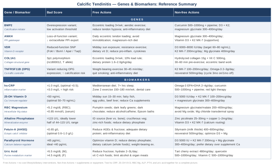
The next layer — one often missed entirely in standard care — is understanding why calcium ends up in the wrong place in the first place. A single book explains this mechanism more clearly than most clinical literature.
The Calcium Paradox: 10 Insights From the Book That Could Change How You Think About Calcific Tendinitis
Vitamin K2 and the Calcium Paradox by Kate Rhéaume-Bleue presents a case that is both well-referenced and genuinely counterintuitive for most readers: the problem in conditions like calcific tendinitis, atherosclerotic plaque, and bone loss isn't calcium intake — it's calcium routing. Calcium ends up in tendons, arteries, and other soft tissues not because there's too much of it, but because the proteins responsible for directing it to bone are inactive due to K2 deficiency. The research she synthesizes reframes several common health assumptions in a way directly relevant to anyone managing calcific deposits.
Insight 1: Calcium Accumulates in Soft Tissues When Routing Proteins Fail
The central paradox is that many people with calcification in soft tissues (tendons, arteries) simultaneously have bone loss. This isn't a coincidence — both happen when calcium routing fails. Calcium enters circulation but isn't directed to bone; instead, it deposits wherever the tissue environment allows. This reframes calcific tendinitis from "too much calcium in the tendon" to "calcium that should be elsewhere, stuck in the tendon."
Insight 2: Matrix Gla Protein Is Your Tendon's Built-In Anti-Calcification Defense
Matrix Gla Protein (MGP) is produced by cells in soft tissues including tendons and blood vessel walls. Its job is to bind calcium and prevent it from forming crystals in connective tissue. It is one of the most potent local inhibitors of soft tissue calcification known. The key word is "produced" — MGP synthesis is not the problem; activation is.
Insight 3: MGP Requires Vitamin K2 for Activation — and Most People Are Deficient
MGP requires carboxylation (modification by vitamin K2-dependent enzymes) to function. Without adequate K2, MGP remains undercarboxylated and biologically inactive — present in tissue but unable to inhibit calcification. Population surveys consistently find that a significant proportion of adults, particularly in Western countries with low fermented food consumption, have insufficient K2 status to maintain fully carboxylated MGP. This is arguably the central mechanistic insight for calcific tendinitis.
Insight 4: Vitamin K1 and Vitamin K2 Are Not Interchangeable
K1 (phylloquinone) and K2 (menaquinones) share a chemical backbone but have very different tissue distribution. K1 is preferentially taken up by the liver (where it activates clotting factors). K2 circulates longer and reaches extrahepatic tissues — including bone, blood vessels, and tendons — where it activates osteocalcin and MGP. Eating leafy greens does not meaningfully compensate for K2 deficiency. Most multivitamins that include vitamin K provide K1 only.
Insight 5: The Rotterdam Study Linked K2 — Not K1 — to Reduced Calcification
The Rotterdam Study, a large long-term Dutch cohort study, found that higher dietary intake of vitamin K2 — but not K1 — was associated with reduced aortic calcification and lower cardiovascular mortality. The effect was attributed to K2's ability to activate MGP in vascular tissue. While this was a cardiovascular finding, the mechanism (MGP activation preventing ectopic calcification) applies equally to tendon tissue. Rhéaume-Bleue uses this study as the epidemiological cornerstone of her argument.
Insight 6: MK-7 Outlasts MK-4 — Why This Matters for Dosing
There are two main supplemental forms of K2: MK-4 (menaquinone-4) and MK-7 (menaquinone-7). MK-4 has a half-life of about 1 hour in circulation; MK-7 has a half-life of approximately 72 hours. For sustained activation of MGP in soft tissues, MK-7 is the pharmacologically superior choice — a single daily dose maintains consistent circulating levels. MK-4 requires multiple daily doses at pharmacological levels (typically 45mg/day as used in Japanese osteoporosis research) to achieve comparable effects.
Insight 7: The D3/K2/Magnesium Triad — Each Amplifies the Others
Vitamin D3 increases calcium absorption from the gut and raises circulating calcium levels — which is beneficial for bone but potentially problematic for soft tissues if K2 is absent to direct that calcium appropriately. This is why Rhéaume-Bleue argues firmly that D3 and K2 must be taken together. Magnesium is the third pillar: it's required for the enzymatic conversion of vitamin D to its active form, and it competes with calcium at tissue binding sites. The trio functions as a system, not as separate interventions.
Insight 8: Natto Is the Best Natural K2 Source — by a Very Wide Margin
Natto (fermented soybean) contains MK-7 at levels roughly 400–800 mcg per 100g serving — orders of magnitude higher than any other food. Aged cheeses and fermented dairy provide small amounts of MK-4 and MK-7, but not at levels sufficient to compensate for true deficiency. Other fermented foods (kimchi, sauerkraut) provide comparatively very little K2. Practically speaking, for anyone not eating natto daily, supplemental MK-7 is the most realistic route to adequate K2 status.
Insight 9: Calcium Supplements Without K2 May Worsen Ectopic Calcification
This is one of the most clinically important and underappreciated insights. Several large-scale studies have found that calcium supplementation in post-menopausal women was associated with increased cardiovascular calcification risk — an effect not seen with dietary calcium. Rhéaume-Bleue argues this is a K2 deficiency problem: supplemental calcium increases circulating levels without K2 to route it to bone, increasing the substrate available for ectopic deposition. Anyone with calcific tendinitis taking calcium supplements who is not also taking K2 should flag this with their prescribing physician.
Insight 10: The Practical Protocol That Flows From This Research
Based on the K2 research reviewed in the book: MK-7 100–200 mcg/day (minimum 100 mcg; 200 mcg for active calcification concerns), Vitamin D3 2000–5000 IU/day depending on current blood levels and VDR status, Magnesium glycinate 300–400mg/day, and dietary calcium preferably over supplements. The protocol is safe for most adults, long-term. The main precaution: individuals on vitamin K-antagonist anticoagulants (warfarin, acenocoumarol) should consult their physician before adding K2, as it may alter drug dosing requirements.
These biochemical and nutritional insights don't replace medical management — but they can significantly change the quality of the conversation you have with your care team. The complementary approaches below offer additional, well-evidenced tools that can work alongside this framework.
What Else Has Human Evidence for Calcific Tendinitis
Low-Level Laser Therapy (Photobiomodulation)
Low-level laser therapy (LLLT), also called photobiomodulation, uses specific wavelengths of red and near-infrared light (typically 630–1000nm) at low energy densities to stimulate cellular mitochondrial activity. In tendinopathy and calcific tendinitis specifically, LLLT may reduce local inflammation, stimulate repair mechanisms in tenocytes, and accelerate calcium crystal resorption during the resorptive phase. The mechanism involves cytochrome c oxidase activation in mitochondria, increasing ATP production and reducing oxidative stress in chronically inflamed tissue.
A randomized, double-blind, placebo-controlled trial published in the Journal of Shoulder and Elbow Surgery examined LLLT applied to the shoulder in patients with calcific tendinitis and found significant reduction in pain and improved functional scores compared to sham treatment at 3 months follow-up. Several systematic reviews in physical therapy literature have consistently identified tendinopathy as one of the strongest evidence bases for LLLT application among musculoskeletal conditions. Studies on PubMed confirm consistent therapeutic benefit with appropriate dosing parameters.
For practical application: seek a physiotherapist or sports medicine clinic that offers class 3b or class 4 laser therapy. Typical protocols involve 6–12 sessions over 3–6 weeks, targeting the affected tendon at 4–8 J/cm² per session. Home-use near-infrared panels are available (Joovv, Mito, etc.) and can provide daily maintenance between clinic visits — though they deliver lower power density than clinical devices. Expect gradual improvement over 4–8 weeks rather than immediate relief. No significant side effects at recommended parameters; avoid directly over the thyroid or during pregnancy.
Massage Therapy
Massage therapy in the context of calcific tendinitis is most relevant for addressing the secondary muscular dysfunction that develops around the shoulder when pain causes guarding, altered movement patterns, and chronic tension in the rotator cuff and periscapular muscles. Direct massage over an active calcific deposit is not the goal — rather, restoring normal tissue extensibility in the surrounding musculature, improving local circulation, and reducing protective muscle spasm all contribute to pain reduction and improved movement quality.
A clinical trial published in the Journal of Bodywork and Movement Therapies examined the effect of soft tissue mobilization and deep transverse friction massage in shoulder tendinopathy patients, finding significant improvements in pain and shoulder range of motion compared to control groups at 8 weeks. Transverse friction massage to the supraspinatus tendon insertion — a technique developed by James Cyriax — is specifically mentioned in the literature as potentially beneficial during the resorptive phase of calcific tendinitis, where it may help stimulate the vascular response needed for crystal clearance.
Practically: work with a licensed massage therapist or physiotherapist trained in deep tissue and sports massage techniques for the shoulder. Avoid aggressive pressure directly on the inflamed deposit during the acute calcific (formative) phase — this can worsen pain and inflammation. In the resorptive or silent phase, gentle transverse friction combined with global shoulder girdle massage (trapezius, pectoralis minor, levator scapulae) 1–2 times per week for 6–8 weeks is a reasonable protocol. Communicate clearly with your therapist about which phase your condition is in.
Mindfulness Meditation / MBSR
Mindfulness-Based Stress Reduction (MBSR) — the structured 8-week program originally developed by Jon Kabat-Zinn — addresses chronic musculoskeletal pain through two distinct pathways relevant to calcific tendinitis. First, chronic pain sensitization: long-standing tendon pain, even with active calcification, involves central sensitization components where the nervous system amplifies pain signals. MBSR has been shown to reduce pain catastrophizing and central sensitization markers in multiple chronic pain conditions. Second, through the stress-inflammation pathway: cortisol dysregulation from chronic stress drives systemic inflammation, which feeds back into the calcification cycle described above.
A systematic review and meta-analysis published in JAMA Internal Medicine (Goyal et al., 2014) found that mindfulness meditation programs produced moderate evidence of improvement in anxiety, depression, and pain — specifically noting musculoskeletal pain as among the better-supported applications. The study (PMID 24395196) remains one of the most cited analyses in clinical mindfulness research and is directly relevant to chronic shoulder pain management.
For practical application: access MBSR through an 8-week in-person course (widely available through hospitals, mindfulness centers, and online via the Mindfulness-Based Stress Reduction program at UMass). Alternatives include app-based programs (Waking Up, Insight Timer, Headspace's pain management series). Aim for 20–45 minutes of daily practice during the program. Expect benefits to accumulate over 6–8 weeks; pain reduction is modest but meaningful, particularly for patients who report high stress reactivity. MBSR is most useful as part of a broader protocol — not as a standalone treatment for calcific tendinitis, but as a meaningful support layer for pain management and inflammatory burden reduction.
Conclusion
Calcific tendinitis sits at the intersection of several biological systems — calcium routing, inflammation signaling, tendon structural integrity, and hormonal calcium regulation — which is precisely why treating it as a simple mechanical problem rarely produces complete or lasting results for everyone. The seven biomarkers covered here (hs-CRP, 25-OH vitamin D, RBC magnesium, alkaline phosphatase, fetuin-A, PTH, and uric acid) give you a real, measurable picture of where your individual biology may be contributing to the problem. The five genetic variants (BMP2, ANKH, VDR, COL1A1, and OPG) add a layer of predisposition context that helps explain why some people's bodies are more prone to calcification in the first place. The K2 and calcium routing research provides an underappreciated framework that most standard care conversations don't include.
The clearest next step: request a targeted blood panel through your GP or functional medicine practitioner covering hs-CRP, 25-OH vitamin D, magnesium (RBC if possible), PTH, alkaline phosphatase, and uric acid. Review your results with the ranges in this article in mind, and bring specific questions to your next clinical appointment. If you have access to consumer genetic testing, a third-party tool can interpret your raw data for the variants discussed here. Better information, acted on methodically, is the most practical path forward.
Musculoskeletal Endocrine & Metabolic
Musculoskeletal: Bone Conditions Tendon & Ligament Conditions
Autoimmune: Inflammatory Conditions