This article contains AI generated content.
· UpdatedCalcium Oxalate Crystal Arthropathy — 5 Genes And 7 Biomarkers To Track
Introduction
If your joint pain has been dismissed as "atypical arthritis," tested negative for gout uric acid crystals, or responded poorly to standard anti-inflammatory treatment, you may be dealing with something more specific: calcium oxalate crystal deposition in joint tissue. This condition is real, increasingly recognized in the medical literature, and genuinely different from other crystal arthropathies in both its causes and its management. It is also frequently missed, partly because most clinical workups do not include oxalate-specific testing and partly because the condition can mimic other diagnoses well enough to send physicians down the wrong path.
Generic advice about joint health rarely reaches the root of what drives calcium oxalate accumulation. The mechanisms here are specific: how efficiently your liver converts glyoxylate to glycine rather than oxalate, how well your kidneys clear the oxalate your body produces and absorbs, whether your gut microbiome still harbors the bacteria that degrade dietary oxalate before it enters your bloodstream, and whether you carry certain genetic variants that quietly tilt your metabolism toward elevated oxalate output. Without understanding those specifics, most interventions provide only partial or temporary relief.
The research in this area has matured considerably over the past decade. There is now a reasonably clear picture of the genes that drive oxalate overproduction, the biomarkers that give you a measurable picture of your oxalate burden at multiple points in the metabolic chain, and the dietary and lifestyle factors that either amplify or dampen crystal deposition risk. This article brings that knowledge together in a practical, organized form that you can use in conversations with your physician.
Better information does not promise a cure. What it does offer is a more precise map — one that replaces vague recommendations with specific, trackable variables. In the sections that follow, you will find the seven most clinically meaningful biomarkers for monitoring your personal oxalate risk, along with actionable plans for each. You will also find a focused look at five genes whose variants significantly alter oxalate metabolism, a distillation of the most important insights from current oxalate nutrition research, and a review of complementary approaches with genuine clinical support. Each section is meant to help you and your care team move toward more targeted decisions.
Summary
This article gives you a working toolkit for understanding calcium oxalate crystal arthropathy from the inside out. The biomarker section covers seven measurable indicators — from the most direct test, 24-hour urinary oxalate, to the often-overlooked active vitamin B6 level that determines how much oxalate your liver actually produces. For each, you will find what it reveals, how to get tested, what it costs, and what to do when results are off. The genetics section profiles five genes — AGXT, GRHPR, HOGA1, SLC26A6, and CASR — that can silently push oxalate or calcium levels upward, sometimes without triggering any rare-disease diagnosis. You will also find a distilled summary of Sally Norton's research challenging several mainstream assumptions about nutrient-dense foods, and three complementary approaches — including microbiome therapy and photobiomodulation — that have meaningful clinical evidence for crystal-driven joint conditions. Every section includes a practical plan: what to do if a marker or gene variant comes back flagged, with and without supplements, including dosage, cycling, and side effects.
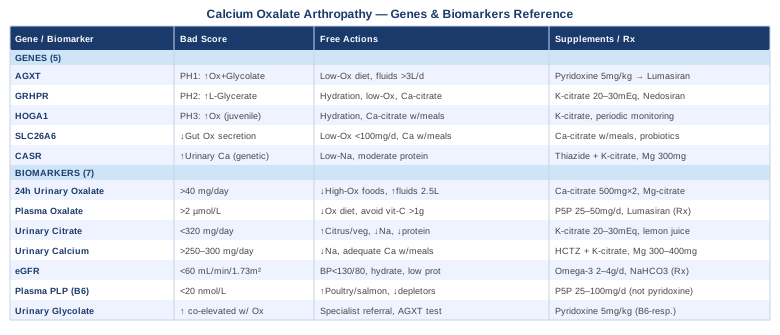
7 Biomarkers to Track
Biomarkers in this context are not just numbers on a lab report. Each one captures a different aspect of your oxalate biology: how much your body is producing, how efficiently it is being excreted, what molecular factors are either protecting or accelerating crystal nucleation, and how your kidneys are coping with the total load. The seven below were selected for clinical relevance, practical measurability, and their ability to directly guide intervention decisions.
Biomarker 1: 24-Hour Urinary Oxalate
Why it matters and what it reveals
The 24-hour urinary oxalate test is the most direct available window into your oxalate metabolism. It measures total daily oxalate excretion, reflecting both what your liver produces endogenously and what your gut absorbs from food. Normal excretion is generally accepted as below 40 mg per day. Values in the 40–80 mg per day range indicate mild to moderate hyperoxaluria; values above 80 mg per day suggest primary enzymatic causes or severe enteric hyperoxaluria and require further investigation. For crystal arthropathy, persistently elevated urinary oxalate signals that systemic oxalate concentration is high enough for deposition in synovial tissue, particularly where local pH, citrate levels, or clearance are suboptimal.
How to measure it
This requires a complete 24-hour urine collection with a preservative container provided by the lab, refrigerated throughout collection and submitted promptly. Most nephrologists, urologists, and metabolic specialists order this routinely; general practitioners can order it as well. Cost ranges from approximately $50 to $150 in the United States depending on the lab and insurance coverage. A comprehensive 24-hour urine metabolic panel — which includes oxalate alongside calcium, citrate, phosphate, sodium, and pH — provides the most diagnostic value per collection and costs $100–$200.
If the score is bad — the plan without supplements
Dietary modification is the first and most powerful lever. Reducing high-oxalate foods — cooked spinach, beets, almonds and almond products, chocolate, black tea, and rhubarb — can meaningfully lower urinary oxalate within two to four weeks in people where dietary absorption is a significant contributor. Targeting at least 2.5 liters of urine output per day dilutes oxalate concentration and reduces crystallization risk. Timing calcium intake across meals (rather than avoiding it) is one of the most underused non-supplement strategies: dietary calcium binds oxalate in the gut, reducing intestinal absorption. Aim for 1,000–1,200 mg of calcium from food per day distributed across all meals.
If the score is bad — the plan with supplements or equipment
Calcium citrate taken with meals (500 mg per dose, twice daily with food) is one of the most evidence-supported interventions for lowering urinary oxalate. It binds dietary oxalate in the intestinal lumen before it can be absorbed. Calcium citrate is preferred over calcium carbonate in this context because the citrate anion itself also inhibits calcium oxalate crystal formation. Magnesium citrate at 200–400 mg per day taken with meals reduces urinary oxalate independently by forming magnesium oxalate complexes that are excreted rather than absorbed. This can be used continuously with periodic serum magnesium monitoring. Probiotic support targeting oxalate-degrading gut bacteria is covered in the complementary approaches section and should be considered alongside dietary change when urinary oxalate remains elevated after four to six weeks of dietary modification.
Biomarker 2: Plasma Oxalate
Why it matters and what it reveals
While urinary oxalate captures what the kidneys are handling, plasma oxalate reflects the systemic concentration — what is actually circulating in the blood and therefore available to deposit in joints and soft tissues. In people with normal kidney function, plasma oxalate is kept very low (typically under 2 µmol/L) because the kidneys clear it efficiently. When kidney function is even mildly impaired, plasma levels can rise well above this threshold, creating conditions where crystal deposition in synovium, cartilage, and bone becomes substantially more likely. This is why calcium oxalate crystal arthropathy disproportionately affects people with chronic kidney disease and why plasma oxalate becomes the more informative marker once eGFR begins to decline.
How to measure it
Plasma oxalate measurement requires a specialized enzymatic or HPLC-based assay not included in standard metabolic panels — it must be specifically requested. Specialty labs including Mayo Clinic Laboratories and ARUP Laboratories offer this test. Cost ranges from $100 to $300. Plasma oxalate is most useful when urinary oxalate is already markedly elevated, when kidney function is meaningfully impaired (eGFR below 45), or when systemic tissue deposition is clinically suspected.
If the score is bad — the plan without supplements
The strategy closely mirrors the urinary oxalate approach: strict dietary oxalate reduction, sustained high fluid intake, and meals timed to include calcium-rich foods for in-gut binding. Avoiding high-dose vitamin C supplementation (above 1,000 mg per day) is particularly critical here — ascorbic acid is metabolized to oxalate in the body, and supplemental doses that are considered routine in some integrative medicine contexts can substantially elevate both plasma and urinary oxalate independent of diet. This is a non-obvious cause of elevated plasma oxalate that is frequently missed.
If the score is bad — the plan with supplements or equipment
Pyridoxal-5-phosphate (P5P) at 25–50 mg per day reduces hepatic oxalate production in individuals with AGXT variants that retain partial B6 responsiveness. For individuals with confirmed primary hyperoxaluria type 1 who have not responded adequately to pyridoxine, lumasiran (Oxlumo) — an FDA-approved siRNA therapeutic — directly suppresses the upstream enzyme glycolate oxidase in the liver, dramatically reducing hepatic oxalate output. This is a prescription medication requiring specialist management. For broader contexts, nephrologist referral at elevated plasma oxalate with reduced eGFR is the most important immediate step; monitoring plasma oxalate every three to six months while managing an elevated baseline is a practical surveillance cadence.
Biomarker 3: Urinary Citrate
Why it matters and what it reveals
Citrate is one of the body's most important natural crystal inhibitors. It complexes with calcium ions in both urine and synovial fluid, reducing the free calcium available to bind oxalate and nucleate crystals. Low urinary citrate — hypocitraturia — is found in a significant proportion of people with calcium oxalate kidney stones and likely plays a parallel protective role in joint crystal biology. Normal values are approximately 550 mg per day or higher in women and 450 mg per day or higher in men; values below 320 mg per day are clearly associated with elevated crystallization risk. Hypocitraturia can result from renal tubular acidosis, a high animal protein diet, chronic diarrhea, chronic metabolic acidosis, or potassium depletion — identifying the cause guides the specific fix.
How to measure it
Urinary citrate is routinely included in comprehensive 24-hour urine metabolic panels and represents an excellent cost-per-insight addition to any oxalate workup. It is rarely ordered alone; pairing it with calcium, oxalate, and pH in a single collection gives the most complete picture of crystal risk. A comprehensive panel typically costs $100–$200.
If the score is bad — the plan without supplements
Increasing dietary potassium through vegetables and citrus fruits raises urinary citrate through renal tubular mechanisms. Reducing animal protein intake lowers the metabolic acid load on the kidney, which directly increases citrate reabsorption. Reducing sodium is also effective — dietary sodium competes with citrate reabsorption at the proximal tubule, and each unit drop in sodium intake tends to raise urinary citrate meaningfully. A Mediterranean-style dietary pattern with high vegetable intake and moderate animal protein consistently improves urinary citrate in stone-forming populations across multiple observational studies.
If the score is bad — the plan with supplements or equipment
Potassium citrate (20–30 mEq per day, split into two or three doses) is the evidence-based standard for correcting hypocitraturia. It provides citrate directly while also alkalizing urine, creating a dual effect on crystal inhibition. Available by prescription at full therapeutic doses and as an over-the-counter supplement at lower doses. Lemon juice concentrate (approximately 120 mL diluted in water daily) modestly raises urinary citrate and serves as a low-cost dietary alternative, though the effect size is smaller than with potassium citrate supplementation. Cycling is generally not required; monitoring via repeat 24-hour urine collection at three-month intervals after initiating treatment provides good feedback on response.
Biomarker 4: Urinary Calcium
Why it matters and what it reveals
Calcium is one of the two essential components of calcium oxalate crystals, and elevated urinary calcium — hypercalciuria — multiplies rather than adds to the risk created by elevated oxalate. Even moderately elevated oxalate in the context of hypercalciuria can produce a supersaturated environment where crystal nucleation becomes likely in both renal and synovial tissue. Normal urinary calcium excretion is typically below 250 mg per day in women and below 300 mg per day in men. Hypercalciuria has three main types: absorptive (excess intestinal calcium uptake, often related to elevated vitamin D activity), resorptive (from elevated parathyroid hormone or accelerated bone turnover), and renal (from impaired tubular calcium reabsorption). Identifying the type is important because it changes which interventions are appropriate and which ones might backfire.
How to measure it
Urinary calcium is included in any standard 24-hour urine metabolic panel, making it straightforward and inexpensive to measure alongside oxalate and citrate. A spot urine calcium-to-creatinine ratio can screen for hypercalciuria but is less reliable than a 24-hour collection. Total panel cost is $100–$200.
If the score is bad — the plan without supplements
Dietary sodium reduction is one of the most effective and underused interventions for hypercalciuria. For each 100 mmol of sodium excreted in urine, the kidney pulls approximately 40 mg of calcium along with it; bringing sodium intake from 4,000 mg to 2,000 mg per day typically reduces urinary calcium by 80–100 mg per day in isolation. Limiting animal protein reduces the acidic metabolic load that drives calcium out of bone. Importantly, very low calcium diets — sometimes wrongly adopted in response to hypercalciuria — worsen crystal risk by increasing intestinal oxalate absorption. Adequate dietary calcium (distributed across meals) is protective, not harmful.
If the score is bad — the plan with supplements or equipment
Hydrochlorothiazide or chlorthalidone (prescription diuretics) are the pharmacological first line for absorptive hypercalciuria, increasing tubular calcium reabsorption and lowering urinary calcium by 30–50%. For individuals not yet requiring prescription diuretics, magnesium citrate or glycinate at 300–400 mg per day can modestly reduce urinary calcium through parathyroid hormone-mediated mechanisms. Thiazide therapy should be paired with potassium citrate to prevent hypokalemia-induced hypocitraturia — a common clinical oversight. For resorptive hypercalciuria driven by secondary hyperparathyroidism, vitamin D optimization is sometimes needed, but must be monitored carefully as excessive vitamin D can paradoxically worsen hypercalciuria by elevating intestinal calcium absorption.
Biomarker 5: eGFR — Estimated Glomerular Filtration Rate
Why it matters and what it reveals
The kidneys are the primary route of oxalate clearance from the body, and their functional capacity directly determines how much oxalate accumulates in plasma and tissues. When eGFR declines — even mildly, from 90+ mL/min/1.73m² to the 60–75 range — the kidney's oxalate clearance capacity falls, and plasma oxalate begins to rise. As plasma oxalate rises, tissue deposition accelerates. In primary hyperoxaluria, this dynamic is acute and life-altering. In secondary and idiopathic forms of oxalate crystal arthropathy, the same principle applies at a slower pace: declining kidney function is an amplifier of crystal deposition risk, not a separate problem. As Peter Attia has emphasized in the context of longevity medicine, eGFR trajectory over time matters as much as a single value — a consistent annual decline of 3–5 points demands investigation, even if the absolute level remains above the clinical threshold for chronic kidney disease.
How to measure it
eGFR is calculated from serum creatinine using the CKD-EPI equation (currently preferred over the older MDRD formula), adjusted for age and sex. It is included in every standard comprehensive metabolic panel and costs $20–$50 as a standalone test. Cystatin C-based eGFR provides more accuracy at borderline values, particularly in muscular individuals or those with very low muscle mass, and costs $50–$100.
If the score is bad — the plan without supplements
Protecting eGFR when it is already declining requires strict blood pressure control (target below 130/80 mmHg with evidence-based agents), sustained hydration, avoidance of nephrotoxic drugs including long-term NSAIDs, and aggressive oxalate reduction to limit the ongoing metabolic burden on the kidney. A moderate protein diet (0.8 g/kg body weight per day) consistently slows the progression of CKD across multiple etiologies in randomized trials. Reducing dietary sodium reduces glomerular hyperfiltration. These steps do not restore lost function, but they can meaningfully slow the rate of further decline.
If the score is bad — the plan with supplements or equipment
No supplement has been proven to restore lost eGFR. However, omega-3 fatty acids (EPA + DHA combined, 2–4 g per day) have modest supportive evidence for slowing CKD progression through anti-inflammatory and hemodynamic mechanisms. Sodium bicarbonate (prescribed, 0.5–1.0 mEq/kg/day in divided doses) slows CKD progression in patients with concurrent metabolic acidosis — a common finding in advanced CKD — and is underused in this population. Referral to nephrology is indicated at eGFR below 45 for general CKD, and earlier (below 60) when primary or enteric hyperoxaluria is suspected, since oxalate-specific interventions can preserve remaining kidney function more effectively the sooner they are initiated.
Biomarker 6: Plasma Pyridoxal-5-Phosphate (Active Vitamin B6)
Why it matters and what it reveals
This is likely the most underrecognized biomarker on this list, and one of the most actionable when it comes back low. Pyridoxal-5-phosphate (PLP) is the biologically active form of vitamin B6 and a required cofactor for the enzyme alanine-glyoxylate aminotransferase (AGXT) — the liver enzyme that converts glyoxylate into glycine rather than allowing it to be oxidized into oxalate. When PLP is insufficient, AGXT operates below its functional capacity, and hepatic oxalate production rises, entirely independently of dietary oxalate intake. This mechanism means that someone eating a nominally healthy diet can still be producing excess oxalate simply because their active B6 levels are low.
Gary Brecka, working in a functional longevity framework informed by genetic and metabolic assessment, has highlighted PLP as one of the most commonly deficient and most correctable metabolic factors in people with chronic inflammatory or pain conditions. Standard "vitamin B6" blood tests often measure total pyridoxine (the inactive precursor), which can be misleadingly normal even when PLP is functionally low — requesting the right test matters.
How to measure it
Request plasma pyridoxal-5-phosphate (PLP) specifically, not a generic "vitamin B6" panel. Available through Quest Diagnostics, LabCorp, and integrative medicine providers. Cost: $50–$150. The generally accepted normal range is 20–125 nmol/L; values below 20 nmol/L represent clear deficiency. Values in the 20–40 nmol/L range may be functionally suboptimal, particularly in individuals who carry partial-loss AGXT variants.
If the score is bad — the plan without supplements
Dietary sources of B6 include poultry, salmon, tuna, potatoes, non-citrus fruits, and fortified cereals. However, reaching therapeutic levels for AGXT function through food alone is difficult once deficiency is established, especially given that chronic inflammation, alcohol use, oral contraceptive use, high-sugar diets, and several common medications (isoniazid, hydralazine, certain anticonvulsants) all actively deplete B6. Addressing these drivers is the essential first step before considering supplementation.
If the score is bad — the plan with supplements or equipment
Supplement with pyridoxal-5-phosphate (P5P) at 25–100 mg per day — not regular pyridoxine. P5P does not require hepatic conversion and is directly usable by AGXT. Starting at 25 mg daily and titrating upward while monitoring plasma PLP every 8–12 weeks is a cautious and practical approach. Important safety note: high-dose pyridoxine (the inactive form) above 200 mg per day over extended periods has been linked to peripheral sensory neuropathy; P5P at 50–100 mg per day does not carry this risk at the same magnitude, but monitoring is still prudent. In patients with confirmed AGXT mutations who are testing B6 responsiveness, doses of up to 5 mg/kg per day under specialist supervision are used in clinical practice — this is a separate, medically supervised protocol distinct from general B6 optimization.
Biomarker 7: Urinary Glycolate
Why it matters and what it reveals
Glycolate is a metabolic sister molecule to oxalate, generated upstream in the same pathway. In Primary Hyperoxaluria Type 1 caused by AGXT mutations, both urinary oxalate and urinary glycolate are elevated simultaneously, because impaired AGXT allows glyoxylate to accumulate and be shunted toward both products. This pattern — co-elevation of glycolate and oxalate — distinguishes PH1 from dietary hyperoxaluria, from enteric hyperoxaluria, and from PH2, where L-glycerate rather than glycolate is the distinguishing metabolite. Testing urinary glycolate becomes particularly important when 24-hour urinary oxalate is markedly elevated above 80–100 mg per day with no adequate dietary or malabsorptive explanation.
How to measure it
Urinary glycolate is measured through quantitative urine organic acid analysis — a specialized test available at Mayo Clinic Laboratories, ARUP Laboratories, and academic nephrology centers with metabolic disease programs. It is typically run as part of a urine organic acid panel or ordered specifically in the context of suspected primary hyperoxaluria. Results are reported normalized to creatinine (µmol/mmol creatinine). Cost: $150–$400. The test is not part of routine primary care workups and generally requires a specialist referral.
If the score is bad — the plan without supplements
Co-elevation of urinary glycolate and oxalate is a clear signal for genetic testing of AGXT mutations before any supplement protocol is designed. Managing primary hyperoxaluria type 1 through diet alone is insufficient — the fundamental problem is endogenous hepatic overproduction, not primarily dietary absorption. The most important non-supplement step is establishing care with a nephrology center or metabolic specialist experienced in primary hyperoxaluria, ideally one affiliated with an oxalate-specific clinical program.
If the score is bad — the plan with supplements or equipment
For confirmed PH1 with a B6-responsive mutation (the p.Gly170Arg variant is the most common example), high-dose pyridoxine under medical supervision (testing protocol: 5 mg/kg/day for three months with pre- and post-measurement of 24-hour urinary oxalate) can reduce urinary oxalate by 30–50% in responsive patients. Non-responders are now candidates for lumasiran (Oxlumo) or nedosiran (Rivfloza) — FDA-approved RNA interference therapies that have transformed the prognosis of primary hyperoxaluria. These are specialist-managed prescription medications. For the broader context of elevated urinary glycolate without confirmed PH1, optimizing plasma PLP levels and reducing dietary oxalate while awaiting specialist evaluation is the appropriate interim protocol.
The Genetic Layer: 5 Key Genes That Shape Your Oxalate Risk
Genetic research in calcium oxalate crystal arthropathy is strongest for the rare, highly penetrant primary hyperoxaluria genes where the evidence is definitive. For more common, polygenic contributors to idiopathic hyperoxaluria, the evidence is earlier and the effects are probabilistic rather than deterministic. Knowing your genetic profile does not determine your fate — but it can guide proactive monitoring, earlier intervention, and more targeted conversations with your physician. This section profiles five genes where the evidence is meaningful and the clinical implications are actionable.
Gene 1: AGXT — The Master Oxalate Regulator
The AGXT gene encodes alanine-glyoxylate aminotransferase, a peroxisomal liver enzyme that sits at the critical decision point for glyoxylate metabolism. When AGXT functions normally, glyoxylate is converted to the harmless amino acid glycine. When AGXT is mutated, glyoxylate accumulates and is diverted into massive oxalate synthesis — the mechanism underlying Primary Hyperoxaluria Type 1, the most severe inherited oxalate disorder. The most common pathogenic variant, p.Gly170Arg, causes the AGXT protein to mislocalize from peroxisomes to mitochondria, where it cannot perform its function. This mislocalization is partially reversible with vitamin B6, making pyridoxine responsiveness testing a standard and meaningful step in PH1 management.
A detailed description of AGXT is available through the NCBI Gene database entry for AGXT. GeneReviews provides a comprehensive clinical summary of Primary Hyperoxaluria Type 1 including mutation spectrum and management.
If the gene variant is present — the plan without supplements
Strict dietary oxalate restriction (targeting below 50 mg per day), sustained high fluid intake targeting urine output above 3 liters per day, and complete avoidance of high-dose vitamin C are the pillars. Spreading calcium intake across meals consistently helps reduce what dietary oxalate does reach the gut. Establishing care with a nephrologist or metabolic specialist familiar with primary hyperoxaluria is essential — this is not a condition manageable through general practice alone given the kidney damage risk.
If the gene variant is present — the plan with supplements or equipment
Pyridoxine responsiveness testing is the first pharmacological step: 5 mg/kg/day of pyridoxine for three months under supervision, with 24-hour urinary oxalate measured before and after. Patients achieving ≥30% reduction in urinary oxalate are classified as B6-responsive and should continue long-term pyridoxine with periodic neurological monitoring. Non-responders and patients with progressive kidney impairment are now strong candidates for lumasiran (Oxlumo), an FDA-approved siRNA therapy targeting hepatic glycolate oxidase. Lumasiran has demonstrated sustained 50–65% reductions in urinary oxalate in clinical trials. Potassium citrate (20–30 mEq per day) should be co-administered to inhibit crystal formation in both urine and synovial tissue.
Gene 2: GRHPR — Primary Hyperoxaluria Type 2
GRHPR encodes glyoxylate reductase/hydroxypyruvate reductase, an enzyme that handles glyoxylate through a pathway distinct from AGXT. Mutations cause Primary Hyperoxaluria Type 2, which is generally less severe than PH1 in terms of kidney damage risk but still produces significant hyperoxaluria and stone burden. The key biochemical distinction from PH1 is that PH2 patients show elevated urinary L-glycerate rather than glycolate as the diagnostic metabolite — making metabolite profiling important for accurate differential diagnosis when genetic testing results are delayed.
Critically, PH2 is not vitamin B6-responsive, since the enzyme involved is GRHPR rather than AGXT. A patient with severe hyperoxaluria and absence of pyridoxine response should have GRHPR sequencing alongside AGXT as a standard step.
If the gene variant is present — the plan without supplements
High fluid intake, low-oxalate diet, calcium citrate with meals, and regular monitoring of kidney function and 24-hour urinary oxalate form the management foundation. Nephrology follow-up is important even when the clinical picture appears less severe than PH1, as stone burden can be substantial.
If the gene variant is present — the plan with supplements or equipment
Potassium citrate (20–30 mEq per day in divided doses) is the pharmacological cornerstone, providing urinary alkalization and direct crystal inhibition. Magnesium citrate (300–400 mg daily) offers additional oxalate-binding support. For severe cases with declining eGFR, nedosiran (Rivfloza) — an siRNA therapy targeting lactate dehydrogenase A, the enzyme responsible for oxalate overproduction in PH2 — has received FDA approval and represents the first disease-specific pharmacological option for this subtype. Specialist referral is required for nedosiran assessment.
Gene 3: HOGA1 — Primary Hyperoxaluria Type 3
HOGA1 encodes 4-hydroxy-2-oxoglutarate aldolase, a mitochondrial enzyme in the hydroxyproline catabolism pathway. Mutations cause Primary Hyperoxaluria Type 3, characterized by hyperoxaluria that often diminishes after adolescence and carries the mildest long-term prognosis of the three primary hyperoxaluria subtypes. That said, significant stone burden and joint crystal deposition can still occur during the hyperoxaluric phase, particularly in childhood and early adulthood.
There is no vitamin B6-responsive mechanism in PH3. The condition is identified through metabolic screening showing hyperoxaluria with normal glycolate and L-glycerate, alongside molecular confirmation of HOGA1 mutation.
If the gene variant is present — plan without and with supplements
Management mirrors the general hyperoxaluria approach: aggressive hydration, low-oxalate diet, calcium citrate with meals to reduce gut absorption, and potassium citrate for urinary alkalization and crystal inhibition. There is no disease-specific pharmacological intervention equivalent to the PH1 B6-pyridoxine or siRNA options. The natural history — with oxalate excretion often decreasing through adulthood — means lifestyle measures are frequently sufficient to prevent serious long-term complications when started early. Periodic monitoring of urinary oxalate and eGFR every six to twelve months is the appropriate ongoing surveillance protocol.
Gene 4: SLC26A6 — The Intestinal Oxalate Transporter
SLC26A6 encodes a chloride/oxalate transporter expressed heavily in the small intestinal epithelium where it normally secretes oxalate from blood back into the gut lumen, facilitating excretion through stool. Reduced-function variants of SLC26A6 impair this secretory mechanism — less oxalate exits through the gut, and more accumulates in plasma for the kidneys (and eventually joints) to handle. Research from rodent knockout models and emerging human genetic association data support the role of SLC26A6 variants in idiopathic calcium oxalate stone disease. This gene represents an important bridge between genetic predisposition and dietary context: people with SLC26A6 loss-of-function variants may tolerate moderate-oxalate foods poorly even without a classic primary hyperoxaluria diagnosis.
If the gene variant is present — the plan without supplements
The practical implication is that dietary oxalate management becomes more consequential than it would be for someone with fully functional intestinal oxalate secretion. A low-oxalate dietary target (under 100 mg per day initially, adjusting based on 24-hour urinary oxalate response) is the primary non-supplement strategy. Monitoring urinary oxalate response to specific dietary changes provides useful personalization data.
If the gene variant is present — the plan with supplements or equipment
Calcium citrate with all meals is particularly important here, as it compensates for the reduced secretory transport by binding gut-lumen oxalate before it can be absorbed. Probiotic support with Lactobacillus acidophilus, Bifidobacterium lactis, and ideally oxalate-degrading strains (see the microbiome section below) reduces the intestinal oxalate burden that SLC26A6 is failing to clear. Daily probiotic use with a reassessment of urinary oxalate at three-month intervals is a practical monitoring cadence. A 2015 randomized trial published in the Journal of Urology demonstrated that specific probiotic combinations reduced urinary oxalate excretion by approximately 20% in hyperoxaluric patients — a clinically relevant effect that is especially meaningful when intestinal secretion is also impaired.
Gene 5: CASR — The Calcium Sensing Receptor
The CASR gene encodes the calcium-sensing receptor expressed in the parathyroid glands, kidney tubules, and gut epithelium. Gain-of-function variants of CASR suppress parathyroid hormone secretion and reduce tubular calcium reabsorption, producing a form of hypercalciuria known as autosomal dominant hypocalcemia type 1. Even lower-penetrance CASR variants that do not cause frank hypocalcemia can modestly increase urinary calcium excretion in ways that tip the calcium oxalate supersaturation balance toward crystallization — particularly when oxalate levels are also elevated. Identifying a CASR variant contextualizes elevated urinary calcium and helps distinguish a genetic set-point shift from dietary, absorptive, or resorptive causes of hypercalciuria before reaching for any specific intervention.
If the gene variant is present — the plan without supplements
Sodium restriction and moderate animal protein reduction are the dietary levers, as both influence the calcium-to-creatinine excretion ratio downstream of the CASR set-point. Adequate — never low — dietary calcium remains protective. Because the CASR variant represents an intrinsic shift in the parathyroid and renal calcium handling axis, dietary modification has a ceiling effect and primarily manages rather than resolves the underlying tendency.
If the gene variant is present — the plan with supplements or equipment
Hydrochlorothiazide or indapamide (prescribed) lower urinary calcium through tubular mechanisms operating downstream of CASR and are effective in this context. For individuals not yet requiring diuretic therapy, magnesium glycinate or citrate at 300–400 mg per day offers modest calcium-lowering benefit. Thiazide therapy should always be combined with potassium supplementation or potassium citrate to prevent hypokalemia-driven hypocitraturia — a frequent clinical oversight that undermines the benefit of the diuretic. Genetic counseling is advisable for first-degree relatives, as CASR variants follow autosomal dominant inheritance patterns.
What Toxic Superfoods Gets Right About Oxalate That Most Doctors Don't Know
Sally Norton is a nutritional health researcher and the author of Toxic Superfoods (2023, Rodale Books), a book that synthesizes decades of biochemical literature on dietary oxalate alongside clinical observation of people with chronic pain, fatigue, and inflammatory conditions. Her central argument is that many foods widely promoted as nutrient-dense health foods — spinach, almonds, cashews, beets, chia seeds, dark chocolate, and sweet potatoes — carry oxalate loads high enough to cause genuine harm in a significant subset of the population, and that the medical community's failure to connect these dietary exposures to specific symptoms has left many people without a diagnosis that addresses a modifiable cause. The following ten insights from her work are particularly relevant to calcium oxalate crystal arthropathy.
1. The Spinach Paradox
One cup of cooked spinach delivers over 750 mg of oxalate — a single serving that can exceed the entire low-oxalate daily target by more than tenfold. Norton documents that the calcium in spinach is almost entirely bound to oxalate and largely unavailable for absorption, meaning the calcium nutritional value commonly cited for spinach is largely illusory. The health halo around spinach has driven many well-intentioned people to substantially increase their oxalate burden while believing they are eating optimally.
2. Oxalate Dumping: The Withdrawal Effect Nobody Warns About
Norton describes a phenomenon she terms "oxalate dumping" — when people suddenly eliminate high-oxalate foods after a period of high intake, the body begins mobilizing stored tissue oxalate, causing a temporary intensification of symptoms including joint pain, fatigue, skin irritation, and brain fog. People who try a low-oxalate diet for two to four weeks, experience worsening symptoms, and conclude the diet is not working are often misreading a transition phase. A gradual, staged dietary reduction over four to twelve weeks — reducing by roughly 10% of oxalate intake per week — is significantly more tolerable.
3. Why Individual Variation Is So Wide
Norton synthesizes research showing that the proportion of dietary oxalate that reaches the bloodstream varies from less than 5% in people with healthy gut microbiomes and adequate Oxalobacter formigenes colonization to over 50% in people with gut dysbiosis, inflammatory bowel conditions, or leaky gut. This variance explains why population-level nutrition research on high-oxalate foods shows mixed results: the same food is essentially benign for one person and genuinely harmful for another, depending entirely on gut health.
4. Silent Tissue Deposition Before Symptoms Appear
Norton draws on autopsy and biopsy literature demonstrating calcium oxalate crystal deposits in joints, tendons, nerve tissue, bone, thyroid, and vessel walls in people who consumed high-oxalate diets for many years without specific symptoms. By the time pain or functional impairment is clinically evident, tissue deposition has typically been occurring silently for years. This has important implications for the timeline of recovery: early dietary change can prevent accumulation; later change must also clear existing deposits, which takes considerably longer.
5. The Almond Flour Problem in Clean Eating
Almonds contain approximately 460 mg of oxalate per cup. Their transformation into almond flour — a staple in paleo, keto, and grain-free cooking — has substantially elevated dietary oxalate intake in populations that believe they are eating especially carefully. A single batch of almond-flour muffins or pancakes can deliver more oxalate than a full day's recommended limit. Norton argues this is an underrecognized consequence of otherwise well-motivated dietary movements.
6. High-Dose Vitamin C Raises Endogenous Oxalate
Ascorbic acid is metabolized to oxalate in the body, and doses above 1,000 mg per day can meaningfully elevate urinary oxalate independent of dietary intake. Norton cites studies showing that 2–4 g of supplemental vitamin C per day — doses common in integrative medicine, anti-aging, and immune optimization contexts — can raise urinary oxalate by 20–30 mg per day in susceptible individuals. For people with genetic variants affecting oxalate metabolism, this increment can be the difference between below-threshold and above-threshold crystal deposition risk.
7. Mineral Wasting Is a Secondary Consequence
Oxalate binds tightly to calcium, magnesium, zinc, and iron in the gastrointestinal tract, reducing their bioavailability. Norton argues that people eating high-oxalate diets can show lab evidence of mineral deficiencies not because they are eating too little of those minerals, but because co-ingested oxalate is binding them before absorption. This secondary mineral depletion can itself drive inflammation and musculoskeletal symptoms, creating a feedback loop that is difficult to untangle without recognizing the root cause.
8. Low-Oxalate Swaps That Preserve Nutritional Quality
Norton provides a practical substitution framework: romaine and arugula replace spinach; macadamia nuts and hemp seeds replace almonds; white potatoes and cauliflower replace sweet potatoes; cacao-free carob and low-oxalate berries (blueberries, raspberries) replace chocolate. This is not a deprivation strategy — it is a lateral move designed to maintain nutrient density without the oxalate cost. She argues that nutritional completeness can be fully maintained on a low-oxalate diet with thoughtful substitution.
9. The Detox Timeline Is Longer Than Clinicians Expect
Based on the tissue half-life of oxalate deposits and clinical observation of recovery trajectories, Norton estimates that meaningful oxalate dumping and tissue clearance can take six months to two years of sustained dietary change. This long timeline is biologically plausible given how slowly crystal deposits mobilize from joint and soft tissue spaces. Expecting improvement within weeks is unrealistic; expecting gradual, non-linear progress over many months aligned with the biology is the correct frame.
10. The Medical Blind Spot — Why Clinicians Miss It
Despite extensive peer-reviewed literature on primary hyperoxaluria, calcium oxalate nephrolithiasis, and renal oxalosis, Norton documents that dietary oxalate is rarely considered as a contributing cause in chronic joint pain, fibromyalgia, or unexplained inflammatory arthritis outside specialist nephrology centers. She attributes this partly to the absence of a pharmaceutical solution for dietary hyperoxaluria (nothing to prescribe means less institutional attention), and partly to the minimal coverage of nutritional biochemistry in standard medical training. Her most actionable conclusion: patients with persistent, unexplained joint pain should specifically ask their physician about a low-oxalate trial and 24-hour urinary oxalate testing before accepting a diagnosis of purely idiopathic arthropathy.
Complementary Approaches with Clinical Support
The following three approaches were selected from the approved list based on their relevance to calcium oxalate crystal arthropathy and the quality of their available clinical evidence. They are not replacements for the biomarker monitoring and dietary strategies described above; they address different dimensions — gut ecology, tissue inflammation, and pain experience — that mainstream management often underserves.
Microbiome-Directed Therapies
Oxalobacter formigenes is an anaerobic intestinal bacterium that uses oxalate as its sole carbon and energy source, thereby degrading substantial amounts of dietary oxalate within the gut before it can be absorbed. People who lack this organism — estimated to be between 40% and 70% of Western adult populations due to antibiotic use and low-fiber diets — absorb substantially more dietary oxalate than those who harbor it. The absence of O. formigenes has been associated with elevated urinary oxalate and increased calcium oxalate stone risk in multiple observational studies, making microbiome status a modifiable variable in oxalate crystal arthropathy management.
A prospective study published in Journal of the American Society of Nephrology found that stone-forming patients lacking O. formigenes had nearly 70% higher odds of recurrent calcium oxalate stones compared to colonized individuals, even after adjustment for dietary oxalate. While direct colonization with live O. formigenes has not yet reached clinical availability, Lactobacillus acidophilus, Bifidobacterium lactis, and certain Lactobacillus gasseri strains possess partial oxalate-degrading capacity and have reduced urinary oxalate by 15–25% in small randomized trials.
Practically, supporting the microbiome for oxalate management involves daily probiotic supplementation with strains documented to have oxalate-degrading activity, combined with prebiotic fiber to sustain colonization. Fermented foods — kefir, sauerkraut, kimchi (ensure these are low in oxalate) — support microbial diversity more broadly. Results are not immediate; microbiome shifts require eight to twelve weeks of consistent supplementation, and benefit should be reassessed through 24-hour urinary oxalate measurement at three months. There are no significant side effects at standard probiotic doses; the main risk is investing time in the intervention without tracking whether urinary oxalate actually responds.
Low-Level Laser Therapy / Photobiomodulation
Low-level laser therapy (LLLT), also called photobiomodulation, applies specific wavelengths of near-infrared and red light (typically 600–1000 nm) to affected tissues at non-thermal intensities. The proposed mechanism involves photon absorption by mitochondrial cytochrome c oxidase, triggering anti-inflammatory signaling cascades, reducing oxidative stress, and promoting tissue repair. For crystal arthropathy specifically, LLLT is not expected to dissolve existing crystal deposits, but it can reduce the synovial inflammation surrounding crystal deposits and modulate the pain response — addressing the suffering component of the condition even when crystals remain present.
A meta-analysis of 22 randomized controlled trials examining LLLT for rheumatic and musculoskeletal joint conditions, published in BMC Musculoskeletal Disorders, found significant reductions in pain scores and improvements in range of motion across multiple joint locations. Effect sizes were moderate but consistent, and no serious adverse events were reported. Studies specifically in crystal arthropathy are sparse; the evidence base is strongest for osteoarthritis and inflammatory arthropathies as proxies.
For practical application to calcium oxalate crystal arthropathy, the most evidence-supported protocol involves daily or every-other-day treatment sessions of 60–120 seconds per affected joint site using a device delivering 100–500 mW at wavelengths between 780 and 860 nm. At-home near-infrared devices in the appropriate power and wavelength range are now commercially available and have been used in home protocols studied in clinical research settings. Sessions can be maintained continuously during active inflammation and reduced to two to three times per week during remission. Contraindications include direct treatment over active malignancy, photosensitizing medications, and the eyes; consultation with a physical therapist familiar with photobiomodulation can guide appropriate positioning for specific joint locations.
Mindfulness Meditation / MBSR
Mindfulness-Based Stress Reduction (MBSR), the structured eight-week program developed by Jon Kabat-Zinn, combines mindfulness meditation, body scan practices, and gentle yoga with psychoeducation about the pain-stress relationship. For crystal arthropathy patients, whose condition is often chronic, unpredictable in symptom severity, and poorly understood by their social and medical environment, MBSR addresses a dimension — the suffering, reactivity, and central sensitization associated with persistent pain — that biomarker tracking and dietary change cannot directly reach. Chronic pain conditions consistently show that the central nervous system's processing of nociceptive signals is a critical variable in pain experience, independent of local tissue damage.
Multiple randomized controlled trials have demonstrated that MBSR reduces chronic pain intensity, pain-related catastrophizing, depression comorbidity, and health-related quality of life impairment across diverse chronic pain populations. A systematic review and meta-analysis published in JAMA Internal Medicine (2014, Goyal et al.) covering 47 trials with over 3,500 participants found moderate evidence for improvements in pain, anxiety, and depression using mindfulness programs. While no trials have specifically enrolled calcium oxalate crystal arthropathy populations, the mechanisms — reduced central sensitization, improved pain tolerance, lower inflammatory stress hormone output — are generalizable.
The standard MBSR protocol is an eight-week group or online program, meeting once per week for two to two-and-a-half hours with daily home practice of 30–45 minutes. Evidence supports that effects accumulate with regular practice; benefit is detectable at two months and increases through six months of sustained engagement. Online MBSR programs (including those modeled on the UMass Medical School curriculum) make this approach accessible without geographic constraints. The only meaningful contraindication is untreated severe psychiatric conditions where intensive introspective practice requires professional supervision; for most arthropathy patients, the risk profile of MBSR is effectively zero.
Conclusion
Calcium oxalate crystal arthropathy sits at the intersection of genetics, metabolism, diet, and kidney physiology — a genuinely multi-variable problem that does not yield to single-factor solutions. What this article has tried to provide is a coherent map of those variables: seven measurable biomarkers that give you a real-time picture of your oxalate burden at different points in the metabolic chain, five genes whose variants quietly shift that picture upstream, and a set of complementary tools that address the dimensions those biomarkers cannot fully reach.
The most important next step is straightforward: if you have not yet had a comprehensive 24-hour urine metabolic panel, request one. It is inexpensive, requires no special preparation, and gives you more actionable information about your specific crystal risk than most clinical conversations about joint pain typically surface. Pair that with a plasma PLP test, and you have already covered two of the most frequently abnormal and most modifiable variables in this condition. From there, the genetic layer adds context, and the dietary and lifestyle strategies add levers. Bring your results to a physician — ideally one with a background in nephrology or metabolic medicine — and use the frameworks in this article to inform what to ask.
Digestive Endocrine & Metabolic
Musculoskeletal: Joint Conditions
Autoimmune: Inflammatory Conditions
Urological: Kidney Conditions