This article contains AI generated content.
· UpdatedCalcium Pyrophosphate Deposition Disease: 4 Genes and 7 Biomarkers to Track
Introduction
Calcium pyrophosphate deposition disease — often called CPPD or pseudogout — tends to get lumped in with other forms of arthritis, treated symptomatically, and left largely unexplained. If you have lived through a sudden, agonizing joint flare that looked like gout but tested negative for urate crystals, or if chondrocalcinosis showed up on imaging without anyone explaining what actually caused it, you likely know the frustration of being told to manage inflammation without anyone asking why the crystals formed in the first place.
CPPD is not a random event. It is the end result of a metabolic imbalance — too much inorganic pyrophosphate accumulating in the joint space, seeding crystals that the immune system then attacks. That imbalance has upstream causes: a gene that transports pyrophosphate too aggressively, an iron overload that disables the enzyme responsible for clearing it, a parathyroid gland working overtime, or a magnesium level so low that normal crystal inhibition breaks down. Standard rheumatology visits rarely explore all of these levers systematically.
Generic advice — rest, NSAIDs, colchicine — addresses the flare, not the root. The underlying condition continues, crystals keep accumulating, and flares become more frequent or more severe over time. A more useful approach is to map the metabolic and genetic terrain that is driving your specific case, then target the factors that are actually modifiable.
That is what this article does. The first section covers seven biomarkers worth tracking — each one revealing a different potential driver of crystal formation — with practical guidance on how to measure them, what to do if they are off, and which interventions have real evidence behind them. A shorter genetics section follows, covering four genes with meaningful clinical links to CPPD. Together, they offer a more precise picture than a symptom diary alone, and a more actionable one than waiting for the next flare.
7 Biomarkers That Reveal What Is Driving Your CPPD
Most CPPD cases are not genetic in origin. They are driven by secondary metabolic disorders that are both detectable and often treatable. The biomarkers below represent the most diagnostically rich and actionable layer of investigation available. Not every one will be relevant to every patient, but running the full panel at least once is the most efficient way to identify which drivers are active in your case.
1. Ionized Serum Calcium
Calcium is the co-ingredient in calcium pyrophosphate crystals. When extracellular calcium concentrations rise, even modestly, crystal nucleation becomes more likely — particularly in cartilage already primed by elevated pyrophosphate levels. Hypercalcemia is one of the most well-documented secondary causes of CPPD, and it is almost always traceable to a fixable upstream issue: primary hyperparathyroidism, excessive calcium supplementation, vitamin D toxicity, or less commonly malignancy. Identifying it early matters because the intervention can be highly effective at reducing recurrence.
How to measure it
Request ionized calcium specifically, not just total calcium. Total calcium is influenced by albumin levels and can mask true hypercalcemia in patients with low protein. Ionized calcium is a direct measure of the biologically active fraction. Cost: $20–$50 at most labs. Optimal range: 4.65–5.20 mg/dL. Anything above 5.3 mg/dL warrants further workup. Many standard metabolic panels include total calcium; push for the ionized fraction if CPPD is in the picture.
If the score is bad, the plan without supplements
The first step is ruling out a parathyroid adenoma — a small benign tumor on the parathyroid glands that drives excess PTH and calcium. This requires a simultaneous PTH measurement (see below) and, if elevated, imaging (sestamibi scan or neck ultrasound). Discontinue any calcium supplements and calcium-fortified foods until levels normalize. Increase fluid intake to 2–3 liters per day, which increases renal calcium clearance. If you are on thiazide diuretics, discuss switching with your physician — thiazides raise serum calcium. Remove vitamin D supplements temporarily if high-dose use is suspected.
If the score is bad, the plan with supplements or equipment
Vitamin K2 (MK-7 form, 100–200 mcg/day) is the most evidence-informed supplement for redirecting calcium away from soft tissues and into bone matrix. It activates osteocalcin and matrix Gla protein, both of which govern calcium routing. Take daily, with a fat-containing meal (K2 is fat-soluble). No cycling needed at standard doses. Side effects are minimal; avoid if on warfarin therapy without physician coordination. Magnesium glycinate or malate at 300–400 mg/day complements K2 by lowering serum calcium modestly and directly inhibiting crystal nucleation. If hypercalcemia is severe (>5.5 mg/dL ionized) or primary hyperparathyroidism is confirmed, surgical evaluation for parathyroidectomy is medically indicated and should not be replaced by supplements alone.
2. RBC Magnesium
Magnesium is probably the most underappreciated factor in CPPD. It serves as a natural crystal inhibitor: adequate extracellular magnesium suppresses calcium pyrophosphate nucleation in cartilage matrix. Clinical research has documented low magnesium as an independent risk factor for CPPD, distinct from other metabolic causes. The relationship is biologically direct — magnesium competes with calcium for crystal lattice sites and activates pyrophosphatase enzymes that clear extracellular pyrophosphate. Many patients with recurrent CPPD are chronically hypomagnesemic without knowing it, largely because standard serum magnesium tests are poor at detecting tissue-level deficiency.
How to measure it
Do not rely on serum magnesium alone. Less than 1% of total body magnesium is in the blood; serum levels stay normal until deficiency is severe. Request RBC (red blood cell) magnesium, which reflects intracellular stores much more accurately. Cost: $40–$90, often requires ordering separately. Optimal RBC magnesium: 5.5–6.5 mg/dL. Anything below 5.2 mg/dL is functionally low even if serum magnesium appears normal. Some integrative medicine labs offer this as part of a full mineral panel.
If the score is bad, the plan without supplements
Dietary magnesium is the most sustainable approach. The richest sources are pumpkin seeds (~150 mg per ounce), dark leafy greens (spinach, Swiss chard), dark chocolate (70%+), almonds, and legumes. Aim for 400–500 mg daily from food. Simultaneously address magnesium wasters: alcohol significantly depletes magnesium, as do proton pump inhibitors (PPIs), loop and thiazide diuretics, and high-dose zinc. Reducing or eliminating any of these can raise RBC magnesium substantially without supplementation. Coffee is a mild magnesium waster; replacing one daily cup with mineral water high in magnesium (Gerolsteiner or similar) can make a measurable difference.
If the score is bad, the plan with supplements or equipment
Magnesium glycinate or malate (300–600 mg/day) is well-tolerated and absorbed. Glycinate is preferable for those with digestive sensitivity; malate is useful when fatigue is also present. Take in the evening — magnesium supports deeper sleep, which in turn reduces cortisol-driven mineral loss. Daily use long-term is appropriate once deficiency is confirmed; no cycling is required. The main side effect is loose stools, which occurs more with magnesium oxide (avoid this form) and less with glycinate or malate. Transdermal magnesium chloride flakes in a warm bath (20 minutes, 3–4 times per week) provides an alternative absorption route with almost no GI side effects. Evidence for transdermal absorption is debated but reported as beneficial by many practitioners treating arthritis.
3. Parathyroid Hormone (PTH)
Primary hyperparathyroidism (PHPT) is one of the three most common secondary causes of CPPD, alongside hemochromatosis and hypothyroidism. An overactive parathyroid gland elevates PTH, raises serum calcium, and may independently stimulate chondrocyte pyrophosphate production. CPPD associated with PHPT tends to be more severe and more diffuse, often affecting multiple joints. Critically, treating PHPT — most often through surgical removal of the adenoma — can dramatically reduce CPPD flare frequency. This makes PTH one of the highest-value biomarkers in a CPPD workup.
How to measure it
Intact PTH (also called iPTH) is the standard clinical test. Cost: $30–$60. Normal range: 15–65 pg/mL. It should always be interpreted alongside simultaneous serum calcium — the combination of high PTH and high calcium is the diagnostic signature of PHPT. Low PTH with high calcium suggests a different cause (malignancy, vitamin D toxicity). Secondary hyperparathyroidism (high PTH, normal or low calcium) is caused by vitamin D deficiency and is a different clinical situation requiring vitamin D repletion rather than surgery.
If the score is bad, the plan without supplements
If PTH is elevated and calcium is also high, parathyroid imaging is the immediate next step — this is not manageable through diet or lifestyle alone. A sestamibi scan with SPECT or a 4D CT scan of the neck will identify an adenoma in most cases. Surgical parathyroidectomy by an experienced surgeon is curative in over 95% of cases and is the most effective intervention for CPPD secondary to PHPT. If PTH is elevated but calcium is normal or low (suggesting secondary hyperparathyroidism from vitamin D deficiency), increasing sun exposure and correcting vitamin D status will lower PTH. Also avoid excessive caffeine and ensure adequate dietary calcium.
If the score is bad, the plan with supplements or equipment
For secondary hyperparathyroidism (vitamin D deficiency pattern): Vitamin D3 at 3000–5000 IU/day combined with K2 MK-7 (100–200 mcg/day) is appropriate; retest after 8–12 weeks. Side effects at these doses are minimal if calcium and kidney function are normal. Do not take vitamin D without K2 when PTH or calcium is being managed — K2 ensures that any calcium mobilized by D3 is directed to bone. For primary hyperparathyroidism, supplements do not replace surgery. Cinacalcet (a prescription calcimimetic) is a medical option for patients not eligible for surgery; discuss with an endocrinologist.
4. Ferritin and Transferrin Saturation
Iron overload is the second major secondary cause of CPPD. In hereditary hemochromatosis — caused by mutations in the HFE gene — excess iron deposits throughout the body, including joint cartilage. Iron acts as a pyrophosphatase inhibitor: it directly blocks the enzyme responsible for clearing extracellular pyrophosphate, allowing PPi to accumulate and crystallize. The knee, wrist (especially the second and third MCP joints), and hip are classic targets. Iron-associated CPPD often presents earlier in life than typical CPPD and tends to be accompanied by fatigue, elevated liver enzymes, and skin bronzing.
How to measure it
Ferritin alone is not enough. Request a full iron panel: serum ferritin + transferrin saturation (TSAT) + TIBC. Cost: $30–$70. Concerning thresholds: ferritin above 300 ng/mL in men or 200 ng/mL in women; transferrin saturation above 45%. If both are elevated, HFE genetic testing (C282Y and H63D variants) is warranted. Ferritin is also an acute-phase reactant — it rises with inflammation — so mildly elevated ferritin in the context of a recent flare does not automatically mean iron overload. Transferrin saturation is more specific for genuine iron excess.
If the score is bad, the plan without supplements
Therapeutic phlebotomy — regular blood donation or medically supervised blood removal — is the primary treatment for iron overload and is surprisingly effective. Each unit removed lowers ferritin by approximately 30–50 ng/mL. Frequency depends on ferritin level but typically begins at every 2–4 weeks, tapering to 2–4 times per year once ferritin reaches the target (50–100 ng/mL for HFE hemochromatosis). Simultaneously: eliminate iron supplements, avoid vitamin C with iron-rich meals (vitamin C enhances iron absorption), reduce red meat consumption, and avoid alcohol (which amplifies hepatic iron uptake). Cast-iron cookware adds meaningful dietary iron and should be replaced.
If the score is bad, the plan with supplements or equipment
IP-6 (inositol hexaphosphate, 1–2 g/day with meals) binds dietary iron in the gut and reduces absorption — a useful adjunct to phlebotomy, not a replacement. It also has mild anti-inflammatory properties. Cycle: 8 weeks on, 4 weeks off; avoid concurrent use with mineral supplements (calcium, zinc, magnesium) as it chelates these as well. Curcumin (500–1000 mg with piperine, twice daily) has mild iron-chelating properties and reduces joint inflammation via NF-κB suppression. Take with food; cycle 8–12 weeks with 4-week breaks; avoid high doses in people with gallstones. Note: formal iron chelation therapy (deferasirox, deferoxamine) is a prescription medical intervention for severe overload.
5. TSH and Free T4
Hypothyroidism is the third major secondary cause of CPPD and is frequently underdiagnosed. Thyroid hormones regulate the activity of alkaline phosphatase and pyrophosphatase enzymes in cartilage. When thyroid function is low, these enzymes slow down, extracellular pyrophosphate is not cleared efficiently, and crystal deposition accelerates. Studies have consistently found a higher prevalence of CPPD — particularly chondrocalcinosis on imaging — among people with overt or subclinical hypothyroidism. Treating underlying hypothyroidism has been associated with reduced crystal burden in some cases.
How to measure it
Request TSH (thyroid-stimulating hormone) plus Free T4 (not just total T4). Cost: $30–$80 depending on panel. Functional optimal range: TSH 1.0–2.5 mIU/L; Free T4 1.2–1.8 ng/dL. Standard lab ranges are broader than functional thresholds — a TSH of 3.8 may fall within "normal" but could still represent sub-optimal thyroid activity in a symptomatic patient. If symptoms include fatigue, constipation, cold intolerance, or brain fog alongside joint issues, also request Free T3 and thyroid antibodies (anti-TPO, anti-TG) to rule out Hashimoto's thyroiditis.
If the score is bad, the plan without supplements
Sleep quality has a direct impact on thyroid hormone production — chronic sleep deprivation suppresses TSH and impairs T3 conversion. Prioritize 7–9 hours. Reduce chronic stress: elevated cortisol inhibits the conversion of T4 to active T3 in peripheral tissues. Selenium-rich foods (2–3 Brazil nuts per day, not more) support the deiodinase enzymes responsible for T4-to-T3 conversion. Ensure adequate iodine through food (seaweed, seafood, iodized salt) without supplementing aggressively unless deficiency is confirmed. If Hashimoto's is present, trial of a gluten-free diet has clinical support in reducing antibodies in a subgroup of patients.
If the score is bad, the plan with supplements or equipment
Selenium 100–200 mcg/day (L-selenomethionine form) is the most evidence-backed supplement for thyroid enzyme support. Cycle: 5 days on, 2 days off, or 8 weeks on, 4 weeks off — selenium accumulates at high doses and chronic excess is toxic. Zinc 15–25 mg/day supports thyroid receptor function; take with food to avoid nausea; cycle similarly to selenium. If labs confirm clinical hypothyroidism, the appropriate treatment is levothyroxine (prescription) — supplements alone will not resolve true hypothyroidism. Some patients do better on combination T4/T3 therapy (liothyronine) or desiccated thyroid extract; discuss with an endocrinologist. Treating the thyroid often reduces CPPD flare frequency within 6–12 months.
6. Alkaline Phosphatase (ALP)
Most CPPD discussions focus on causes of too much pyrophosphate. But there is a genetic condition — hypophosphatasia — where the problem is inadequate clearance due to extremely low alkaline phosphatase activity. ALP in bone and cartilage normally hydrolyzes extracellular pyrophosphate; when it is genetically deficient, PPi accumulates massively, causing severe CPPD often presenting in early adulthood. Meanwhile, elevated ALP points to liver disease, Paget's disease, or malignancy — all of which can produce secondary joint and metabolic effects. ALP is therefore a two-directional signal, and its interpretation depends on context.
How to measure it
ALP is part of a standard comprehensive metabolic panel (CMP), making it one of the cheapest biomarkers to obtain — often free within a routine blood draw. Normal adult range: 44–147 IU/L, though labs vary. ALP below 40 IU/L with recurrent CPPD strongly suggests hypophosphatasia and should prompt ALPL gene testing. ALP above 200 IU/L without a known cause requires investigation for liver disease (check GGT, ALT, AST) or bone-specific conditions. Fractionated ALP testing can distinguish bone vs. liver origin if the source is unclear.
If the score is bad, the plan without supplements
If ALP is very low (potential hypophosphatasia): avoid high-dose vitamin B6 supplementation, which can paradoxically accumulate to toxic levels in hypophosphatasia due to impaired degradation. Reduce alcohol, which suppresses ALP activity. Consult a metabolic bone disease specialist — hypophosphatasia has disease-specific treatments (asfotase alfa, an enzyme replacement therapy). If ALP is elevated: investigate and address the underlying condition directly. Reducing alcohol (major cause of elevated ALP through liver stress) should be an immediate first step.
If the score is bad, the plan with supplements or equipment
For low ALP: Zinc 15–25 mg/day and vitamin B6 (pyridoxine, 25–50 mg/day) are cofactors for ALP activity. In hypophosphatasia, B6 should be used with caution (start low, monitor B6 plasma levels). For elevated ALP from metabolic causes, milk thistle (silymarin, 140–420 mg/day standardized extract) has evidence supporting hepatic ALP normalization in non-alcoholic liver disease contexts — cycling: 8–12 weeks with 4-week breaks. Berberine (400–500 mg, 3×/day) is relevant if metabolic syndrome accompanies elevated ALP — it has demonstrated ALP-lowering effects in NAFLD trials. Cycling: 8–12 week cycles with 2–4 week breaks; side effects include GI discomfort and mild blood glucose lowering.
7. Serum Phosphate
Phosphate and pyrophosphate share metabolic pathways, and disruptions in phosphate homeostasis ripple into pyrophosphate metabolism. Chronic hypophosphatemia — low serum phosphate — is associated with increased risk of crystal-related joint disease, partly because low phosphate alters the phosphate-to-pyrophosphate ratio in the extracellular matrix. Antacid overuse (aluminum-containing antacids bind phosphate in the gut), malabsorption, and poor dietary intake are common and underappreciated causes of low phosphate in the general population. Hyperphosphatemia, usually in the context of chronic kidney disease, creates a different metabolic risk profile affecting mineral crystal deposition throughout the body.
How to measure it
Serum inorganic phosphate is part of most basic metabolic panels. Cost: included in a standard BMP or CMP, $15–$40 standalone. Normal range: 2.5–4.5 mg/dL. Optimal: 3.0–4.0 mg/dL. Phosphate is best drawn fasting in the morning — levels dip 1–2 hours after a carbohydrate-heavy meal due to insulin-driven cellular uptake. If phosphate is consistently below 2.5, kidney phosphate wasting (Fanconi syndrome, X-linked hypophosphatemia) should be ruled out. In this case, urinary phosphate excretion testing is needed.
If the score is bad, the plan without supplements
Eliminate phosphate-binding antacids (calcium carbonate, aluminum hydroxide) if they are being used chronically without medical necessity — they are a common and overlooked cause of dietary phosphate depletion. Adequate protein intake ensures phosphate intake from food, since protein-rich foods (dairy, legumes, meat, eggs) are the primary dietary phosphate sources. Vitamin D optimization improves intestinal phosphate absorption independently. Reducing excessive sugar and refined carbohydrate intake helps by stabilizing insulin, which otherwise drives phosphate into cells and lowers serum levels acutely.
If the score is bad, the plan with supplements or equipment
If hypophosphatemia is dietary or antacid-related, phosphate supplementation through food first — dairy, legumes, seeds — is preferable to supplements. When supplementation is needed, it is typically prescription-grade and monitored (neutral phosphate solutions). Vitamin D3 (2000–4000 IU/day) with K2 supports phosphate reabsorption in the gut and kidney tubules without directly supplementing phosphate. For hyperphosphatemia in kidney disease context, phosphate binders (sevelamer, lanthanum carbonate) are prescription-only and medically supervised. Do not use high-dose phosphate supplements without lab guidance — hyperphosphatemia carries its own calcification risks.
The Genetic Side: 4 Key Genes Linked to CPPD
Biomarker tracking reveals what is happening metabolically right now. Genetic analysis reveals what the body is predisposed to doing regardless of metabolic context. In familial CPPD — which presents earlier, hits multiple joints, and runs in families — genetic variants are often the primary driver rather than a secondary metabolic condition. Even in non-familial cases, common variants in genes regulating pyrophosphate metabolism can lower the threshold at which secondary conditions trigger crystal deposition.
ANKH: The Gatekeeper of Extracellular Pyrophosphate
ANKH encodes a transmembrane protein responsible for transporting inorganic pyrophosphate (PPi) from the intracellular space — where it is generated as a byproduct of cellular metabolism — to the extracellular matrix of cartilage and bone. Gain-of-function mutations in ANKH result in excessive PPi export, flooding the joint space with the key ingredient in CPPD crystal formation. ANKH mutations cause familial chondrocalcinosis type 2 (CCAL2) and have been identified in multiple pedigrees across different populations.
The landmark 2002 study by Pendleton and colleagues identified ANKH as the CCAL2 gene, with affected family members developing symptomatic chondrocalcinosis as early as their twenties. Genetic testing through whole exome sequencing or targeted ANKH panel testing is available through specialty labs (typically $200–$600 depending on panel scope).
If the gene is bad, the plan without supplements
Since ANKH gain-of-function mutations drive excess PPi export, the counterpart strategy is optimizing all downstream PPi clearance mechanisms. This means ensuring adequate alkaline phosphatase activity (through the cofactors zinc and B6), optimizing thyroid function (which drives pyrophosphatase enzyme activity in cartilage), and maintaining adequate hydration (which promotes synovial fluid turnover and crystal dilution). Low-impact exercise — walking, swimming, cycling — promotes joint fluid circulation and mildly stimulates cartilage metabolism. Alcohol avoidance matters: alcohol impairs multiple metabolic enzymes that clear PPi.
If the score is bad, the plan with supplements
Magnesium glycinate (400–600 mg/day) is the most directly relevant supplement — magnesium inhibits calcium pyrophosphate crystal nucleation directly, acting as a crystal poison even when PPi levels are elevated. Daily use, no cycling required at standard doses. Vitamin K2 MK-7 (200 mcg/day) activates matrix Gla protein in cartilage, which reduces pathological calcification; daily, fat-soluble, no cycling. N-acetylcysteine (NAC, 600 mg, 3×/day) has been investigated in crystal-related inflammation and may reduce oxidative stress in ANKH-affected cartilage tissue — cycle 8–12 weeks with 4-week breaks; side effects are minimal (mild GI effects at high doses). Frequency: daily for magnesium and K2; cyclical for NAC.
ENPP1: The Pyrophosphate Generator
ENPP1 (ectonucleotide pyrophosphatase/phosphodiesterase 1) generates extracellular PPi by cleaving nucleoside triphosphates like ATP in the joint matrix. It is the primary source of extracellular pyrophosphate in cartilage. Gain-of-function activity in ENPP1 leads to PPi overproduction; this pathway is linked to familial chondrocalcinosis type 1 (CCAL1). Beyond direct crystal disease, ENPP1 variants — particularly the K121Q polymorphism (rs1044498) — are widely studied in the context of insulin resistance and metabolic syndrome. This creates a mechanistic link between metabolic dysregulation and joint crystal accumulation that is clinically underappreciated.
If the gene is bad, the plan without supplements
ENPP1 overactivity is sensitive to metabolic state: insulin resistance upregulates ENPP1 expression and activity, creating a feedback loop that worsens both metabolic health and crystal disease simultaneously. Regular resistance training (3×/week, 45–60 minutes) improves insulin sensitivity and has been shown to reduce ENPP1 overexpression in metabolically compromised tissue. Reducing fructose and ultra-processed carbohydrate intake lowers the substrate load driving ATP turnover and extracellular PPi generation. Time-restricted eating (14–16 hour fasting window) may help through improved metabolic signaling.
If the score is bad, the plan with supplements
Berberine (400–500 mg, 3×/day) is arguably the most relevant supplement here given its dual action on both insulin signaling and AMPK activation — pathways directly relevant to ENPP1 expression. Clinical trials in metabolic syndrome show meaningful improvements in insulin sensitivity. Cycle: 8–12 weeks on, 2–4 weeks off. Side effects: GI discomfort (especially initially), mild hypoglycemia — take with food. Do not combine with metformin without physician guidance. Alpha-lipoic acid (ALA, 300–600 mg/day) complements berberine via independent insulin-sensitizing and anti-oxidant pathways; cycle similarly; avoid in thiamine deficiency. Magnesium remains relevant as it reduces PPi crystal nucleation even when generation is excessive.
HFE: The Iron Overload Connection
The HFE gene regulates the interaction between the hemochromatosis protein and transferrin receptor, controlling iron absorption in the gut. The two most clinically important variants are C282Y and H63D. Homozygosity for C282Y (approximately 1 in 200 people of Northern European ancestry) causes hereditary hemochromatosis — progressive iron overload that deposits throughout the body including joint cartilage. Iron directly inhibits pyrophosphatase enzymes in the joint, allowing PPi to accumulate. CPPD in HFE-positive patients often affects joints atypically, including MCPs, wrists, and hips, before other signs of iron overload become obvious.
If the gene is bad, the plan without supplements
Therapeutic phlebotomy is the cornerstone. Regular blood donation (every 8–12 weeks once iron is at target) is free, effective, and associated with reduced joint symptoms over time. Avoid iron-fortified foods, iron supplements, and vitamin C taken alongside iron-rich meals. Reduce red meat to 1–2 servings per week. Alcohol dramatically worsens iron loading in HFE patients — complete avoidance is advised in confirmed hemochromatosis. Tea (containing tannins) and coffee consumed with meals reduce iron absorption naturally and are appropriate dietary adjustments. HFE genetic testing costs $100–$200 and is a one-time test.
If the score is bad, the plan with supplements
IP-6 (inositol hexaphosphate, 2 g/day with meals) binds dietary iron in the gut, reducing absorption; cycle 8 weeks on, 4 weeks off; do not take alongside zinc or calcium supplements as it chelates these too. Curcumin (500–1000 mg with 5–10 mg piperine, twice daily) has mild chelating activity and significant anti-inflammatory effects — relevant for reducing joint inflammation driven by iron-activated NF-κB signaling. Cycle: 8–12 weeks with 4-week breaks; avoid in gallbladder disease; may thin blood at high doses. Do not use high-dose supplemental vitamin C in HFE patients — it dramatically increases iron absorption.
NT5E (CD73): The Purine Recycling Link
NT5E encodes CD73, a surface enzyme that converts extracellular AMP to adenosine and phosphate. CD73 plays a regulatory role in purine metabolism within joint tissue and helps modulate the balance between nucleotide turnover products, including pyrophosphate. Loss-of-function mutations in NT5E cause arterial calcification due to CD73 deficiency (ACDC), a condition characterized by calcium pyrophosphate crystal deposition in joints and vessels. While ACDC is rare, NT5E polymorphisms that reduce CD73 activity may represent a milder shared-mechanism contributor to CPPD risk, particularly in patients whose disease does not fit neatly into secondary causes.
If the gene is bad, the plan without supplements
Adenosine signaling through the CD73 pathway is suppressed by chronic high caffeine intake — caffeine blocks adenosine receptors globally. Reducing caffeine to 1–2 cups daily may allow adenosine-mediated anti-inflammatory signaling to normalize partially. Adequate sleep is critical: adenosine accumulates during wakefulness and drives sleep pressure — impaired NT5E function may alter local adenosine regulation, making sleep quality even more important for joint tissue recovery. Low-purine diet principles (reducing organ meats, certain seafood, excessive meat) reduce the substrate load for extracellular nucleotide turnover. Zinc and magnesium from food support metalloenzyme function broadly.
If the score is bad, the plan with supplements
Zinc (15–25 mg/day) as a metalloenzyme cofactor supports NT5E and related ecto-nucleotidase function. Take with food; zinc at high doses (>40 mg) depletes copper, so monitor if using long-term — consider a zinc-copper ratio (8:1). Methylation support — methylfolate (5-MTHF, 400–800 mcg/day) and methylcobalamin B12 (500–1000 mcg/day) — supports the broader enzyme expression network that includes NT5E; relevant especially if MTHFR variants are also present. Daily use; no cycling needed at standard doses. Side effects are minimal; avoid methyl forms in those with histamine intolerance or known sensitivities — use hydroxocobalamin and folinic acid instead.
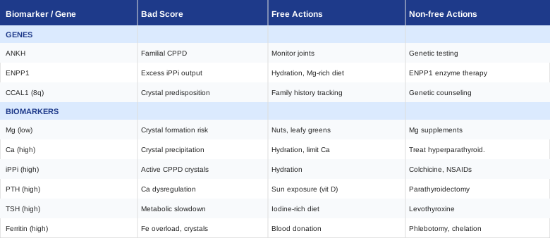
Moving from genetics to practical reference, the table below summarizes the key action points across all 4 genes and 7 biomarkers covered in this article.
A Book That May Reframe How You Think About This Condition
Outlive: The Science and Art of Longevity by Peter Attia, MD (2023) is primarily about preventing the "four horsemen" of chronic disease. But its underlying framework — track biomarkers aggressively, intervene early, treat the metabolic environment rather than waiting for symptoms — translates directly to how a thoughtful patient should approach CPPD. The book does not discuss crystal arthritis explicitly, but the principles map precisely onto the investigation framework described in this article.
Here are the ten most relevant takeaways from Outlive applied to CPPD:
1. The Standard of Care Is Optimized for Disease, Not Health
Attia's central argument: medicine waits for disease to be diagnosable before acting. But the metabolic dysfunction that causes conditions like CPPD — hyperparathyroidism, iron overload, hypothyroidism — often exists silently for years. Tracking biomarkers before symptoms are severe is not hypochondria; it is rational risk management.
2. Serum Tests Miss Intracellular Reality
Just as Attia emphasizes that standard lipid panels miss small LDL particle count (ApoB), standard serum magnesium misses cellular magnesium depletion. The analogy is direct: the number on your standard panel can be normal while the biologically relevant value is deficient. Always request the more informative test.
3. Insulin Resistance Affects Every Tissue, Including Joints
Attia devotes significant attention to insulin resistance as a root driver of metabolic disease. In CPPD, insulin resistance links to ENPP1 overexpression and increased pyrophosphate production. Controlling blood glucose and improving insulin sensitivity through exercise and diet does not just protect cardiovascular health — it may directly reduce PPi generation in joint tissue.
4. Exercise Is the Most Potent Intervention Available Without Prescription
Attia is emphatic: no single drug does what consistent exercise does for metabolic health. For CPPD, resistance training improves insulin sensitivity (reducing ENPP1 activity), promotes joint fluid circulation, and maintains muscle mass that protects joints from mechanical load. Zone 2 cardio (30–45 minutes, 3–5×/week at a pace where you can hold a conversation) is the specific protocol he advocates for metabolic improvement.
5. Iron Overload Is Chronically Underscreened
Attia discusses ferritin tracking as an underperformed test in standard care. For CPPD patients, the diagnostic yield of a full iron panel (including transferrin saturation) is high enough to justify it as a first-order test — not a specialty referral for those who have already failed standard treatment.
6. Sleep Is Not a Lifestyle Luxury, It Is Metabolic Maintenance
Attia presents extensive evidence that poor sleep raises cortisol, impairs glucose metabolism, and elevates systemic inflammation. For CPPD, this matters because cortisol suppresses T3 thyroid conversion (worsening hypothyroidism risk), depletes magnesium, and prolongs inflammatory flares. Targeting 7–9 hours with consistent sleep-wake timing is a non-trivial intervention.
7. Supplements Are the Last Tool, Not the First
Attia is deliberately skeptical of supplements as primary interventions. In the CPPD context, this means fixing the underlying cause first — treating the parathyroid adenoma, managing the iron overload, normalizing the thyroid — and using supplements as targeted support for residual gaps, not as replacements for addressing root causes.
8. Genetic Testing Adds Precision, Not Fatalism
Attia uses genetic data (including ApoE genotype for cardiovascular risk) as a tool to sharpen intervention strategy, not to predict inevitable outcomes. The same frame applies to ANKH, ENPP1, and HFE variants — knowing your genetic predisposition tells you which interventions to prioritize more aggressively, not which outcomes to accept passively.
9. Regular Monitoring Creates Feedback Loops
The argument is not to test once and move on. Attia emphasizes quarterly or semi-annual biomarker tracking as the mechanism by which interventions get refined. For CPPD, this means re-running the 7-biomarker panel after 8–12 weeks of targeted intervention to see which levers moved and which need adjustment.
10. The Goal Is Healthspan, Not Just Lifespan
CPPD rarely kills anyone, but it steals function — painful joints, reduced range of motion, and the anxiety of unpredictable flares. Attia's frame of optimizing for functional life years is directly applicable here: the goal is not just to reduce flare frequency but to protect joint integrity, maintain mobility, and avoid the cascade of inactivity, deconditioning, and decline that untreated crystal disease can initiate.
Evidence-Based Complementary Approaches for Joint Crystal Disease
Biomarker optimization and genetic awareness address root causes. The following complementary practices address the joint environment, pain experience, and inflammation amplification that make CPPD harder to live with between and during flares. Each has meaningful human clinical evidence, though none has been studied in large CPPD-specific trials.
Tai Chi
Tai chi is a Chinese mind-body practice combining slow, deliberate movement sequences with breathing and balance work. For CPPD, its relevance lies in multiple overlapping mechanisms: it maintains joint range of motion without the mechanical loading that aggravates inflamed cartilage, reduces systemic inflammatory markers (CRP, IL-6), and improves proprioception — the joint position sense that deteriorates in chronic arthritis and predisposes to re-injury. CPPD commonly affects knees and wrists; both benefit from the low-impact loading that tai chi provides without risking acute flares.
A 2016 randomized controlled trial published in the New England Journal of Medicine (Wang et al.) compared tai chi to physical therapy in knee osteoarthritis — a condition that frequently coexists with CPPD — and found comparable improvements in pain, function, and quality of life. The tai chi group showed additional benefits in depression scores and balance. While no equivalent RCT exists specifically for CPPD, the cartilage and synovial environment is sufficiently similar to make these findings directly translatable.
For practical application: begin with a beginner tai chi series (Yang style is the most accessible), 3–4 sessions per week of 30–40 minutes each. Online instructed programs or community classes both work. During acute flares, pause or reduce intensity significantly — tai chi should be practiced in the sub-acute or remission phases. Benefits tend to accumulate over 8–12 weeks of consistent practice; do not expect rapid symptom relief in the first two weeks.
Mindfulness-Based Stress Reduction (MBSR)
MBSR — the 8-week structured program developed by Jon Kabat-Zinn — addresses chronic pain through a fundamentally different mechanism than most medical interventions. Rather than suppressing the pain signal, it changes the relationship the nervous system has with it, reducing both the subjective suffering and the cortisol-driven inflammation amplification that worsens joint disease. For CPPD patients, the stress-inflammation axis is clinically relevant: cortisol suppresses T3 conversion (worsening hypothyroidism), promotes magnesium excretion, and amplifies cytokine signaling that extends flare duration. MBSR addresses all of these indirectly by lowering the chronic stress baseline.
Multiple randomized trials support MBSR for chronic inflammatory conditions. A systematic review in Arthritis Care and Research found that mindfulness-based interventions reduced pain, fatigue, and psychological distress in rheumatic disease patients, with effects that persisted at 6-month follow-up. Evidence for CPPD specifically is absent, but the shared inflammatory pathology makes the transfer reasonable. MBSR also measurably reduces cortisol levels and inflammatory cytokines in practice populations.
The standard protocol is 8 weeks of 2.5-hour weekly group sessions plus a one-day retreat and daily home practice (45 minutes). Shorter modified versions exist and maintain most of the benefit. Many hospitals and community health centers offer MBSR programs; online platforms (Palouse Mindfulness offers a free evidence-based program) provide an accessible starting point. The practice requires commitment and is most effective when begun during a period of relative symptom stability rather than in the middle of an acute flare.
Low-Level Laser Therapy (Photobiomodulation)
Photobiomodulation (PBM) uses near-infrared or red light (typically 630–1000 nm) delivered at non-thermal doses to stimulate mitochondrial function and reduce inflammatory signaling in tissue. At joints, this translates to reduced cytokine production (particularly TNF-α and IL-1β), improved microcirculation, and reduced pain sensitivity. CPPD flares are mediated by NLRP3 inflammasome activation and subsequent IL-1β release — the same pathway that PBM has demonstrated inhibitory effects on in tissue models and small human trials.
The Ottawa Panel Clinical Practice Guidelines on electrophysical agents in arthritis include low-level laser therapy as a conditionally recommended intervention for joint pain and inflammation, based on pooled evidence from multiple randomized trials. While existing trials focus primarily on osteoarthritis and rheumatoid arthritis, the anti-inflammatory mechanism is not disease-specific. A 2022 systematic review in Lasers in Medical Science found significant reductions in pain and functional impairment in inflammatory joint conditions treated with PBM at 810–904 nm, 3–5 sessions per week.
For practical application: professional-grade PBM devices used by physiotherapists and sports medicine clinics can be accessed by referral (typical cost: $50–$100 per session, 6–12 sessions for initial course). Consumer near-infrared devices for home use have become more affordable ($200–$600 for panels); these operate at lower power than clinical devices and require longer sessions (10–20 minutes per joint area). Target the affected joints directly. Use outside of acute flares to reduce baseline inflammation; during acute flares, the joint may be too sensitive for direct application. Do not use over active infection, malignancy, or directly on thyroid tissue.
Conclusion
Calcium pyrophosphate deposition disease has clear upstream causes — and those causes are both detectable and often modifiable. The 7 biomarkers covered in this article represent the most valuable diagnostic layer available: ionized calcium and PTH for parathyroid dysfunction, RBC magnesium for crystal inhibition capacity, ferritin and transferrin saturation for iron overload, thyroid hormones for enzymatic crystal clearance, alkaline phosphatase for PPi catabolism, and serum phosphate for metabolic balance. Four genes — ANKH, ENPP1, HFE, and NT5E — add a layer of precision for cases with familial patterns or unexplained recurrence.
The most useful next step is straightforward: request the full biomarker panel at your next visit, push for the more informative tests (ionized calcium, RBC magnesium) rather than settling for standard panel proxies, and bring the findings to a physician or specialist willing to investigate secondary causes rather than managing symptoms alone. Genetics, complementary practices, and lifestyle optimization layer on top of — not instead of — that metabolic investigation. Better information does not guarantee better outcomes, but it makes better decisions possible.
Musculoskeletal: Bone Conditions Joint Conditions
Endocrine & Metabolic: Thyroid Conditions
Autoimmune: Inflammatory Conditions