This article contains AI generated content.
· UpdatedCartilage-Hair Hypoplasia: 5 Genes and 6 Biomarkers to Track
Introduction
Living with cartilage-hair hypoplasia, or caring for someone who does, means navigating a condition that most doctors encounter once in a career, if ever. CHH is a rare autosomal recessive skeletal dysplasia caused by mutations in the RMRP gene, which encodes the RNA subunit of the RNase MRP enzyme. The result is a constellation of features — disproportionate short stature, fine sparse hair, varying degrees of immune deficiency, and an elevated lifetime risk for certain cancers — that rarely fit neatly into standard clinical algorithms. Generic reassurance that everything is "being monitored" is not always enough when you have a condition this specific.
The challenge with CHH is that its severity varies enormously from person to person, even among individuals carrying the same RMRP mutation. Some patients have near-normal immune function; others present with severe combined immunodeficiency requiring bone marrow transplantation in childhood. Some have significant skeletal complications; others remain relatively mobile for decades. This variability is not random noise — it reflects real biological differences in how the body compensates at the molecular and cellular level, and understanding those differences is where modern genetics and biomarker science can genuinely help.
Generic advice — "eat well, stay active, get regular check-ups" — is not wrong, but it glosses over the specific biological terrain of CHH. The immune deficiency in CHH is not like other immune deficiencies. The skeletal fragility is not the same as osteoporosis. The anemia seen in some patients has its own mechanism rooted in RMRP dysfunction. Addressing these features requires a targeted approach, one built around the specific markers and genetic factors that actually govern disease expression in this condition.
This article takes that more specific approach. The biomarker section below gives you a practical framework for tracking the six measurements most likely to reveal what is happening in your body right now and guide actionable decisions. The genetics section shows you which gene variants interact with RMRP dysfunction and what compensation strategies exist for each. Both strategies are grounded in published human research. Neither promises a cure. But better information, tracked consistently and discussed with the right specialists, genuinely does lead to better decisions.
Summary
This article covers six core biomarkers that every person with CHH should track, including T-lymphocyte subsets, NK cell counts, serum immunoglobulins, 25-OH vitamin D, IGF-1, and alkaline phosphatase — with specific thresholds, cost ranges, and improvement plans for each. The genetics section then maps five key genes — RMRP, TERC, VDR, STAT3, and DNMT3B — explaining what each does, how it intersects with CHH biology, and what can be done when a variant is unfavorable, both with and without supplementation. Beyond biomarkers and genes, the article includes a summary of the most impactful research insights from leading CHH investigators, complementary approaches with clinical evidence for immune and skeletal support, and a practical closing action plan. If you have been told to "just monitor things," this article shows you what to monitor, why, and what to do about it.
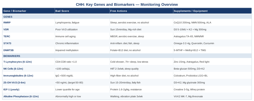
6 Biomarkers to Track When You Have Cartilage-Hair Hypoplasia
The value of biomarker tracking in CHH lies not just in spotting problems but in catching drift before it becomes a crisis. Immune function in CHH can deteriorate gradually, vitamin D can fall without symptoms, and growth factor abnormalities can compound skeletal vulnerability over time. The six markers below cover the most clinically meaningful biological terrain in CHH, based on what is known about the disease's pathophysiology and on the practices of leading immunologists and metabolic specialists.
Biomarker 1: T-Lymphocyte Subsets (CD3+, CD4+, CD8+)
Why it matters: The immune deficiency in CHH is primarily T-cell mediated. RMRP dysfunction impairs ribosome biogenesis and mitochondrial DNA replication, both of which are essential for the rapid proliferation of T cells during immune activation. In CHH, the thymus may be underdeveloped, and the T-cell pool is often smaller, less diverse, and ages prematurely. This is not a theoretical risk — CHH patients have significantly higher rates of severe infections, autoimmune complications, and lymphoma, all of which are tied to T-cell dysregulation.
CD4+ T cells (helper cells) coordinate the broader immune response and support antibody production. CD8+ T cells (cytotoxic cells) handle direct viral and cancer cell killing. The CD4:CD8 ratio is a useful summary statistic: a ratio below 1.0 suggests immune senescence or chronic activation. In healthy adults, this ratio is typically 1.5–2.5. CHH patients often run lower, sometimes significantly so.
How to measure it: T-cell subset panels are ordered as flow cytometry of peripheral blood. They are typically run as part of an immunology workup and are available at most hospital labs and major reference labs. Cost range: $150–$400 depending on the panel depth and whether it includes NK cells. If you have a known CHH diagnosis, your immunologist can order a comprehensive lymphocyte subset panel that includes CD3+, CD4+, CD8+, CD19+ (B cells), and CD56+ (NK cells) in a single draw.
Monitoring frequency: Every 6–12 months if stable; every 3 months if there has been a recent infection, new symptoms, or a change in treatment. Baseline values matter enormously here — your trend over time is more informative than any single result.
If the score is bad, the plan without supplements: Minimize immunosuppressive exposures, which include chronic psychological stress, sleep deprivation under 7 hours, and excessive cardio above 80% VO2max (this transiently suppresses T-cell function). Cold exposure in moderate forms — ending a shower with 30–60 seconds of cold water — has been shown in small studies to increase NK cell count and reduce infection frequency. Prioritize sleep quality rigorously, as deep sleep is when T-cell trafficking and renewal peaks. Avoid unnecessary antibiotic use, which disrupts the gut-immune axis and may further impair lymphocyte diversity.
If the score is bad, the plan with supplements or equipment: Zinc picolinate or bisglycinate at 15–25 mg/day supports thymic function and T-cell maturation; it is one of the most evidence-backed supplements for immune cell number and function. Cycle on for 3 months, off for 1 month to avoid copper depletion. Watch for nausea at higher doses. Astragalus membranaceus (standardized extract, 500–1000 mg/day) has preliminary human data supporting T-cell proliferation and NK cell activation; cycle for 8 weeks, pause for 4. Photobiomodulation (low-level laser/red light therapy) directed at the sternum and thymus region has early mechanistic evidence for thymic stimulation, though clinical data in CHH is absent — it represents a low-risk adjunct. Sessions of 10–15 minutes, 3x/week at wavelengths of 630–850 nm, are commonly used protocols.
Biomarker 2: NK Cell Count (CD56+/CD16+)
Why it matters: Natural killer cells are the immune system's first responders to viral-infected and malignant cells. They do not require prior sensitization, which makes them especially important in CHH, where adaptive T-cell responses are already compromised. Reduced NK cell number or function leaves a critical gap in cancer surveillance — relevant given that CHH patients carry a 7- to 10-fold elevated lifetime risk for lymphoma and a significant basal cell carcinoma risk.
NK cell counts below 100 cells/µL in adults are considered low. CHH patients commonly show both reduced count and impaired killing function (measurable via NK cell cytotoxicity assays, though these are less routinely ordered).
How to measure it: NK cells are typically included in comprehensive lymphocyte panels via flow cytometry (same draw as T-cell subsets). Cost: included in the comprehensive lymphocyte panel ($150–$400). If only a basic CBC was ordered, NK cells will not appear — you must specifically request a lymphocyte subset panel.
Monitoring frequency: Every 6–12 months, synchronized with T-cell subsets.
If the score is bad, the plan without supplements: High-intensity interval training (HIIT) at 2–3 sessions per week has consistently shown acute and sustained increases in NK cell count and cytotoxicity in clinical studies. The mechanism involves catecholamine-driven NK cell mobilization from the spleen and bone marrow. For CHH patients with skeletal limitations, low-impact HIIT alternatives — stationary cycling, rowing, or water-based interval work — achieve comparable immune effects. Sleep, again, is essential: NK cell count is demonstrably lower after a single night of under 6 hours sleep.
If the score is bad, the plan with supplements or equipment: Beta-glucan (from oats or Saccharomyces cerevisiae, 500 mg/day) has multiple randomized controlled trials supporting NK cell activation; it binds dectin-1 receptors on innate immune cells and upregulates killing capacity. Take consistently for 8–12 weeks, then reassess. Vitamin D3 + K2 at therapeutic doses (see below) also directly supports NK cell function via VDR signaling. Red light therapy at 850 nm applied over the abdomen (spleen region) for 10 minutes, 4x/week has shown some preclinical evidence for NK mobilization and warrants consideration as a low-risk adjunct.
Biomarker 3: Serum Immunoglobulins (IgG, IgA, IgM)
Why it matters: While CHH primarily affects T-cell immunity, B-cell function and antibody production are also variably impaired. Some CHH patients develop hypogammaglobulinemia — low levels of one or more immunoglobulin classes — which substantially increases susceptibility to bacterial infections, particularly encapsulated bacteria like Streptococcus pneumoniae and Haemophilus influenzae. Monitoring immunoglobulin levels guides decisions about vaccination schedules, prophylactic antibiotics, and, in severe cases, intravenous immunoglobulin (IVIG) replacement therapy.
IgG is the most clinically significant for infection protection. IgG below 500 mg/dL in adults is generally considered the threshold for considering IVIG. IgA deficiency is also common in CHH and impairs mucosal immunity — the first line of defense in the respiratory and gastrointestinal tracts.
How to measure it: Serum protein electrophoresis (SPEP) or quantitative immunoglobulin levels ordered as a simple blood draw. Cost: $50–$150 for quantitative IgG/IgA/IgM. This is a standard immunology lab test available at virtually all clinical labs.
Monitoring frequency: Every 6–12 months. More frequent if on IVIG therapy, if there has been a recent significant infection, or if levels have been trending down.
If the score is bad, the plan without supplements: Optimizing gut health is the most underappreciated non-pharmacological lever for immunoglobulin production. The gut-associated lymphoid tissue (GALT) is responsible for a large proportion of IgA secretion. A high-fiber, diverse diet increases short-chain fatty acid production, which supports IgA-secreting plasma cells. Avoiding alcohol entirely is important — even moderate alcohol consumption demonstrably reduces IgA secretion. Stress management is also relevant: cortisol acutely suppresses IgA secretion in mucosal tissues.
If the score is bad, the plan with supplements or equipment: Colostrum (bovine, 2–4 g/day) contains secretory IgA and growth factors and has human evidence for supporting mucosal immunity; useful as an adjunct when IgA is specifically low. Probiotics at high-dose multi-strain formulations (particularly Lactobacillus rhamnosus GG and Bifidobacterium longum) have RCT evidence for increasing fecal IgA and reducing respiratory infection rates. Use for 12-week cycles with a 4-week break. In patients with significant hypogammaglobulinemia, IVIG or subcutaneous immunoglobulin (SCIG) replacement, prescribed by an immunologist, remains the only truly effective intervention — no supplement replaces this.
Biomarker 4: 25-Hydroxyvitamin D (25-OH Vitamin D)
Why it matters: Vitamin D is not just a bone nutrient — it is a steroid hormone that directly regulates over 200 genes, including many governing immune cell differentiation, T-regulatory cell function, and inflammatory signaling. For CHH specifically, the intersection of vitamin D with both skeletal and immune dimensions makes it a uniquely high-leverage biomarker. The VDR (vitamin D receptor) gene, covered in the genetics section, has variants that affect how efficiently vitamin D is utilized at the cellular level, meaning CHH patients with VDR polymorphisms may need higher serum levels to achieve the same biological effect.
In skeletal dysplasias, suboptimal vitamin D accelerates cartilage matrix degradation, impairs mineralization, and increases fracture risk. In the immune compartment, vitamin D deficiency below 30 ng/mL has been consistently associated with reduced T-regulatory cell frequency, higher inflammatory cytokine levels, and impaired antimicrobial peptide production in the skin and mucosa.
How to measure it: A standard serum 25-OH vitamin D blood test. Cost: $30–$80 at commercial labs; often covered by insurance with a documented deficiency diagnosis. Optimal range for CHH patients, given dual skeletal and immune relevance, is 50–80 ng/mL — considerably higher than the "normal" laboratory range of 30 ng/mL, which reflects bone health only. Peter Attia and other longevity-focused clinicians commonly target 50–60 ng/mL as a functional optimum.
Monitoring frequency: Every 3–6 months while adjusting dose; every 6 months once stable.
If the score is bad, the plan without supplements: Deliberate sun exposure of 15–20 minutes daily to large skin surface areas (arms, legs, back) between 10 AM and 3 PM produces approximately 10,000–20,000 IU of vitamin D3 in fair-skinned individuals in summer months. This is almost never sufficient to correct deficiency alone, but it meaningfully contributes. Dietary sources — fatty fish, egg yolks, liver — add 200–400 IU per serving and should be regular fixtures in the diet.
If the score is bad, the plan with supplements or equipment: Vitamin D3 + K2 (to prevent calcium misplacement) is the standard correction protocol. Doses range from 2,000 IU/day for maintenance to 5,000–10,000 IU/day for correction of deficiency; the appropriate dose depends on baseline level and VDR genotype. Always take with a fatty meal for maximum absorption. Retest at 90 days to adjust dose. Magnesium glycinate at 300–400 mg/day is important alongside D3, as magnesium is a required cofactor for vitamin D activation in the liver and kidneys — without it, supplemental D3 has limited effect. Side effects of excessive D3 include hypercalcemia (rare below 10,000 IU/day); K2 mitigates this risk. Cycling: No cycling required; vitamin D is a daily maintenance supplement for most CHH patients given persistent needs.
Biomarker 5: IGF-1 (Insulin-Like Growth Factor 1)
Why it matters: IGF-1 is the primary mediator of growth hormone action in peripheral tissues, and it is central to skeletal growth, muscle anabolism, and tissue repair. In CHH, the primary growth limitation is intrinsic skeletal — the cartilage itself is abnormal, and IGF-1 cannot correct this. However, IGF-1 levels remain clinically relevant because they predict bone density maintenance, muscle mass preservation, and metabolic resilience over time. Low IGF-1 in CHH adults compounds the existing skeletal fragility and is associated with higher fracture risk, accelerated muscle loss, and fatigue.
IGF-1 levels also track indirectly with nutritional adequacy — chronic low protein intake, caloric restriction, or malabsorption all suppress IGF-1. In CHH patients with Hirschsprung disease or other gastrointestinal complications, this is a genuine concern.
How to measure it: A single morning fasting blood draw for serum IGF-1 (also called somatomedin C). Cost: $50–$150. Age-adjusted reference ranges apply; for adults, the functional target is generally the upper half of the age-matched reference range. Low IGF-1 in the lower quartile for age warrants investigation.
Monitoring frequency: Annually in stable adults; every 6 months in children with CHH or in adults with new musculoskeletal symptoms.
If the score is bad, the plan without supplements: Protein intake is the single most impactful dietary driver of IGF-1. Aim for 1.6–2.0 g of protein per kg of body weight per day, distributed across meals. Resistance training — even low-impact forms like resistance bands or swimming — is the most potent non-pharmacological stimulus for IGF-1 secretion; 3 sessions per week of moderate intensity is effective. Deep, uninterrupted sleep (7–9 hours) is essential, as growth hormone (which drives IGF-1 production) is secreted primarily in slow-wave sleep. Intermittent fasting protocols beyond 16 hours suppress IGF-1 in the long term and should be avoided in CHH patients already at risk for low levels.
If the score is bad, the plan with supplements or equipment: Creatine monohydrate at 3–5 g/day supports muscle protein synthesis and secondarily supports IGF-1 by enabling more effective resistance training stimulus; take daily without cycling. Whey protein as a post-exercise supplement (25–40 g) acutely boosts IGF-1 via its high leucine content. Deer velvet antler extract (a traditional supplement claimed to contain IGF-1 precursors) has very limited and low-quality human evidence; not recommended. In cases of growth hormone deficiency confirmed by provocative testing (distinct from CHH itself), recombinant human growth hormone therapy under endocrinologist supervision is an option, though its benefits in skeletal dysplasia are modest.
Biomarker 6: Alkaline Phosphatase (ALP) with Bone-Specific Isoform
Why it matters: Alkaline phosphatase reflects osteoblast activity — the bone-building cells. In CHH, the cartilage matrix is disordered at a fundamental level, and bone remodeling dynamics are chronically abnormal. Tracking ALP over time provides a window into how active bone turnover is at any given moment. Persistently elevated ALP suggests excessive or disordered bone resorption and formation cycling; abnormally low ALP is associated with hypophosphatasia, a condition that can co-exist with or complicate skeletal dysplasias and significantly worsens bone quality.
Bone-specific ALP (bALP), measured separately from total ALP (which includes liver and intestinal isoforms), is more precise for skeletal monitoring. Paired with serum phosphate and calcium, it gives a more complete picture of mineralization adequacy.
How to measure it: Total ALP is included in a standard comprehensive metabolic panel. Bone-specific ALP requires a separate immunoassay. Cost: ALP as part of CMP is $20–$50; bone-specific ALP separately is $80–$150. Add serum calcium, phosphate, and magnesium in the same draw for full context.
Monitoring frequency: Every 6–12 months. More frequently during periods of rapid skeletal change, growth spurts in children, or following fractures.
If the score is bad, the plan without supplements: Weight-bearing activity, even gentle walking, stimulates osteoblast activity and normalizes ALP trends over time. In CHH with limited weight-bearing capacity, aquatic exercise or vibration plate therapy (whole-body vibration, 25–50 Hz, 10–15 minutes per session, 3x/week) has clinical evidence for stimulating bone formation markers including bALP. Adequate dietary calcium (1000–1200 mg/day from food sources primarily) combined with vitamin D optimization is foundational.
If the score is bad, the plan with supplements or equipment: Vitamin K2 (MK-7 form) at 100–200 µg/day activates osteocalcin and directs calcium into bone matrix rather than soft tissue; take with fat-containing meals. Magnesium threonate or glycinate at 300–400 mg/day supports both bone mineralization and ALP activity. Silicon (as orthosilicic acid, 10 mg/day) has human trial evidence for increasing bALP and improving collagen cross-linking in bone; cycle 12 weeks on, 4 weeks off. Whole-body vibration platforms, used for 10 minutes daily, are one of the most evidence-supported non-pharmacological interventions for increasing bone mineral density in skeletal dysplasia patients with limited exercise capacity.
Moving from what to track to why your genes predict your response, the next section maps the five gene variants most likely to shape how CHH expresses in your body.
5 Genes That Shape Cartilage-Hair Hypoplasia Expression
Understanding the genetics of CHH does not mean becoming a molecular biologist. It means knowing which biological systems your specific variants may be stressing, and where targeted interventions can meaningfully compensate. This section covers the RMRP gene and four modifier genes that interact with CHH biology.
Gene 1: RMRP — The Causative Gene
The RMRP gene encodes the RNA component of the RNase MRP complex, an enzyme that processes ribosomal RNA (rRNA) and mitochondrial RNA. Mutations in RMRP are the direct cause of CHH. The landmark 2001 study by Ridanpää et al. identified RMRP mutations as the genetic basis of CHH, showing that this single gene impairs ribosome biogenesis, mitochondrial DNA replication, and cell cycle progression in proliferating cells — explaining why tissues with rapid cell turnover (cartilage, lymphocytes, hair follicles) are preferentially affected.
Over 100 different RMRP mutations have been identified. The most common in Finnish patients is the 70A>G transition, but other populations carry different founder mutations. Genotype-phenotype correlations exist but are imperfect — the same mutation can produce very different severity in different individuals.
If the gene is bad, the plan without supplements: RMRP cannot be "fixed" by lifestyle alone, but its downstream effects can be meaningfully modulated. Mitochondrial function support is the most direct lever: mitochondrial dysfunction downstream of RMRP is central to energy metabolism and lymphocyte replication capacity. High-quality sleep (the primary context for mitochondrial repair), regular aerobic exercise at moderate intensity (which stimulates mitochondrial biogenesis via PGC-1α), and avoidance of mitochondrial toxins (alcohol, heavy smoking, chronic high fructose intake) are all relevant. Ribosomal stress from RMRP dysfunction is also worsened by amino acid deprivation; ensuring adequate daily protein intake directly reduces the burden on remaining ribosomal capacity.
If the score is bad, the plan with supplements or equipment: CoQ10 (ubiquinol form) at 200–400 mg/day directly supports the mitochondrial electron transport chain; particularly useful given RMRP's role in mitochondrial RNA processing. No cycling needed; take with fat-containing meals. NAD+ precursors (NMN at 500 mg/day or NR at 300–500 mg/day) support mitochondrial biogenesis via the SIRT1/PGC-1α pathway; take in the morning to align with circadian NAD+ rhythms. Alpha-lipoic acid at 300–600 mg/day acts as a mitochondrial antioxidant and co-factor; cycle 3 months on, 1 month off. Evidence base for these supplements in CHH specifically is extrapolated from mitochondrial disease research broadly.
Gene 2: VDR (Vitamin D Receptor)
VDR polymorphisms — particularly the FokI, BsmI, and TaqI variants — affect how effectively cells respond to vitamin D signaling. Unfavorable VDR variants reduce the binding affinity between the vitamin D-VDR complex and DNA response elements, meaning that even adequate serum vitamin D levels may produce suboptimal immune and bone effects. In CHH, where both immune function and bone health are already compromised, VDR insufficiency is a compounding vulnerability.
If the gene is bad, the plan without supplements: Higher sun exposure and dietary vitamin D represent practical compensations. Magnesium status is critical — magnesium is required for vitamin D activation and without it, even good VDR variants cannot use available vitamin D effectively.
If the score is bad, the plan with supplements or equipment: Patients with unfavorable VDR variants typically need serum 25-OH vitamin D levels in the 60–80 ng/mL range (rather than the standard 40–50 ng/mL target) to achieve equivalent downstream signaling. This generally requires 5,000–10,000 IU/day of D3 combined with K2 (100–200 µg MK-7) and magnesium (300–400 mg/day). VDR genotyping is available through consumer genetic testing (e.g., 23andMe raw data interpreted through tools such as Genetic Genie or Rhonda Patrick's published VDR analysis frameworks). Frequency: daily, no cycling.
Gene 3: TERC (Telomerase RNA Component)
TERC encodes the RNA template used by telomerase to extend chromosome ends (telomeres). This gene is relevant in CHH because RMRP dysfunction is associated with accelerated telomere shortening in lymphocytes — an epigenetic form of immune aging. Patients with TERC variants that reduce telomerase activity may age their immune cells faster, experience more severe lymphopenia, and face higher lymphoma risk. Telomere length measurement in peripheral blood lymphocytes has been studied as a CHH disease marker.
If the gene is bad, the plan without supplements: Chronic psychological stress is one of the strongest documented drivers of telomere shortening. Mindfulness-based stress reduction (MBSR), even practiced for 8 weeks at standard protocol intensity, has shown measurable attenuation of telomere shortening rate in clinical studies. Aerobic exercise at moderate intensity consistently associates with longer leukocyte telomere length in population studies. Sleep quality — particularly preserving slow-wave sleep — also directly protects telomere integrity.
If the score is bad, the plan with supplements or equipment: Astragalus extract (TA-65, a standardized cycloastragenol form) has the strongest commercial evidence for telomerase activation in humans; studies using 250–1000 mg/day showed modest but measurable increases in telomerase activity and lymphocyte telomere length. However, TA-65 is expensive and evidence in CHH-specific immune populations is absent. Standard astragalus root extract at 500 mg/day is a lower-cost alternative with weaker but more accessible evidence. NAD+ precursors (NMN/NR, as above) also support telomere maintenance indirectly via SIRT1 activation, which modulates telomerase activity. Cycle astragalus for 12 weeks on, 4 weeks off to assess response.
Gene 4: STAT3 (Signal Transducer and Activator of Transcription 3)
STAT3 variants influence how efficiently immune cells respond to cytokine signaling, particularly IL-6 and IL-10 pathways. In CHH, where T-cell function is already impaired, unfavorable STAT3 variants can amplify inflammatory dysregulation and impair T-regulatory cell differentiation — contributing to both the autoimmune features seen in some CHH patients and the difficulty in resolving infections. STAT3 gain-of-function mutations (distinct from polymorphisms) are a known cause of combined immune deficiency overlapping with CHH phenotypes.
If the gene is bad, the plan without supplements: Reducing chronic low-grade inflammation is the primary lever for managing STAT3 hyperstimulation. This means minimizing ultra-processed food, stabilizing blood glucose (avoiding glycemic spikes), adequate sleep, and stress reduction. A dietary pattern high in omega-3 fatty acids and polyphenols directly modulates STAT3 activity — multiple studies confirm that fish oil reduces phospho-STAT3 signaling in inflammatory pathways.
If the score is bad, the plan with supplements or equipment: Omega-3 fatty acids (EPA + DHA) at 2–4 g/day of combined EPA+DHA have robust evidence for reducing IL-6-driven STAT3 activation; take with meals. Quercetin at 500–1000 mg/day is a natural STAT3 inhibitor with human safety data; cycle 8 weeks on, 4 weeks off. Curcumin (phosphatidylcholine-complexed or nanoparticle form) at 500–1000 mg/day similarly modulates STAT3 and NF-κB; low bioavailability with standard forms makes enhanced formulations essential. Side effects: omega-3 at high doses may prolong bleeding time; relevant before any surgical procedures.
Gene 5: DNMT3B (DNA Methyltransferase 3 Beta)
DNMT3B is an epigenetic writer enzyme responsible for de novo DNA methylation during immune cell development. Variants in DNMT3B affect T-cell and B-cell maturation, and notably, ICF syndrome (immunodeficiency-centromeric instability-facial anomalies) is caused by DNMT3B mutations — a condition that overlaps phenotypically with CHH's immune features. In CHH, DNMT3B variants may modulate how severely the primary RMRP mutation affects lymphocyte development and differentiation capacity.
If the gene is bad, the plan without supplements: Epigenetic methylation patterns respond significantly to dietary methyl donors. A diet rich in folate (dark leafy greens, legumes), choline (eggs, liver), and methionine (animal proteins) provides the methyl groups that DNMT3B requires to function. Adequate B12 is essential for the methylation cycle to run effectively. Avoiding alcohol is critical — alcohol is one of the most potent dietary disruptors of DNA methylation and directly impairs DNMT3B activity.
If the score is bad, the plan with supplements or equipment: Methylfolate (5-MTHF) at 400–800 µg/day combined with methylcobalamin (B12) at 500–1000 µg/day and trimethylglycine (TMG) at 1–3 g/day supports the methyl donor pool that DNMT3B draws upon. This combination — sometimes called a methylation support stack — requires attention to individual MTHFR status (see Gary Brecka's widely accessible work on methylation genetics); patients who are MTHFR C677T homozygous need higher doses of methylfolate specifically. Take daily; no cycling required. Excess methyl donors can occasionally cause overstimulation symptoms (anxiety, irritability) — start at lower doses and titrate.
Key Research Insights on Cartilage-Hair Hypoplasia That Change How You Think About the Condition
The most valuable framework shift for understanding CHH comes not from a single book but from the body of work produced by Outi Mäkitie and colleagues at the University of Helsinki and Karolinska Institute — arguably the most published research group on CHH globally — synthesized alongside the immune deficiency literature. Here are the ten most clinically impactful insights from this research landscape.
1. CHH is More Than Bones — It is a Systemic Disease of Rapidly Dividing Cells
Because RMRP affects ribosome biogenesis and cell cycle progression, any tissue requiring rapid cell proliferation is vulnerable. Cartilage (chondrocytes), lymphocytes, and hair follicle cells are affected most visibly, but the gut epithelium, hematopoietic stem cells, and telomere maintenance systems are also involved. This is why CHH requires multi-system surveillance, not just orthopedic monitoring.
2. Immune Deficiency in CHH Exists on a Spectrum That Can Worsen Over Time
A key finding from longitudinal studies is that CHH-related immune deficiency is not fixed at birth. Some patients have near-normal immune function in early childhood but develop progressive T-cell lymphopenia in adulthood due to accelerated thymic involution and telomere shortening. This makes periodic re-evaluation of immune status in adulthood as important as in childhood.
3. Lymphoma Risk in CHH is Real and Underappreciated
CHH patients have a 7- to 10-fold increased risk of lymphoma compared to the general population. Non-Hodgkin lymphoma, particularly of T-cell origin, is the most common malignancy. This risk warrants vigilance for B symptoms (unexplained fevers, night sweats, weight loss) and should inform decisions about immunosuppressive therapies.
4. Bone Marrow Transplantation Can Correct the Immune Component — Not the Skeletal One
Hematopoietic stem cell transplantation (HSCT) has been used successfully in CHH patients with severe combined immunodeficiency and can restore immune function. Importantly, it does not affect the underlying skeletal dysplasia — the cartilage abnormalities persist post-transplant. This distinction is important for setting realistic expectations.
5. Anemia in CHH Has a Specific Mechanism
Some CHH patients develop macrocytic anemia resembling the anemia of Diamond-Blackfan syndrome. This is not iron deficiency anemia — it reflects impaired erythroid progenitor proliferation due to RMRP dysfunction. Treating it with iron is ineffective and potentially harmful if iron stores are already adequate. Recognizing the mechanism avoids unnecessary and potentially counterproductive supplementation.
6. Hirschsprung Disease Occurs in a Significant Minority of CHH Patients
Approximately 15–20% of CHH patients have Hirschsprung disease (congenital colonic aganglionosis), reflecting RMRP's role in enteric nervous system development. Unrecognized Hirschsprung disease produces gut dysmotility, nutrient malabsorption, and dysbiosis that compound the immune and nutritional challenges of CHH.
7. The Severity of Skeletal Dysplasia Does Not Predict Immune Severity
Clinically, one might assume that more severe short stature predicts more severe immune deficiency. Research has not confirmed this. Skeletal and immune phenotype can dissociate completely within the same family carrying identical mutations. This is a critical insight: never use physical appearance as a proxy for immune risk assessment.
8. Autoimmune Phenomena Are Recognized But Underdiagnosed in CHH
Despite immune deficiency, CHH patients can also develop autoimmune disease — a seemingly paradoxical finding explained by deficient T-regulatory cell function. Autoimmune cytopenias, inflammatory bowel disease-like presentations, and psoriasis have all been documented. The immune system in CHH is dysregulated, not simply depleted.
9. Vaccination Schedules Need to be Individualized
Live attenuated vaccines (MMR, varicella, yellow fever) carry real risk in CHH patients with T-cell immunodeficiency and should not be given without first documenting T-cell number and function. Non-live vaccines are generally safe but may produce suboptimal antibody responses — post-vaccination titers should be checked.
10. Genetic Counseling Has High Practical Value Given the Recessive Inheritance Pattern
CHH is autosomal recessive with a carrier frequency of approximately 1 in 76 in Finnish populations and varies in other ethnic groups. Siblings of CHH patients have a 25% risk of being affected if both parents carry RMRP mutations. Genetic counseling and prenatal diagnosis are available and clinically actionable — this is not just intellectual information.
Complementary and Lifestyle-Based Approaches With Clinical Relevance for CHH
Standard medical care remains the backbone of CHH management. The approaches below are chosen specifically because they have meaningful human evidence for the biological domains most affected in CHH: immune function, skeletal integrity, and systemic inflammation. None of these replace medical monitoring or immunological follow-up.
Microbiome-Directed Therapies
The gut microbiome is a major regulator of immune function, accounting for approximately 70–80% of immune cell residence and a substantial proportion of secretory IgA production. In CHH, where immune deficiency and gut complications (including Hirschsprung disease in some patients) converge, microbiome health is a clinically meaningful variable. Dysbiosis in CHH patients can amplify systemic inflammation, impair mucosal immunity, and worsen nutrient absorption — all of which compound the condition's existing challenges.
A specific evidence-based protocol for immune-focused microbiome optimization involves a combination of high-dose multi-strain probiotics (Lactobacillus rhamnosus GG + Bifidobacterium longum at ≥10 billion CFU/day) combined with a prebiotic-rich diet (inulin, resistant starch, diverse plant fiber). A 2021 systematic review in Nutrients confirmed that probiotic supplementation in primary immunodeficiency patients improved secretory IgA levels and reduced upper respiratory infection frequency. Fecal microbiota transplantation (FMT) has emerging evidence for immune reconstitution in post-HSCT patients but remains experimental for CHH.
Practically, CHH patients should introduce probiotics gradually (to avoid transient bloating), prioritize fermented foods (plain yogurt, kefir, sauerkraut) as daily dietary staples, and avoid unnecessary antibiotic use that destroys microbial diversity. Those with Hirschsprung-related surgical history should consult their gastroenterologist before starting high-dose probiotics, as altered gut anatomy changes microbial dynamics.
The Autoimmune Protocol (Sarah Ballantyne, PhD)
Because CHH involves immune dysregulation — not simply immune deficiency, but also a risk of autoimmune phenomena — the Autoimmune Protocol (AIP) developed by Sarah Ballantyne has genuine conceptual relevance. The AIP is a dietary and lifestyle elimination protocol designed to reduce gut permeability ("leaky gut"), remove common dietary triggers of immune activation (grains, legumes, nightshades, dairy, eggs initially), and systematically identify foods that drive individual inflammatory responses.
Ballantyne's AIP approach includes not just dietary changes but sleep optimization, stress management, and movement — all of which have direct relevance to the immune and skeletal challenges of CHH. Multiple case series and a 2017 pilot trial in Inflammatory Bowel Diseases showed significant clinical improvements in autoimmune bowel disease using the AIP framework. The evidence base is not CHH-specific, but the mechanistic logic applies to any condition involving immune dysregulation and gut-immune axis impairment.
The AIP is a structured elimination-reintroduction protocol: the elimination phase runs for a minimum of 4–6 weeks, during which all potential dietary triggers are removed. Reintroduction is then done one food at a time, with symptom tracking. For CHH patients with suspected food-driven inflammatory flares or autoimmune symptoms, this protocol provides a systematic and non-pharmacological way to identify individual triggers. It should be undertaken with nutritional supervision to avoid deficiencies, particularly during the elimination phase.
Mindfulness-Based Stress Reduction (MBSR)
Chronic psychological stress is a documented suppressor of NK cell count, T-cell proliferative capacity, secretory IgA, and telomere length — all of which are already under pressure in CHH. Living with a rare, visible, and chronic condition carries a significant psychological burden, and this burden is not merely quality-of-life issue; it is a physiological one that directly feeds back into immune function.
MBSR, the standardized 8-week protocol developed by Jon Kabat-Zinn at the University of Massachusetts Medical School, has been tested in immune-relevant populations including cancer patients, HIV patients, and autoimmune disease patients. A 2003 randomized trial published in Psychosomatic Medicine showed measurable increases in antibody titers to influenza vaccine in MBSR participants compared to controls — a direct measure of immune function improvement. The standard MBSR protocol involves 8 weeks of once-weekly group sessions (2.5 hours each), a day-long retreat, and 45 minutes of daily home practice.
For CHH patients, MBSR is accessible via in-person programs at hospitals and meditation centers, or through validated online formats (Palouse Mindfulness is a free, evidence-based online version). The commitment is real — 45 minutes daily is not trivial — but the physiological return for immune function, cortisol regulation, and telomere preservation makes it one of the most evidence-supported non-pharmacological interventions available for managing the stress-immune connection in CHH.
Low-Level Laser Therapy / Photobiomodulation
Photobiomodulation (PBM) using red and near-infrared light (630–850 nm) stimulates mitochondrial cytochrome c oxidase, increasing cellular ATP production, reducing oxidative stress, and modulating inflammatory signaling. Given that RMRP dysfunction in CHH directly impairs mitochondrial function, PBM represents a mechanistically coherent adjunct. Early clinical evidence in musculoskeletal conditions and immune modulation supports exploring its role in CHH's specific biological context.
In skeletal applications, a 2018 systematic review in Photobiomodulation, Photomedicine, and Laser Surgery found consistent evidence for reduced joint pain and improved function in musculoskeletal disorders, with anti-inflammatory effects mediated through reduced IL-1β and TNF-α. For CHH, the most directly relevant application is cartilage and joint support in the metaphyseal regions most affected by the dysplasia.
A practical PBM protocol for CHH involves a tabletop or panel device (660–850 nm, minimum 50 mW/cm²), applied for 10–15 minutes per session to affected joints and the sternum/thymus region, 4–5 times per week. Devices range from $200 (handheld units) to $1,500+ (full-body panels). Evidence specifically in CHH is absent — this is a mechanistically-grounded extrapolation. No known toxicity at recommended parameters; avoid direct eye exposure.
Breathing-Based Therapies
Controlled breathing practices — including the Wim Hof method, slow resonance breathing at 0.1 Hz (approximately 6 breaths per minute), and CO2 tolerance training — have documented effects on autonomic nervous system balance, cortisol modulation, and innate immune activation. For CHH patients managing chronic immune vulnerability, stress-related immune suppression, and systemic inflammation, breathing-based interventions offer a daily, zero-cost tool with real physiological reach.
The most evidence-supported protocol is slow resonance breathing at 5–6 breaths per minute (approximately 5 seconds inhale, 5 seconds exhale), practiced for 20 minutes daily. A 2017 RCT in Frontiers in Human Neuroscience confirmed increased heart rate variability (HRV) — a marker of parasympathetic tone — in participants using this protocol, with downstream effects on inflammatory cytokine profiles. Higher HRV consistently correlates with better immune regulatory function.
For CHH patients with limited exercise capacity due to skeletal features, breathing practices represent one of the most accessible high-return practices available. Starting at 10 minutes daily, building to 20 minutes, using free smartphone apps (Breathwrk, Othership) for pacing is a low-barrier entry. The Wim Hof technique (cyclic hyperventilation + breath holds) has separate evidence for NK cell and neutrophil activation but involves breath-holding maneuvers that require caution in patients with cervical spine instability — a common complication in skeletal dysplasias. Consult your specialist before attempting breath-hold protocols if you have any cervical or spine involvement.
Conclusion
Cartilage-hair hypoplasia is a condition that demands more specificity than generic advice can offer. The six biomarkers covered here — T-lymphocyte subsets, NK cell count, serum immunoglobulins, 25-OH vitamin D, IGF-1, and alkaline phosphatase — give you a practical monitoring framework grounded in how CHH actually works biologically. The five gene variants — RMRP, VDR, TERC, STAT3, and DNMT3B — explain why the same diagnosis presents so differently in different people, and what you can do about each unfavorable variant, with and without supplementation.
The most useful next step is not to implement everything at once. Start with the biomarkers: a comprehensive lymphocyte panel, serum immunoglobulins, 25-OH vitamin D, and IGF-1 can be drawn in a single blood test and give you immediate, actionable data. Bring the results to an immunologist with CHH experience — ideally one connected to a skeletal dysplasia center. The information in this article is a framework for those conversations, not a substitute for them. Better information leads to better questions, and better questions lead to better care.