This article contains AI generated content.
· UpdatedCauda Equina Syndrome - 4 Genes And 6 Biomarkers To Track
Recovering from Cauda Equina Syndrome (CES) is an experience defined by profound uncertainty. After the emergency decompression surgery is complete, many patients find themselves sent home with a list of vague instructions and the daunting reality of residual symptoms. Numbness in the groin and buttocks—often referred to as saddle anesthesia—weakness in the legs, foot drop, and fluctuating bladder or bowel control are not just physically limiting; they are emotionally exhausting. Sitting in silence, waiting for nerves to heal, and wondering if normal sensation will ever return can make the recovery path feel completely out of your control.
The standard advice offered to post-operative spine patients is often far too general to address the complexity of this condition. Generic guidelines tend to treat Cauda Equina Syndrome as if it were typical lower back pain, recommending standard core exercises, general stretching, or routine anti-inflammatories. However, a compressed lumbosacral nerve root requires a highly specific microenvironment to heal. The cellular challenges of nerve repair—such as resolving nerve root ischemia, clearing chemical cytokines, and rebuilding the protective myelin sheath—cannot be addressed by a cookie-cutter rehabilitation program.
To rebuild nerve function effectively, we must look beneath the surface at the biological and mechanical drivers of recovery. This article takes a deep, science-aware approach to managing your recovery by focusing on objective, trackable markers and structural genetics. By understanding the cellular indicators of axonal damage and inflammation, alongside the structural genes that dictate disc resilience, you can work with your medical team to build a proactive, personalized recovery plan.
There is grounded hope for recovery, even though the nervous system regenerates at a slow pace. Armed with precise biological data and targeted movement strategies, you can make informed decisions that actively support your nerves. In the following sections, we explore six critical biomarkers to monitor, four influential genes that shape your recovery capacity, ten spine-stabilizing principles from clinical biomechanics, and five evidence-based complementary modalities designed to help you regain control of your health.
Summary
This comprehensive recovery guide is designed for individuals navigating the aftermath of Cauda Equina Syndrome. It details six essential cellular biomarkers—such as Neurofilament Light Chain (NfL) and Tumor Necrosis Factor-Alpha (TNF-α)—that directly measure nerve damage and localized inflammation, offering practical plans to address poor scores with and without supplements. The article also investigates four primary structural and neurotrophic genes, including COL11A1 and BDNF, outlining how to compensate for genetic bottlenecks that affect disc strength and nerve growth. Additionally, you will learn the top ten spine-stabilizing biomechanical principles developed by Dr. Stuart McGill, followed by five clinically supported complementary therapies, including photobiomodulation and Mindfulness-Based Stress Reduction, to build a complete pathway toward neurological healing.
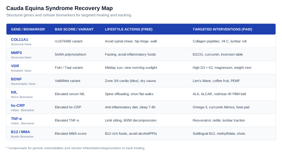
To monitor how well your spinal nerve roots are recovering and to detect ongoing inflammation or axonal stress, we must look at specific biological markers in the blood. The following six biomarkers offer a window into your nervous system's healing timeline.
Tracking Cellular Health: 6 Crucial Biomarkers to Monitor for Nerve Recovery
Biomarker 1: Neurofilament Light Chain (NfL)
Neurofilament Light Chain (NfL) is a structural scaffolding protein found exclusively within the axons of neurons. When the nerve roots of the cauda equina are subjected to severe physical compression or prolonged oxygen deprivation (ischemia), the axonal membrane is compromised, and NfL is released into the surrounding cerebrospinal fluid, eventually leaking into the bloodstream. Monitoring blood levels of NfL provides a direct, highly sensitive readout of active nerve fiber degeneration. This marker allows you to distinguish between chronic, stable nerve irritation and active, ongoing structural damage, making it incredibly useful for verifying that decompression surgery was successful or detecting a potential re-herniation early.
How to Measure It
Serum NfL is measured via a blood draw using highly sensitive immunoassay platforms (typically the Simoa assay). It is offered by specialized neurological laboratories and forward-thinking functional medicine clinics. The cost of a serum NfL test ranges from $150 to $300, and it is usually ordered to monitor axonal health over time.
If the Score is Bad: The Plan Without Supplements
An elevated NfL score indicates active, ongoing nerve fiber breakdown. The primary non-supplement strategy is complete mechanical offloading of the lumbosacral spine to prevent micro-motion from irritating the healing nerve roots. You must implement a strict spine hygiene protocol: eliminate all forward bending, twisting, and heavy lifting from your daily routine. Incorporate short, frequent walks (5 to 10 minutes, 3 to 4 times a day) on flat, even surfaces to promote circulation to the lumbar region without stressing the joints. Additionally, practice mid-day supine decompression: lie flat on your back with your knees bent and supported by a bolster for 15 to 20 minutes to reduce intradiscal pressure and allow the compressed nerves a quiet, load-free environment to rest.
If the Score is Bad: The Plan With Supplements or Equipment
To support axonal repair and protect compromised nerve cells from further degeneration, target the metabolic pathways of the nerves: - Alpha-Lipoic Acid (ALA): Take 600 mg twice daily on an empty stomach. ALA is a powerful antioxidant that improves nerve conduction velocity and helps restore mitochondrial function within the damaged axons. Take this dose for 12 weeks, then reduce to 300 mg daily for maintenance. Potential side effects include mild heartburn or acid reflux; avoid if you have a known thiamine deficiency. - Acetyl-L-Carnitine (ALCAR): Take 500 mg twice daily. ALCAR assists in the transport of fatty acids into the mitochondria, optimizing energy production within the nerve cells to fuel the regeneration process. Cycle this supplement by taking it for 8 weeks, followed by a 2-week break. Side effects can include mild insomnia if taken late in the evening. - Equipment: Use a home Photobiomodulation (PBM) / Low-Level Laser Therapy (LLLT) belt emitting red (660 nm) and near-infrared (850 nm) wavelengths. Apply the light directly to the skin over the lumbar spine for 12 to 15 minutes daily (targeting an energy density of 4 to 10 J/cm²). PBM stimulates cytochrome c oxidase within the neuronal mitochondria, increasing the ATP production required for axonal repair.
Biomarker 2: High-Sensitivity C-Reactive Protein (hs-CRP)
High-Sensitivity C-Reactive Protein (hs-CRP) is a standard blood marker that measures systemic inflammation in the body. While hs-CRP is produced by the liver in response to various inflammatory signals, elevated systemic inflammation directly impacts the local microenvironment of the healing cauda equina. High circulating levels of inflammatory molecules sensitize the spinal nerve roots, making them hyper-reactive to even minor mechanical pressures. A high hs-CRP score indicates that systemic inflammation is actively impeding the body's natural nerve repair mechanisms and prolonging neuropathic pain.
How to Measure It
Measured via a standard hs-CRP blood test. It is highly routine, widely available, and can be ordered by any primary care physician or obtained through online direct-to-consumer laboratory services. The cost is very low, typically ranging between $20 and $50.
If the Score is Bad: The Plan Without Supplements
Transition immediately to a strict anti-inflammatory dietary pattern. Eliminate all refined sugars, industrial seed oils, processed meats, and alcohol, as these foods directly stimulate liver CRP production. Prioritize wild-caught fatty fish, extra virgin olive oil, leafy green vegetables, and antioxidant-rich berries. Establish a strict sleep routine to secure 7 to 8 hours of deep, restorative sleep nightly, as sleep deprivation causes a sharp rise in inflammatory cytokines. Practice daily diaphragmatic breathing (the 4-7-8 technique) for 10 minutes morning and night; this exercises the vagus nerve, which activates the body's cholinergic anti-inflammatory pathway to lower systemic inflammation.
If the Score is Bad: The Plan With Supplements or Equipment
To suppress systemic inflammatory cascades and lower circulating CRP, use target compounds: - High-Dose Omega-3 Fatty Acids (EPA/DHA): Take 3,000 mg to 4,000 mg of combined EPA/DHA daily with meals. Omega-3 fatty acids serve as the direct precursors for specialized pro-resolving mediators (SPMs) that actively shut down the inflammatory response. Take this continuously; consult your physician if you are on blood-thinning medications, as high-dose omega-3s can have a mild anti-platelet effect. - Curcumin Phytosome (Meriva): Take 500 mg twice daily. This specific lecithin-formulated curcumin has superior bioavailability and downregulates key inflammatory transcription factors like NF-kB. Cycle this supplement by taking it for 12 weeks, followed by a 2-week break. Side effects are rare but can include mild digestive upset. - Equipment: Use a Far-Infrared Heating Pad on your lower back for 20 minutes daily. The deep-penetrating infrared heat increases local blood flow, helping to clear inflammatory cellular debris from the damaged spinal tissues and promote local healing.
Biomarker 3: Tumor Necrosis Factor-Alpha (TNF-α)
Tumor Necrosis Factor-Alpha (TNF-α) is a primary pro-inflammatory cytokine that plays a direct role in disc-mediated nerve damage. When a lumbar disc herniates, the inner nucleus pulposus material is squeezed out and exposed to the immune system. The immune cells identify this foreign material and release a massive flood of TNF-α. This cytokine causes severe chemical radiculitis—a chemical burn that inflames and irritates the adjacent cauda equina nerve roots, even if physical compression has been surgically relieved. High levels of TNF-α are strongly associated with intense, burning neuropathic pain and delayed nerve recovery.
How to Measure It
Measured using a serum TNF-α ELISA blood test. It is a specialized test available through major diagnostic laboratories, with costs generally ranging between $80 and $150.
If the Score is Bad: The Plan Without Supplements
Avoid any activity that increases intradiscal pressure, as this can force more inflammatory nucleus pulposus fluid onto the nerve roots. Limit continuous sitting to a maximum of 20 minutes, using a lumbar support cushion to maintain a neutral spine. Avoid bending forward to pick up objects; instead, squat down while keeping your back completely upright. Practice the "90/90 decompression" position: lie flat on your back on a rug or mat, and place your lower legs on a chair or ottoman so your hips and knees are bent at a 90-degree angle. Rest in this position for 15 minutes twice daily to decompress the discs and reduce local cytokine accumulation.
If the Score is Bad: The Plan With Supplements or Equipment
To inhibit the production of TNF-α and protect the nerve roots from chemical irritation: - Trans-Resveratrol: Take 250 mg to 500 mg daily with a fat-containing meal. Resveratrol is a potent polyphenol that directly inhibits the expression of TNF-α and protects disc cells from inflammatory breakdown. Cycle this by taking it for 8 weeks, followed by a 2-week break. Side effects may include mild stomach discomfort. - Stinging Nettle Root (Urtica dioica): Take 300 mg twice daily. Stinging nettle root contains compounds that act as natural TNF-α inhibitors. Cycle the supplement by taking it for 6 weeks, followed by a 1-week break. Avoid this supplement if you have chronically low blood pressure. - Equipment: Utilize a lumbar traction device or decompression table (under the supervision of a physical therapist) for 10 minutes at low tension, 3 times a week. Gentle traction opens up the disc space, lowering the intradiscal pressure that drives chemical cytokine release.
Biomarker 4: Interleukin-6 (IL-6)
Interleukin-6 (IL-6) is a dual-acting cytokine that plays a prominent role in chronic inflammatory states. In spinal nerve injuries, persistently elevated levels of IL-6 are associated with the transition from acute pain to chronic, centralized neuropathic pain. High IL-6 levels stimulate the immune cells in the dorsal root ganglion, causing the spinal cord and brain to become hypersensitive to pain signals (central sensitization). Monitoring IL-6 helps you determine if your recovery is stalled by a persistent, systemic inflammatory loop that prevents the cauda equina from entering a restorative, regenerative phase.
How to Measure It
Measured via a standard serum IL-6 blood test. It is widely available through commercial diagnostic laboratories and typically costs between $70 and $130.
If the Score is Bad: The Plan Without Supplements
Engage in regular, low-intensity cardiovascular exercise. Zone 2 exercise—such as walking at a pace where you can comfortably carry on a conversation—stimulates the release of anti-inflammatory myokines (like Interleukin-10) from your muscles, which naturally suppress circulating IL-6. Walk for 20 to 30 minutes daily on a flat, smooth path. Incorporate passive heat stress by using a dry sauna at 140°F to 160°F (60°C to 70°C) for 15 to 20 minutes, 3 times a week. The heat induces heat shock proteins that help resolve cellular stress and lower systemic IL-6 levels.
If the Score is Bad: The Plan With Supplements or Equipment
To suppress the cellular pathways that generate IL-6 and calm spinal hypersensitivity: - Quercetin Phytosome: Take 250 mg to 500 mg daily. Quercetin is a bioflavonoid that acts as a natural senolytic and mast cell stabilizer, reducing the release of IL-6. Take this daily for 12 weeks, followed by a 4-week break. Side effects are rare but can include mild, temporary headaches. - Apigenin: Take 50 mg to 100 mg daily, approximately 30 to 60 minutes before bed. Apigenin inhibits the inflammatory pathways that drive IL-6 production and supports GABA pathways to improve sleep quality. Take this continuously. May cause mild drowsiness, which is helpful for sleep. - Equipment: Use a Pulsed Electromagnetic Field (PEMF) mat. Laying on a PEMF mat at low frequencies (1 to 15 Hz) for 20 minutes twice daily helps to downregulate pro-inflammatory cytokines like IL-6 and restores the healthy electrical charge of damaged cell membranes.
Biomarker 5: Vitamin D (25-hydroxyvitamin D)
Vitamin D is a critical secosteroid hormone that regulates over 900 genes in the human body, many of which are directly involved in nervous system function and structural healing. Vitamin D stimulates the synthesis of neurotrophic factors (like BDNF and Nerve Growth Factor) that are mandatory for nerve regeneration and the remyelination of damaged nerve fibers. Furthermore, vitamin D is vital for maintaining the bone mineral density of the vertebral bodies and supporting the health of the cartilaginous disc endplates. Low levels of vitamin D are strongly linked to accelerated disc degeneration, spinal stenosis, and heightened pain sensitivity.
How to Measure It
Measured via a routine 25-hydroxyvitamin D [25(OH)D] blood test. This is an extremely common, highly affordable test costing between $30 and $60, and it is easily ordered during annual physical exams.
If the Score is Bad: The Plan Without Supplements
Maximize your skin's natural production of vitamin D by spending 15 to 30 minutes in direct, midday sunlight daily without sunscreen (ensuring your arms and legs are exposed, and being careful never to sunburn). Sensitize your hormone receptors by viewing outdoor morning sunlight within 30 minutes of waking. Increase your intake of vitamin D-rich foods, including wild-caught salmon, Atlantic mackerel, sardines canned in oil, pasture-raised egg yolks, and cod liver oil.
If the Score is Bad: The Plan With Supplements or Equipment
To rapidly raise your vitamin D levels and support spinal remodeling: - Vitamin D3 + Vitamin K2 (MK-7): Take 5,000 IU to 10,000 IU of Vitamin D3 daily, always paired with 100 mcg to 200 mcg of Vitamin K2. Vitamin K2 is vital; it activates the proteins that direct calcium into your bones and teeth, preventing the calcium from depositing in your blood vessels or spinal ligaments. Take this with a meal containing healthy fats. Retest your blood levels every 3 months, adjusting the dose to maintain a target serum level of 50 to 80 ng/mL. - Magnesium Glycinate: Take 200 mg to 400 mg daily before bed. Magnesium is a mandatory cofactor for the enzymes that activate vitamin D in the body. Side effects can include mild loose stools if the dose is too high. - Equipment: If sunlight is unavailable due to season or latitude, use a narrowband UVB medical lamp designed for vitamin D synthesis, following the manufacturer's exposure times closely.
Biomarker 6: Methylmalonic Acid (MMA) / Vitamin B12
Vitamin B12 is the primary nutrient required for the maintenance and repair of the myelin sheath—the protective coating that insulates the cauda equina nerve roots. When nerves are compressed, demyelination occurs, which disrupts nerve signals and causes numbness and muscle weakness. Measuring Methylmalonic Acid (MMA) is the gold standard for detecting functional vitamin B12 deficiency inside the cells. If MMA levels are high, it means your cells do not have enough B12 to repair the myelin sheath, even if your standard serum B12 blood test results appear normal.
How to Measure It
MMA can be measured through a specialized serum blood test or a urine test. It is available through major clinical laboratories and typically costs between $80 and $150.
If the Score is Bad: The Plan Without Supplements
Aggressively increase your dietary intake of bioavailable vitamin B12 by consuming animal-derived foods such as grass-fed beef liver, wild clams, oysters, sardines, pasture-raised beef, lamb, and eggs. If you follow a plant-based diet, incorporate high-quality nutritional yeast fortified with B12, but recognize that animal sources contain the most bioavailable forms. Completely avoid alcohol, as it severely damages the stomach lining and reduces B12 absorption. Additionally, check if you are taking medications like metformin or proton pump inhibitors (PPIs), which are known to block B12 absorption in the gut.
If the Score is Bad: The Plan With Supplements or Equipment
To saturate your nervous system with the active form of B12 needed for myelin repair: - Methylcobalamin (Sublingual B12): Take 1,000 mcg to 5,000 mcg daily. Sublingual tablets dissolve under the tongue and absorb directly into the bloodstream, bypassing any potential absorption issues in the gut. Take daily for 3 months, then retest your MMA levels. Side effects are virtually non-existent because excess B12 is safely excreted in the urine. - L-Methylfolate (5-MTHF): Take 400 mcg to 800 mcg daily. Methylfolate works in tandem with methylcobalamin in the methylation cycle, supporting the repair of peripheral nerve pathways. - Equipment: If you are experiencing severe neurological symptoms (such as foot drop or progressive numbness) and your MMA remains high, work with your doctor to receive intramuscular B12 injections (1,000 mcg of methylcobalamin) once or twice weekly for 4 to 6 weeks to quickly saturate your nerve tissues.
While tracking active inflammatory and degenerative biomarkers gives us real-time data on nerve healing, understanding your genetic blueprint allows you to identify the underlying structural vulnerabilities that may have predisposed you to this condition in the first place.
The Genomic Blueprint: 4 Key Genes Influencing Spinal Structure and Nerve Repair
Your genetic makeup plays a massive role in how resilient your intervertebral discs are to mechanical stress, and how efficiently your peripheral nerves regenerate after a compression injury. The following four genes are critical factors in spinal and neurological health.
Gene 1: COL11A1 (Collagen Type XI Alpha 1 Chain)
The COL11A1 gene provides instructions for making type XI collagen, a minor but crucial structural collagen found in the cartilage and the intervertebral discs. Type XI collagen acts as a structural scaffold, organizing the larger type II collagen fibers within the nucleus pulposus and the outer annulus fibrosus. Certain genetic variants (such as the rs1676486 polymorphism) lead to weaker collagen fibers, making the spinal discs less resilient to mechanical shear and axial loading. This structural vulnerability increases the risk of disc herniation, which can compress the cauda equina nerve roots. To compensate for this genetic weakness, you must build muscular spinal stability to take the physical load off the vulnerable collagen structures.
If the Gene is Bad: The Plan Without Supplements
Avoid all activities that place high shear stress on the spine, such as heavy deadlifts, squats, or twisting under load. Focus on building "torsional stiffness" in the core using neutral spine mechanics. Restructure your daily habits: hip-hinge for all bending, use the "log roll" technique to get out of bed, and avoid sitting in soft, unsupportive couches that force the lumbar spine into flexion. Engage in low-impact walking on flat terrain to stimulate passive nutrient exchange within the disc without mechanical wear.
If the Gene is Bad: The Plan With Supplements or Equipment
- Hydrolyzed Collagen Peptides (Type I and II): 10 to 15 grams daily. Consuming bioavailable collagen peptides provides the specific amino acid building blocks (glycine, proline, hydroxyproline) needed for disc matrix repair. Take daily, mixed into water or tea. Take continuously. - Vitamin C (Ascorbic Acid): 500 mg twice daily. Vitamin C is a mandatory enzymatic cofactor for the hydroxylation of proline and lysine during collagen synthesis. Take continuously. Side effects include mild stomach upset if taken on an empty stomach. - Silica (Choline-Stabilized Orthosilicic Acid): 5 mg to 10 mg daily. Silica promotes collagen cross-linking and strengthens connective tissues. Take continuously. - Equipment: Use an Ergonomic Lumbar Support Chair at your desk and a Spine-Aligning Lumbar Roll in your car. This maintains the natural lumbar lordosis, reducing the mechanical burden on the structurally weak collagen fibers of the lower spine.
Gene 2: MMP3 (Matrix Metalloproteinase-3)
The MMP3 gene encodes matrix metalloproteinase-3, an enzyme responsible for breaking down extracellular matrix proteins, including collagen and proteoglycans, in the spinal discs. While this breakdown is a normal part of tissue remodeling, certain genetic variants (such as the 5A/6A promoter polymorphism) cause an overproduction of MMP3. This leads to accelerated enzymatic degradation of the disc matrix, causing the discs to dehydrate, lose height, and herniate more easily. To compensate for high MMP3 activity, you must implement strategies that suppress chronic low-grade inflammation and inhibit excessive matrix metalloproteinase production.
If the Gene is Bad: The Plan Without Supplements
Incorporate intermittent fasting or time-restricted feeding (e.g., a 16-hour fast, 8-hour eating window). Fasting activates cellular autophagy and naturally downregulates matrix-degrading enzymes, including MMP3. Avoid foods that trigger systemic inflammation, particularly industrial trans fats, high-fructose corn syrup, and refined carbohydrates, as inflammation upregulates MMP3 expression. Practice moderate, non-impact spinal mobilization exercises (like gentle cat-camel stretches, keeping within a pain-free range) to encourage fluid flow in the discs without mechanical friction.
If the Gene is Bad: The Plan With Supplements or Equipment
- Epigallocatechin Gallate (EGCG / Green Tea Extract): 400 mg daily. EGCG is a potent natural inhibitor of matrix metalloproteinases, helping protect the disc matrix from enzymatic breakdown. Cycle 12 weeks on, 2 weeks off. Avoid taking on an empty stomach to prevent liver strain. - Curcumin (C3 Complex): 500 mg daily. Curcumin downregulates the inflammatory cytokines that stimulate MMP3 production. Take daily. - Equipment: A Lumbar Inversion Table (cautiously, setting the angle to no more than 15 to 30 degrees) can be used for 5 to 10 minutes daily. This gentle traction pulls fluid back into the dehydrated discs and counteracts the enzymatic degradation of the disc core.
Gene 3: VDR (Vitamin D Receptor)
The VDR gene encodes the receptor for vitamin D, which is responsible for intracellular vitamin D signaling. Specific polymorphisms in this gene (such as FokI, TaqI, and BsmI) alter the shape of the receptor, reducing its sensitivity and binding efficiency. As a result, even if you have normal levels of vitamin D in your blood, your cells cannot utilize it effectively. This lead to impaired bone mineral density in the vertebral bodies, weakened vertebral endplates, and accelerated disc degeneration. To compensate, you must maintain higher-than-average blood levels of vitamin D and supply the cofactors necessary to maximize receptor binding.
If the Gene is Bad: The Plan Without Supplements
Focus on maximizing natural vitamin D synthesis and absorption through lifestyle. Spend 20 to 30 minutes in direct midday sunlight daily, exposing a large surface area of your skin. Optimize your circadian biology by viewing morning sunlight within 30 minutes of waking, which helps sensitize your hormone receptors. Consume foods high in calcium and fat-soluble vitamins, such as grass-fed dairy (if tolerated), sardines with bones, wild-caught salmon, and egg yolks, to support the structural remodeling of your vertebrae.
If the Gene is Bad: The Plan With Supplements or Equipment
- High-Dose Vitamin D3 (Cholecalciferol): 5,000 IU to 10,000 IU daily to saturate the less-sensitive receptors. Aim to keep your blood levels at the upper end of the optimal range (70–80 ng/mL). Take with a fat-rich meal. - Vitamin K2 (MK-7): 100 mcg to 200 mcg daily. This is critical to prevent ectopic calcification and guide calcium directly into the bone matrix. - Magnesium Bisglycinate: 300 mg to 400 mg daily before bed. Magnesium is required for the conversion of vitamin D into its active form and enhances VDR sensitivity. - Equipment: Use a Weight Vest (cautiously, starting with only 2–5% of your body weight) during short, upright walks. The gentle vertical load stimulates osteoblast activity in the vertebral bodies, compensating for genetic bone density vulnerabilities.
Gene 4: BDNF (Brain-Derived Neurotrophic Factor)
The BDNF gene encodes brain-derived neurotrophic factor, a protein that plays a vital role in the survival, growth, and maintenance of neurons, including peripheral nerve fibers and the spinal cord. The Val66Met polymorphism in the BDNF gene results in a significantly reduced release of BDNF in response to physical activity or stress. Individuals with this variant have a slower rate of nerve regeneration and myelination following cauda equina compression, and may experience more persistent neuropathic pain. To compensate, you must engage in lifestyle habits and use supplements that strongly upregulate BDNF production.
If the Gene is Bad: The Plan Without Supplements
Engage in regular physical exercise that stimulates BDNF synthesis. Cardiovascular exercise, particularly intervals on a stationary upright bike (which protects the spine from impact and flexion), is highly effective. Aim for 20 to 30 minutes, 3 times a week, pushing your heart rate into Zone 3 or 4 for short bursts. Incorporate regular heat stress, such as a Finnish sauna session (20 minutes at 170°F / 80°C, 3 times a week), which has been shown to raise BDNF levels through the upregulation of heat shock proteins.
If the Gene is Bad: The Plan With Supplements or Equipment
- Lion's Mane Mushroom (Hericium erinaceus): 500 mg to 1,000 mg daily of a standardized extract containing active hericenones and erinacines. Lion's Mane stimulates the synthesis of Nerve Growth Factor (NGF) and BDNF, supporting myelination and axonal repair. Take daily; cycle 8 weeks on, 2 weeks off. Side effects are rare but watch for mild skin itching. - Whole Coffee Fruit Extract (WCFE): 100 mg daily. Clinical trials show that WCFE can increase circulating BDNF levels by up to 140%. Take in the morning to avoid sleep interference. - Equipment: Use a Pulsed Electromagnetic Field (PEMF) Device over the lumbosacral region. Low-frequency PEMF stimulation has been shown in clinical models to upregulate BDNF and NGF expression in damaged nerve pathways, accelerating functional recovery.
While molecular biomarkers and genetic testing provide a cellular framework for recovery, physical stabilization of the lumbar spine is the mechanical foundation upon which all neural healing occurs.
Rebuilding the Spine: Key Takeaways from Dr. Stuart McGill's Spine Rehabilitation Protocols
To protect the healing cauda equina nerve roots from mechanical compression and friction, you must eliminate excessive spinal movement and build core endurance. Dr. Stuart McGill's clinical work offers ten critical principles that challenge conventional spine care and lay out a clear path to recovery.
1. Banish Flexion and Embrace the Hip Hinge
Bending forward at the waist (lumbar flexion) puts immense pressure on the posterior aspect of the intervertebral discs, squeezing the nucleus pulposus outward toward the cauda equina nerve roots. To spare your spine, you must replace all waist bending with a clean "hip hinge." This movement involves keeping the spine in a neutral position while pivoting entirely at the hip joints, keeping the weight close to the body's center of gravity.
2. Build Stability with the McGill Big Three
The McGill Big Three is a trio of exercises designed to build endurance and stability in the musculature surrounding the spine without exposing the lumbar segments to dangerous motion. The exercises are: - The McGill Curl-up: lying supine with one leg straight, one knee bent, hands under the lumbar spine to preserve the natural curve, and lifting the head and shoulders slightly off the floor. - The Side Bridge: supporting the body on the elbow and knees (or feet) to strengthen the obliques and quadratus lumborum. - The Bird Dog: on hands and knees, extending the opposite arm and leg while maintaining a completely flat, stable back.
3. Pinpoint and Eliminate Your Pain Triggers
Rather than labeling your condition as general "back pain," you must identify the specific postures, loads, or movements that trigger your radicular symptoms. For instance, is your pain triggered by sitting (flexion-intolerant), standing (extension-intolerant), or walking (load-intolerant)? Once identified, you can systematically remove these triggers from your daily life, giving the inflamed cauda equina nerve roots a quiet environment in which to heal.
4. Prioritize Core Stiffness Over Flexibility
A common misconception is that stretching the lower back is beneficial for spinal recovery. In reality, stretching the lumbar spine destabilizes the joints and exposes the compressed nerve roots to micro-movements that delay healing. The core's true function is to act as a brace or corset that prevents motion. Focus on building muscle endurance and structural stiffness rather than chasing flexibility.
5. Implement the Interval Walking Routine
Passive, flat walking is a powerful rehabilitation tool. McGill recommends a specific walking protocol: 3 short, fast-paced walks (10 to 15 minutes each) spread throughout the day, rather than one long, exhausting walk. Fast walking swings the arms and hips in a way that naturally flushes the nerve pathways and creates a gentle, dynamic pumping action that helps rehydrate the spinal discs.
6. Adopt the Virtual Surgery Mindset
In the acute phase of recovery, treat your spine as if you have just undergone major surgery, even if you did not. This "virtual surgery" concept means eliminating any movements that stretch, twist, or load the spine to its limits. By behaving as if your spine is fragile and healing, you protect the delicate nerve fibers from micro-trauma, allowing the initial inflammatory cascade to subside.
7. Stop Performing Knee-to-Chest Stretches
Many physical therapy programs recommend pulling your knees to your chest to "stretch out" the lower back. McGill warns that this stretch triggers the stretch reflex in the back muscles, providing temporary pain relief while actually flattening the lumbar lordosis and squeezing the disc material backward, directly compressing the cauda equina. Replace this stretch with neutral spine lying.
8. Protect the Spine in the Vulnerable Morning Window
Overnight, your spinal discs absorb water and swell, making them highly pressurized and vulnerable during the first two hours after waking. Bending, twisting, or lifting heavy objects early in the morning carries a much higher risk of causing a disc herniation. Avoid aggressive exercises during this window, and instead, focus on tall posture and light, upright walking.
9. Restore Hip Mobility to Unburden the Lumbar Spine
If your hips are stiff and lack mobility, your body will compensate by forcing the lumbar spine to bend and twist during daily movements. To keep your lower back stable and quiet, you must restore clean movement to your hip joints. Perform gentle hip mobilization exercises, such as the 90/90 stretch or passive hip extensions, ensuring the movement occurs entirely at the hip joint without bending the lumbar spine.
10. Progress from Stabilization to Functional Movement
Recovery is a gradual progression. Start with basic muscle bracing in bed, move to the McGill Big Three, progress to flat walking, and finally transition to functional movements like squatting and lifting using a hip hinge. Never rush the progression; if a movement triggers radicular pain or numbness in the saddle area, immediately step back to the previous stage of stabilization.
While physical stabilization and cellular optimization form the core of a recovery protocol, complementary clinical modalities can provide additional support for nerve regeneration, pain modulation, and tissues healing.
Complementary and Alternative Modalities for Nerve Recovery
The following five complementary modalities have demonstrated meaningful scientific evidence for supporting nerve regeneration, managing chronic neuropathic pain, or reducing spinal muscle tension. Each should be approached with caution and integrated under professional guidance.
1. Low-Level Laser Therapy / Photobiomodulation
Low-level laser therapy (LLLT), also known as photobiomodulation (PBM), involves applying specific wavelengths of red and near-infrared light to damaged tissue. In the recovery of cauda equina syndrome, this therapy is highly relevant because the light penetrates deep into the spinal tissues, stimulating the mitochondria within injured nerve cells. This energy boost accelerates adenosine triphosphate (ATP) synthesis, which is crucial for fuel-dependent axonal regeneration and myelin sheath repair.
Clinical protocols typically utilize wavelengths between 800 nm and 850 nm for deep tissue penetration, delivering an energy density of 4 to 10 J/cm² over the affected lumbar segments. Systematic reviews indexed on PubMed demonstrate that this therapy increases the number of myelinated fibers, improves the organization of the myelin sheath, and accelerates functional recovery in damaged peripheral nerves.
To apply this therapy realistically, seek a clinical practitioner who uses a Class 3B or Class 4 cold laser device. Ensure the laser is positioned directly over the L4-S1 spinal levels where the cauda equina nerve roots originate. A typical schedule consists of 2 to 3 sessions per week for 6 to 8 weeks, making sure to immediately discontinue the treatment if you experience a temporary increase in neuropathic pain.
2. Mindfulness-Based Stress Reduction (MBSR)
Mindfulness-Based Stress Reduction (MBSR) is a structured program that teaches mindfulness meditation, body awareness, and gentle movement. For individuals recovering from cauda equina syndrome, the intense anxiety surrounding bowel/bladder function and potential permanent nerve damage can trigger a state of constant fight-or-flight. MBSR helps downregulate the sympathetic nervous system, reducing the emotional amplification of neuropathic pain and decreasing systemic inflammatory cytokines.
The standard MBSR protocol involves an 8-week course consisting of weekly group sessions, a full-day retreat, and daily home meditation practices. A landmark randomized clinical trial published in JAMA, which can be found in the PubMed database, demonstrated that MBSR significantly reduced pain intensity and improved functional limitations in adults suffering from chronic low back pain compared to standard medical care.
To apply this cautiously, enroll in a certified 8-week MBSR program, either online or in person. During the gentle yoga and body-scan exercises, strictly avoid any physical postures that require bending, twisting, or compressing your lumbar spine. Focus entirely on the breathing and mental awareness components, adapting all physical movements to keep your spine in a neutral, supported position.
3. Progressive Muscle Relaxation (PMR)
Progressive Muscle Relaxation (PMR) is a somatic technique that involves systematically tensing and then releasing specific muscle groups throughout the body. Following a severe spinal injury like cauda equina syndrome, the body naturally develops protective muscle guarding—chronic tension in the lower back, hips, and gluteal muscles designed to prevent movement. While protective in the acute phase, chronic muscle guarding can lead to painful muscle spasms and restrict blood flow to the healing nerve roots.
The standard PMR protocol consists of tensing a muscle group for 5 to 7 seconds, then releasing the tension and focusing on the sensation of relaxation for 15 to 20 seconds, progressing from the feet up to the face. Clinical reviews indexed on PubMed suggest that PMR is a safe and effective adjunct therapy that reduces chronic low back pain scores and improves overall sleep quality.
Apply PMR cautiously by performing the technique while lying supine on a firm mattress with a pillow supporting your knees to keep your spine neutral. When tensing the muscles of your lower back, abdomen, and gluteals, apply only 10% to 20% of your maximum contraction force to avoid triggering muscle spasms or spinal micro-motion. Perform the routine for 15 minutes before bed to aid sleep and reduce nocturnal muscle tension.
4. Microbiome-Directed Therapies
Microbiome-directed therapies involve modulating the gut microbiota through diet, prebiotics, and targeted probiotic strains to influence systemic health. This modality is relevant because of the "gut-spine axis," where gut dysbiosis triggers a leaky gut, releasing lipopolysaccharides (LPS) into the bloodstream and elevating systemic cytokines like TNF-α and IL-6. This systemic inflammation directly irritates the cauda equina nerve roots and accelerates intervertebral disc degeneration.
Interventions typically involve taking high-potency probiotics containing strains of Bifidobacterium and Lactobacillus, alongside dietary prebiotics like inulin or green banana flour. Mendelian randomization and animal models indexed on PubMed show a clear causal connection between specific gut bacterial taxa and disc degeneration, showing that restoring gut microbial balance can reduce inflammatory markers and protect spinal structures.
To apply this, work with a practitioner to complete a stool analysis or begin by slowly introducing prebiotic-rich foods into your diet. Add a high-quality, multi-strain probiotic (minimum 20 billion CFU daily) and monitor for gas or bloating. Focus on a high-fiber, low-sugar diet to sustain beneficial, anti-inflammatory gut species, ensuring any dietary changes are introduced gradually.
5. Qigong
Qigong is a traditional Chinese practice combining gentle, slow movements, breathing exercises, and focused meditation. For cauda equina syndrome recovery, Qigong is highly beneficial because it encourages energy and fluid flow through the pelvis and legs without subjecting the lower back to impact or heavy loads. The slow movements help rebuild motor control, balance, and leg strength, which are often compromised by cauda equina nerve injury.
A common Qigong form used for spinal health is Baduanjin (Eight Silken Movements), which focuses on gentle stretching and alignment. Clinical trials and reviews indexed on PubMed indicate that Qigong practices lead to significant improvements in pain relief, functional movement, and quality of life for individuals with chronic spinal pain.
To practice safely, learn from an instructor who understands spinal conditions, or perform the exercises while seated if your balance or leg strength is compromised. Ensure that you do not perform any deep back bends or extreme rotations. Focus on the breathing and gentle arm movements, keeping your lower back in a stable, neutral alignment, and immediately stop if you feel any tingling or weakness in your legs.
Integrating these physical and biological modalities creates a comprehensive strategy that supports both the structure of your spine and the regeneration of your nerves.
Conclusion
Recovering from Cauda Equina Syndrome requires moving beyond generic back pain advice and addressing the specific biological and mechanical factors that influence nerve regeneration and disc health. By tracking cellular biomarkers like Neurofilament Light Chain and systemic inflammatory cytokines, you can obtain objective data on your body's healing progress. Understanding your genetic baseline—including key structural genes like COL11A1 and metabolic receptors like VDR—helps you identify potential structural weak points and customize your nutritional and lifestyle choices to bypass these bottlenecks. When combined with the spine-sparing principles of Dr. Stuart McGill and evidence-based complementary therapies, this data-driven approach shifts your recovery from a process of passive waiting to one of active, informed management. The next smart step is to work with your healthcare team to establish your baseline biomarker and genetic profiles, evaluate your daily spinal biomechanics, and implement a structured, gradual recovery plan tailored to your unique biology.