This article contains AI generated content.
· UpdatedCeliac Disease Arthropathy: 5 Genes And 7 Biomarkers To Track
Introduction
Joint pain that comes alongside a celiac diagnosis often gets treated as a side note. The conversation quickly shifts back to intestinal health, antibody levels, and dietary compliance — and the arthropathy gets filed under "it should improve once you go gluten-free." Sometimes it does. Often, it does not, or not fully, and the person is left managing pain that no one seems willing to investigate in depth.
The problem is not that gluten-free advice is wrong. The problem is that it is not sufficient on its own for many people. Two individuals with the same celiac diagnosis can have completely different inflammatory profiles, different genetic risk factors, and different reasons why their joints are reacting the way they are. Blanket guidance cannot account for that variation.
This article takes a more granular approach. Rather than repeating what is already known, it focuses on what can be measured and what those measurements can tell you. There are specific genetic variants that increase the likelihood of arthropathy in celiac disease, and there are blood markers that reflect ongoing immune activity, nutritional deficiency, and inflammatory load in ways that symptoms alone cannot capture. Both layers of information lead to better decisions.
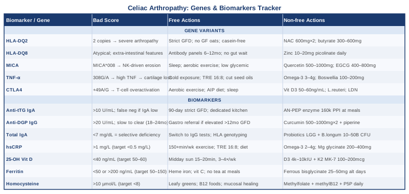
Two complementary paths are covered here. The first — and most immediately actionable — is a set of seven biomarkers worth tracking regularly, with practical guidance on what each one reveals, how to test for it affordably, and what to do when a result is abnormal. The second is a look at five gene variants that help explain why some celiac patients develop significant joint disease and others do not, including response plans for each. The article also covers a book that reframes celiac disease from a gut condition into a systemic immune disorder, and a set of evidence-based complementary approaches with specific protocols.
Summary
This article covers seven biomarkers — anti-tTG IgA, anti-DGP IgG, total IgA, hsCRP, vitamin D, ferritin, and homocysteine — with detailed protocols for when each comes back abnormal, both with and without supplementation. It then covers five gene variants (HLA-DQ2, HLA-DQ8, MICA, TNF-α, and CTLA4) that shape how severe celiac arthropathy becomes, and what can be done about each one. Beyond the core tracking strategies, you will find a summary of Gluten Freedom by Dr. Alessio Fasano — whose research fundamentally changed how the medical community understands celiac disease — with ten key insights that challenge standard clinical assumptions. Finally, five complementary modalities are covered, including the Autoimmune Protocol, microbiome therapy, yoga, mindfulness, and low-level laser therapy, each grounded in specific clinical evidence for joint and autoimmune applications. The goal throughout is fewer guesses and more targeted decisions.
7 Biomarkers to Track in Celiac Disease Arthropathy
Most people with celiac disease are tested at diagnosis, told to go gluten-free, and scheduled for annual antibody checks. That is a reasonable starting point, but it leaves a significant amount of useful information untouched — especially when joint symptoms are part of the picture. The seven markers below capture different aspects of what drives arthropathy in celiac disease: immune activation, nutritional depletion, inflammatory load, and barrier dysfunction. Together, they give a much clearer picture than any single marker or symptom report can provide.
1. Anti-tTG IgA (Anti-Tissue Transglutaminase IgA)
Why it matters
Anti-tTG IgA is the primary diagnostic and monitoring marker for celiac disease, but its relevance to arthropathy specifically is often overlooked. The immune system in celiac disease attacks not just intestinal tissue but tissue transglutaminase wherever it appears in the body — including in synovial joint tissue, muscle, and neurological structures. This means the same antibody that damages villi can directly contribute to joint inflammation. Persistently elevated anti-tTG IgA after diagnosis is a reliable signal that immune activation is ongoing, and ongoing immune activation in a joint predicts continued synovial damage.
How to measure it
Anti-tTG IgA is available through any major diagnostic lab. Cost ranges from $30 to $80 without insurance. Reference range: below 4 U/mL in most labs; above 10 U/mL is clearly elevated. Always order total IgA alongside this test, because IgA deficiency — which is 10 to 15 times more common in celiac patients than the general population — will produce a false negative and completely invalidate the result.
If the score is bad, the plan without supplements
The first step is a thorough dietary audit with a registered dietitian who specializes in celiac disease. Hidden gluten sources account for persistent elevation far more often than deliberate dietary transgression: shared cookware and cutting boards, processed foods with ambiguous labeling, certain medications and supplements with gluten excipients, cosmetics containing wheat derivatives, and certified gluten-free oats (which a subset of celiac patients cannot tolerate even when processed correctly). A 90-day strict audit — eliminating all ambiguous products, cooking only on dedicated equipment, eating only at home or in well-vetted restaurants — will reliably lower anti-tTG IgA if diet is the issue.
If the score is bad, the plan with supplements or equipment
No supplement replaces dietary compliance, but for situations where perfect gluten avoidance is structurally difficult (travel, institutional food environments), enzyme preparations containing prolyl endopeptidase (AN-PEP) can degrade small amounts of gluten in the stomach before it reaches the intestine. Clinical studies have examined dosing around 160,000 PPI units taken at the start of a meal. This is a harm-reduction tool for accidental exposure, not a license to consume gluten intentionally. See relevant clinical evidence on PubMed. No significant side effects have been reported in short-term use.
2. Anti-DGP IgG (Deamidated Gliadin Peptide Antibodies)
Why it matters
Anti-DGP IgG is a newer and more specific marker that captures immune reactivity to the chemically modified form of gliadin that the immune system recognizes most aggressively. It is particularly important in two circumstances: when total IgA is low (making IgA-based tests unreliable), and when seronegative or atypical celiac disease is suspected. From an arthropathy standpoint, IgG-mediated immune activity in synovial tissue may operate independently of gut mucosal healing, which means joint symptoms can persist even as intestinal markers normalize. Tracking anti-DGP IgG separately can reveal immune activity that anti-tTG IgA is no longer detecting.
How to measure it
Available as part of a combined celiac panel from any standard lab. Cost: $40–$90. Reference range: below 20 U/mL in most assays. An important clinical note: anti-DGP IgG takes longer to normalize on a strict gluten-free diet than anti-tTG IgA — sometimes 18 to 24 months — so a gradual decline over time is more meaningful than expecting rapid normalization.
If the score is bad, the plan without supplements
Persistent elevation at 12 months on a confirmed strict gluten-free diet warrants referral to a gastroenterologist with celiac expertise for evaluation of refractory celiac disease. This is a more serious condition requiring biopsy-based assessment and sometimes immunosuppressive treatment. For joint-specific management, rheumatology co-management helps determine whether ongoing arthropathy is driven by active immune disease versus established structural joint damage that has become independent of celiac activity.
If the score is bad, the plan with supplements or equipment
Curcumin as a standardized extract (500–1000 mg twice daily with food, paired with piperine for bioavailability) has modest but consistent anti-inflammatory effects on IgG-driven immune pathways and is well tolerated at these doses. Cycling at 8–12 week intervals is a practical approach; most people tolerate it continuously at standard doses. High doses above 2g/day can cause gastrointestinal discomfort in some people. This is a supportive measure, not an alternative to investigating the underlying cause of elevated antibodies.
3. Total IgA
Why it matters
Total IgA should be measured alongside every celiac antibody panel, yet it is frequently omitted. The reason it matters: selective IgA deficiency (serum IgA below 7 mg/dL) occurs in roughly 1 in 500 people in the general population but is approximately 10 to 15 times more common in celiac disease. When IgA is deficient, both anti-tTG IgA and anti-EMA tests will return false negatives, meaning celiac disease can go undetected for years. In an arthropathy context, this translates to ongoing undetected gluten exposure, ongoing immune activation in joints, and progressive damage that could have been intercepted earlier.
How to measure it
Serum IgA is a standard test, typically $20–$40. Normal adult range: 70–400 mg/dL. Selective IgA deficiency is defined as below 7 mg/dL. This is not a test to recheck frequently — IgA status is generally stable — but it needs to be known before any antibody-based monitoring is interpreted.
If the score is bad, the plan without supplements
If IgA is low or absent, the diagnostic approach shifts entirely to IgG-based markers: anti-DGP IgG and anti-gliadin IgG. HLA-DQ2/DQ8 genotyping offers another pathway to confirm or rule out genetic susceptibility without relying on antibody levels. No lifestyle or dietary intervention restores IgA deficiency, which is structurally encoded.
If the score is bad, the plan with supplements or equipment
IgA deficiency itself is not correctable through supplementation. However, supporting mucosal immune health through daily probiotics — particularly Lactobacillus rhamnosus GG and Bifidobacterium longum in combinations providing 10–50 billion CFU — may improve the resilience of mucosal defenses even when secretory IgA is structurally limited. This is a supportive measure for overall gut-immune function, not a correction of the underlying deficiency.
4. High-Sensitivity CRP (hsCRP)
Why it matters
High-sensitivity CRP is the most accessible systemic inflammation marker and one of the most clinically informative for distinguishing the drivers of joint pain. In celiac arthropathy, elevated hsCRP suggests active autoimmune-driven synovitis that is still responsive to immune management. Normal CRP with persistent pain suggests either established mechanical joint damage, nutrient-deficiency-driven joint fragility, or another concurrent process. Knowing which is dominant changes the entire approach. Peter Attia has emphasized targeting hsCRP below 0.5 mg/L as an optimal systemic health benchmark, well below the conventional "normal" cutoff of less than 10 mg/L that most labs use.
How to measure it
The high-sensitivity version specifically — standard CRP is not sensitive enough to detect chronic low-grade inflammation. Cost: $15–$40 at standard labs. Optimal target: below 1.0 mg/L for inflammation management; below 0.5 mg/L as a precision medicine goal. Always measure fasting and outside of any active infection or acute illness, both of which will produce acute spikes that are not informative about chronic autoimmune activity.
If the score is bad, the plan without supplements
The most evidence-backed lifestyle interventions for lowering hsCRP in celiac arthropathy are, in rough order of impact: rigorous gluten avoidance (the anti-inflammatory benefit of dietary compliance is well documented across multiple studies), regular moderate-intensity aerobic exercise at 150+ minutes per week, sleep optimization to 7–9 hours consistently, and an anti-inflammatory dietary pattern centered on fatty fish, leafy greens, berries, and olive oil while reducing refined grains and processed seed oils. Time-restricted eating (16:8 pattern) consistently lowers CRP in clinical trials independent of caloric intake.
If the score is bad, the plan with supplements or equipment
Omega-3 fatty acids (EPA + DHA combined, 2–4 grams per day) have among the strongest evidence for reducing hsCRP of any supplement, across multiple randomized controlled trials. Choose products with third-party testing for heavy metals and at least 60% combined EPA/DHA content by weight. Take with a meal containing fat. No cycling needed; long-term use is well-supported. Magnesium glycinate (200–400 mg at bedtime) reduces inflammatory signaling in magnesium-deficient individuals and is well tolerated. Vitamin D optimization — addressed in the next biomarker — also consistently lowers CRP when baseline levels are below 30 ng/mL.
5. 25-Hydroxyvitamin D
Why it matters
Vitamin D deficiency is nearly universal in newly diagnosed celiac disease. The proximal duodenum, which is specifically damaged by celiac disease, is also the primary site of fat-soluble vitamin absorption. This means vitamin D depletion is a near-inevitable consequence of untreated or poorly controlled celiac disease, and it does not resolve quickly even after the diet is corrected — intestinal healing takes 12 to 24 months or more. Beyond its well-known role in bone metabolism, vitamin D functions as a critical immune modulator, suppressing the Th1 and Th17 inflammatory responses that drive autoimmune joint disease. Low vitamin D amplifies synovial inflammation, worsens pain perception thresholds, and correlates with extra-intestinal manifestations of celiac disease including arthropathy. See relevant studies on PubMed.
How to measure it
Serum 25-hydroxyvitamin D. Cost: $30–$60. Most conventional labs flag anything above 30 ng/mL as sufficient, but researchers and clinicians including Peter Attia target 40–70 ng/mL for optimal immune and musculoskeletal function, with 50–60 ng/mL as a practical target. Measure twice yearly — spring and autumn — to track seasonal variation, which is clinically significant at northern latitudes.
If the score is bad, the plan without supplements
Regular midday sun exposure — 15 to 20 minutes on arms and legs, without sunscreen, three to four times per week — raises vitamin D meaningfully in lighter-skinned individuals in favorable latitudes. Dietary sources (fatty fish, egg yolks, fortified dairy alternatives) contribute modestly but are rarely sufficient to correct significant deficiency on their own. Tracking sun exposure as part of a daily habit is low-cost and has no downside within these parameters.
If the score is bad, the plan with supplements or equipment
Vitamin D3 (cholecalciferol) is the standard supplementation form. For celiac patients with documented deficiency, loading doses of 4,000–10,000 IU per day may be used under supervision, with retesting at 12 weeks to calibrate. Always co-administer vitamin K2 (100–200 mcg as MK-7 form) when using high-dose D3, to support proper calcium routing and reduce arterial calcification risk. Once serum levels are maintained in the target range, a maintenance dose of 2,000–5,000 IU daily is typically sufficient. No cycling is required; this is a long-term maintenance strategy. Toxicity is unlikely below 10,000 IU/day in adults but retesting is essential before sustained high-dose use.
6. Serum Ferritin and Iron Studies
Why it matters
Iron-deficiency anemia is the single most common presenting symptom of celiac disease in adults, driven by impaired iron absorption in the proximal small intestine — the zone most devastated by villous atrophy. But ferritin, the storage form of iron, matters for reasons that extend beyond anemia. Low ferritin independently increases fatigue, impairs immune regulation, and elevates the inflammatory tone in joint tissue. There is also an interpretation trap: ferritin is an acute-phase reactant, meaning active celiac disease can inflate ferritin levels, masking underlying depletion. Thomas Dayspring has highlighted that ferritin above 200 ng/mL in the context of systemic inflammation carries its own risk — elevated ferritin can reflect iron-driven oxidative stress, not iron sufficiency.
How to measure it
A complete iron panel includes serum ferritin, serum iron, TIBC (total iron binding capacity), and transferrin saturation. Cost: $40–$80 for the full panel. Functional target range for ferritin: 50–150 ng/mL. Transferrin saturation below 20% indicates functional iron deficiency even if ferritin appears borderline. Measure every six months during active celiac management.
If the score is bad, the plan without supplements
Intestinal healing is the highest-leverage intervention: only healed mucosa can absorb iron effectively. Dietary sources of heme iron — red meat, poultry, shellfish — are absorbed at 15–35% efficiency versus 2–8% for non-heme plant sources. Vitamin C paired with non-heme iron foods significantly improves absorption. Avoiding coffee, tea, and calcium within 60 minutes of iron-rich meals prevents major absorption inhibition.
If the score is bad, the plan with supplements or equipment
Ferrous bisglycinate (iron chelate) is the preferred supplemental form for celiac patients — better tolerated and better absorbed than standard ferrous sulfate. Dosing every other day rather than daily has been shown to improve net absorption in clinical studies, because daily dosing raises hepcidin (the iron-regulatory hormone) and blocks absorption the following morning. See evidence on alternate-day iron dosing. Practical dose: 25–50 mg elemental iron on alternating days, taken on an empty stomach or with vitamin C, away from calcium, zinc, or magnesium. Retest every 8–12 weeks to guide duration.
7. Homocysteine
Why it matters
Elevated homocysteine is one of the most underrecognized consequences of ongoing or poorly managed celiac disease. Homocysteine — a sulfur-containing amino acid — rises when folate, vitamin B12, and vitamin B6 are deficient, all of which are commonly malabsorbed in celiac disease due to proximal intestinal damage. Beyond its cardiovascular implications, elevated homocysteine directly promotes synovial inflammation and contributes to cartilage degradation, making it directly relevant to joint disease. Levels above 15 µmol/L are associated with worsened joint pathology in multiple autoimmune conditions. Most people with active celiac arthropathy have never had their homocysteine measured, which represents a significant gap in standard monitoring.
How to measure it
Fasting serum homocysteine. Cost: $30–$60. Conventional labs flag above 15 µmol/L as elevated, but many precision medicine practitioners (including Peter Attia) target below 8–10 µmol/L for optimal long-term health. Measure annually at minimum; more frequently when B-vitamin malabsorption is suspected or has been documented.
If the score is bad, the plan without supplements
Address the root: malabsorption from active celiac disease. Confirming mucosal healing through repeat antibody monitoring and, if indicated, follow-up endoscopy is the upstream fix. Once absorption normalizes, dietary sources of folate (dark leafy greens, legumes), B12 (animal proteins, particularly liver), and B6 (poultry, fish, potatoes) will typically bring homocysteine back into range without supplementation.
If the score is bad, the plan with supplements or equipment
A methylated B-vitamin complex is the most efficient supplemental intervention: methylfolate (L-5-MTHF, 400–800 mcg), methylcobalamin (B12, 500–1000 mcg), and pyridoxal-5-phosphate (B6, 25–50 mg). Methylated forms bypass the MTHFR enzyme, which many people — including a disproportionate share of autoimmune patients — cannot activate efficiently. Take daily with food; no cycling needed. Important constraint: B6 doses above 100–200 mg per day sustained long-term can cause peripheral neuropathy — stay within functional supplement ranges. Retest homocysteine at 8–12 weeks to confirm response.
5 Gene Variants That Shape the Celiac-Arthropathy Connection
Genetic testing cannot change a diagnosis or replace dietary management, but it provides a layer of explanation that clinical markers alone cannot. For celiac arthropathy specifically, the gene variants below help answer a persistent question: why do some celiac patients develop significant joint disease while others with the same diagnosis and comparable dietary compliance never have joint symptoms? The answer is partly in these five genes, and knowing which variants you carry shapes both the interpretation of your clinical markers and the choice of targeted interventions.
1. HLA-DQ2 (HLA-DQA1*05 / HLA-DQB1*02)
HLA-DQ2 is the single most important genetic risk factor for celiac disease: approximately 90–95% of celiac patients carry it. The HLA-DQ2 protein is expressed on antigen-presenting cells throughout the body — not just in the gut — and it binds deamidated gliadin peptides with unusually high affinity, presenting them to CD4+ T cells and triggering the autoimmune cascade. HLA-DQ2 homozygosity — carrying two copies rather than one — is associated with earlier disease onset, more severe intestinal damage, and a higher burden of extra-intestinal manifestations including arthropathy. See relevant literature on PubMed.
If the gene is bad — the plan without supplements
HLA status cannot be changed, but its downstream effects can be modulated significantly. For DQ2 homozygotes, the tolerance threshold for gluten is lower than for heterozygotes — even trace-level exposure that would be clinically silent in another celiac patient may sustain immune activation in synovial tissue. Practical steps: dedicated gluten-free kitchen equipment, stringent evaluation of any processed food labeled gluten-free, and consideration of eliminating certified gluten-free oats (which approximately 10% of celiac patients react to). A 90-day trial eliminating casein-containing dairy may also help, as some evidence suggests cross-reactivity between casein peptides and gliadin in high-risk HLA genotypes.
If the gene is bad — the plan with supplements or equipment
N-acetyl cysteine (NAC, 600 mg twice daily) has shown capacity to reduce T-cell-driven oxidative stress in autoimmune contexts, and it supports glutathione replenishment, which is chronically depleted in active celiac disease. Cycle 8–12 weeks on with a 2–4 week break. Butyrate supplementation (sodium butyrate, 300–600 mg per day) supports intestinal barrier integrity and dampens pro-inflammatory NF-κB signaling downstream of HLA-DQ2 activation. Both are low-risk supplements with strong mechanistic rationale; neither replaces strict dietary management.
2. HLA-DQ8 (HLA-DQA1*03 / HLA-DQB1*0302)
HLA-DQ8 is the second major genetic risk variant, carried by approximately 5–10% of celiac patients who do not carry HLA-DQ2. Clinically, HLA-DQ8-associated celiac disease tends to present with more atypical and extra-intestinal features — arthropathy, dermatitis herpetiformis, and neurological manifestations appear proportionally more often than in DQ2-positive disease, even when gut symptoms are mild. This makes HLA-DQ8 specifically relevant to any celiac patient whose joint disease seems disproportionate to their digestive symptoms.
If the gene is bad — the plan without supplements
Dietary management principles are the same, but the atypical presentation pattern means regular monitoring through antibody testing is especially important — waiting for digestive symptoms to indicate disease activity will systematically miss the signal. Every 6–12 month antibody panels (anti-tTG IgA, anti-DGP IgG) regardless of how the gut feels is the appropriate surveillance approach for HLA-DQ8-positive individuals.
If the gene is bad — the plan with supplements or equipment
Zinc supplementation (10–20 mg elemental zinc as zinc picolinate or glycinate, daily with food) supports intestinal barrier repair and modulates T-helper cell balance, both directly relevant to HLA-DQ8-driven immune activation. Monitor copper levels if using for more than three months, since chronic zinc supplementation above 40 mg/day can induce copper deficiency — at standard doses this is not a concern, but worth tracking in the context of ongoing celiac-related nutrient depletion.
3. MICA (MHC Class I Chain-Related Gene A)
MICA is a stress-induced protein expressed on epithelial and synovial cells in response to cellular stress — including the stress caused by gluten-driven immune activation in gut and joint tissue. Specific MICA gene variants, particularly MICA*008 (found in strong genetic linkage with HLA-B8), are associated with more severe villous atrophy and with elevated risk of arthropathy independent of HLA-DQ2/DQ8 status. MICA proteins activate NK cells and cytotoxic T lymphocytes in synovial tissue, driving cartilage erosion that can become structurally independent of ongoing gluten exposure over time. See relevant research on PubMed.
If the gene is bad — the plan without supplements
MICA expression is upregulated by cellular stress, which means reducing systemic and gut epithelial stress is the most direct intervention available. This involves rigorous gluten avoidance (the primary stressor), consistent moderate aerobic exercise (which reduces systemic oxidative stress over time), sleep optimization (sleep deprivation is a potent inducer of cellular stress pathways), and elimination of smoking, which independently increases MICA expression through NF-κB activation. A low-glycemic dietary pattern further reduces the metabolic stress signals that induce MICA upregulation.
If the gene is bad — the plan with supplements or equipment
Quercetin (500–1000 mg per day as a standardized extract, paired with bromelain or piperine for bioavailability) has demonstrated capacity to suppress NF-κB-mediated inflammation, the signaling pathway most upstream of MICA induction. Cycle at 8 weeks on, 2 weeks off. EGCG from green tea extract (400–800 mg per day standardized to 50% EGCG) has similar mechanistic support and is well tolerated at these doses. Both are reasonable additions for MICA-positive individuals with persistent joint inflammation despite dietary compliance.
4. TNF-α (Tumor Necrosis Factor Alpha Gene Polymorphisms)
The TNF-α gene contains functional polymorphisms — most notably TNF-308G/A (rs1800629) — where the "A" allele is a high-producing variant associated with significantly elevated TNF-α secretion in response to immune stimulation. TNF-α is a central mediator of both systemic and joint inflammation: it directly degrades collagen in joint cartilage, stimulates osteoclast activity (driving bone erosion), and maintains the inflammatory milieu in the synovium that prevents tissue repair. In celiac disease, high-producing TNF-α genotypes are associated with more severe intestinal damage and greater systemic inflammatory burden. See relevant studies on PubMed. This variant helps explain why some celiac patients develop erosive joint disease that does not fully resolve with dietary management alone.
If the gene is bad — the plan without supplements
High-TNF-α genotypes respond meaningfully to several behavioral interventions with direct evidence for lowering circulating TNF-α: regular cold water exposure (cold showers or cold plunges, 3–5 minutes at under 15°C, three to four times per week) acutely lowers circulating TNF-α through neuroimmune pathways; time-restricted eating (16:8 pattern) reduces metabolic inflammation that amplifies TNF-α expression; elimination of refined seed oils rich in linoleic acid (soybean, corn, sunflower) reduces arachidonic acid substrate for pro-inflammatory eicosanoids. Sleep quality optimization remains foundational — even a single poor night of sleep measurably raises circulating TNF-α in clinical studies.
If the gene is bad — the plan with supplements or equipment
Omega-3 fatty acids (EPA + DHA, 3–4 grams per day) are among the best-studied dietary interventions for reducing TNF-α production, competing directly with arachidonic acid for pro-inflammatory enzyme access. Boswellia serrata extract standardized to 20% AKBA content (100–200 mg per day) has shown TNF-α and NF-κB inhibition in human arthritis trials at these doses. Cycle Boswellia at 12 weeks on, 4 weeks off; side effects at standard doses are minimal. For severe TNF-α-driven arthropathy unresponsive to dietary and supplement interventions, biologic TNF inhibitors (adalimumab, etanercept) are established medical treatments — a conversation with a rheumatologist is appropriate if lifestyle and supplement approaches have not produced adequate joint relief.
5. CTLA4 (Cytotoxic T-Lymphocyte Antigen 4)
CTLA4 encodes an immune checkpoint receptor that downregulates T-cell activation after an immune response is mounted. Loss-of-function polymorphisms in CTLA4 — particularly CTLA4 +49A/G — impair this braking mechanism, leaving T-cell responses chronically activated and inadequately suppressed. CTLA4 variants are associated with multiple autoimmune diseases simultaneously: celiac disease, autoimmune thyroiditis, rheumatoid arthritis, and type 1 diabetes. In celiac arthropathy, impaired CTLA4 function means even trace-level gluten exposure triggers a disproportionately amplified T-cell response in synovial tissue, contributing to more severe and more persistent joint disease than the level of dietary exposure would predict.
If the gene is bad — the plan without supplements
Supporting immune checkpoint regulation through lifestyle is the primary lever available. Consistent moderate aerobic exercise independently upregulates regulatory T-cell populations and improves immune self-restraint over time. Gut barrier repair — through strict dietary adherence and nutritional repletion — reduces the systemic immune burden that overwhelms impaired checkpoints. Minimizing immune system stressors including chronic sleep deprivation, excessive alcohol, and sustained psychological stress preserves what checkpoint function exists. A 90-day autoimmune protocol elimination diet (removing grains, dairy, legumes, nightshades, and eggs) can help reset inflammatory baseline for CTLA4-variant carriers with persistently elevated immune activity.
If the gene is bad — the plan with supplements or equipment
Vitamin D3 optimized to 50–60 ng/mL directly upregulates CTLA4 expression and regulatory T-cell differentiation — one of the strongest mechanistic arguments for aggressive vitamin D optimization in any autoimmune context. Probiotics containing Lactobacillus reuteri DSM 17938 (10 billion CFU per day, ongoing) have demonstrated capacity to increase regulatory T-cell populations in clinical settings. Low-dose naltrexone (LDN, 1.5–4.5 mg at bedtime) is increasingly used off-label to modulate immune checkpoint activity in autoimmune disease, including in celiac patients with persistent extra-intestinal manifestations; a physician familiar with its use is needed to prescribe and monitor it appropriately.
What Dr. Alessio Fasano's Research Reveals About Celiac Disease That Most Doctors Miss
Dr. Alessio Fasano is among the world's foremost authorities on celiac disease and gut permeability. His research has fundamentally reshaped how the medical community understands celiac disease — not as a purely digestive condition, but as a systemic autoimmune disorder with roots in intestinal barrier dysfunction, the microbiome, and immune dysregulation. His 2014 book Gluten Freedom, along with his extensive published research accessible through the NIH literature database, contains insights that continue to challenge standard clinical assumptions. The ten points below synthesize his most impactful findings.
1. Celiac Disease Is Not a Food Intolerance — It Is an Autoimmune Disease
Most clinical conversations frame celiac disease as a dietary sensitivity that happens to trigger immune reactions. Fasano's framework is more precise: celiac disease is a systemic autoimmune disorder with a genetic prerequisite (HLA-DQ2/DQ8), an environmental trigger (gluten), and a biological enabling mechanism (intestinal permeability). This distinction matters because it shifts the management goal from "tolerate gluten-free eating" to "manage an autoimmune disease that happens to have a known dietary trigger."
2. Intestinal Permeability Is the Gateway — and It Is Measurable
Fasano's research identified zonulin as the first known regulator of intestinal permeability. In genetically susceptible individuals, gliadin triggers zonulin release, which opens the tight junctions between intestinal cells and allows gliadin peptides — and other luminal contents — to cross into systemic circulation. This "leaky gut" is not metaphorical; it is a measurable physiological event. Importantly, serum zonulin levels can be tested clinically and may correlate with disease activity and extra-intestinal manifestations.
3. Arthropathy Is Part of the Disease, Not a Complication
Fasano's systemic model explains why joint, neurological, and skin manifestations occur: the same immune mechanisms that damage intestinal villi operate throughout the body wherever tissue transglutaminase is expressed. Arthropathy is not a secondary complication of poor nutrition — it is part of the autoimmune disease expression, driven by the same antibodies and T-cell responses that attack the gut.
4. The Microbiome Is a Third Variable in Celiac Disease
Genetic susceptibility plus gluten does not fully predict who develops celiac disease. Research from Fasano's group and others has shown that the gut microbiome composition modulates whether an at-risk individual develops clinical disease. Celiac patients have characteristic microbiome dysbiosis — reduced Bifidobacterium and Faecalibacterium prausnitzii, increased Bacteroides and Prevotella — that is not fully corrected by a gluten-free diet alone.
5. Gluten-Free Diet Alone Does Not Fully Restore Gut Permeability
One of the most clinically important findings is that intestinal barrier function often remains partially impaired in celiac patients who are compliant with a gluten-free diet. Residual permeability continues to allow systemic immune stimulation, which may explain why extra-intestinal manifestations — including joint symptoms — persist in some patients despite confirmed dietary compliance and normalized antibody levels.
6. Non-Celiac Gluten Sensitivity Is a Real and Distinct Condition
Fasano's research helped legitimize non-celiac gluten sensitivity (NCGS) as a biological — not psychological — phenomenon. NCGS involves an innate immune response to gluten without the adaptive immune response that defines celiac disease. It is not the same condition, but it shares the intestinal permeability mechanism and can produce joint pain, fatigue, and neurological symptoms. Distinguishing the two requires proper testing, not trial-and-error.
7. Timing of Gluten Introduction in Infancy May Influence Risk
Research has suggested that the timing, amount, and manner of first gluten exposure in genetically at-risk infants influences whether celiac disease develops and when. Early gut colonization patterns and breastfeeding status appear to modulate this risk. While this is not immediately actionable for adults already diagnosed, it reinforces the microbiome's role in disease initiation and highlights that genetic risk is not deterministic.
8. Seroconversion Can Occur in Adulthood — Even Late in Life
Celiac disease is not exclusively a childhood condition. Fasano's epidemiological research has documented that seroconversion — the development of celiac antibodies in previously negative individuals — occurs throughout adulthood and even into old age. For people with unexplained arthropathy who tested negative for celiac disease years ago, repeat testing may be warranted, particularly in HLA-DQ2 or DQ8-positive individuals.
9. The "Safe Threshold" for Gluten Is Not Universal
Regulatory standards define foods as gluten-free at below 20 ppm. Fasano's research has contributed to evidence that this threshold, while protective for most celiac patients, is not universally safe — particularly for HLA-DQ2 homozygotes and individuals with very high anti-tTG titers. Some patients continue to mount measurable immune responses to gluten loads well below 20 ppm, which has implications for refractory cases and persistent arthropathy.
10. The Goal Is Not Just Antibody Normalization — It Is Full Mucosal and Systemic Healing
The benchmark in much of standard clinical practice is antibody normalization (particularly anti-tTG IgA returning to normal). Fasano's framework emphasizes that this is a minimum standard, not a complete measure of recovery. Mucosal healing — confirmed by biopsy or high-resolution imaging — takes longer and is often incomplete even in compliant patients. Systemic inflammation markers, microbiome composition, and nutrient status need to be tracked separately because antibody normalization does not guarantee resolution of extra-intestinal disease.
Complementary Approaches for Celiac Disease Arthropathy
Dietary management and targeted supplementation are the foundation of celiac arthropathy care, but several complementary modalities have meaningful clinical evidence for reducing joint symptoms and supporting immune regulation. The five approaches below are selected specifically for their relevance to autoimmune joint disease and gut-immune dysfunction.
The Autoimmune Protocol (AIP)
The Autoimmune Protocol, developed and systematically documented by Dr. Sarah Ballantyne in The Paleo Approach, is a structured elimination and reintroduction dietary protocol designed specifically for autoimmune conditions. It removes foods that challenge gut barrier integrity and stimulate immune activity — including all grains (including gluten-free grains), legumes, nightshades, dairy, eggs, nuts, seeds, and processed foods — while emphasizing nutrient-dense whole foods that support intestinal healing and immune regulation. The rationale is directly applicable to celiac arthropathy: if residual gut permeability is sustaining systemic immune activation even on a standard gluten-free diet, AIP addresses the broader dietary inputs that maintain that permeability.
Clinical support for AIP in autoimmune conditions is growing. A prospective open-label study published in Inflammatory Bowel Diseases (Konijeti et al., 2017) demonstrated clinically meaningful reductions in intestinal inflammation in Crohn's disease and ulcerative colitis following the AIP protocol. While celiac arthropathy-specific trials are lacking, the mechanism is shared: gut barrier repair, reduction of immune stimulation from food antigens, and anti-inflammatory macronutrient patterning. The study reference is accessible through PubMed search.
Practical application: a standard AIP elimination phase runs 30–90 days, followed by systematic reintroduction of eliminated foods every three to five days, tracking joint symptoms and gastrointestinal response. This is a demanding protocol; working with a practitioner familiar with AIP significantly improves adherence and accuracy of reintroduction. It is not intended as a permanent diet but as a diagnostic and therapeutic tool. Given the overlapping mechanisms with celiac disease, it is one of the highest-relevance complementary strategies available.
Microbiome-Directed Therapies
The gut microbiome is not peripheral to celiac arthropathy — it is central to it. Celiac disease is characterized by consistent microbiome dysbiosis: reduced populations of Bifidobacterium, Faecalibacterium prausnitzii (a primary butyrate producer), and Lactobacillus species, alongside elevated gram-negative bacteria that contribute to intestinal permeability and systemic immune activation. Critically, this dysbiosis persists to a significant degree even in celiac patients following a gluten-free diet, which means it is not entirely corrected by diet alone. Since butyrate-producing bacteria directly support tight junction integrity and reduce the inflammatory tone that drives joint disease, targeted microbiome restoration has direct relevance to arthropathy outcomes.
Clinical evidence for probiotic intervention in celiac disease and autoimmune joint disease is accumulating. A randomized controlled trial examining Lactobacillus rhamnosus GG in celiac patients found modulation of immune markers and improved gut barrier function markers compared to placebo (Klemenak et al., 2015, accessible via PubMed). Separate evidence supports Bifidobacterium infantis for reducing systemic inflammatory cytokines in celiac patients.
A practical microbiome support protocol for celiac arthropathy: a high-diversity probiotic containing Lactobacillus rhamnosus GG, Bifidobacterium longum, and Lactobacillus acidophilus (10–50 billion CFU, daily with food), combined with a prebiotic fiber strategy emphasizing cooked-and-cooled resistant starch, inulin-containing vegetables (chicory, garlic, leeks), and green-tipped bananas. Avoid probiotic interruption during antibiotic courses — use high-dose probiotic supplementation throughout antibiotic treatment rather than waiting until it ends. Fermented foods (plain kefir from coconut milk if dairy-sensitive, kimchi, sauerkraut) provide additional strain diversity and are well tolerated by most celiac patients who have confirmed that the specific product is gluten-free.
Mindfulness Meditation and MBSR
Mindfulness-Based Stress Reduction (MBSR) is a structured eight-week program developed by Dr. Jon Kabat-Zinn that combines meditation, body scanning, and mindful movement to reduce chronic pain and stress reactivity. Its relevance to celiac arthropathy is twofold: chronic psychological stress independently activates the HPA axis and upregulates pro-inflammatory cytokines including TNF-α and IL-6, worsening joint symptoms; and pain catastrophizing — the tendency to amplify pain perception under psychological distress — is a well-documented modifier of arthropathy severity that is directly addressed by mindfulness training.
Multiple randomized controlled trials have demonstrated MBSR efficacy for chronic musculoskeletal pain and autoimmune-related fatigue. A 2017 systematic review in JAMA Internal Medicine confirmed moderate-strength evidence for mindfulness meditation in reducing chronic pain intensity and improving pain-related quality of life. Specific to autoimmune arthritis, mindfulness interventions have shown reductions in disease activity scores, pain ratings, and fatigue independent of medication changes (see relevant studies).
Practical application: the standard MBSR program requires 8 weekly group sessions of 2.5 hours plus a day-long retreat, with 45 minutes of daily home practice. Free and low-cost structured programs are available through academic medical centers, Insight Timer, and specific MBSR apps. For celiac arthropathy specifically, the most relevant practices are body scan meditation (which reduces pain amplification), mindful movement (gentle yoga adapted for joint limitations), and open monitoring meditation (which builds tolerance for discomfort without reactive escalation). Starting with 10–15 minutes of guided meditation daily and building toward 30–45 minutes produces measurable neurological changes within 8 weeks of consistent practice.
Yoga
Yoga combines physical postures, breathing techniques, and mindful awareness in a way that addresses multiple aspects of autoimmune arthropathy simultaneously — joint mobility, muscular support for inflamed joints, systemic inflammation reduction through parasympathetic activation, and stress modulation that directly impacts cytokine profiles. For celiac arthropathy, which often affects peripheral joints including knees, ankles, and small joints of the hands and feet, yoga offers targeted mobility work that pharmacological approaches cannot replicate.
A randomized controlled trial examining yoga in rheumatoid arthritis — an autoimmune arthropathy with mechanistic parallels to celiac-related joint disease — found significant improvements in disease activity scores, pain ratings, and quality of life measures after 8 weeks of structured yoga practice (Badsha et al., 2009). A systematic review in Rheumatology International covering yoga across autoimmune rheumatological conditions concluded that yoga produced meaningful improvements in pain, fatigue, and mental health without safety concerns when adapted to disease stage. See yoga and autoimmune arthropathy literature.
Practical guidance for celiac arthropathy: begin with restorative or gentle yoga styles (Yin yoga, Restorative yoga, Hatha at a beginner level) rather than vigorous styles, particularly during flares. Three sessions per week of 30–60 minutes has been the typical protocol in clinical studies showing benefit. Individual posture modification is essential — joint hypermobility (common in celiac patients with connective tissue involvement) requires different adaptations than joint stiffness. An instructor with experience in chronic pain or autoimmune conditions is preferable for the initial learning period.
Low-Level Laser Therapy (Photobiomodulation)
Low-level laser therapy (LLLT), also called photobiomodulation, applies specific wavelengths of red and near-infrared light to tissue to stimulate mitochondrial function, reduce local inflammation, and promote cellular repair. For joint disease specifically, LLLT has been most studied in osteoarthritis and rheumatoid arthritis — both involving synovial inflammation and cartilage degradation that overlap mechanistically with celiac arthropathy. The anti-inflammatory effect is mediated through cytochrome c oxidase activation, nitric oxide modulation, and reduction of pro-inflammatory prostaglandins in synovial tissue.
A systematic review published in The Cochrane Database (Brosseau et al., 2005, updated in subsequent meta-analyses) found moderate-quality evidence for LLLT reducing pain and improving function in rheumatoid arthritis and knee osteoarthritis, with no significant adverse effects across included studies. Wavelengths between 810 and 940 nm at doses of 4–8 joules per point applied to affected joints produced the most consistent results. See relevant evidence on PubMed.
Practical application: clinical LLLT devices are available in physiotherapy and sports medicine settings; home devices using red (630–660 nm) and near-infrared (830–850 nm) LEDs are increasingly accessible and studied. For celiac arthropathy affecting specific joints — knees, wrists, ankles — targeted application of 10–20 minutes per joint, three to five times per week during active inflammation periods, is a reasonable protocol drawn from clinical study parameters. Home devices range from $50 to $500 depending on power output; higher-powered clinical devices (above 500 mW) produce faster responses but require professional settings. Safety considerations are minimal — avoid applying directly over the eyes and use with caution over active infections.
Conclusion
Celiac disease arthropathy is manageable, but it responds to precision — not to broad generalizations. The biomarkers covered here give you a monitoring framework that goes well beyond annual antibody checks: they capture ongoing immune activation, nutritional depletion, and systemic inflammation in ways that explain why joint symptoms persist and point toward what to address first. The genetic layer adds context that makes the same markers mean different things in different people.
The most important next step is not to implement everything at once, but to start with the most accessible and informative data points: anti-tTG IgA alongside total IgA, hsCRP, vitamin D, and ferritin form a practical first panel that can be ordered through any standard lab and interpreted without specialist referral. From there, adding homocysteine and anti-DGP IgG gives a more complete picture, and HLA genotyping rounds out the long-term risk stratification.
Use this information to have a more specific conversation with your gastroenterologist, rheumatologist, or primary care physician — not as a substitute for that conversation. Bringing concrete biomarker targets and the questions they raise changes the quality of clinical encounters and increases the likelihood of getting the level of investigation that celiac arthropathy actually warrants.
Musculoskeletal: Joint Conditions
Autoimmune: Inflammatory Conditions Autoimmune Digestive Conditions