This article contains AI generated content.
· UpdatedCellulitis: 5 Genes and 6 Biomarkers to Track
Introduction
If you have dealt with cellulitis more than once, you already know that the standard advice — treat it fast, keep your skin clean, avoid cuts — only goes so far. For many people, the infection keeps returning despite doing everything right. That is not a failure of willpower or hygiene. It is often a reflection of underlying biology that standard care does not address.
The frustrating reality is that cellulitis looks the same from the outside in almost every patient, so most treatment protocols are identical. But underneath that uniform red, warm, swollen skin, there are meaningful individual differences: how your skin barrier holds up under stress, how your innate immune system recognizes bacterial invaders, how efficiently your lymphatic system clears fluid and pathogens, and how loudly your body's inflammatory signals fire. These differences are partly genetic and partly measurable through blood markers.
Generic protocols are designed for the average patient. If your biology sits at one end of several distributions at once — a weaker skin barrier, a slower immune recognition gene, reduced lymphatic capacity — then average recommendations will leave you underprotected. Understanding your specific profile changes that.
This article approaches cellulitis from two angles that most clinical advice overlooks. The first is a practical biomarker framework: six blood markers that reveal your current inflammatory state, infection severity, nutritional resilience, and differential diagnosis clarity. The second is a genetic overview of five gene variants that shape your baseline susceptibility. Together, they give you something far more useful than generic prevention tips: a biological map you can actually act on.
Summary
This article covers six of the most clinically relevant biomarkers for cellulitis — including CRP, procalcitonin, D-dimer, and IL-6 — explaining what each one reveals, how to measure it affordably, and what to do when the result is out of range, both with and without supplements. It then examines five genes (FLG, TLR2, IL1RN, VEGFR3, and MBL2) that may explain why some people are far more prone to recurrent infections than others, with practical compensation strategies for each risk variant. Beyond the biology, you will find a breakdown of the key principles from Peter Attia's Outlive and how they apply to chronic infection prevention, plus three complementary approaches — manual lymphatic drainage, photobiomodulation, and microbiome-directed therapy — that have meaningful clinical backing for this condition. Whether you have had cellulitis once or are dealing with your fifth recurrence, this framework gives you a smarter starting point.
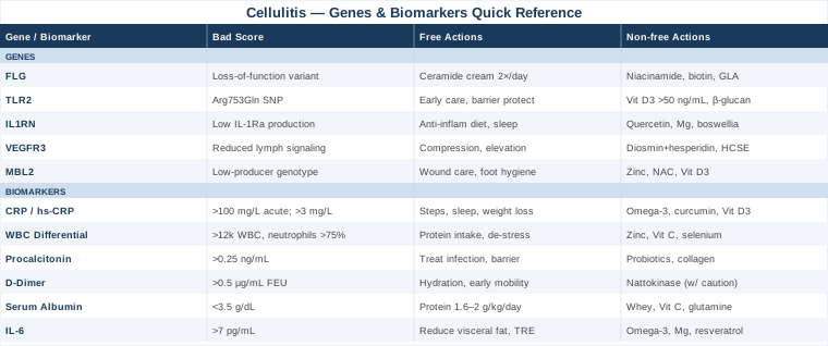
6 Biomarkers Worth Testing If You Have Recurrent Cellulitis
Biomarker testing offers something that most cellulitis management does not: objective, measurable data you can track over time. The six markers below were selected because they each answer a distinct clinical question — not just whether you have an infection, but how severe it is, whether it is truly bacterial, whether a dangerous mimic is present, and whether your body has the nutritional reserves to heal. Peter Attia and Thomas Dayspring have both emphasized that the value of biomarker tracking is not in a single snapshot but in trends. That principle applies directly here.
1. C-Reactive Protein (CRP) and High-Sensitivity CRP
Why it matters: CRP is a protein produced by the liver in response to inflammation. In cellulitis, it rises sharply within 24 to 48 hours of infection onset and drops steadily as the infection resolves. Serial CRP measurements are one of the most reliable ways to confirm that treatment is working, or to flag treatment failure before it becomes obvious clinically. Chronically elevated hs-CRP between episodes also signals persistent low-grade inflammation that may increase susceptibility to recurrence.
What it reveals: An acute CRP above 100 mg/L typically indicates significant bacterial infection. Values between 10 and 50 mg/L suggest mild to moderate inflammation. Hs-CRP below 1 mg/L between episodes is the target for general health; levels above 3 mg/L suggest ongoing systemic inflammation that impairs immune surveillance.
How to measure it: Standard CRP is included in most comprehensive metabolic panels or can be ordered as a standalone test. Cost: $10–$30 in most labs. High-sensitivity CRP (hs-CRP) is slightly more expensive at $25–$60 and is the preferred version for baseline tracking between infections.
If CRP is elevated between episodes — the plan without supplements
Chronic low-grade CRP elevation often traces back to three modifiable factors: visceral adiposity, sleep disruption, and sedentary behavior. Reducing waist circumference through caloric control has a linear relationship with CRP reduction. Targeting 7 to 9 hours of sleep consistently is one of the most powerful anti-inflammatory levers available without any intervention. Walking 8,000 to 10,000 steps daily reduces hs-CRP in observational and intervention studies. If a specific infection source (dental infection, gut dysbiosis, subclinical sinusitis) is found, addressing it may produce a sharp and lasting drop.
If CRP is elevated between episodes — the plan with supplements
Omega-3 fatty acids (EPA + DHA): 2–4 g per day of combined EPA + DHA from fish oil has consistent meta-analytic evidence for reducing hs-CRP. Take with the largest meal of the day to maximize absorption. No cycling needed. Monitor for blood-thinning effects if on anticoagulants. Curcumin with piperine: 500–1,000 mg of bioavailable curcumin (with piperine or in liposomal form) daily has shown modest CRP-reducing effects in randomized trials. Cycle off after 8–12 weeks if used long-term; avoid in those with gallstones. Vitamin D3 + K2: Deficiency is common and independently associated with elevated CRP. Target 25-OH vitamin D above 40 ng/mL. Dose accordingly (typically 2,000–5,000 IU/day D3 with 100–200 mcg K2 MK-7). Retest after 90 days.
2. White Blood Cell Count with Differential
Why it matters: The total white blood cell (WBC) count and its differential — breaking down neutrophils, lymphocytes, monocytes, and eosinophils — is often the first objective indicator of an active bacterial infection. In cellulitis, neutrophilia (elevated neutrophil count) with a left shift (increased band cells) indicates the bone marrow is ramping up acute bacterial defense. It also helps distinguish cellulitis from conditions that mimic it.
What it reveals: A WBC above 12,000 cells/μL with more than 75% neutrophils strongly supports a bacterial etiology. A normal WBC with lymphocyte dominance makes viral or inflammatory (non-infectious) mimics more likely. Tracking WBC over 48–72 hours on antibiotic treatment gives a rapid read on treatment adequacy.
How to measure it: Included in a complete blood count (CBC) with differential. Cost: $10–$40. Widely available. Order at onset and repeat 48–72 hours into treatment.
If WBC differential is abnormal between episodes — the plan without supplements
Persistently low WBC or a poor neutrophil-to-lymphocyte ratio between infections may indicate nutritional deficiencies, chronic stress, or subclinical bone marrow suppression. Prioritize adequate protein intake (at least 1.6 g/kg body weight daily), zinc-rich foods (meat, shellfish, legumes), and reducing chronic psychological stress, which is well-documented to suppress neutrophil function. Correcting iron deficiency if present also restores normal WBC production.
If WBC differential is abnormal between episodes — the plan with supplements
Zinc: 15–30 mg/day elemental zinc supports neutrophil function. Take with food; cycle off after 8 weeks to avoid copper depletion (supplement 2 mg copper if using long-term). Vitamin C: 500–1,000 mg/day is well-studied for supporting neutrophil activity and phagocytosis. Take split doses; excess is cleared renally. Selenium: 100–200 mcg/day (selenomethionine form preferred) has evidence for supporting immune cell maturation. Do not exceed 400 mcg/day.
3. Procalcitonin (PCT)
Why it matters: Procalcitonin is arguably the most useful biomarker for distinguishing true bacterial skin infections from non-infectious mimics. Studies show that approximately 30% of patients diagnosed with lower-limb cellulitis are actually presenting with conditions like stasis dermatitis, lipodermatosclerosis, or contact dermatitis — none of which respond to antibiotics. Procalcitonin rises specifically and rapidly in systemic bacterial infection, while remaining low in viral illness and inflammatory skin conditions.
What it reveals: PCT below 0.1 ng/mL makes significant bacterial infection unlikely. Values above 0.25 ng/mL support bacterial infection; values above 0.5 ng/mL suggest more systemic bacterial involvement. Serial measurements every 48 hours are used to guide antibiotic duration in some protocols.
How to measure it: Available as a standalone blood test. Cost: $30–$80. Not always included in standard panels — ask your physician specifically. More commonly used in inpatient settings but increasingly available outpatient.
If procalcitonin is elevated — the plan without supplements
PCT elevation confirms active infection that needs appropriate antibiotic management. The no-supplement priority here is ensuring the underlying infection source is fully treated and that structural risk factors are addressed: managing lymphedema, repairing skin barrier breaks, controlling diabetes if present. Persistently elevated PCT after completing antibiotics warrants deeper investigation for occult bacterial focus.
If procalcitonin is elevated — the plan with supplements
After antibiotic treatment is complete, supporting the post-infection recovery matters. Probiotics (Lactobacillus rhamnosus GG, 10 billion CFU/day for 4–6 weeks): antibiotics disrupt gut flora, which in turn affects systemic immune regulation. Probiotics during and after treatment mitigate this effect. Collagen peptides (10–15 g/day): support dermal repair and skin barrier reconstruction, reducing entry points for future infection.
4. D-Dimer
Why it matters: This biomarker is not primarily a cellulitis marker — it is a safety net. Deep vein thrombosis (DVT) is one of the most dangerous conditions that can be mistaken for cellulitis, and missing it is life-threatening. D-dimer is a fibrin degradation product that rises sharply in the presence of clot. Because DVT and cellulitis can present identically on visual inspection — warm, red, swollen lower leg — D-dimer testing at first presentation is critical when clinical suspicion for DVT exists.
What it reveals: A negative D-dimer (below 0.5 μg/mL FEU) effectively rules out DVT in patients with low-to-moderate pre-test probability. A positive D-dimer requires further imaging (Doppler ultrasound). Note that D-dimer rises in many other conditions including infection itself, so it is more useful as a negative rule-out than a positive confirmation.
How to measure it: Standalone blood test. Cost: $20–$60. Always measure at initial presentation of a lower-limb swelling/redness episode if DVT cannot be clinically excluded.
If D-dimer is elevated — the plan without supplements
Elevated D-dimer needs medical evaluation before any self-directed approach. If DVT is confirmed, anticoagulation is required and supersedes all other considerations. If DVT is ruled out and D-dimer is mildly elevated due to cellulitis itself, focus on hydration (adequate fluid intake reduces blood viscosity) and early mobility (avoiding prolonged immobility during recovery).
If D-dimer remains elevated after infection resolution — the plan with supplements
Nattokinase: 2,000–4,000 FU/day is studied as a mild fibrinolytic agent in cardiovascular contexts. Evidence in post-infection D-dimer elevation is limited — use cautiously and not in combination with anticoagulants. Discuss with your physician first. Hydration and movement remain the most evidence-backed interventions for mild clot risk in outpatient settings.
5. Serum Albumin
Why it matters: Albumin is not a direct infection marker, but it is one of the most reliable predictors of wound healing capacity and immune resilience. Low albumin (hypoalbuminemia) is independently associated with worse cellulitis outcomes, higher recurrence rates, and longer healing times. The reasoning is direct: albumin reflects nutritional protein status and liver synthetic function, both of which determine the body's ability to mount and resolve an immune response and repair damaged tissue. Thomas Dayspring has emphasized albumin as an underused clinical signal in general health monitoring.
What it reveals: Normal serum albumin is 3.5–5.0 g/dL. Levels below 3.5 g/dL indicate hypoalbuminemia, often from malnutrition, chronic inflammation, liver disease, or kidney protein loss. Values below 3.0 g/dL are associated with significantly impaired healing.
How to measure it: Included in comprehensive metabolic panel (CMP). Cost: $15–$40. Inexpensive and widely available. Measure at baseline and recheck if recovery is slow.
If albumin is low — the plan without supplements
The most direct intervention is increasing dietary protein to 1.6–2.0 g/kg of body weight daily, distributed across meals. Adequate caloric intake overall matters — albumin falls with caloric restriction even when protein seems sufficient. Reducing processed food and ultra-processed carbohydrates lowers the inflammatory burden on the liver. Treating any underlying cause (kidney protein loss, liver dysfunction) is essential before protein supplementation will have lasting effect.
If albumin is low — the plan with supplements
Whey protein (30–40 g/day): highest biological value protein source, proven to raise albumin in malnourished patients in multiple randomized trials. Take once or twice daily. Vitamin C (500 mg twice daily): supports collagen synthesis and albumin utilization. Glutamine (10–15 g/day): conditionally essential amino acid during infection/stress; supports gut integrity and albumin synthesis. Use for 4–6 weeks post-infection then evaluate.
6. Interleukin-6 (IL-6)
Why it matters: IL-6 is a pro-inflammatory cytokine that sits at the center of the acute phase response in cellulitis. It signals the liver to produce CRP and other acute phase reactants, mobilizes white blood cells, and drives fever. Chronically elevated IL-6 between infections points to a state of smoldering systemic inflammation that significantly impairs immune tolerance and increases the biological cost of each new infection. It is a more upstream signal than CRP.
What it reveals: Normal serum IL-6 is below 7 pg/mL. Elevation between episodes of cellulitis suggests obesity-related adipose inflammation, gut barrier dysfunction, or persistent viral/bacterial burden (such as CMV or H. pylori) that is maintaining an inflammatory state.
How to measure it: Ordered as a standalone cytokine panel or inflammatory panel. Cost: $40–$150. Less commonly included in standard workups — request specifically. Some concierge medicine and functional medicine labs offer it routinely.
If IL-6 is elevated between episodes — the plan without supplements
Visceral fat is the leading driver of chronic IL-6 elevation in otherwise healthy people, because adipocytes are direct IL-6 producers. Reducing visceral fat through a combination of moderate caloric deficit, resistance training, and consistent aerobic exercise is the most evidence-supported intervention. Time-restricted eating (10–12 hour eating window) has emerging data for reducing adipose-derived IL-6 independent of weight loss. Improving gut barrier integrity through dietary fiber and fermented foods also addresses a common source of inflammatory cytokine signaling.
If IL-6 is elevated between episodes — the plan with supplements
Omega-3s (EPA + DHA, 3–4 g/day): consistent evidence for lowering IL-6 in randomized trials, particularly in overweight individuals. Magnesium glycinate (300–400 mg/day at night): magnesium deficiency is associated with elevated IL-6; repletion has modest anti-inflammatory effects. Resveratrol (250–500 mg/day): some evidence from randomized trials for IL-6 reduction. Take with food. Cycle after 12 weeks. Avoid at high doses in those with hormone-sensitive conditions.
Moving from what your blood can tell you to what your DNA may predict, the genetic picture of cellulitis susceptibility is less actionable day-to-day but reveals structural vulnerabilities that no amount of clean living will fully override — and knowing them lets you compensate more precisely.
5 Genes That May Shape Your Risk for Cellulitis
Genetic testing for common variants is now accessible through direct-to-consumer platforms (23andMe, AncestryDNA) or clinical-grade tests via companies like Genomind or Color Genomics. For the variants below, raw data download plus interpretation through tools like Genetic Lifehacks or SelfDecode can reveal your specific status. Interpreting these variants is probabilistic, not deterministic — a risk variant raises susceptibility, it does not guarantee disease.
1. FLG — The Skin Barrier Gene
What it is: The FLG gene encodes filaggrin, a structural protein essential for building the outermost layer of skin — the cornified envelope. Filaggrin aggregates keratin filaments and maintains the skin's water retention and barrier function. Loss-of-function variants in FLG are the most common genetic cause of impaired skin barrier integrity in Europeans and Asians, present in roughly 10% of the general population and significantly higher in those with atopic dermatitis.
Why it matters for cellulitis: An intact skin barrier is the first line of defense against Streptococcus pyogenes and Staphylococcus aureus — the two primary cellulitis-causing bacteria. When filaggrin is reduced or absent, the skin's acidic pH rises, water loss increases, and microscopic fissures appear that provide bacterial entry points. Studies link FLG mutations to significantly elevated risk of recurrent skin infections, including cellulitis, even in individuals without obvious eczema. Irvine et al. documented this link extensively in the dermatological literature.
If FLG is impaired — the plan without supplements
Moisturization as medicine: Applying a high-quality emollient (ceramide-containing or petrolatum-based) twice daily to the lower legs is the single most effective non-supplement intervention for compensating for FLG insufficiency. It physically substitutes for the water-retention and barrier function that filaggrin provides. Avoid soap on affected areas; use gentle, fragrance-free cleansers. Keep skin hydrated from the inside with adequate water intake. Address any skin cracks immediately — even microtrauma matters.
If FLG is impaired — the plan with supplements
Niacinamide (500–1,000 mg/day oral; also topical 4–5%): upregulates ceramide and free fatty acid synthesis in the skin, partially compensating for FLG dysfunction. Well-tolerated; no significant cycling needed. Biotin (5–10 mg/day): supports keratinocyte function and skin barrier protein synthesis; evidence is modest but low-risk. Evening primrose oil or borage oil (1–2 g/day GLA): gamma-linolenic acid modulates epidermal lipid composition and has randomized trial evidence for improving skin barrier function in atopic conditions. Take with food; use for 12 weeks then reassess.
2. TLR2 — The Bacterial Recognition Gene
What it is: Toll-like receptor 2 (TLR2) is a pattern recognition receptor that sits on the surface of immune cells and keratinocytes. Its specific role is to detect components of gram-positive bacterial cell walls — including peptidoglycan from Streptococcus and lipoteichoic acid from Staphylococcus. When TLR2 detects these signals, it rapidly triggers innate immune activation, recruiting neutrophils and initiating the inflammatory cascade that contains the infection.
Why it matters for cellulitis: Several TLR2 single-nucleotide polymorphisms (SNPs), including the commonly studied Arg753Gln variant, are associated with reduced signaling efficiency. Individuals carrying these variants may have a slower or attenuated initial immune response to gram-positive bacteria, giving pathogens more time to establish infection before containment begins. Evidence in this area is primarily from studies of other streptococcal infections (endocarditis, bacteremia), but the mechanism directly applies to skin infections.
If TLR2 is impaired — the plan without supplements
Early treatment and aggressive prevention: If your innate immune response is slower at recognizing bacterial invasion, the window between infection onset and meaningful immune containment is wider. This means: do not wait to see a physician if you notice early redness spreading from a wound. It also means prioritizing skin barrier maintenance more intensively (see FLG plan). Reduce Staphylococcus nasal carriage if you test positive (mupirocin nasal ointment per physician guidance) as this is a common self-inoculation source.
If TLR2 is impaired — the plan with supplements
Vitamin D3 (maintain 25-OH vitamin D above 50 ng/mL): vitamin D directly upregulates TLR2 expression and its downstream signaling. This is one of the most compelling immune-genetics interactions in human data. Test, then supplement to target. Beta-glucan (250–500 mg/day from oat or yeast sources): binds to related pattern recognition receptors and primes innate immune readiness. Evidence in infection prevention is growing. Cycle 8 weeks on, 4 weeks off.
3. IL1RN — The Inflammatory Modulation Gene
What it is: The IL1RN gene encodes interleukin-1 receptor antagonist (IL-1Ra), a naturally occurring protein that competes with interleukin-1β (IL-1β) for receptor binding and therefore acts as a brake on the IL-1 inflammatory cascade. Variants in IL1RN that reduce IL-1Ra production leave the IL-1β response less well-regulated — meaning inflammation can become disproportionately intense or prolonged.
Why it matters for cellulitis: The IL-1 pathway drives much of the tissue damage in cellulitis — the swelling, redness, pain, and eventual tissue injury are largely IL-1β-mediated. When IL-1Ra is genetically low, the inflammatory response in each episode tends to be more severe and takes longer to resolve. This gene is also relevant to recurrence risk: sustained low-grade IL-1 signaling between episodes may impair local tissue immunity.
If IL1RN is impaired — the plan without supplements
Anti-inflammatory diet architecture: Reducing dietary ultra-processed food, refined carbohydrates, and seed oils (high linoleic acid) consistently lowers IL-1β signaling. Increasing cold-water fatty fish (salmon, mackerel, sardines) 3–4 times per week, and whole-food polyphenol sources (berries, green tea, olive oil), provides direct anti-IL-1 substrate. Sleep optimization is non-negotiable here: sleep deprivation acutely elevates IL-1β.
If IL1RN is impaired — the plan with supplements
Quercetin (500–1,000 mg/day with food): inhibits IL-1β synthesis in preclinical and human studies. Take in a bioavailable form (quercetin phytosome or with bromelain). Cycle 8 weeks on, 4 off. Magnesium glycinate (300–400 mg/day): magnesium deficiency upregulates IL-1β; repletion provides measurable modulation. Boswellic acid (AKBA, 200–300 mg/day): inhibits inflammatory pathways overlapping with IL-1 signaling. Use for 12-week cycles.
4. VEGFR3 (FLT4) — The Lymphatic Architecture Gene
What it is: Vascular endothelial growth factor receptor 3, encoded by FLT4, is the primary receptor driving lymphatic vessel development and maintenance. Loss-of-function mutations in this gene are the leading cause of Milroy disease (primary congenital lymphedema) and are implicated in milder forms of lymphatic insufficiency that may not produce overt lymphedema but still impair lymphatic drainage capacity.
Why it matters for cellulitis: Lymphedema is one of the strongest independent risk factors for cellulitis. Even subclinical lymphatic insufficiency — the kind you might not notice unless you look carefully at leg puffiness after a long flight or standing day — creates an ideal environment for bacterial growth: protein-rich interstitial fluid, impaired immune cell trafficking, and reduced local antibody delivery. People with primary lymphedema have a lifetime recurrent cellulitis risk well above 50%. VEGFR3 variants that reduce signaling efficiency may explain why some people develop subclinical lymphedema and unexplained recurrent cellulitis without a clear precipitating injury.
If VEGFR3 is impaired — the plan without supplements
Compression and elevation: If lymphatic drainage is structurally limited, mechanical support is the most direct compensation. Medical-grade compression stockings (20–30 mmHg for mild insufficiency, 30–40 mmHg for more significant swelling) during prolonged standing or travel significantly reduce interstitial fluid accumulation. Elevating the legs above heart level for 30 minutes in the evening reduces overnight fluid redistribution. Manual lymphatic drainage performed by a certified therapist should be considered for those with documented lymphatic insufficiency.
If VEGFR3 is impaired — the plan with supplements
Diosmin + hesperidin (600–1,000 mg/day of the combination, e.g., Daflon): a well-studied flavonoid combination for chronic venous and lymphatic insufficiency with randomized controlled trial evidence. Reduces leg swelling and heaviness. Long-term use appears safe; some reports of mild GI side effects. Horse chestnut seed extract (HCSE, standardized to 50 mg aescin twice daily): Cochrane review-level evidence for reducing venous leg edema; likely benefits mild lymphatic insufficiency through similar mechanisms.
5. MBL2 — The Complement System Gene
What it is: Mannose-binding lectin (MBL) is a pattern recognition protein of the innate immune system that activates the complement cascade upon binding to carbohydrate patterns on bacterial surfaces — including Staphylococcus aureus and Streptococcus pyogenes. The MBL2 gene has well-characterized codon variants (A/B alleles at codons 54 and 57) that dramatically reduce circulating MBL levels. Homozygous low-producers may have less than 10% of normal MBL activity.
Why it matters for cellulitis: MBL deficiency is associated with increased susceptibility to bacterial infections in multiple human studies, particularly in early childhood and in adults with additional immune compromise. For cellulitis, the specific relevance is that MBL normally helps opsonize bacteria in skin interstitial fluid before neutrophils arrive — a low-MBL environment gives bacteria a longer head start. Population studies link MBL2 low-producer genotypes to recurrent skin and soft tissue infections.
If MBL2 is impaired — the plan without supplements
Minimize exposure windows: Since complement-mediated opsonization is slower, the priority is reducing bacterial exposure opportunities: meticulous wound care (any skin break, no matter how small, deserves immediate cleaning and covering), reducing nasal MRSA/MSSA carriage if relevant, and being especially vigilant about foot hygiene including between toes. Avoid immunosuppressive medications unless absolutely necessary.
If MBL2 is impaired — the plan with supplements
Zinc (15–30 mg/day): zinc is required for MBL synthesis and complement system function. Zinc-deficient individuals have measurably reduced MBL levels that partially restore with supplementation. Cycle with copper as above. N-acetylcysteine (600 mg twice daily): supports glutathione synthesis, which is required for healthy complement system function. Modest evidence; well-tolerated for 8-week cycles. Vitamin D3 (target 50 ng/mL 25-OH vitamin D): vitamin D has multiple complement-supporting effects and is one of the highest-value interventions for people with multiple immune gene variants.
Understanding your biomarker picture and genetic risk is powerful — but it becomes even more so when placed in a broader framework for how chronic conditions are prevented rather than just treated. The approach outlined in one widely read book on longevity medicine offers exactly that kind of framework.
What Peter Attia's Longevity Framework Reveals About Managing Recurrent Cellulitis
Outlive: The Science and Art of Longevity by Peter Attia, MD, is not about cellulitis. But its core argument — that modern medicine waits too long, measures too little, and misses the opportunity to intervene before damage becomes irreversible — maps directly onto the problem of recurrent skin infections. Here are the ten insights from that book most directly applicable to cellulitis prevention.
1. Track biomarkers longitudinally, not just at crisis points
Attia argues that a single normal result tells you almost nothing; it is the trend over time that reveals risk. For cellulitis patients, this means measuring CRP, albumin, and other relevant markers every 6–12 months between episodes, not only when infection is suspected. A CRP rising from 0.8 to 3.2 mg/L over two years is a warning worth acting on, even if both values are technically "normal."
2. Insulin resistance is a hidden driver of infection susceptibility
Attia dedicates significant space to what he calls MTOR and insulin signaling dysregulation as root causes of multiple chronic conditions. Elevated fasting insulin and impaired glucose regulation directly suppress immune function and impair wound healing. Check fasting insulin (target below 6 μU/mL) in addition to standard glucose and HbA1c.
3. Zone 2 cardio is the most underused anti-inflammatory intervention
Sustained low-intensity aerobic exercise (maintaining a pace where you can hold a conversation but not easily sing) for 45–60 minutes, 3–4 times per week, drives mitochondrial adaptation that measurably reduces systemic IL-6 and CRP. Attia is specific: Zone 2 is not a workout, it is infrastructure. Its anti-inflammatory effects accumulate over months, not weeks.
4. Muscle mass is immune insurance
Low skeletal muscle mass (sarcopenia) is independently associated with impaired immune response, slower wound healing, and worse outcomes in infection. This is because muscle is the primary reservoir of glutamine — the amino acid that fuels immune cells during periods of acute stress. Resistance training 2–3 times per week, prioritizing compound lifts and adequate protein, is a direct immune-support strategy.
5. Sleep is not optional maintenance — it is active immune repair
During deep sleep (NREM stage 3), the body clears metabolic waste from tissues, restores immune cell function, and consolidates immune memory. Attia cites sleep as the variable with the highest return on investment in longevity medicine. For cellulitis patients, sleep below 7 hours consistently impairs neutrophil function, elevates IL-6, and reduces vaccine-like immune memory of previously encountered pathogens.
6. Visceral fat is an independent inflammatory organ
Attia describes visceral adipose tissue (VAT) as metabolically active — continuously secreting IL-6, TNF-alpha, and leptin in quantities that disrupt immune regulation. DEXA scan or waist-to-height ratio (target below 0.5) is the most accessible way to quantify this. Reducing VAT is one of the highest-leverage actions for lowering chronic infection susceptibility.
7. Early prevention outperforms late treatment by a factor of ten
The book's central thesis is the Marginal Decade concept — the choices made in your 40s and 50s determine the quality of your last decade of life. Applied to cellulitis: the habits that prevent a first recurrence are far more effective than treatments after the fifth. Structural interventions (compression, barrier maintenance, biomarker monitoring) implemented before the next episode is exponentially more valuable than reactive care.
8. Nutrient deficiencies are common, invisible, and consequential
Attia screens extensively for vitamin D, magnesium, omega-3 index, and iron in nearly all patients — because population-level deficiency in these nutrients is common and each has measurable immune consequences. For cellulitis patients: an omega-3 index below 8%, vitamin D below 40 ng/mL, or ferritin below 30 ng/mL are all correctable factors that subtly impair immune defense.
9. Stress physiology impairs immune function through HPA axis dysregulation
Chronic psychological stress elevates cortisol, which directly suppresses lymphocyte and neutrophil function. Attia discusses HRV (heart rate variability) as a real-time measure of autonomic and immune resilience. Tracking HRV with a wearable device and targeting trends (not absolute numbers) gives an actionable feedback loop for stress-immune interactions.
10. Personalization over population averages
Attia's core argument against standard of care medicine is that population-derived "normal ranges" obscure individual optimization targets. The takeaway for cellulitis patients: do not accept "your labs are normal" as sufficient. Interpret your markers in the context of your history, your genetics, and your trend — not just against a reference range built for the average person.
Complementary Approaches With Clinical Backing for Cellulitis
Beyond biomarkers and genetics, there are a small number of complementary modalities with meaningful human clinical evidence relevant to cellulitis management and recurrence prevention. The three below were selected because they address real mechanisms — lymphatic drainage, tissue repair, and skin microbiome — rather than offering generic wellness benefits.
Manual Lymphatic Drainage and Massage Therapy
Manual lymphatic drainage (MLD) is a specialized massage technique developed by Emil Vodder in the 1930s, designed to stimulate the superficial lymphatic network. Unlike conventional massage, MLD uses extremely light pressure with specific directional strokes that follow the anatomy of lymphatic collectors. Its relevance to cellulitis is direct: lymphedema is one of the most potent risk factors for recurrent cellulitis, and MLD is the gold-standard physical treatment for lymphedema management.
A Cochrane review of complete decongestive therapy (CDT, which includes MLD) for lymphedema-related leg edema found significant reduction in limb volume and tissue fibrosis, both of which reduce the anaerobic, protein-rich interstitial environment that bacteria exploit. The 2014 systematic review by Huang et al. in the International Journal of Nursing Sciences documented MLD's effectiveness in reducing cellulitis recurrence in lymphedema patients when used as part of a CDT protocol.
Practically, a course of 10–15 MLD sessions with a certified therapist (ask for CDT certification), followed by self-MLD practice and compression garment use between sessions, is the standard protocol. Frequency: acute phase, 5 sessions per week; maintenance, 1–2 per week. MLD should be avoided in active, untreated cellulitis (active infection is a contraindication), but resumed aggressively once the acute episode resolves to reduce residual edema.
Low-Level Laser Therapy and Photobiomodulation
Low-level laser therapy (LLLT), also called photobiomodulation (PBM), uses near-infrared and red-light wavelengths (typically 630–1,000 nm) to penetrate skin and stimulate mitochondrial cytochrome c oxidase, triggering anti-inflammatory signaling, accelerated ATP production, and improved tissue oxygenation. It does not generate significant heat, distinguishing it from surgical lasers.
Multiple randomized controlled trials have examined LLLT in wound healing and post-infectious tissue repair. A systematic review published in Photomedicine and Laser Surgery documented consistent acceleration of wound closure and reduction of local inflammation markers with 830 nm infrared application. For post-cellulitis tissue recovery — particularly in areas with residual induration or fibrosis — LLLT has documented ability to reduce inflammatory cytokines in connective tissue and improve lymphatic vessel function, directly addressing two cellulitis risk factors simultaneously. Evidence in direct cellulitis prevention is emerging rather than definitive, but the mechanism is sound and risk is negligible.
A realistic protocol: 830 nm device (available as consumer-grade panels or clinic devices), applied to affected skin areas for 10–15 minutes per session, 4–5 sessions per week during recovery, then 2–3 sessions per week for prevention. Ensure device delivers at least 20–100 mW/cm² at the skin surface. Eye protection is required. Avoid over active infection sites.
Microbiome-Directed Therapies
The skin microbiome — the community of bacteria, fungi, and other microorganisms living on skin surfaces — plays an active role in defending against pathogenic bacteria. Staphylococcus epidermidis, a common commensal skin bacterium, produces antimicrobial peptides that directly inhibit S. aureus growth. Disruption of this community through antibiotic overuse, harsh cleansers, or topical antiseptic overuse removes this natural protection. The gut microbiome influences skin infection susceptibility through systemic immune regulation, the gut-skin axis.
Research published in Science Translational Medicine by Nakatsuji et al. demonstrated that topical application of S. epidermidis strains producing NASO-derived antimicrobial peptides reduced S. aureus colonization in human subjects — a direct proof-of-concept for microbiome-directed prevention of the leading cellulitis pathogen. Oral probiotic supplementation with Lactobacillus rhamnosus and Bifidobacterium longum strains has shown modulation of systemic inflammatory markers in randomized trials, with indirect implications for skin immune defense.
Practically: stop using antibacterial soap on intact skin — it removes protective commensals without benefit. Use gentle pH-balanced cleansers (pH 5.5). After antibiotic courses, use oral probiotics (multi-strain, 10–20 billion CFU, L. rhamnosus GG included) for 4–6 weeks. Increase dietary fiber from diverse plant sources (30+ different plants per week is the target from the British Gut Project data) to feed commensal organisms. If you have recurrent perianal or interdigital infections, targeted topical probiotic products are in clinical development, though not yet widely available.
Conclusion
Cellulitis is not simply a matter of bad luck or inadequate hygiene. For people who experience it repeatedly, the explanation is almost always biological: a skin barrier that needs more support, an immune recognition system that fires more slowly, a lymphatic network that cannot clear fluid efficiently enough, or an inflammatory baseline that never fully resets between episodes. None of these are character flaws — they are measurable, and in many cases, modifiable.
The most useful next step depends on where you are. If you have had cellulitis more than once, the highest-priority action is getting a baseline biomarker panel: CRP, CBC with differential, procalcitonin, albumin, D-dimer, and ideally hs-CRP and IL-6. These give you a picture of your current state. If you have access to genetic testing, looking at variants in FLG, TLR2, IL1RN, FLT4, and MBL2 gives you a longer-term map of structural risk.
From there, the plans outlined above — whether dietary, behavioral, supplemental, or procedural — are not substitutes for medical care but genuine additions to it. Discuss your biomarker results and this framework with a physician who is willing to look at the whole picture. The goal is not to treat the next infection faster. It is to understand your biology well enough to make that next infection far less likely.
Skin: Inflammatory Skin Conditions Infectious Skin Conditions
Autoimmune: Inflammatory Conditions
Infectious: Bacterial Infections