This article contains AI generated content.
· UpdatedCentral Core Disease Genes and Biomarkers: 4 Genes and 7 Biomarkers to Track
Getting a diagnosis that includes the words "central core disease" often raises more questions than it answers. A muscle biopsy shows cores. A weakness pattern fits. But the label itself does not tell you which gene is responsible, what the practical risks are day to day, or what — if anything — can be done about it. Generic advice about "eating well" or "staying active" is not wrong, but it is not built for a condition where the wrong kind of exertion can trigger muscle breakdown, and where a routine anesthetic can become dangerous without the right precautions.
This article goes a level deeper. Central core disease is, in the overwhelming majority of cases, a story about one gene — RYR1 — and a small number of related genes that produce overlapping muscle findings. Understanding which gene is involved, what it does inside a muscle cell, and what has actually been tested in people (not just mice) changes the kind of decisions that are possible: which precautions matter, which supplements have real evidence versus theoretical appeal, and which biomarkers are worth tracking over time.
None of this amounts to a cure. Central core disease is a structural and genetic condition, and no supplement or lifestyle change rewrites a ryanodine receptor. But there is a meaningful difference between "nothing can be done" and "here is what is actually known, what has been tried, and what a sensible monitoring and management plan looks like." That distinction is worth having, and it is where better information starts to translate into better decisions.
This article works through two complementary approaches. First, it looks at the genes most often responsible for central core disease and related core myopathies — what each one does, how it is usually managed, and what a realistic plan looks like with and without supplements or equipment. Second, it covers the biomarkers worth tracking over time, from a simple blood test to specialized muscle imaging, along with what each one can and cannot tell you. A shorter section then pulls in relevant ideas from longevity science, and a final section looks at complementary therapies that have genuine, if often modest, supporting evidence.
Summary
Central core disease is caused by variants in a small handful of genes — most often RYR1, and less commonly SELENON (SEPN1), ACTA1, and MYH7 — each of which disrupts a different part of how muscle cells handle calcium or build their contractile machinery. Knowing which gene is involved changes real decisions: anesthesia precautions, exercise dosing, respiratory monitoring, and even whether the heart needs to be screened. This article breaks down what each gene actually does, what a sensible management plan looks like with and without supplements or equipment, and which of the tried therapies (like N-acetylcysteine or salbutamol) have real trial data behind them versus just theoretical promise.
Beyond genetics, seven practical biomarkers are worth understanding — from a basic creatine kinase blood test to muscle MRI, respiratory function testing, and the genetic panel itself — including what each costs, how it's measured, and what to do if a result is abnormal. A shorter section translates ideas from Peter Attia's longevity framework into a version that respects the real constraints of a muscle-based genetic condition, and a final section looks at which complementary therapies, such as respiratory muscle training, actually have supporting human evidence for neuromuscular disease. Read on for the specifics on each gene, each biomarker, and the therapies that are (and are not) worth pursuing.
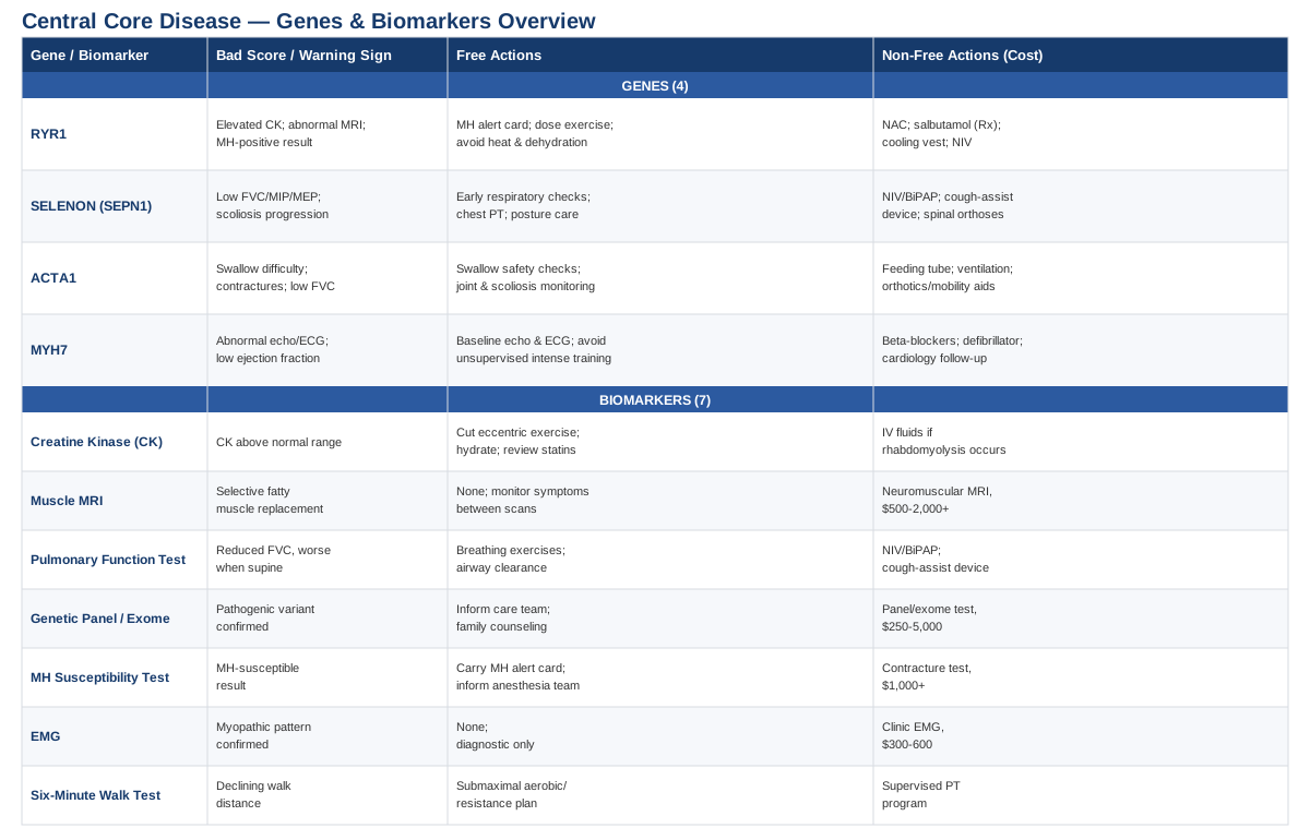
The Genes Behind Central Core Disease
Central core disease is a congenital myopathy — a muscle disease present from birth or early childhood, defined by round or oval "core" regions visible on muscle biopsy where the normal internal structure of the muscle fiber is disrupted. It sits within a broader family of conditions called RYR1-related myopathies, and understanding the genetics matters because it directly shapes safety decisions, not just curiosity about mechanism.
RYR1: The Gene Behind Most Cases
The vast majority of central core disease is caused by variants in RYR1, the gene that codes for ryanodine receptor 1, a calcium release channel embedded in the membrane of the sarcoplasmic reticulum inside skeletal muscle cells. When a nerve signal tells a muscle to contract, RYR1 is what opens to release stored calcium into the cell, which is what actually triggers the contraction. According to MedlinePlus Genetics, over 100 different RYR1 variants have been linked to central core disease alone, and they tend to work in one of two opposite ways: some make the channel "leaky," continuously losing calcium, while others block calcium from passing through when it is actually needed. Either way, the result is muscle fibers that cannot contract normally, along with more subtle effects on calcium handling that show up under physiological stress like heat, exertion, or certain anesthetic drugs.
RYR1-related myopathy is most often inherited in an autosomal dominant pattern, meaning one altered copy of the gene is enough to cause disease, according to MedlinePlus Genetics. A smaller subset of cases is autosomal recessive, generally causing a more severe presentation. Because the same gene is the leading genetic cause of malignant hyperthermia susceptibility — a life-threatening reaction to certain anesthetic agents — an RYR1 diagnosis is not purely academic. It is the reason people with central core disease are told to carry a medical alert card and to inform every anesthesiologist they see, detailed in the GeneReviews chapter on malignant hyperthermia susceptibility.
Muscle imaging adds a second layer of information beyond the gene itself. According to the archived GeneReviews chapter on central core disease, MRI studies have found a consistent, selective pattern of muscle involvement in RYR1-related myopathy, with the quadriceps, sartorius, adductor magnus, soleus, gastrocnemius, and peroneal muscles typically showing the most fatty replacement. A 2018 genetic and clinical study of newly identified RYR1 variants, published in PMC, reinforced that a genetics-first diagnostic approach — combined with muscle MRI and biopsy — gives the most reliable picture, since imaging patterns and severity can vary considerably even among people with the same gene.
If the Gene Is Bad, the Plan Without Supplements
The first and most important step has nothing to do with diet or exercise: anyone with a confirmed or suspected RYR1 pathogenic variant should carry documentation of their malignant hyperthermia risk and make sure every surgical or dental team is aware of it before any anesthesia is administered. This single precaution has outsized safety value compared to anything else on this list.
Exercise still matters, but it needs to be dosed rather than maximized. A 2024–2025 exercise physiology study of people with RYR1-related myopathies, published in PMC, found that adults reached only about 62% of expected peak oxygen uptake and children about 49%, with an anaerobic threshold averaging roughly 3.4 metabolic equivalents — comparable to the effort of grocery shopping. In other words, many people with this condition are already working near their functional ceiling during ordinary daily tasks. The same study explicitly flagged exercise-induced muscle breakdown as a documented risk, which argues for an individualized, submaximal training plan rather than a "push through it" approach. A reasonable structure is two to three sessions per week of low-to-moderate intensity aerobic activity (walking, stationary cycling) paired with light resistance work, staying below perceived exhaustion, with built-in rest days and closer monitoring after any increase in intensity or duration.
Heat and dehydration are worth taking seriously given the shared biology with malignant hyperthermia — calcium handling in muscle is already abnormal, so avoiding prolonged heat exposure, staying well hydrated around activity, and stopping promptly if unusual muscle pain, dark urine, or excessive fatigue appears are sensible, low-cost precautions. It's also worth flagging certain medications, particularly statins, to a physician, since they can independently raise creatine kinase and compound muscle symptoms.
If the Gene Is Bad, the Plan With Supplements or Equipment
The most rigorously tested supplement for RYR1-related myopathy is N-acetylcysteine (NAC), an antioxidant that looked promising in animal models of the disease. A randomized, double-blind, placebo-controlled trial in 33 participants, published in PMC, dosed NAC at roughly 30 mg/kg/day (up to 2,700 mg/day) divided into three daily doses for six months. The honest result: NAC did not reduce the oxidative stress marker it was designed to target, and the improvement in six-minute walk distance (about 24 meters) did not reach statistical significance. It was safe and well tolerated, with no major side effects reported, but the trial is a good example of why it's worth distinguishes preclinical promise from confirmed human benefit — this is not a supplement to expect results from outside of a research context.
Salbutamol (albuterol), a prescription beta-2 agonist normally used for asthma, has been explored off-label in small pilot studies and case reports for central core and multiminicore disease, as summarized in a 2021 review of RYR1-related myopathy therapies in PubMed. Reported doses in these small studies were in the range of a few milligrams several times daily, with some participants reporting subjective improvements in strength and stamina. This is a prescription medication, not a supplement, and it carries real side effects — tremor, rapid heart rate, and palpitations are common — so it requires physician supervision and is not something to self-initiate; the evidence base remains limited to small, largely uncontrolled studies.
On the equipment side, a few options have solid physiological rationale even without condition-specific trials: cooling vests for anyone who is heat-sensitive or exercises in warm environments, non-invasive ventilation if respiratory muscle involvement develops, and ankle-foot orthoses or standing frames to support mobility as needed. The same 2021 review also notes that gene-targeted and small-molecule therapies (including an investigational drug called ARM210/Surlorian) are in active clinical trials, which is worth knowing about but should be discussed with a specialist rather than pursued independently, since these remain experimental.
SELENON (SEPN1): The Gene Behind Respiratory-Predominant Core Myopathy
SELENON (also called SEPN1) codes for selenoprotein N, a protein involved in protecting cells from oxidative damage and in regulating calcium during early muscle development, according to MedlinePlus Genetics. Variants in this gene cause a recessive form of core-related myopathy — most often multiminicore disease, but also rigid spine muscular dystrophy — that tends to follow a distinctive pattern: respiratory muscle weakness, particularly of the diaphragm, that is often disproportionately severe compared to limb weakness, along with early spinal rigidity and scoliosis.
This mismatch between how strong someone looks in the limbs and how weak their breathing muscles actually are is the single most important clinical fact about SELENON-related disease, and it should reshape monitoring priorities from the point of diagnosis.
If the Gene Is Bad, the Plan Without Supplements
Respiratory monitoring needs to start early and continue even if someone appears to be walking and moving reasonably well, because the diaphragm can be affected well before limb weakness becomes obvious. This means regular pulmonary function testing (discussed further in the biomarkers section below), attention to sleep quality and morning headaches (a subtle sign of nocturnal hypoventilation), and prompt referral to a pulmonologist familiar with neuromuscular disease if any of these appear. Spinal rigidity should be monitored with regular orthopedic follow-up for scoliosis, since the combination of a stiff spine and weak respiratory muscles can compound breathing difficulty as curvature progresses. Chest physiotherapy and breathing exercises, referenced as a standard part of management in the GeneReviews management guidance for RYR1-related core myopathies (much of which is shared practice across this gene family), are a reasonable low-cost daily habit.
If the Gene Is Bad, the Plan With Supplements or Equipment
Once nocturnal hypoventilation is confirmed on testing, non-invasive ventilation (NIV/BiPAP) used nightly is standard of care and can meaningfully improve sleep quality, daytime energy, and long-term respiratory outcomes; the main downsides are mask discomfort and occasional skin pressure areas, both manageable with proper mask fitting. A mechanical insufflation-exsufflation ("cough assist") device is used as needed, particularly during respiratory illness, to help clear secretions when a weak cough can't do it alone. Spinal orthoses may be recommended by an orthopedic specialist to slow scoliosis progression.
On supplementation, it's worth being direct: because SELENON encodes a selenoprotein, selenium supplementation has an understandable theoretical appeal. However, there is no clinical evidence that supplementing selenium corrects the underlying defect, and excess selenium carries real toxicity risks, including hair loss, gastrointestinal symptoms, and nerve damage. This is a case where the biologically intuitive fix is not supported by evidence, and self-supplementing is not advisable outside of a formal research study.
ACTA1: The Gene Behind the More Severe End of the Spectrum
ACTA1 codes for skeletal alpha-actin, one of the two core proteins (alongside myosin) that physically slide past each other to generate muscle contraction, as described by MedlinePlus Genetics. ACTA1 variants most often cause nemaline myopathy, but they can also produce actin-accumulation myopathy, cap myopathy, and congenital fiber-type disproportion — the latter of which overlaps clinically with the core myopathy spectrum. Because actin is a structural protein rather than a regulatory channel, ACTA1-related disease tends to present earlier and, in its most severe forms, more seriously than typical RYR1-related central core disease, though milder ACTA1 phenotypes exist and overlap significantly with the general core myopathy presentation.
If the Gene Is Bad, the Plan Without Supplements
Given how variable severity can be, the practical plan centers on close multidisciplinary surveillance rather than a single fixed protocol: monitoring feeding and swallowing safety (with a formal swallow study if there's any concern, since aspiration risk is real in more severe presentations), tracking joint contractures, scoliosis, and hip development, and involving a pulmonology team from early on given the respiratory vulnerability seen across this gene family.
If the Gene Is Bad, the Plan With Supplements or Equipment
Equipment needs are driven by severity and can include non-invasive or, less commonly, invasive ventilation, a feeding tube or gastrostomy if swallowing safety is compromised, and orthotics, standing frames, or power mobility devices to support function as a child grows. Honestly, there is no supplement shown to influence actin function in muscle. The most promising research direction is gene-targeted therapy, still in preclinical and early experimental stages according to the same 2021 therapeutic review referenced above, and worth discussing with a specialist as a long-term prospect rather than a current option.
MYH7: The Gene That Connects Muscle to the Heart
MYH7 codes for beta-myosin heavy chain, the motor protein found in both the heart and in slow-twitch ("type 1") skeletal muscle fibers. According to MedlinePlus Genetics, MYH7 variants can cause myosin storage myopathy and congenital fiber-type disproportion — both of which overlap with the core myopathy picture — but the same gene is also one of the most common causes of hypertrophic and dilated cardiomyopathy. This dual role is the single most important thing to know if MYH7 is the gene identified: a presentation that looks purely skeletal can come with a silent cardiac risk that has nothing to do with how the muscles feel day to day.
If the Gene Is Bad, the Plan Without Supplements
A baseline echocardiogram and ECG at diagnosis are not optional extras — they are the single most consequential step for anyone with an MYH7-related myopathy, given how common cardiac involvement is with this gene. Because MYH7-related cardiomyopathies are often dominantly inherited, cascade screening of close relatives is also worth raising with a genetic counselor. Until cardiac status is clearly characterized, it's sensible to avoid unsupervised high-intensity endurance training, since undiagnosed cardiomyopathy changes the risk calculus around strenuous exercise.
If the Gene Is Bad, the Plan With Supplements or Equipment
If hypertrophic or dilated cardiomyopathy is confirmed, management follows standard cardiology protocols — typically beta-blockers or other rate/rhythm-focused medications, and in higher-risk cases an implantable defibrillator — decisions that belong entirely to a cardiologist, not to a self-directed supplement plan. On the skeletal muscle side, the approach mirrors other congenital myopathies: physical therapy two to three times weekly, orthotic review every six to twelve months during growth, and routine respiratory monitoring. There is no supplement that changes myosin function; the leverage here is almost entirely in early cardiac detection.
Seven Biomarkers Worth Tracking
Genetic diagnosis answers "which gene," but it doesn't answer "how am I doing right now" or "is this getting better, worse, or staying the same." That's where a smaller set of practical biomarkers earns its place — some cheap and routine, others specialized and used more sparingly.
Creatine Kinase (CK)
CK is an enzyme that leaks out of damaged muscle cells into the bloodstream, making it a simple window into ongoing muscle breakdown, described by MedlinePlus. In RYR1-related myopathy specifically, CK is useful for catching subclinical exertional muscle breakdown before it becomes a bigger problem. How to measure it: a standard blood draw, often available for $10–30 out of pocket or bundled into a routine metabolic panel. If it's elevated: without supplements, the plan is to scale back high-intensity or eccentric exercise, prioritize hydration around activity, and review any new medications (especially statins) with a physician; there is no supplement proven to lower exercise-related CK elevation in this population, though clinically significant rhabdomyolysis is managed medically with IV fluids, not a home remedy.
Muscle MRI
A muscle MRI using T1-weighted and STIR sequences can reveal the selective pattern of fatty muscle replacement characteristic of RYR1-related myopathy — often sparing certain muscles while affecting others in a fairly predictable pattern, as detailed in the GeneReviews chapter on central core disease. It's also useful for confirming a diagnosis non-invasively and tracking structural change over years. How to measure it: performed at a center with neuromuscular imaging experience, typically costing $500–2,000+ depending on region and insurance coverage. Because the same exercise capacity study cited earlier found relative stability over a six-month window, MRI is generally repeated only every few years or when there's a meaningful change in symptoms, not on a frequent schedule.
Pulmonary Function Testing
Spirometry and related respiratory measures catch weakness of the diaphragm and chest wall muscles, which matters enormously for SELENON-related disease and can also affect RYR1-related myopathy over time, per MedlinePlus. How to measure it: performed in a clinic, typically $50–300, ideally measured both sitting and lying down since diaphragm weakness often shows up more clearly when supine. Frequency: every six to twelve months as a baseline, more often if any decline is detected. If results are abnormal: without equipment, breathing exercises and airway clearance techniques are reasonable starting points; a systematic review and meta-analysis of respiratory muscle training in neuromuscular disease, published in PMC, found meaningful improvements in forced vital capacity and inspiratory/expiratory pressures across 16 randomized trials. With equipment, non-invasive ventilation and a cough-assist device become appropriate once hypoventilation or ineffective cough clearance is documented.
Diagnostic Genetic Panel or Exome Sequencing
This is arguably the most important biomarker of all, since it identifies the specific gene and variant responsible, which then determines every other decision in this article — anesthesia risk, respiratory monitoring priorities, and whether cardiac screening is needed. How to measure it: a blood or saliva sample analyzed via a targeted neuromuscular gene panel or broader exome sequencing, typically $250–5,000 depending on the test and insurance coverage, and often covered when clinically indicated. A 2018 study in PMC specifically recommended a genetics-led diagnostic approach, combined with imaging and biopsy, as the most reliable strategy. This is generally a one-time test, though reanalysis every few years can be worthwhile as gene databases and variant interpretation improve.
Malignant Hyperthermia Susceptibility Testing
Not every RYR1 variant confers malignant hyperthermia risk, but many do, and getting this right is a genuine safety issue during any future surgery. The gold standard is the caffeine-halothane contracture test performed on a fresh muscle biopsy sample, available only at a small number of specialized centers, as described in the GeneReviews chapter on malignant hyperthermia susceptibility. How to measure it: contracture testing costs upward of $1,000 and requires a specialized center; a more accessible alternative is confirming that the specific variant already identified through genetic testing is one of the known MH-causative variants, which is far cheaper if the panel above has already been done. This is essentially a one-time determination that should then travel with someone as part of their medical record.
Electromyography (EMG)
EMG uses a fine needle electrode to measure the electrical activity of muscle, helping confirm that weakness is coming from the muscle itself (myopathic) rather than from the nerves supplying it (neurogenic), according to MedlinePlus. How to measure it: performed in a clinic setting, typically $300–600, with some discomfort during needle insertion and possible mild soreness afterward. This is usually done once during the initial diagnostic workup and repeated only if the clinical picture changes unexpectedly.
Six-Minute Walk Test
This simple, low-cost functional test — measuring how far someone can walk in six minutes — has become a standard outcome measure in RYR1-related myopathy research, including the NAC trial and exercise capacity study referenced earlier, precisely because it captures real-world function in a way lab values can't. How to measure it: performed in a hallway with a stopwatch and, ideally, a pulse oximeter, essentially free during a routine clinic visit. Frequency: worth tracking every six to twelve months as a trend line rather than a single snapshot. If distance is declining: without equipment, an individualized aerobic and resistance program dosed below the roughly 3.4 MET anaerobic threshold identified in the exercise capacity study is a sensible target, cycled with rest days and reassessed regularly rather than pushed to fatigue, given the documented risk of exercise-induced muscle breakdown in this population.
What a Longevity Framework Gets Right (and Where It Needs Adjusting)
Peter Attia's book Outlive: The Science and Art of Longevity isn't written with a rare congenital myopathy in mind — its central argument is that most people are managing "Medicine 2.0" diseases of aging reactively when they could be preventing them proactively, with muscle and cardiorespiratory fitness treated as central, measurable levers. That framework doesn't map perfectly onto a genetic muscle disease, but several of its core ideas are genuinely useful here, provided the intensity is scaled down to match what a compromised calcium-handling or contractile system can actually tolerate.
Muscle Is Framed as an Organ of Longevity, Not Just Strength
Attia repeatedly returns to the idea that muscle mass and strength are not vanity metrics but some of the strongest predictors of long-term survival available. Large-scale data backs this up directly: the Prospective Urban Rural Epidemiology (PURE) study of nearly 140,000 people, published in PubMed, found that grip strength was inversely associated with all-cause and cardiovascular mortality, and was actually a stronger predictor than systolic blood pressure. For someone with a muscle-affecting genetic condition, this reframes preserved strength not as a bonus but as a legitimate health priority worth protecting deliberately.
VO2 Max Might Be the Single Best Vital Sign
Attia calls cardiorespiratory fitness the most powerful, modifiable predictor of lifespan he knows of. A cohort study of over 122,000 people undergoing treadmill testing, published in PMC, found that fitness was inversely associated with mortality with no observed upper limit of benefit, and that the mortality difference between low and high fitness groups rivaled or exceeded that of smoking or diabetes. For someone with RYR1-related myopathy already operating near their functional ceiling during daily tasks, this doesn't mean chasing an elite VO2 max — it means treating whatever aerobic capacity is safely achievable as worth protecting and tracking over time, in the same spirit as the six-minute walk test above.
The "Centenarian Decathlon" Reframes the Goal
Rather than training for an arbitrary standard, Attia suggests picking the specific physical tasks you want to still perform decades from now — carrying groceries, getting off the floor, climbing stairs — and reverse-engineering training from there. For a congenital myopathy, this is arguably more useful than any generic fitness target: it centers the plan on preserved function and independence rather than a number on a chart, and gives physical therapy concrete, personally meaningful goals to work toward.
Zone 2 Training Builds Capacity Without Maxing Out
Attia's emphasis on low-intensity, "zone 2" aerobic training — conversational-pace effort that builds mitochondrial and metabolic capacity without high mechanical or metabolic strain — happens to align unusually well with the exercise caution already discussed for RYR1-related myopathy. This is, if anything, a case where the longevity literature's "go easier, more often" philosophy is a safer match for a muscle disease than the more common "go hard" fitness culture.
Strength Training Matters, But the Dose Has to Be Individualized
Attia is unambiguous that strength training is non-negotiable for long-term function. For most healthy readers, that means progressively heavier loads. For someone with a core myopathy, the same principle applies but the progression needs to be much more conservative, monitored with CK trends rather than by feel, and coached by a physical therapist familiar with neuromuscular disease rather than a general strength program.
Stability Prevents the Injuries That Actually Derail People
A recurring point in the book is that falls and injuries, not diseases, often end someone's functional independence — which makes balance and movement-quality work as important as raw strength. This is directly relevant to congenital myopathies, where reduced proximal muscle strength already raises fall risk; simple balance and gait-focused physical therapy is a low-risk, high-value addition.
Medicine 3.0 Means Screening Before Symptoms Force the Issue
Attia's argument for proactive, early screening over reactive treatment is essentially the same philosophy behind the biomarker section above: checking pulmonary function and cardiac status before there's an obvious problem, rather than waiting for a crisis, is the practical translation of "Medicine 3.0" into this specific condition.
Sleep Is Treated as a Performance and Recovery Lever
The book spends real time on sleep as foundational to metabolic health and recovery. For anyone with respiratory muscle involvement (particularly SELENON-related disease), this point carries extra weight — poor sleep quality may be an early sign of nocturnal hypoventilation rather than a purely lifestyle issue, which is one more reason to raise persistent fatigue or morning headaches with a physician rather than dismissing them.
Emotional Health Is Part of the Stack, Not an Afterthought
Attia is candid that physical optimization means little without attending to mental and emotional health, an easy thing to overlook when a plan is dense with biomarkers and gene names. Living with a genetic condition that carries real constraints — on exercise, on anesthesia, on unpredictability — is a legitimate stressor worth acknowledging and addressing directly, including with professional support if needed.
Train for the Decade That Matters Most, Not the One You're In
The book's closing idea — that today's choices are training for a specific future decade of life, not just the present — is a useful reframe for a condition that can otherwise feel like it's only about damage control. Preserving function now is what makes a wider range of choices possible later, whether or not the underlying gene ever changes.
Complementary Approaches Worth Considering
A handful of complementary therapies have genuine supporting evidence for neuromuscular disease broadly, even though condition-specific trials in central core disease itself are essentially nonexistent given how rare it is. The honest caveat throughout this section is that the evidence below comes from the wider neuromuscular disease population, not from central core disease specifically, and should be treated as reasonably well-supported by extension rather than proven for this exact condition.
Breathing-Based Therapies
Respiratory muscle training is directly relevant here because respiratory weakness is one of the most consequential complications across the RYR1/SELENON myopathy spectrum, and it's one of the few complementary approaches with a real trial base behind it in neuromuscular disease generally.
A systematic review and meta-analysis of respiratory muscle training across neuromuscular conditions, covering 16 randomized controlled trials and 457 participants published in PMC, found consistent improvements in forced vital capacity and maximal inspiratory and expiratory pressures compared to control groups. A separate pediatric trial in children with neuromuscular disorders, published in PMC, used a protocol of 30 breaths at 30% of maximum inspiratory pressure using an electronic threshold device, performed twice daily, and reported no adverse events.
For someone with a core myopathy, this translates into a low-risk, twice-daily inspiratory muscle training routine using an inexpensive handheld threshold device, ideally introduced under the guidance of a respiratory therapist and tracked against periodic pulmonary function testing rather than adopted blindly. Because respiratory involvement can be disproportionate in SELENON-related disease specifically, this is one of the more genuinely worthwhile additions in this entire article, not just a generic wellness suggestion.
Massage Therapy
Massage therapy is a reasonable addition for muscle stiffness, discomfort, and general tension that can accompany chronic weakness and altered gait patterns, though it does nothing to address the underlying calcium-handling or contractile defect.
An evidence map covering multiple systematic reviews of massage therapy for pain, published in PMC, found consistent but low-to-moderate strength evidence of benefit for musculoskeletal pain across several conditions, without any evidence specific to congenital myopathies. Reported mechanisms include reduced local inflammation and modulation of pain processing pathways, rather than any structural muscle repair.
Realistically, this means massage is worth considering as a comfort-focused addition — perhaps every one to two weeks, or as needed for muscle tension — rather than a disease-modifying therapy, and it should be applied gently given the underlying muscle fragility rather than with the deep, aggressive pressure sometimes used for athletic recovery.
Biofeedback
Biofeedback uses real-time visual, auditory, or tactile cues to help someone consciously adjust a physical pattern — most commonly gait, posture, or muscle activation — that they otherwise couldn't perceive or correct on their own.
The evidence base here comes mostly from cerebral palsy and stroke rehabilitation rather than congenital myopathy specifically; studies of biofeedback-driven gait retraining in children with cerebral palsy, reviewed in PMC, have shown that real-time feedback can measurably improve step length and movement quality during physical therapy sessions. This is a meaningfully different population from central core disease, so the extrapolation here is more cautious than for breathing-based therapies.
Applied practically, biofeedback is best thought of as a tool a physical therapist might use during a session focused on gait or posture retraining — for example, using visual feedback to help correct compensatory movement patterns that develop around proximal weakness — rather than a standalone home therapy, and its value is likely more about movement efficiency and fall prevention than about the underlying muscle disease itself.
Bringing It Together
Central core disease is, at its core, a story about a small number of genes that disrupt how muscle cells handle calcium or build their contractile machinery — most often RYR1, with SELENON, ACTA1, and MYH7 accounting for related and overlapping presentations. Knowing which gene is involved is the single highest-leverage piece of information available, because it directly shapes anesthesia precautions, exercise limits, respiratory monitoring, and in the case of MYH7, cardiac screening that has nothing to do with how the muscles feel day to day. No supplement reverses a pathogenic RYR1 variant, but real, evidence-based decisions — about exercise dosing, respiratory support, and which biomarkers to track — meaningfully change how the condition is managed and how much of it stays predictable rather than reactive.
The most useful next step is rarely dramatic. It's confirming the specific gene involved if that hasn't been done, establishing a baseline on the biomarkers most relevant to that gene (pulmonary function and cardiac screening in particular), and building an exercise and monitoring plan with a physician or physical therapist who understands the real constraints involved. From there, tracking trends over time — a six-minute walk distance, a CK level, an FVC reading — turns an abstract diagnosis into something concrete enough to actually manage.