This article contains AI generated content.
· UpdatedChondral Delamination - 5 Genes and 6 Biomarkers to Track
When Standard Advice Leaves You Without Answers
If you have received a diagnosis of chondral delamination, you probably walked out of the appointment with more questions than clarity. You were told to reduce impact, pursue physical therapy, and perhaps consider a procedure down the line. What you were likely not told is why this happened in your joint specifically, why your cartilage has separated the way it has, or what your body's current biology says about your trajectory.
Chondral delamination is not simple cartilage thinning. It refers to the structural separation or peeling of the cartilage layer — often at the interface between calcified and non-calcified zones, or between the cartilage and subchondral bone — under forces the tissue should theoretically tolerate. Some people sustain significant mechanical trauma and recover without delamination. Others develop it from what appears to be ordinary loading. That difference is not random. It reflects variations in cartilage matrix composition, inflammatory tone, genetic predispositions, and metabolic environment that standard imaging simply cannot reveal.
Generic recommendations — lose weight, move less aggressively, strengthen the surrounding muscles — are not wrong, but they are incomplete. Two people with the same MRI finding may be driven by entirely different biology. One may have chronically elevated cartilage-degrading enzymes. Another may carry genetic variants that impair collagen production or amplify inflammation. These distinctions matter enormously when it comes to choosing what you actually do about it.
This article approaches chondral delamination from two complementary angles. The first examines six biomarkers — measurable signals from blood and urine — that reflect how fast your cartilage is breaking down, how inflamed the joint environment is, and whether your current interventions are shifting those signals. The second explores five genetic variants with the strongest evidence for influencing cartilage vulnerability, along with what each one means in practice. Alongside these, you will find a synthesis of recent science on connective tissue recovery and a curated set of complementary approaches backed by human clinical evidence. None of this replaces clinical care. But better biological information consistently leads to more targeted — and more genuinely effective — decisions.
6 Biomarkers That Reveal What Is Actually Happening in Your Cartilage
Biomarkers translate silent biology into readable numbers. For a condition like chondral delamination, where damage often progresses without pain proportional to its severity, they offer something that clinical assessment and imaging alone cannot provide: a window into the rate and nature of ongoing breakdown. The six markers below cover cartilage matrix degradation, joint inflammation, enzymatic activity, and synovial health. Together, they build a dynamic biological picture of what is driving the damage and whether that picture is improving.
1. CTX-II — The Primary Signal of Cartilage Collagen Breakdown
C-terminal telopeptide of type II collagen (CTX-II) is the most specific biomarker currently available for measuring active articular cartilage degradation. Type II collagen forms the structural backbone of cartilage, and when matrix metalloproteinases cleave it — as occurs in delamination — CTX-II fragments are released and excreted in urine. Elevated levels indicate that cartilage is being consumed faster than it is being rebuilt, regardless of pain intensity at any given moment.
Longitudinal research has established CTX-II as a meaningful predictor of structural progression. Garnero and colleagues demonstrated in Arthritis and Rheumatism that high baseline urinary CTX-II predicted cartilage loss over two years independently of BMI and pain severity. Because it is non-invasive, relatively inexpensive, and sensitive enough to respond to interventions within weeks, it is one of the most practical monitoring tools available for tracking whether your current approach is actually changing the biological trajectory of the condition.
How to Measure It
CTX-II is measured from a second-morning urine sample — first void discarded — and normalized to creatinine concentration to account for dilution. Available through LabCorp, Quest Diagnostics, and functional medicine laboratories. Cost: $40 to $120. Values should be interpreted relative to age- and sex-matched reference ranges; results in the upper quartile for your demographic warrant attention and intervention. Retest every 3 to 6 months to assess trend and response to changes.
If the Score Is High, the Plan Without Supplements
The most immediate lever is reducing high-impact, high-shear mechanical loading. Running on hard surfaces, jumping, pivoting sports, and heavily loaded deep squats produce the mechanical stress that drives CTX-II elevation in compromised cartilage. Replace these with cycling, swimming, or pool walking, which maintain the cyclic compressive loading that cartilage requires for nutrient exchange — cartilage is avascular and depends entirely on the pumping action of loading and unloading — without generating destructive shear forces. Aim for 30 to 45 minutes of low-impact movement daily, not rest.
Address body weight if relevant. Each kilogram of body weight adds approximately three kilograms of compressive force across the knee joint during daily activity. A 5 to 7% weight reduction produces measurable improvement in cartilage loading parameters and correlates with CTX-II reduction in overweight individuals. Targeted physical therapy focusing on quadriceps strength, hip abductor activation, and lower limb mechanical alignment removes the root causes of uneven cartilage stress distribution.
If the Score Is High, the Plan With Supplements or Equipment
Hydrolyzed collagen peptides (10 g/day taken with 50 mg vitamin C, 30 to 60 minutes before weight-bearing exercise) increase the bioavailable precursors for cartilage matrix synthesis at the time of peak mechanical stimulus. Keith Baar's research at UC Davis showed this timed combination significantly elevated collagen synthesis markers compared to placebo controls. Shaw et al., 2017, American Journal of Clinical Nutrition. Take daily for at least 12 weeks before retesting CTX-II. Side effects are minimal — occasional mild GI discomfort.
Glucosamine sulfate (1,500 mg/day) combined with chondroitin sulfate (1,200 mg/day) has demonstrated structural benefits — specifically slowing of joint space narrowing — in the subgroup of patients with documented moderate-to-severe cartilage loss. Use together in 3-month cycles with 1 month off. Those with shellfish allergies should use synthetic glucosamine; GI sensitivity is the primary side effect concern. On the equipment side, a properly fitted unloader knee brace (assessed by an orthotist) can mechanically redistribute load away from the delaminated compartment — a non-pharmacological intervention that directly reduces the mechanical CTX-II driver at the site of damage.
2. COMP — The Structural Integrity Marker
Cartilage Oligomeric Matrix Protein (COMP) is a non-collagenous glycoprotein released into synovial fluid and blood when the cartilage matrix is mechanically stressed or structurally damaged. Unlike CTX-II, which specifically tracks collagen breakdown, COMP reflects broader matrix disruption — including damage to the pericellular and interterritorial matrix regions that contribute to the structural cohesion lost in delamination. In healthy cartilage, serum COMP rises transiently after intense loading and returns to baseline within hours. In damaged cartilage, it remains persistently elevated, indicating continuous structural compromise.
Lohmander and colleagues demonstrated in foundational work that elevated serum COMP could detect cartilage damage before structural changes became visible on standard imaging — positioning it as an early-warning marker. Subsequent research confirmed its correlation with cartilage volume loss on MRI and with symptom severity, making it useful both for characterizing current structural status and for tracking response to intervention over time.
How to Measure It
COMP is measured via a serum blood draw — collected in the morning on a rest day, as COMP rises transiently for several hours after exercise and would produce a misleadingly high result if collected post-activity. Available through rheumatology practices and specialty labs; not part of standard general-practice panels. Cost: $80 to $180. Serum COMP above approximately 12 ng/mL is generally considered elevated, though laboratory-specific reference ranges apply. Retest every 3 to 6 months.
If the Score Is High, the Plan Without Supplements
Reduce matrix stress while preserving the loading that cartilage depends on for nutrition. Cycling remains optimal — the pedaling motion generates the pumping compression that cartilage needs without the shear or torsion that damages matrix proteins and elevates COMP. Eliminate activities that combine compression and rotation simultaneously: lateral cutting, pivoting, and twisting sports are particularly problematic. Neuromuscular control training — proprioceptive exercises, balance work, and single-leg stability drills — reduces the micro-instability that generates micro-trauma-driven COMP release. Recovery quality is directly linked to COMP trends: matrix maintenance is most active during periods of systemic rest, so sleep quality and psychological recovery are not peripheral concerns.
If the Score Is High, the Plan With Supplements or Equipment
Undenatured type II collagen (UC-II, 40 mg/day) works through oral tolerance — regular low-dose exposure to intact type II collagen via the gut trains the immune system to reduce autoimmune-mediated attack on cartilage, a mechanism distinct from supplementing collagen building blocks. A randomized controlled trial published in the International Journal of Medical Sciences showed UC-II significantly reduced knee pain and improved function compared to a glucosamine and chondroitin combination. Take continuously; no cycling required. Side effects are minimal — rare GI sensitivity.
Boswellia serrata extract standardized to AKBA (acetyl-11-keto-beta-boswellic acid), at 100 to 250 mg AKBA per day, inhibits 5-lipoxygenase (5-LOX), reducing leukotriene-driven inflammation that directly damages the non-collagenous matrix proteins reflected by COMP. Use for 3-month cycles with 1 month off. Side effects: mild GI effects in some individuals; caution with anticoagulant medications. PRP (platelet-rich plasma) injections deliver concentrated growth factors intra-articularly, promoting chondrocyte-driven matrix repair. Evidence is strongest for early-to-moderate cartilage pathology. Cost: $500 to $2,000 per series; coverage varies. Discuss with an orthopedic sports medicine specialist.
3. hs-CRP — Systemic Inflammation That Silently Damages Cartilage
High-sensitivity C-reactive protein (hs-CRP) does not specifically measure joint inflammation — it reflects the overall systemic inflammatory burden. But that burden has direct consequences for cartilage: in elevated inflammatory states, circulating cytokines activate chondrocyte-mediated matrix destruction and impair the repair responses that should follow damage. Even low-grade chronic inflammation — hs-CRP consistently between 1 and 3 mg/L — is associated with accelerated cartilage loss and worse clinical outcomes across joint pathologies.
Peter Attia consistently positions hs-CRP as a tier-one monitoring biomarker in longevity and tissue-protection protocols, noting its sensitivity to lifestyle modification and its broad predictive value for musculoskeletal as well as cardiovascular outcomes. For chondral delamination, persistently elevated hs-CRP is a signal that systemic inflammation is contributing to local joint damage — and that addressing it at the system level may be as important as any locally targeted intervention.
How to Measure It
hs-CRP is a standard blood test available at any clinical laboratory. Cost: $15 to $40. Optimal for joint protection is below 1 mg/L; values consistently above 3 mg/L represent a significant systemic inflammatory burden. Do not test during acute illness or infection — results will be misleadingly elevated. Retest every 3 to 6 months to monitor trend.
If the Score Is High, the Plan Without Supplements
The four highest-leverage lifestyle variables for hs-CRP are sleep duration and quality, dietary pattern, exercise load calibration, and psychological stress. Chronic sleep below 7 hours per night measurably elevates inflammatory cytokines — addressing sleep is often the fastest route to meaningful hs-CRP improvement. An anti-inflammatory dietary pattern, rich in colorful vegetables, extra-virgin olive oil, fatty fish, and legumes while low in ultra-processed foods and refined carbohydrates, reliably reduces hs-CRP by 30 to 50% over 6 to 12 weeks in intervention studies. Moderate aerobic exercise is strongly anti-inflammatory; poorly recovered overtraining is the opposite — the dose and recovery quality both matter. Chronic psychological stress sustains elevated cortisol that paradoxically promotes low-grade systemic inflammation through altered immune regulation over time.
If the Score Is High, the Plan With Supplements or Equipment
Omega-3 fatty acids (EPA + DHA combined, 2 to 4 g/day from fish oil or triglyceride-form algae oil) are the most consistently evidence-supported supplement for reducing hs-CRP. Take daily with a fat-containing meal. At doses above 3 g/day, inform your physician about mild blood-thinning effects. Long-term use is supported by evidence; cycling is not required.
Curcumin with piperine (500 to 1,000 mg curcumin + 5 to 20 mg piperine, twice daily) modulates NF-kB, the primary transcription factor driving systemic inflammatory gene expression, at a mechanistic level well-supported by human trials. Use in 12-week cycles with 4 weeks off. Mild GI discomfort is possible at higher doses; caution with anticoagulants. Vitamin D3 (2,000 to 5,000 IU/day, titrated to serum 25-OH-D of 40 to 60 ng/mL) consistently lowers hs-CRP when correcting from deficiency — one of the most cost-effective systemic anti-inflammatory interventions available.
4. IL-6 — The Cytokine Behind Cartilage Matrix Destruction
Interleukin-6 (IL-6) is a pro-inflammatory cytokine with particularly destructive effects in the joint environment. Elevated IL-6 activates RANKL signaling, accelerating subchondral bone resorption and destabilizing the bone-cartilage interface — the very zone where delamination originates. It simultaneously upregulates matrix metalloproteinase enzymes that digest cartilage collagen and suppresses the anabolic signaling needed for matrix repair. IL-6 is produced by adipose tissue, immune cells, and the synovial membrane, making it a direct convergence point between metabolic health, systemic inflammation, and joint-local biology.
Because IL-6 responds rapidly to sleep deprivation, exercise, visceral fat, and stress, it can serve as a sensitive feedback marker for lifestyle interventions — showing measurable improvement within weeks when the right changes are made consistently.
How to Measure It
Serum IL-6 is measured via blood draw, ideally on a rest day and in a fasted state — IL-6 rises acutely with exercise and transiently with meals. Less commonly ordered in standard general practice; request through a functional medicine or rheumatology clinic. Cost: $40 to $100. Optimal: below 3 pg/mL; values above 7 to 10 pg/mL are clinically significant in the context of joint disease. Retest every 3 to 6 months.
If the Score Is High, the Plan Without Supplements
Reducing visceral fat is the single highest-leverage intervention for chronically elevated IL-6. Abdominal adipose tissue is not a passive consequence of inflammation — it is an active, continuous generator of IL-6. Targeted resistance training 2 to 3 times per week is particularly effective at reducing visceral fat while building the peri-articular muscle mass that reduces joint load. For those with chondral damage, this training should use joint-appropriate approaches: blood flow restriction (BFR) training, seated cable machines, and water resistance exercises reduce load at the cartilage surface while still providing effective systemic metabolic stimulus. Sleep remains critical: even a single night of significantly disrupted sleep raises IL-6 detectably; chronic restriction maintains it chronically elevated.
If the Score Is High, the Plan With Supplements or Equipment
Omega-3 EPA+DHA and curcumin-piperine (as described above under hs-CRP) directly suppress IL-6 downstream signaling and form the foundation of a supplement protocol here. Resveratrol (250 to 500 mg/day of trans-resveratrol in a high-absorption form such as liposomal or bioperine-enhanced) has demonstrated IL-6 reduction in human trials in metabolic and inflammatory populations. Cycle 8 weeks on, 4 weeks off. GI discomfort and drug interactions are the primary safety considerations; inform your physician.
Far-infrared or traditional sauna use (3 to 4 sessions per week, 15 to 20 minutes at 70 to 90°C) has been associated with reductions in chronic IL-6 through heat-shock protein pathways in population and observational data. Far-infrared models offer a lower-cost home entry point. Avoid sauna if cardiovascular conditions are present; start conservatively and build duration gradually.
5. MMP-13 — The Enzyme That Degrades Type II Collagen
Matrix metalloproteinase 13 (MMP-13) is the primary collagenase enzyme responsible for cleaving type II collagen fibers in articular cartilage. It is activated by inflammatory cytokines — especially IL-1β and TNF-α — and by excessive mechanical loading. Critically, MMP-13 is particularly active at the tidemark region of cartilage, the calcified-to-uncalcified interface where chondral delamination most frequently initiates. Understanding this enzyme's activity level provides insight into whether the degradative machinery driving structural failure is currently active.
Unlike CTX-II, which measures the product of MMP-13 activity (released collagen fragments), measuring MMP-13 directly reflects the enzymatic capacity for destruction — useful for identifying whether the degradative cascade is actively running, independent of how much collagen has already been lost.
How to Measure It
Serum MMP-13 is available through specialty and functional medicine laboratories; it requires a specific request and is not part of standard panels. Cost: $80 to $200. Elevated values above the lab's reference range, particularly when simultaneously elevated CTX-II is also present, create a concerning picture of active, enzyme-mediated cartilage destruction. Retest every 3 to 6 months. Access is best through a rheumatology or sports medicine clinic familiar with biomarker-guided joint management.
If the Score Is High, the Plan Without Supplements
MMP-13 gene expression is primarily controlled by NF-kB and AP-1 transcription factors, both of which respond rapidly to dietary and metabolic inputs. Eliminating refined seed oils, ultra-processed foods, processed meats, and added sugars removes the principal dietary NF-kB activators. Time-restricted eating — condensing food intake into a 8 to 10-hour window daily without changing caloric intake — has been shown in human studies to reduce systemic MMP activity by lowering the overall inflammatory tone, specifically through improved metabolic cycling. This does not require aggressive caloric restriction; timing alone achieves meaningful anti-inflammatory effects.
If the Score Is High, the Plan With Supplements or Equipment
EGCG (epigallocatechin gallate, 400 to 800 mg/day from standardized decaffeinated green tea extract) has demonstrated specific MMP-13 inhibition in chondrocyte cell studies and relevant human inflammatory data. Cycle 12 weeks on, 4 weeks off. Always take with food — never on an empty stomach, as concentrated EGCG can cause hepatic stress at higher doses on an empty gut. Choose decaffeinated extract if caffeine sensitivity is a concern; disclose all supplements to your physician.
Curcumin-piperine (500 to 1,000 mg curcumin twice daily) and resveratrol (250 to 500 mg/day, 8 weeks on / 4 weeks off) both inhibit NF-kB-driven MMP-13 transcription through complementary pathways and work synergistically when combined at appropriate doses. Physician-supervised low-dose doxycycline (20 mg twice daily) has been studied specifically as an MMP inhibitor — distinct from its antibiotic action. The LINK trial demonstrated that sub-antimicrobial doxycycline slowed joint space narrowing in a targeted OA population. This requires a prescription; carefully discuss antibiotic resistance risk and duration limits with your physician before pursuing this option.
6. Serum Hyaluronic Acid — The Synovial Lubricant Marker
Hyaluronic acid (HA) is the primary viscoelastic component of synovial fluid, providing both joint lubrication and cushioning by forming a viscous boundary layer between articular surfaces. It also serves as the structural backbone for aggrecan-based proteoglycan aggregates within the cartilage matrix itself. In joints with chondral delamination, the exposed cartilage at the delamination edge experiences dramatically increased friction — adequate HA concentration is essential for keeping that friction controlled. When HA is depleted or when synovitis is present, the feedback loop of friction, damage, and further inflammation accelerates the delamination process.
Paradoxically, elevated serum HA often signals active synovitis — the inflamed synovial membrane releasing excess HA into the bloodstream — while the functional HA within the joint space may simultaneously be insufficient. Context and clinical correlation are essential for interpreting this marker.
How to Measure It
Serum HA is measured via blood draw through specialty rheumatology and functional medicine laboratories. Cost: $60 to $150. Synovial fluid HA can be directly assessed during joint aspiration by an orthopedist if effusion is present. Elevated serum HA in the presence of joint swelling strongly suggests synovitis. Low-to-normal serum HA with persistent mechanical symptoms and known cartilage damage may indicate lubricant depletion within the joint. Retest after 6 to 12 months of intervention.
If the Marker Indicates Synovitis, the Plan Without Supplements
Control intra-articular inflammation as the primary goal. Ice and elevation applied for 15 to 20 minutes after activity reduce acute synovial inflammatory mediators reliably and without side effects. Identifying and eliminating activities that consistently produce visible joint swelling is critical — joint effusion inhibits quadriceps activation through a well-documented reflex inhibition pathway, creating a dangerous cycle of weakness, instability, and accelerated cartilage damage. Manual lymphatic drainage performed by a trained physiotherapist can reduce synovial fluid accumulation between flares. Keep a simple loading diary to identify which activities specifically trigger effusion; these are the inputs to modify first.
If the Marker Is Abnormal, the Plan With Supplements or Equipment
Oral hyaluronic acid (80 to 200 mg/day of high-molecular-weight HA supplement) has shown consistent modest benefits on joint comfort and synovial HA dynamics in controlled trials; a meta-analysis in PLOS ONE supported its use for knee OA symptom management. Take daily with no cycling required. Side effects are minimal. Intra-articular HA injections (viscosupplementation) provide direct replacement of depleted synovial HA. Evidence is strongest for patients with documented HA deficiency and mild-to-moderate cartilage pathology. Typically 3 to 5 injections per series; cost $300 to $1,500 depending on product and clinic. Higher-molecular-weight formulations tend to provide longer-lasting relief. Discuss protocol and candidacy with an orthopedic specialist.
5 Gene Variants That May Shape Your Cartilage Biology
Understanding the genetic backdrop of chondral delamination does not mean your outcome is predetermined. Gene variants that influence cartilage metabolism, inflammatory tone, and matrix quality shift biological probabilities — not certainties. Many of them can be substantially compensated for through targeted nutrition, supplementation, and lifestyle strategies that directly modulate the pathways involved. The five genes below represent the most clinically relevant and best-studied variants in the context of articular cartilage vulnerability.
Genetic testing is accessible through direct-to-consumer platforms like 23andMe combined with third-party interpretation tools (Genetic Genie, Promethease, or StrateGene), or through clinical genomics panels ordered by a physician or functional medicine practitioner. Cost: $100 to $500 depending on depth and platform.
COL2A1 — The Type II Collagen Blueprint
COL2A1 encodes type II collagen, the primary structural protein of articular cartilage. Variants in this gene can result in collagen fibers that are structurally weaker, less tightly crosslinked, or produced at reduced efficiency — all of which increase susceptibility to the mechanical shear that initiates delamination. More severe mutations in COL2A1 cause recognized collagen disorders such as spondyloepiphyseal dysplasia. More common SNPs in and around COL2A1 have been associated with increased osteoarthritis risk and altered cartilage fiber architecture in population-level genetic studies, suggesting a gradient of vulnerability even among common variants.
If the Gene Variant Is Present, the Plan Without Supplements
The strategic principle for COL2A1 variants is protect before loading. Adopt a permanent low-shear movement foundation: swimming, cycling, elliptical training, and flat-terrain walking should replace running on hard surfaces, pivoting sports, and heavily loaded axial compression exercises. Resistance training remains essential — stronger quadriceps, hamstrings, glutes, and hip abductors absorb forces that would otherwise be transferred directly to the vulnerable cartilage surface. Choose approaches that minimize joint shear: leg press over barbell squat, cable exercises over free-weight pivoting movements, water resistance for lower extremity work. The goal is preserving existing structural integrity while building the muscular protection around it.
If the Gene Variant Is Present, the Plan With Supplements or Equipment
Collagen peptides (10 g/day with 50 mg vitamin C, 30 to 60 minutes before loading) provide the proline and glycine substrate for collagen synthesis that is particularly important when COL2A1 genetic efficiency is reduced. Take continuously; side effects are minimal. Vitamin C (500 to 1,000 mg/day) is an essential cofactor for prolyl hydroxylase, the enzyme that hydroxylates proline during collagen maturation — without adequate vitamin C, collagen crosslinking is impaired even when the building blocks are present. This makes vitamin C non-negotiable rather than optional for COL2A1-variant individuals. Custom orthotics assessed by a sports podiatrist optimize lower limb mechanical alignment and reduce torsional shear forces transmitted to the knee — particularly relevant when malalignment is contributing to the specific delamination pattern observed on imaging.
ACAN — The Aggrecan Gene and Cartilage Hydration
ACAN encodes aggrecan, the large proteoglycan molecule responsible for giving articular cartilage its water-retaining and shock-absorbing properties. Aggrecan attracts and holds water within the cartilage matrix by way of its negatively charged glycosaminoglycan side chains, creating the hydrostatic pressure that allows cartilage to resist compressive loads without permanent deformation. Variants in ACAN — including those associated with disproportionate short stature and early-onset joint degeneration — reduce this water-retention capacity, leaving cartilage stiffer, less able to distribute load, and more prone to structural interface failure under impact forces.
If the Gene Variant Is Present, the Plan Without Supplements
Hydration is directly therapeutic for ACAN-variant individuals — adequate daily water intake (2 to 3 liters) helps maintain the water content that aggrecan is working to retain within the matrix. Equally important is consistent low-impact daily movement: the pumping action of regular loading and unloading cycles drives fluid and nutrients into the matrix that aggrecan organizes. Extended sedentary periods are particularly damaging when ACAN function is reduced, because the cartilage dehydrates faster and loses its cushioning capability more quickly during immobility than in individuals with normal aggrecan expression.
If the Gene Variant Is Present, the Plan With Supplements or Equipment
Chondroitin sulfate (1,200 mg/day) provides glycosaminoglycan building blocks that are structural components of the aggrecan molecule, offering partial compensation for reduced endogenous production. Combined with glucosamine sulfate (1,500 mg/day), this pair has demonstrated slowing of joint space narrowing in OA trial data. Use in 3-month cycles with 1 month off; mild GI effects are the most common side effect. Oral hyaluronic acid (100 mg/day) supports the adjacent proteoglycan network within the matrix. Hydrotherapy or pool-based exercise as a primary movement modality is ideal for ACAN-variant individuals — water supports body weight while still providing sufficient cyclic joint loading to stimulate aggrecan-dependent fluid cycling and cartilage nutrition.
MMP13 — When the Body's Remodeling Enzyme Becomes a Threat
Genetic variants in the MMP13 gene — particularly promoter polymorphisms that increase transcriptional responsiveness to inflammatory signals — leave some individuals with chronically elevated MMP-13 enzyme activity relative to their actual inflammatory burden. This means that ordinary inflammatory stimuli, which would not significantly activate MMP-13 in a person without this variant, trigger disproportionate type II collagen degradation. At the tidemark region where delamination characteristically initiates, this creates a biochemically hostile environment for maintaining cartilage structural integrity over time.
If the Gene Variant Is Present, the Plan Without Supplements
Because MMP13 transcription is controlled by NF-kB and AP-1 signaling — both of which respond directly to dietary quality, sleep, and stress — a rigorously anti-inflammatory lifestyle is the most direct compensatory strategy available. For MMP13-variant individuals, a Mediterranean-pattern diet, 7 to 9 hours of consistent sleep, active psychological stress management, and avoidance of joint mechanical overloading are not optional background recommendations. They are targeted compensatory mechanisms for a specific genetic vulnerability. Treating them as such — with the same adherence you would give a medication — changes their effectiveness substantially.
If the Gene Variant Is Present, the Plan With Supplements or Equipment
EGCG (400 to 800 mg/day, cycled 12 weeks on / 4 weeks off, taken with food), curcumin-piperine (500 to 1,000 mg twice daily, 12-week cycles), and resveratrol (250 to 500 mg/day, 8 weeks on / 4 weeks off) collectively modulate MMP-13 expression through NF-kB inhibition, AP-1 modulation, and sirtuin pathway activation. Using two of these together at appropriate doses creates a more complete inhibitory effect than any single compound alone. A physician-supervised trial of low-dose doxycycline (20 mg twice daily) as an MMP inhibitor may be worth discussing with a rheumatologist when MMP13 genetic risk is confirmed alongside elevated serum MMP-13 levels. Protocol duration and antibiotic resistance risk should be carefully reviewed.
GDF5 — The Cartilage Repair Growth Factor Gene
GDF5 (growth differentiation factor 5) is one of the most well-replicated and robustly validated genetic associations with osteoarthritis and cartilage vulnerability discovered through genome-wide association studies. A common single nucleotide polymorphism (+104T/C) in the GDF5 gene promoter reduces its transcriptional expression, leading to diminished cartilage repair capacity and impaired joint tissue response to mechanical stress. GDF5 belongs to the TGF-β superfamily and plays central roles in chondrocyte differentiation, cartilage matrix maintenance, and joint tissue homeostasis throughout adult life. Miyamoto et al., Nature Genetics, 2007, established this association across Japanese, British, and Greek cohorts, making it among the most replicated cartilage vulnerability loci identified to date.
If the Gene Variant Is Present, the Plan Without Supplements
Since GDF5 supports chondrocyte mechanosensing and anabolic response to loading, strategies that stimulate chondrocytes through appropriate mechanical input without generating destructive shear help compensate for reduced intrinsic repair signaling. Blood flow restriction (BFR) training is particularly valuable here: by applying inflatable cuffs that partially restrict venous outflow while preserving arterial inflow, BFR allows high-intensity muscle stimulus — and the associated growth factor release — at very low external loads (20 to 30% of one-rep maximum). This generates minimal shear at the cartilage surface while still producing the mechanical environment that stimulates anabolic chondrocyte activity. Use commercially available BFR cuffs for quadriceps, hamstring, and hip exercises; 4 sets of 15 to 30 repetitions, 3 to 4 sessions per week. Consult a qualified trainer or physiotherapist for initial cuff pressure guidance.
If the Gene Variant Is Present, the Plan With Supplements or Equipment
Vitamin D3 (2,000 to 5,000 IU/day, titrated to serum 25-OH-D of 40 to 60 ng/mL) modulates SMAD signaling, which runs in parallel to GDF5/TGF-β downstream pathways and shares effector mechanisms with them. This makes vitamin D supplementation particularly targeted for GDF5-variant individuals, as it provides a compensatory anabolic signal through an adjacent pathway. PRP (platelet-rich plasma) injections release concentrated TGF-β1 into the joint environment, partially compensating for reduced endogenous GDF5 signaling — arguably the most mechanistically targeted clinical intervention available for confirmed GDF5-variant individuals with documented cartilage damage. Discuss appropriateness, injection protocol, and cost ($500 to $2,000 per series) with an orthopedic sports medicine specialist.
IL1B — The Inflammation Amplifier
IL1B encodes interleukin-1 beta, one of the most potent and cartilage-destructive inflammatory cytokines active in the joint environment. A functional variant (rs1143634) in the IL1B gene is associated with substantially higher IL-1β production in response to inflammatory stimuli — effectively lowering the threshold at which the inflammatory cascade is amplified. Because IL-1β directly activates MMP-13 expression, suppresses collagen and aggrecan synthesis in chondrocytes, and triggers chondrocyte apoptosis, carrying this variant creates a biological amplification effect at every step of the cartilage-destructive cascade — and this effect is compounded in a joint already compromised by delamination.
If the Gene Variant Is Present, the Plan Without Supplements
The gut-joint axis deserves specific, targeted attention for IL1B-variant individuals. Gut dysbiosis generates lipopolysaccharide (LPS) from gram-negative bacterial cell walls, which is one of the most potent triggers of IL-1β release from macrophages and monocytes. This creates a biologically direct pathway from gut microbiome composition to joint inflammation. Prioritize a fiber-rich, fermented-food diet — 30 or more grams of diverse dietary fiber daily, regular fermented foods (kefir, kimchi, sauerkraut, yogurt), and prebiotic-rich sources such as garlic, leeks, and chicory. Critically, avoid the common error of chronic NSAID use for joint pain management: NSAIDs damage the gut lining, worsen intestinal permeability, and paradoxically increase LPS-driven IL-1β production over time through gut dysbiosis — counterproductive for IL1B-variant individuals specifically.
If the Gene Variant Is Present, the Plan With Supplements or Equipment
Omega-3 EPA+DHA (2 to 4 g/day) shifts arachidonic acid metabolism toward anti-inflammatory resolvins and protectins that directly suppress IL-1β signaling. Boswellia AKBA extract (100 to 250 mg AKBA daily) specifically inhibits the 5-LOX pathway downstream of IL-1β. Both can be taken continuously. For severe cases with confirmed IL1B high-risk genotype and documented ongoing cartilage deterioration despite optimized lifestyle and supplementation, a rheumatologist may consider Anakinra (IL-1 receptor antagonist, prescription only), which is used in inflammatory arthropathies and for which evidence in OA is emerging. This is not a first-line approach; injection site reactions, increased infection susceptibility, and cost require careful discussion with a specialist.
The Science of Collagen and Connective Tissue Recovery: 10 Insights That Challenge the Standard Model
Over the past decade, a body of research on connective tissue biology — championed by scientists like Keith Baar at UC Davis and communicated broadly through Huberman Lab podcast episodes on recovery and tissue synthesis — has fundamentally changed how experts think about cartilage, tendon, and ligament healing. Much of it directly contradicts the traditional advice of rest and passive recovery for joint conditions. Here are the ten most actionable and evidence-grounded insights from that body of work.
1. Timing Collagen Intake Around Exercise Changes Everything
The idea that you can take a supplement at any time of day and expect the same result has been directly challenged by connective tissue research. Baar's group showed that consuming gelatin or collagen peptides with vitamin C approximately 60 minutes before joint-loading exercise creates a spike in circulating amino acid precursors that is specifically available when the mechanical stimulus triggers matrix synthesis. Outside of this timing window, the substrate is metabolized for general energy without the same connective tissue-directed effect. This is arguably the most impactful practical shift for anyone supplementing collagen for joint conditions.
2. Vitamin C Is Not Optional — It Is the Crosslinking Catalyst
Type II collagen cannot achieve its structural strength without the enzyme-mediated hydroxylation of proline and lysine residues — a process that requires vitamin C as a non-negotiable cofactor. In states of vitamin C insufficiency (which is more common than frank deficiency and easy to overlook), collagen synthesis proceeds but produces structurally inferior fibers that are less stable under mechanical load. For anyone working to support cartilage integrity, even marginal vitamin C insufficiency undermines every other collagen-targeted strategy.
3. Brief Loading Is the Trigger — Longer Is Not Better for Synthesis
Connective tissue research shows that brief, structured mechanical loading (6 to 10 minutes of joint-specific exercise) is sufficient to trigger the mechanosensing cascade that initiates matrix synthesis — and that longer sessions do not proportionally increase this signal. The window of elevated synthesis activity after a loading stimulus is time-limited. This makes short, frequent, purposeful loading sessions more effective for cartilage matrix maintenance than infrequent long sessions, particularly for those with compromised cartilage who cannot tolerate extended loading.
4. Tendons and Cartilage Respond Far More Slowly Than Muscle
Skeletal muscle adapts to training stimulus within days to weeks. Connective tissues — tendons, ligaments, and cartilage — operate on a timeline of months to years. This mismatch is the source of many overuse injuries and failed rehabilitation programs: the muscular system is ready to load more aggressively long before the connective tissue can safely accommodate it. For chondral delamination specifically, the practical implication is that patience is not passive — it is a required component of the recovery protocol.
5. Cold Water Immersion Timing Can Work Against Connective Tissue Repair
Cold water immersion immediately after exercise is popular for reducing muscle soreness and perceived fatigue, but emerging evidence suggests it blunts the inflammatory signaling that drives connective tissue adaptation. The acute post-exercise inflammatory response — which many people try to suppress — appears to be a necessary part of the repair cascade for tendons and potentially cartilage matrix. Applying cold therapy in the immediate post-exercise window may reduce the adaptation signal at the connective tissue level. If cold is used for recovery, the evidence suggests delaying it by at least 4 to 6 hours after the training session.
6. The Inflammatory Signal After Loading Is Not the Enemy
This extends the cold immersion point into a broader principle: the controlled inflammatory response to mechanical loading is not pathological — it is the initiation of repair. The problem in chondral delamination is not that inflammation exists after loading; it is that chronic, unresolved inflammation drives ongoing degradation. Aggressively suppressing post-exercise inflammation with NSAIDs or ice immediately after loading may impair the acute repair signal. Distinguishing post-exercise inflammation (acute, proportional, resolving) from chronic joint inflammation (persistent, elevated, unresolved) matters enormously for intervention strategy.
7. Heat Exposure Promotes Connective Tissue Recovery Through Distinct Mechanisms
Heat stress — through sauna, hot baths, or local heat application — activates heat-shock proteins that support cellular resilience and assist in protein refolding and matrix organization. In contrast to the systemic anti-inflammatory effects described above, heat-induced heat-shock protein upregulation supports tissue repair rather than suppressing it. Regular sauna exposure (3 to 4 sessions per week) is associated with reduced systemic inflammatory markers and may promote a joint environment more conducive to matrix maintenance. Evidence remains observational and mechanistic rather than from large cartilage-specific RCTs, but the risk-benefit profile is favorable for most people.
8. Sleep Is When Connective Tissue Does Most of Its Repair
Growth hormone release — the primary endocrine signal for connective tissue anabolism — is concentrated in the first half of the night during deep sleep stages. Chronic sleep restriction substantially reduces GH pulse amplitude and total nightly secretion. For cartilage specifically, where the turnover timescale is already very slow, reducing the primary anabolic endocrine signal during what should be the prime repair window makes a meaningful difference in net matrix balance. Sleeping 7 to 9 hours, consistently, with sleep quality optimized through temperature, darkness, and consistent timing, is not a lifestyle luxury in this context — it is a biological requirement for positive matrix turnover.
9. Gelatin Versus Isolated Collagen Peptides — Both Have Value but Different Profiles
Hydrolyzed collagen peptides (di- and tripeptides) are efficiently absorbed and specifically traffick to connective tissues in studies tracking their fate after digestion. Gelatin provides the same amino acid profile with less processing but in a form that may be less bioavailable for some individuals. Baar's research primarily used vitamin C-enriched gelatin; subsequent work has validated similar effects with hydrolyzed peptides. Both are valid approaches; hydrolyzed peptides are more practically convenient for most people. What matters more than the form is the timing and the vitamin C cofactor — these are the variables where consistent science exists.
10. Loading Pattern Quality Matters More Than Quantity
The direction, speed, and magnitude of mechanical loading at the cartilage surface determine whether that loading is anabolic (stimulating matrix synthesis) or catabolic (triggering degradation and MMP release). High-quality neuromuscular control, proper alignment, and appropriate movement mechanics transform the same external load into a very different experience at the cartilage surface. For chondral delamination, investing in movement quality — through skilled physiotherapy, gait retraining, and neuromuscular rehabilitation — may deliver more cartilage-protective benefit than any supplement or intervention that ignores how the joint is actually being used.
Complementary Approaches With Meaningful Human Evidence
Several complementary modalities have been investigated specifically in populations with joint and cartilage conditions, with evidence quality ranging from observational to randomized controlled trials. The three below offer the most clinically relevant human data for the specific challenges of chondral delamination: pain management, proprioception, and systemic inflammation.
Low-Level Laser Therapy (Photobiomodulation)
Low-level laser therapy (LLLT), also called photobiomodulation, uses specific wavelengths of red or near-infrared light to stimulate cellular mitochondrial activity and reduce inflammatory mediator release in target tissues. In the context of joint conditions, the proposed mechanism involves reducing synovial inflammation, downregulating pro-inflammatory cytokines locally, and promoting ATP production in chondrocytes — which may support matrix maintenance activity. It is a non-thermal, non-invasive intervention with a favorable safety profile.
A systematic review and meta-analysis by Bjordal and colleagues, published in BMC Musculoskeletal Disorders, examined LLLT for osteoarthritis and found significant pain reduction and functional improvement in randomized trials using appropriate wavelengths (780 to 860 nm) and dosing parameters. Effect sizes were modest but consistent, with the strongest evidence for knee applications. The review noted that incorrect dosing (too low or too high fluence) could negate the effect, making device quality and parameters relevant.
For chondral delamination, LLLT is most practically applicable as an adjunct for managing local synovial inflammation and acute pain flares rather than as a primary structural intervention. Seek a physiotherapist or sports medicine clinic with a Class IV or properly parameterized Class IIIb clinical LLLT device. A typical protocol involves 3 to 5 sessions per week for 4 to 6 weeks; home devices exist but vary substantially in quality and effective dosing parameters. Evidence remains at the moderate level for this specific application — it is a reasonable adjunct, not a standalone solution.
Tai Chi
Tai chi is a low-impact movement practice that combines slow, controlled movements with weight shifting and postural awareness, generating significant demands on balance, proprioception, and lower limb neuromuscular control — all of which are directly relevant to chondral delamination management. By improving joint position sense and co-contraction of stabilizing muscles, tai chi addresses the neuromuscular instability that contributes to uneven cartilage loading and micro-trauma-driven COMP elevation.
Chenchen Wang and colleagues at Tufts University conducted a well-designed randomized controlled trial (published in Arthritis Care and Research, 2009) comparing tai chi to a physical therapy and wellness education program in patients with knee osteoarthritis. Tai chi participants showed significant improvements in pain, physical function, depression, and self-efficacy over 12 weeks, with benefits sustained at 48-week follow-up. A subsequent systematic review confirmed these findings across multiple trials, noting particular benefit for proprioception and balance — outcome measures directly relevant to the joint instability that complicates chondral delamination.
Practically, tai chi is accessible through community classes, supervised sessions at rehabilitation centers, or online video programs. Yang-style and Sun-style are the most extensively studied forms for joint health. Starting with 2 to 3 sessions per week of 30 to 45 minutes, under the supervision of a qualified instructor initially, is the recommended approach. Progression should be gradual; certain movements involve knee loading that should be modified in acute phases. The evidence is sufficient to recommend tai chi as a meaningful complement to biomarker-guided interventions, particularly for improving the neuromuscular protection of the joint.
Mindfulness Meditation and MBSR
Mindfulness-based stress reduction (MBSR) is an 8-week structured program combining mindfulness meditation, body scanning, and gentle yoga developed by Jon Kabat-Zinn. It is relevant to chondral delamination not only as a pain management tool but through its direct effects on chronic stress, which — as discussed throughout this article — drives cortisol dysregulation, elevated IL-6, and persistent hs-CRP. Addressing the psychological component of chronic joint pain through MBSR targets multiple biomarkers simultaneously.
A randomized controlled trial by Zautra and colleagues, published in Pain, demonstrated that MBSR significantly reduced pain sensitivity, improved physical function, and reduced inflammatory biomarkers in patients with chronic musculoskeletal conditions compared to active control. Zeidan and colleagues at Wake Forest University have published mechanistic work showing that mindfulness specifically modulates pain processing in the anterior cingulate cortex and thalamus, reducing both pain intensity ratings and pain-related neural activity. These are not placebo effects — they reflect measurable neurological and neuroimmunological changes.
The most practical approach is completing a formal 8-week MBSR program (available online through certified instructors and platforms such as the UMASS Center for Mindfulness), followed by a daily practice of 15 to 25 minutes using body scanning or breath-focused meditation. The inflammatory benefits are dose-dependent — consistency matters more than session length. MBSR is particularly appropriate for individuals whose pain is amplified by anxiety, hypervigilance, or sleep disruption, which are common complications of chronic joint conditions. Evidence is strong enough in musculoskeletal pain to recommend MBSR confidently as a complementary approach with real biological — not just psychological — relevance.
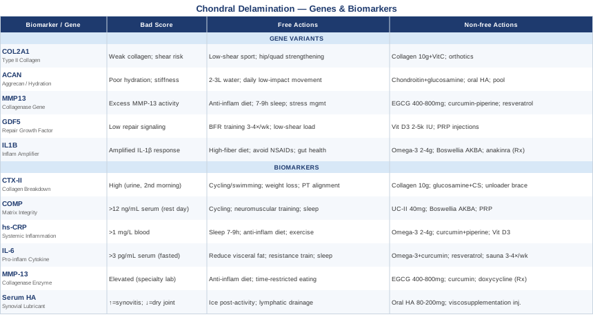
Where to Go From Here
Chondral delamination is a complex structural problem with biological drivers that vary from person to person. The most important takeaway from this article is that tracking — whether through biomarkers, genetic analysis, or careful symptom logging — gives you leverage that generic advice cannot. When you know which specific signals are elevated, which enzymatic pathways are overactive, and which genetic predispositions you are working around, you can make decisions that are genuinely calibrated to your biology rather than to a population average.
Start with what is accessible. A complete biomarker baseline — CTX-II, COMP, hs-CRP, and IL-6 — is achievable through a functional medicine physician or a sports medicine specialist at relatively low cost and provides an immediate diagnostic picture. Layer in genetic analysis when budget allows. Build the non-supplement foundation first — movement pattern quality, sleep, diet, and stress management consistently outperform supplements when the basics are not in place. Add targeted supplementation and complementary approaches from there, cycling appropriately and retesting biomarkers to verify that what you are doing is actually shifting the numbers.
The goal is not to chase a cure but to create an internal environment where cartilage damage slows, repair capacity is optimized, and the joint has the best possible conditions for structural stability over time. That is achievable — and the information to do it smarter than the standard model exists. The next step is simply to start measuring.
Musculoskeletal: Joint Conditions Sports Injuries
Autoimmune: Inflammatory Conditions Connective Tissue Conditions