This article contains AI generated content.
· UpdatedChondral Flap Lesion of the Knee: 7 Biomarkers and 6 Genes to Track
Introduction
A chondral flap lesion of the knee is one of those injuries that tends to slip between diagnostic categories. The cartilage is partially detached from the underlying bone, but because it does not show on a standard X-ray and MRI findings can be subtle depending on lesion size and orientation, many people spend months — or even years — with unexplained catching sensations, intermittent swelling, and pain that does not follow an obvious pattern. If you have been told your knee looks structurally acceptable yet something is clearly wrong, that disconnect is real, not imagined.
What makes these lesions particularly challenging to manage is that articular cartilage has an extremely limited blood supply, which means its intrinsic repair capacity is modest. Yet this limitation is not absolute. The biological environment surrounding the lesion — the level of synovial inflammation, the activity of catabolic enzymes, the quality of the local extracellular matrix, the body's vitamin D and omega-3 status — all determine whether a flap lesion stays stable, progresses toward full detachment, or finds conditions that allow partial stabilization over time. Generic advice like "rest, ice, and avoid aggravating activities" addresses none of these underlying factors.
This article takes a more targeted approach. By examining specific blood and urine biomarkers, it becomes possible to understand what is actively driving cartilage breakdown in a given individual and what interventions might slow that process or improve the biological conditions for repair. The genetics section adds another layer: certain gene variants affect how cartilage is built, how quickly it degrades under mechanical stress, and how well the body responds to protective interventions like vitamin D supplementation and anti-inflammatory protocols.
The goal here is not to promise outcomes, but to give you genuinely useful information that goes beyond generic advice. Better data leads to better decisions — and understanding your own biology is one of the most actionable steps available when managing a chondral flap lesion.
Summary
This article covers seven measurable biomarkers — CTX-II, COMP, hs-CRP, IL-6, vitamin D, MMP-3, and the omega-3 index — that directly reflect cartilage breakdown rate, catabolic enzyme activity, inflammation, and repair capacity. For each marker, you will find what an abnormal result actually means for cartilage biology, how to measure it and at what cost, and concrete plans both with and without supplements for moving the number in the right direction. The genetics section covers six variants — in COL2A1, GDF5, ACAN, MMP-3, VDR, and IL-1B — that shape how your cartilage is built and how fast it degrades, along with targeted compensation strategies. You will also find a condensed summary of ten key insights from sports medicine science on tissue repair and inflammation that challenge some conventional assumptions, and three evidence-based complementary approaches — photobiomodulation, tai chi, and mindfulness-based stress reduction — that have meaningful clinical backing for knee joint conditions. If even two or three of these angles apply to your situation, acting on them is likely to matter more than any single intervention in isolation.
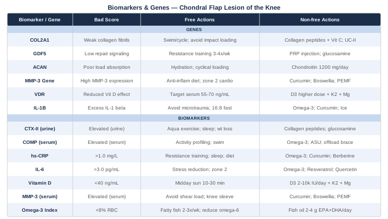
7 Biomarkers to Track for a Chondral Flap Lesion of the Knee
Tracking biomarkers shifts the management of a chondral flap lesion from reactive — waiting to see if the knee gets worse — to proactive, building a real-time picture of the biological environment inside the joint. The seven markers below were chosen because each captures a distinct dimension of cartilage health: breakdown rate, catabolic enzyme activity, systemic and local inflammation, hormonal support of chondrocytes, and the anti-inflammatory fatty acid status that governs how well the body resolves tissue damage. Measuring them once establishes a baseline; tracking them at three to six-month intervals reveals whether your interventions are actually working.
CTX-II (C-terminal Cross-linked Telopeptide of Type II Collagen)
CTX-II is the most specific cartilage degradation marker currently available in clinical practice. When type II collagen — the structural backbone of articular cartilage — is cleaved by matrix metalloproteinases, fragments are released into synovial fluid, enter circulation, and are excreted in urine. An elevated CTX-II does not simply reflect general joint stress; it signals that the cartilage matrix is being actively dismantled at a molecular level. In the context of a chondral flap, where the lesion edges are already mechanically vulnerable, this matters enormously. Research by Garnero and colleagues published in Arthritis and Rheumatism established urinary CTX-II as a predictive marker for cartilage loss rate in knee pathology. While a chondral flap is not osteoarthritis, the downstream collagen degradation pathways are biologically identical.
How to measure it
Urinary CTX-II, normalized to urinary creatinine, is measured from a second-morning urine sample (first morning void discarded). Specialty labs such as Quest Diagnostics and LabCorp offer this test; some functional medicine and sports medicine panels include it. Cost: $80–$150. Most conventional orthopedic practices do not routinely order it — you may need to request it specifically or work with a sports medicine or functional medicine physician.
If the score is elevated — plan without supplements
Mechanical loading drives CTX-II upward. Reducing compressive and shear forces on the affected compartment is the highest-leverage intervention. Even a 5% reduction in body weight measurably reduces knee joint loading. Avoid high-impact activities during the active phase — running on hard surfaces, jumping, deep squatting with load. Replace these with aquatic exercise or stationary cycling, which maintain cardiovascular fitness and quadriceps tone without axial compressive load. Sleep is also a critical but often overlooked variable: most cartilage matrix remodeling and anabolic repair signaling occurs during slow-wave sleep. Seven to nine consistent hours of quality sleep, combined with an anti-inflammatory dietary pattern — reduced refined carbohydrates, seed oils, and ultra-processed foods; increased fatty fish, leafy greens, and polyphenol-rich fruits — lowers CTX-II measurably over eight to twelve weeks.
If the score is elevated — plan with supplements or equipment
Hydrolyzed collagen peptides: 10–15 g daily taken with 500 mg vitamin C approximately 45–60 minutes before loading activity. Vitamin C is a required cofactor for collagen cross-linking. A randomized trial published in Nutrients showed that specific collagen peptides reduced urinary CTX-II and improved joint comfort. Cycle: continuous use for 12 weeks minimum; retest CTX-II at that point. Side effects: generally well-tolerated; occasional mild digestive discomfort. Glucosamine sulfate: 1,500 mg/day in split doses. Long-term OA trials have shown measurable reductions in CTX-II with sustained use. Minimum effective cycle: three months. Side effects: mild GI upset in some individuals; avoid shellfish-derived glucosamine if allergic. Curcumin with piperine: 500–1,000 mg curcuminoids twice daily with meals. Inhibits the NF-κB pathway, which is the primary transcriptional driver of MMP-mediated collagen breakdown. Cycle: continuous use, retest at 12 weeks. Side effects: may potentiate anticoagulant medications; take with food to reduce GI irritation. Lateral wedge insoles (for medial compartment lesions): Shift mechanical loading away from the medial side, reducing CTX-II-driving contact stress at the lesion site. A physiotherapist can confirm which compartment the lesion occupies and fit appropriately.
COMP (Cartilage Oligomeric Matrix Protein)
COMP is a structural glycoprotein found almost exclusively in cartilage and tendon tissue. When cartilage is mechanically stressed or enzymatically degraded, COMP fragments are rapidly released into synovial fluid and then into blood — making it one of the earliest markers to rise following cartilage injury. This speed of response makes it particularly valuable for monitoring a chondral flap that is subject to intermittent mechanical stress during daily activities. Studies published in the Annals of the Rheumatic Diseases have linked persistently elevated serum COMP to faster structural progression in knee joint pathology. In athletes with known cartilage lesions, measuring COMP before and two to four hours after specific activities provides direct information about how much matrix disruption each activity is causing.
How to measure it
Serum COMP from a fasting blood draw, ideally in the morning before physical activity to avoid post-exercise spikes. Available through specialty labs and some academic medical center panels. Cost: $100–$200. Not available through most standard primary care panels; request through a sports medicine or functional medicine physician.
If the score is elevated — plan without supplements
Activity profiling is the most practical first step. Since COMP rises acutely and proportionally with mechanical loading, identifying which specific activities drive the largest spikes allows you to prioritize what to modify. Swimming and aquatic walking produce very low COMP excursion compared to running or stair descent under load. Neuromuscular training focused on quadriceps strength and dynamic knee stability reduces peak joint loading during walking, reducing both the magnitude and duration of COMP elevation following activity. A physiotherapist with experience in cartilage rehabilitation can structure a progressive loading program that builds loading tolerance without exceeding the lesion's current threshold.
If the score is elevated — plan with supplements or equipment
Omega-3 fatty acids: 2–4 g EPA+DHA daily. Reduce synovial inflammatory signaling, which blunts COMP release in response to loading. Triglyceride-form fish oil taken with a fatty meal for optimal absorption. Cycle: continuous; meaningful anti-inflammatory benefit apparent at eight to twelve weeks. Side effects: fishy aftertaste (refrigerate capsules), mild anticoagulant effect at high doses. Avocado-Soybean Unsaponifiables (ASU): 300 mg/day standardized extract. Multiple European trials have shown reductions in cartilage biomarkers and structural progression markers with ASU. Often combined with glucosamine and chondroitin for additive effect. Cycle: three months minimum, continuous is acceptable. Side effects: well-tolerated. Compartment-offloading knee brace: For lesions confined to one compartment (typically confirmed by MRI), a functional offloading brace redistributes mechanical stress away from the lesion site during activity. This reduces peak COMP excursion per unit of activity and allows continued function without driving progressive damage. Prescription-level braces (e.g., DJO Unloader One, Ossur Unloader One Everyday) are measurably more effective than over-the-counter options.
High-Sensitivity CRP (hs-CRP)
Chronic low-grade systemic inflammation is one of the primary environmental drivers of cartilage matrix catabolism. hs-CRP is a standard, inexpensive inflammatory marker that reflects the current level of systemic inflammation. While it is not cartilage-specific, it is a reliable proxy for the catabolic environment inside the knee joint: when hs-CRP is elevated, the expression of pro-inflammatory interleukins and MMPs within the synovium tends to be elevated as well. Peter Attia consistently identifies hs-CRP as one of the most important routine markers to monitor, particularly in people managing chronic inflammatory conditions affecting musculoskeletal tissue.
How to measure it
Standard fasting blood draw. Available universally through any laboratory. Often included in comprehensive metabolic or cardiac risk panels, or orderable as a standalone test. Cost: $15–$40. Context matters: a recent infection, physical injury, or intense exercise bout can transiently elevate hs-CRP; ideally measure during a stable, illness-free period.
Optimal target
Below 1.0 mg/L is optimal for joint health. 1.0–3.0 mg/L indicates a moderate inflammatory burden; above 3.0 mg/L indicates high systemic inflammation that warrants active intervention.
If the score is elevated — plan without supplements
Sleep quality has a larger and more underappreciated effect on hs-CRP than most people realize: a single night of significantly disrupted sleep raises it measurably, and chronic mild sleep restriction maintains it at a persistently elevated level. Body composition matters equally: visceral adipose tissue is itself a cytokine-secreting organ that continuously drives CRP upward. Structured resistance training three to four times per week lowers hs-CRP significantly over twelve to sixteen weeks through multiple mechanisms, including reduced visceral fat, improved insulin sensitivity, and downregulation of pro-inflammatory gene expression. A Mediterranean or anti-inflammatory dietary pattern — high in polyphenols, omega-3-rich fish, fiber; low in refined carbohydrates and trans fats — consistently reduces hs-CRP across diverse populations.
If the score is elevated — plan with supplements or equipment
Omega-3 fatty acids: 3–4 g EPA+DHA daily reduces hs-CRP by 20–40% in many individuals over eight to twelve weeks. This is one of the most robustly supported supplement interventions for systemic inflammation across multiple meta-analyses. Curcumin with piperine: Multiple meta-analyses confirm meaningful reductions in CRP with 500–1,000 mg curcuminoids twice daily. Berberine: 500 mg twice daily. Lowers NF-κB activity and has shown anti-inflammatory effects across several trials. Cycle: twelve weeks on, four weeks off to avoid gut microbiome adaptation. Side effects: GI discomfort in some; interactions with certain medications — check with your physician. Magnesium glycinate: 300–400 mg at night. Low magnesium is associated with elevated CRP; correction is inexpensive and low-risk. Continuous use. Side effects: loose stools at higher doses.
Interleukin-6 (IL-6)
IL-6 is a pivotal pro-inflammatory cytokine in joint biology. Within the synovial environment of a chondral flap, elevated IL-6 promotes the expression of MMP-1, MMP-3, and MMP-13 — the collagenases and stromelysins that directly cleave type II collagen and aggrecan, the two principal structural components of articular cartilage. IL-6 also suppresses chondrocyte anabolic signaling, reducing the cell's capacity to synthesize new matrix. An elevated serum IL-6 is therefore not simply a marker of inflammation; it signals an active catabolic state at the cellular level, one that is working against any effort the lesion has to stabilize.
How to measure it
Serum IL-6 from a fasting blood draw. Not universally available through standard panels; functional medicine labs and specialty reference labs offer it. Because IL-6 spikes acutely with illness, exercise, and psychological stress, timing and context of the blood draw matter significantly. Cost: $50–$120.
Optimal target
Below 3.0 pg/mL in the absence of acute illness.
If the score is elevated — plan without supplements
Lifestyle levers for IL-6 closely overlap with those for hs-CRP. Specifically, chronic psychological stress maintains elevated cortisol, which — counterintuitively — drives inflammatory signaling over the long term through glucocorticoid receptor desensitization and NF-κB upregulation. Structured mind-body practices (addressed in the complementary section below) produce documented reductions in IL-6 over eight to twelve weeks. Reduction of visceral fat through caloric management and resistance training is one of the most powerful long-term drivers of lower IL-6.
If the score is elevated — plan with supplements or equipment
Omega-3 fatty acids: Directly suppress IL-6 transcription through EPA-derived mediators. 3–4 g EPA+DHA daily. Continuous use; reassess at three months. Resveratrol: 250–500 mg/day with food. Inhibits the STAT3 and NF-κB pathways that drive IL-6 production. Cycle: twelve weeks on, four weeks off. Side effects: generally well-tolerated; potential interaction with blood-thinning medications. Cold water immersion: Post-exercise cold exposure (10–15°C water temperature, 10–15 minutes, three to four times per week) has shown reductions in post-exercise IL-6 in trained individuals in several controlled studies. Not appropriate during the acute post-injury phase without physician clearance. Quercetin: 500 mg twice daily. IL-6 inhibitor via MAPK pathway suppression. Cycle: eight weeks on, two weeks off. Well-tolerated; may interact with antibiotics at very high doses.
25-Hydroxyvitamin D (Vitamin D)
Vitamin D receptors are expressed on chondrocytes, and vitamin D plays a documented regulatory role in chondrocyte differentiation, type II collagen synthesis, and the expression of cartilage-protective growth factors including TGF-β. Studies linking low vitamin D levels to worse cartilage outcomes have been replicated across multiple large cohorts. Thomas Dayspring and Peter Attia both emphasize that the clinically meaningful optimal range for vitamin D is not the population mean (which reflects widespread deficiency) but rather 40–60 ng/mL — a target that many individuals in northern climates, office-based workers, and darker-skinned individuals fall significantly short of without intentional supplementation.
How to measure it
25-OH vitamin D from a standard fasting blood draw. Widely available at any laboratory. Test in autumn or early winter for your true baseline; summer sun exposure often masks a latent deficiency. Cost: $30–$70.
If the score is low — plan without supplements
Sensible sun exposure is the most natural correction. Ten to thirty minutes of midday sun on significant skin surface area (arms, legs, abdomen if possible), three to five times per week during spring and summer months, can maintain vitamin D levels in temperate climates. At latitudes above approximately 40 degrees north, UVB exposure is insufficient from October through March, making supplementation essentially mandatory for maintenance.
If the score is low — plan with supplements or equipment
Vitamin D3: 2,000–5,000 IU/day for maintenance; individuals who are deficient (below 30 ng/mL) may require 5,000–10,000 IU/day under medical supervision to correct levels. Always pair with vitamin K2 (MK-7 form): 100–200 µg/day to direct calcium appropriately to bone rather than soft tissue. Retest at three months to confirm adequate response. Side effects: hypercalcemia is possible at sustained doses above 10,000 IU/day without monitoring, particularly in those with granulomatous conditions — periodic testing is prudent. Magnesium glycinate or malate: 300–400 mg/day. Magnesium is required for the enzymatic conversion of vitamin D to its active form. Many individuals are deficient in both simultaneously; correcting magnesium first often dramatically improves vitamin D response to supplementation.
MMP-3 (Matrix Metalloproteinase-3 / Stromelysin-1)
MMP-3 is a protease that cleaves aggrecan, fibronectin, and laminin, and also activates MMP-13 — the primary type II collagen-degrading enzyme in cartilage. It is significantly elevated in joints with active cartilage damage and is a useful index of how aggressively the enzymatic environment is working against cartilage integrity. Serum and synovial MMP-3 levels correlate with joint space narrowing rate in knee pathology research. In the context of a chondral flap, a persistently elevated MMP-3 indicates that the molecular environment favors further matrix loss, even if the flap itself has not progressed structurally on imaging.
How to measure it
Serum MMP-3 from a standard blood draw. Available through specialty and rheumatology reference labs; less commonly through primary care panels. Cost: $75–$160. Elevated MMP-3 is also used in the diagnosis and monitoring of rheumatoid arthritis, so it is occasionally included in auto-immune panels.
If the score is elevated — plan without supplements
Mechanical shear stress and synovial inflammation are the two primary drivers of MMP-3 upregulation. Activity modification targeting high-shear loading scenarios (rapid deceleration, pivoting, stair descent under load) reduces the mechanical stimulus for MMP-3 release. Compression sleeves (20–30 mmHg knee sleeve) reduce synovial effusion, which decreases the concentration of pro-inflammatory mediators in the joint that upregulate MMP-3. Prolonged low-grade repetitive loading — extended standing on hard floors, repetitive stair climbing with loaded backpacks — is also worth addressing because it maintains chronic MMP-3 upregulation even below the threshold of pain.
If the score is elevated — plan with supplements or equipment
Curcumin with piperine: Directly inhibits MMP-3 and MMP-13 transcription by suppressing the NF-κB and AP-1 promoter binding activity. 1,000 mg curcuminoids twice daily. Continuous use; reassess at twelve weeks. Boswellia serrata (AKBA fraction): 300–400 mg standardized extract (65% boswellic acids) twice daily. Clinical trial data support reduction in MMP activity and cartilage degradation markers in knee conditions. Cycle: continuous or twelve weeks on, four weeks off. Side effects: occasional mild GI discomfort; generally well-tolerated. PEMF (Pulsed Electromagnetic Field) therapy: Home devices (Oska Pulse, Bemer, or clinical-grade units) have shown downregulation of MMP-3 in cell culture studies and preliminary clinical work in knee OA. Typically eight to twenty minutes per session directly over the affected knee, daily, for eight to twelve weeks. Device cost: $200–$2,000 for home units. Non-invasive, no known significant side effects. Sub-antimicrobial dose doxycycline: 40 mg modified-release formulation daily is an established MMP inhibitor used clinically in periodontal disease and has been studied in joint conditions. Prescription-only; discuss with your physician if MMP-3 remains persistently elevated despite lifestyle and supplement interventions.
Omega-3 Index (EPA + DHA in Red Blood Cells)
The omega-3 index measures the percentage of EPA and DHA within red blood cell membranes — reflecting average omega-3 status over the preceding three to four months, making it far more stable and informative than a single plasma measurement. EPA and DHA are the direct precursors to specialized pro-resolving mediators (SPMs) — a class of lipid compounds that actively resolve inflammation, promote macrophage clearance of cellular debris, and create conditions favorable to tissue repair. In the context of cartilage, a low omega-3 index correlates with higher circulating levels of IL-6 and TNF-α, faster cartilage degradation markers, and poorer joint outcomes across multiple cohort studies. Peter Attia places the omega-3 index among his highest-priority routine markers, and it is increasingly central to functional medicine panels for musculoskeletal conditions.
How to measure it
A simple finger-prick dried blood spot test (OmegaQuant and similar labs provide home collection kits). Also available through blood draw at specialty labs. Cost: $50–$100 with home collection widely available and requiring no physician order in most jurisdictions.
Optimal target
Above 8% is the target supported by anti-inflammatory and cardiovascular research. Below 4% is associated with high inflammatory burden and poor tissue-repair conditions. The majority of Western adults without fish oil supplementation test between 4–6%.
If the score is low — plan without supplements
Dietary change alone can improve the omega-3 index over three to six months. Two to three servings per week of wild-caught fatty fish (salmon, mackerel, sardines, herring, anchovies) provides meaningful EPA+DHA. Equally important but often overlooked: reducing linoleic acid (omega-6) intake from processed vegetable oils (soybean, sunflower, corn oil) improves the EPA/AA tissue ratio even without increasing absolute omega-3 intake.
If the score is low — plan with supplements or equipment
Triglyceride-form fish oil: 2–4 g EPA+DHA daily. Triglyceride form (e.g., Nordic Naturals, Carlson) absorbs significantly better than ethyl ester form. Take with a fatty meal. Cycle: continuous; retest omega-3 index at four months to confirm response. Side effects: fishy aftertaste (refrigerate capsules), mild anticoagulant effect at high doses — discuss with physician if on warfarin or similar. Phospholipid-form omega-3s (krill oil or herring roe oil): For individuals with persistently low index despite triglyceride fish oil, the phospholipid carrier form may improve cellular incorporation. 500 mg–1 g EPA+DHA equivalent daily. Higher per-dose cost but potentially more efficient for some individuals. Algal DHA oil: Vegan alternative, DHA-dominant. 1–2 g DHA daily. Less EPA than fish-derived sources; adequate for baseline maintenance but less potent for acute inflammation resolution.
The Genetic Layer: 6 Variants That Shape Cartilage Risk and Repair
Genetics does not determine outcomes — but it does define the biological starting conditions. Understanding which gene variants influence cartilage construction, maintenance, and degradation allows you to target compensatory interventions more precisely. Most of the following variants can be identified through direct-to-consumer genetic testing (23andMe, AncestryDNA) combined with third-party interpretation tools (Genetic Genie, Rhonda Patrick's tools, or consultation with a functional medicine physician), or through clinical genomic panels. The research here is primarily observational and GWAS-based; where strong human evidence exists, it is noted; where evidence remains preliminary, that is stated explicitly.
COL2A1 (Type II Collagen Alpha-1 Chain)
What it does: COL2A1 encodes the alpha-1 chain of type II collagen — the primary structural protein of articular cartilage. Heterozygous variants in COL2A1 are strongly associated with early-onset cartilage pathology, including chondrodysplasias and premature joint degeneration. Even subtler polymorphisms in regulatory regions of COL2A1 affect the quality and organization of the collagen fibril network, influencing how cartilage handles mechanical stress. Human GWAS studies have identified COL2A1 region variants as among the most consistently replicated genetic factors in knee cartilage volume and OA risk.
If this gene variant is suboptimal — plan without supplements
Because COL2A1 affects the structural integrity of collagen fibrils, mechanical environment is the most critical modifiable factor. Reducing cumulative high-impact loading over a lifetime (avoiding running on hard surfaces, emphasizing swimming and cycling), maintaining lean body mass to reduce joint loading, and prioritizing neuromuscular control of the knee (strong VMO, good proprioception) all reduce the mechanical demands placed on a collagen network that may be intrinsically less resilient. Frequency: these are permanent lifestyle orientations, not short-term interventions. Side effects: none.
If this gene variant is suboptimal — plan with supplements or equipment
Hydrolyzed collagen peptides with vitamin C: 15 g daily with 500 mg vitamin C before loading. Type II specific peptides stimulate chondrocyte synthesis through a different mechanism than structural collagen supplementation. Undenatured type II collagen (UC-II): 40 mg/day — works through oral tolerization to reduce the autoimmune-like cartilage inflammation that can worsen poor-quality collagen. Cycle: minimum six months. Side effects: generally well-tolerated. Vitamin C: 500–1,000 mg/day standalone — required for hydroxylation of proline and lysine during collagen fibril formation; a COL2A1 risk variant does not change this requirement but may make it more consequential. Frequency: daily, continuous.
GDF5 (Growth Differentiation Factor 5)
What it does: GDF5 encodes a signaling protein critical for cartilage and joint development, and for chondrocyte differentiation during repair responses. The rs143383 single nucleotide polymorphism in the GDF5 promoter region is one of the most replicated and robustly validated genetic risk factors for knee OA in human populations. The T allele at this position reduces GDF5 transcription in joint tissues, lowering cartilage repair signaling. Importantly, a landmark study by Miyamoto et al. published in Nature Genetics confirmed this association across multiple Asian and European populations.
If this gene variant is suboptimal — plan without supplements
GDF5 is a growth factor — it signals chondrocytes to proliferate and synthesize matrix. Physical activity (specifically controlled loading and resistance training) upregulates GDF5 expression in cartilage tissue in animal models and is one of the few non-pharmacological signals that partially compensates for reduced GDF5 transcription. Progressive resistance training targeting the quadriceps and hip abductors, structured around the principle of gradually increasing load below pain threshold, should be performed three to four times per week. This simultaneously reduces joint loading during ambulation and provides the mechanical stimulus that drives endogenous GDF5 upregulation. Frequency: ongoing, lifelong.
If this gene variant is suboptimal — plan with supplements or equipment
Platelet-Rich Plasma (PRP) injection: Growth factors delivered via PRP (including TGF-β and PDGF) partially substitute for reduced endogenous GDF5 signaling and have shown measurable improvements in cartilage biomarkers and symptoms in RCTs for knee cartilage pathology. Discuss with an orthopedic sports medicine physician. Protocol varies; typically one to three injections spaced four to six weeks apart. Cost: $500–$2,000 per injection. Side effects: temporary post-injection flare (24–72 hours), low infection risk. Glucosamine sulfate + chondroitin: Support the downstream substrate availability for matrix synthesis that GDF5 promotes. 1,500 mg glucosamine + 1,200 mg chondroitin daily, minimum three months. Side effects: mild GI effects possible.
ACAN (Aggrecan)
What it does: ACAN encodes aggrecan, the major proteoglycan of articular cartilage responsible for water retention and compressive load distribution. Variants in the ACAN gene affect the length and composition of the aggrecan core protein, directly influencing the cartilage's capacity to absorb and distribute mechanical force. Shorter aggrecan repeat lengths (VNTR polymorphisms) are associated with early disc and cartilage degeneration. This is an area of active research, and evidence in humans for specific actionable variants is early-stage; if you have an ACAN variant flagged, treat it as a signal to prioritize compressive load management rather than a definitive prediction.
If this gene variant is suboptimal — plan without supplements
Cartilage water content (which aggrecan regulates) is sensitive to hydration status and loading patterns. Consistent hydration throughout the day — not just during exercise — is relevant. Cartilage fluid exchange is driven by cyclical loading and unloading; walking, light cycling, and aquatic exercise all provide this cyclical stimulus without compressive overload. Avoid prolonged static loading (extended kneeling, crouching) which flattens cartilage water content without the rebound loading needed for re-imbibition. Frequency: ongoing daily habits.
If this gene variant is suboptimal — plan with supplements or equipment
Chondroitin sulfate: 1,200 mg/day. Provides direct substrate for aggrecan side-chain synthesis and has shown protective effects on cartilage proteoglycan content in imaging studies. Cycle: minimum three months, continuous use reasonable. Side effects: well-tolerated; occasional mild GI effects. Hyaluronic acid (oral or injectable): Oral HA (e.g., 80–200 mg/day of food-grade HA) supports synovial fluid quality, which works in concert with aggrecan for cartilage lubrication. Injectable HA (viscosupplementation) can be considered for symptomatic management; discuss with your orthopedic physician. Side effects: injectable HA may cause temporary local reaction.
MMP-3 Gene (rs679620 Variant)
What it does: A functional polymorphism in the MMP-3 promoter region (rs679620, 5A allele) is associated with higher baseline MMP-3 gene expression. Individuals carrying the 5A allele produce more MMP-3 in response to inflammatory stimuli, potentially accelerating cartilage matrix degradation. This variant is directly relevant to anyone with an elevated serum MMP-3 biomarker — it may explain part of why MMP-3 remains elevated despite standard interventions, and it shifts the intervention priority toward MMP-3-specific suppression strategies described in the biomarker section above.
If this gene variant is suboptimal — plan without supplements
Anti-inflammatory dietary patterns that directly reduce the transcriptional stimulus for MMP-3 are the primary non-supplement approach: reduced sugar and refined carbohydrates (which drive NF-κB), increased polyphenol-rich foods (blueberries, green tea, extra virgin olive oil), and consistent aerobic exercise at moderate intensity (zone 2) which has documented MMP-suppressive effects through anti-inflammatory adaptations. Frequency: daily dietary practice; zone 2 exercise three to four times per week, 30–45 minutes per session.
If this gene variant is suboptimal — plan with supplements or equipment
The supplement approach mirrors the MMP-3 biomarker plan above: curcumin with piperine (1,000 mg twice daily), Boswellia serrata (300–400 mg twice daily), and PEMF therapy (8–20 minutes daily over the affected joint) are the most evidence-supported options for MMP-3 gene-driven risk. Cycle: twelve weeks, reassess serum MMP-3.
VDR (Vitamin D Receptor)
What it does: The VDR gene encodes the receptor through which vitamin D exerts its effects in cells — including chondrocytes. Common VDR polymorphisms (BsmI, TaqI, FokI, ApaI) affect receptor binding efficiency and downstream gene transcription, meaning that two individuals with identical serum vitamin D levels may have very different intracellular vitamin D responses depending on VDR variants. FokI ff genotype, for example, produces a longer, less efficient receptor protein. In the context of cartilage, suboptimal VDR function impairs chondrocyte differentiation and collagen synthesis even when serum vitamin D is adequate by conventional thresholds.
If this gene variant is suboptimal — plan without supplements
Maintaining vitamin D levels at the higher end of the optimal range (55–70 ng/mL) rather than simply meeting the lower threshold (40 ng/mL) partially compensates for reduced receptor efficiency through mass action — more ligand drives more activation even when receptor affinity is reduced. Sensible sun exposure as described in the biomarker section remains the best natural driver.
If this gene variant is suboptimal — plan with supplements or equipment
Target higher vitamin D supplementation doses to achieve serum levels of 55–70 ng/mL rather than 40–50 ng/mL if VDR variants are suboptimal. Pair with vitamin K2 (MK-7) 200 µg/day and magnesium 400 mg/day. Some functional medicine practitioners also add vitamin A (retinol) in modest amounts (2,500–5,000 IU/day) to support VDR activity through RXR heterodimerization — only under guidance. Cycle: maintain consistently; reassess 25-OH vitamin D levels every six months. Side effects: discussed in biomarker section above.
IL-1B (Interleukin-1 Beta)
What it does: IL-1B encodes interleukin-1 beta, a cytokine that directly suppresses collagen synthesis in chondrocytes and upregulates MMP-1, MMP-3, and MMP-13. The rs1143634 and rs16944 polymorphisms in IL-1B are associated with higher baseline IL-1B production in response to joint stress. In individuals carrying these variants, the inflammatory response to cartilage trauma — including a chondral flap — is amplified and prolonged, creating a more persistently catabolic joint environment. Evidence for these specific variants in joint pathology is moderate and primarily from European OA cohorts; it should be interpreted with context rather than alarm.
If this gene variant is suboptimal — plan without supplements
IL-1B is acutely triggered by mechanical damage and synovial trauma. Avoiding activities that generate mechanical microtrauma to the lesion — high-impact loading, pivoting on a locked knee, any activity that reproducibly causes joint swelling — is the priority. Consistent intermittent fasting (16:8 or similar) has shown IL-1B suppression in several metabolic studies and is a low-risk strategy for someone carrying high-expression IL-1B variants. Zone 2 aerobic training also reduces baseline IL-1B over time through its anti-inflammatory systemic effects.
If this gene variant is suboptimal — plan with supplements or equipment
Omega-3 fatty acids: 3–4 g EPA+DHA daily directly suppresses IL-1B via the EPA→resolvin E1 pathway. Curcumin: 500–1,000 mg twice daily suppresses IL-1B transcription through NF-κB inhibition. Quercetin: 500 mg twice daily. Cryotherapy or ice compression (applied to the affected knee for 15–20 minutes after loading activity, three to four times per week) is one of the simplest and most evidence-consistent ways to reduce local IL-1B activity after activity-related microtrauma. Cycle for supplements: twelve weeks, then reassess IL-6 and hs-CRP as proxy markers.
What Tissue Repair Science Tells Us About Cartilage Healing
Built to Move: The Ten Essential Habits to Help You Move Freely and Live Fully by physical therapist Kelly Starrett and Juliet Starrett (2023) is one of the most practically useful recent books on joint health, mobility, and connective tissue maintenance. Drawing on decades of clinical work with elite athletes and everyday patients, and grounding its recommendations in exercise science and physiology research, it challenges several assumptions in conventional orthopedic advice. The ten insights below are the ones most directly applicable to a chondral flap lesion.
1. Sitting Is a Loading Problem, Not Just a Posture Problem
Prolonged static sitting places the knee joint in a position of sustained compression without the fluid-exchange cycling that cartilage depends on for nutrient delivery and waste clearance. Cartilage has no blood supply; it relies entirely on the mechanical "pump" of cyclical loading and unloading to move nutrients in and metabolic waste out. The Starretts recommend a daily movement floor — ten minutes of low-intensity walking or movement for every hour of sitting. This is not about posture; it is about keeping cartilage metabolically active.
2. Strength Is Protective Load Management, Not Cosmetic
Strong quadriceps, hip abductors, and glutes do not simply look better — they absorb peak mechanical force before it reaches the articular surface. Every pound of quadriceps strength reduces knee joint contact force during walking by approximately three to four times that amount. The book argues, convincingly, that most joint injuries exist in part because the surrounding musculature was never built to match the demands placed on it. Progressive resistance training below the threshold of pain is not risky for a chondral flap; avoiding it is.
3. Breathing and HRV Are Inflammation Regulators
The vagal tone of the nervous system — measurable through heart rate variability — is a direct regulator of systemic inflammation. Low HRV is associated with elevated IL-6 and CRP. Nasal breathing, slow exhale-extended breathing patterns (e.g., 4-second inhale, 8-second exhale), and consistent aerobic exercise all improve vagal tone and have measurable anti-inflammatory downstream effects. The Starretts include breathing as a first-tier recovery tool, not a supplementary relaxation technique.
4. Sleep Is the Non-Negotiable Repair Window
Cartilage does not have an active healing process during waking hours — it is primarily protected and managed. The anabolic window for connective tissue repair occurs during slow-wave sleep, when growth hormone pulsatility is highest. Collagen synthesis is sleep-dependent. The book cites research showing that athletes who chronically undersleep (below 7 hours) show elevated cartilage degradation markers compared to age-matched peers sleeping 8+ hours. Sleep is the cheapest and most evidence-supported cartilage intervention available.
5. Joint Position Matters More Than Stretch Duration
Conventional flexibility advice focuses on duration of stretch. Starrett's approach focuses on achieving full joint range of motion under mild load — what they call "loaded mobility." For the knee, this includes controlled eccentric loading through the full flexion range. This mechanical stimulus signals chondrocytes to maintain matrix density in the parts of the cartilage that are typically underloaded in people who avoid bending the knee fully because of pain. Avoiding end-range knee position chronically leaves portions of cartilage metabolically deprived.
6. Protein Adequacy Is a Structural Prerequisite
Connective tissue synthesis requires amino acid substrate. The book emphasizes that most adults — particularly older adults and those restricting calories — are chronically underconsuming protein relative to what tissue repair demands. Their recommendation aligns with Attia's: 1.6–2.2 g of protein per kg of lean body mass per day, with timing distributed across meals rather than concentrated. Collagen precursor amino acids (glycine, proline, hydroxyproline) are specifically required for type II collagen synthesis and are found in lower quantities in muscle meat than in bone broth, skin, or collagen supplements.
7. Zone 2 Training Is the Metabolic Anti-Inflammatory Intervention
Zone 2 aerobic exercise — steady-state cardio at a pace where nasal breathing is possible and conversation is comfortable — produces a distinctly different hormonal and inflammatory signature from high-intensity exercise. It reduces baseline IL-6 and CRP over twelve to sixteen weeks, improves mitochondrial density in muscle (reducing fatigue-related joint loading), and maintains cartilage hydration through gentle cyclical loading. For someone with a chondral flap, zone 2 on a stationary bike or in a pool provides maximum anti-inflammatory benefit at minimum joint stress. Target 150–180 minutes per week.
8. The Microbiome Connects to Joint Inflammation
Starrett and colleagues have increasingly incorporated gut health into their joint health framework. Short-chain fatty acids produced by gut bacteria (particularly butyrate from fiber fermentation) directly suppress intestinal and systemic NF-κB activity — the same pathway that drives MMP and cytokine expression in the synovium. A fiber-rich diet (30+ grams of diverse fiber daily) supports the bacterial populations that produce these anti-inflammatory metabolites. This is emerging science in joint conditions, but the safety and breadth of benefit from dietary fiber optimization make it a low-barrier, high-value habit.
9. Cold and Heat Sequencing Affects Recovery Quality
The book is specific about timing: cold immediately after strength training blunts the adaptation signal (mTOR-mediated) from that session — so do not ice the knee immediately after a strength workout if the goal is to build cartilage-protective muscle. Cold is most appropriate after purely aerobic work or as a standalone anti-inflammatory protocol on rest days. Contrast therapy (alternating cold and heat) has shown better inflammatory marker clearance than cold or heat alone in some sports medicine studies and is worth exploring systematically.
10. Blood Flow Restriction Training Opens a New Option
Blood flow restriction (BFR) training — applying a pneumatic cuff or specialized wrap to the limb and performing low-load resistance exercises (20–30% of 1RM) — achieves similar strength and hypertrophy adaptations to heavy loading at a fraction of the joint compressive force. Several studies in post-surgical knee patients have shown BFR training to be safe and effective for building quadriceps strength without driving cartilage biomarker elevation. For someone managing a chondral flap who cannot tolerate conventional loading, BFR represents a clinically validated bridge strategy.
Evidence-Based Complementary Approaches for Knee Cartilage Health
The following three approaches were selected from a longer list based on the availability of meaningful human clinical evidence specifically relevant to knee joint conditions, and the practical feasibility of incorporating them alongside a standard medical plan.
Low-Level Laser Therapy and Photobiomodulation
Photobiomodulation (PBM) uses specific wavelengths of red and near-infrared light (typically 630–1,000 nm) to stimulate mitochondrial function in target tissue. In chondrocytes and synovial cells, PBM at these wavelengths has been shown to reduce inflammatory cytokine production, decrease MMP activity, and support ATP synthesis — all of which are directly relevant to the catabolic environment surrounding a chondral flap. Several in vitro studies have specifically shown that near-infrared PBM reduces IL-1B-induced MMP-13 expression in chondrocytes, making this one of the mechanistically best-justified complementary approaches for cartilage.
A systematic review and meta-analysis published in Lasers in Medical Science evaluated LLLT for knee OA and found significant reductions in pain and functional impairment compared to placebo, particularly with near-infrared wavelengths (780–860 nm) at adequate energy doses. The Ottawa Panel clinical practice guidelines for LLLT in OA also list it among recommended modalities for knee pain, noting Grade A evidence for pain reduction.
Clinically available through physiotherapy and sports medicine clinics; home devices (Joovv, PlatinumLED BioMax, Kineon Move+) are increasingly accessible at $200–$1,000. For knee cartilage, treat directly over the knee joint, 5–10 minutes per session, once daily for an initial eight to twelve-week trial. Keep expectations calibrated: PBM reduces the catabolic environment and pain; it does not mechanically reattach a flap. Side effects are minimal — avoid direct ocular exposure. It is not a replacement for orthopedic evaluation.
Tai Chi
Tai chi is a mind-body practice originating from Chinese martial arts traditions, characterized by slow, controlled movements through full joint range, combined with weight shifting, body awareness, and breathwork. For knee joint conditions, tai chi addresses multiple relevant dimensions simultaneously: quadriceps and hip abductor activation, proprioceptive training, controlled cartilage loading through range of motion, and stress reduction that secondarily lowers inflammatory biomarkers. It demands none of the high compressive forces that damage cartilage, making it an accessible option even in the presence of an active chondral lesion.
A landmark randomized controlled trial by Wang et al. published in the Annals of Internal Medicine in 2016 (PMID 27159061) compared twelve weeks of tai chi to physical therapy in 204 patients with knee OA and found that tai chi produced equivalent or superior improvements in pain, function, and quality of life — with effects maintained at 52 weeks. A secondary analysis also showed improvements in depression and fatigue, which contribute to the pain amplification seen with chronic joint conditions. This is among the strongest comparative-effectiveness trials in the knee joint literature.
A practical tai chi program for someone with a chondral flap should begin with a Yang-style short form or a seated modification if the knee is acutely symptomatic. Classes led by certified tai chi for health instructors are available in most communities; online programs (e.g., Tai Chi for Arthritis by Paul Lam) are clinically tested and validated. Two to three sessions per week of 45–60 minutes, for a minimum of twelve weeks, is the protocol supported by the strongest evidence. Progress gradually into deeper stances as the knee tolerates loading. Avoid forcing full knee flexion if it triggers sharp pain.
Mindfulness-Based Stress Reduction (MBSR)
MBSR is an eight-week structured program developed by Jon Kabat-Zinn that combines mindfulness meditation, body scan practices, and gentle yoga. Its relevance to a chondral flap lesion operates through two mechanisms: first, direct anti-inflammatory effects (cortisol regulation, vagal tone improvement, and documented reductions in IL-6 and CRP with regular practice); and second, central pain modulation — chronic joint pain sensitizes central nervous system pain processing, meaning that even a stable lesion generates amplified pain signals. MBSR addresses this central sensitization component in ways that no biomarker intervention or supplement can.
A systematic review of MBSR for chronic musculoskeletal pain conditions, published in JAMA Internal Medicine and summarized in NIH-indexed meta-analyses, found consistent reductions in pain severity, pain-related disability, and psychological distress compared to wait-list or active control conditions. Inflammatory marker reductions (particularly IL-6) with MBSR have been documented in several studies. While most trials focused on low back pain or osteoarthritis rather than chondral flap lesions specifically, the mechanisms of central sensitization and inflammatory cytokine modulation are shared.
The MBSR program is widely available through hospitals, community centers, and online platforms (Sounds True, the MBSR Online course). The standard protocol is eight weeks of weekly group sessions (2.5 hours) plus a one-day retreat, and 40–60 minutes of daily home practice. A more accessible entry point is a structured daily meditation practice of ten to twenty minutes using an app such as Waking Up or Insight Timer, combined with a body scan practice specifically focused on the affected knee before sleep. Key outcomes to monitor over eight weeks: sleep quality, pain catastrophizing score (PCS questionnaire), and subjective fatigue — all of which respond measurably before structural changes occur.
Conclusion
A chondral flap lesion of the knee does not exist in a biological vacuum. It sits within a larger environment shaped by inflammation levels, catabolic enzyme activity, hormonal status, genetic architecture, and daily mechanical habits — most of which are measurable and many of which are actionable. The seven biomarkers covered here give you a real-time window into what is happening in that environment. The six genetic variants explain why some individuals face a more challenging biological starting point and need targeted compensation. The tissue repair science reminds you that sleep, zone 2 training, protein adequacy, and movement variety are not optional lifestyle add-ons — they are the foundation on which everything else depends.
The next smart step is not to act on everything at once. Start with the most accessible: get hs-CRP, vitamin D, and an omega-3 index measured. These three tests are inexpensive, available without a specialist, and each gives you an immediately actionable result. If any of them is outside the optimal range, the plans outlined in this article give you a clear starting point. Bring the results to a sports medicine physician or functional medicine practitioner who can help you interpret them in the context of your specific lesion, loading history, and overall health picture. Better information, consistently acted on, is the most reliable path forward.
Musculoskeletal: Joint Conditions Sports Injuries
Autoimmune: Inflammatory Conditions Connective Tissue Conditions