This article contains AI generated content.
· UpdatedChondrocalcinosis: 4 Genes And 6 Biomarkers To Track
Introduction
If you've been told you have chondrocalcinosis — or calcium pyrophosphate deposition disease (CPPD) — the conversation with your doctor probably ended somewhere around "it looks like arthritis on your X-ray" and "we can manage the flares." That answer is not wrong. It is just incomplete in ways that matter.
What rarely gets discussed is that chondrocalcinosis almost always has a reason. In a substantial portion of cases, it is a downstream signal of something measurable and correctable upstream: a mineral imbalance, a metabolic disruption, a thyroid problem, iron overload, or in some people, a genetic predisposition that quietly accumulates risk over years. Each of these has a different root cause, and none of them show up in a standard joint examination.
Generic advice — eat more vegetables, move more, try anti-inflammatories during flares — isn't harmful, but it misses the point when the real driver is persistently low magnesium, undetected hyperparathyroidism, or a slow-progressing hemochromatosis gene variant. Treating the flare without addressing the metabolic cause is like mopping the floor while the tap is still running.
This article takes a more targeted approach. It covers the six most actionable biomarkers to track if you have or suspect chondrocalcinosis — each one mapped to a known upstream mechanism — and the four genes with the strongest clinical evidence for this condition, along with practical strategies for each. Beyond that, you'll find a summary of longevity science thinking that directly applies to this condition, and a selection of complementary approaches with genuine human evidence behind them. The goal is better information leading to better decisions — not promises, but a clearer map.
Summary
This article approaches chondrocalcinosis from angles most rheumatology visits never reach. The biomarker section covers six specific lab tests — serum magnesium, parathyroid hormone, ferritin and iron saturation, thyroid function (TSH), phosphate with alkaline phosphatase, and high-sensitivity CRP — explaining what each reveals, how to test it, and what to do if the number is off. For each marker, you'll find a plan with and without supplements, including specific doses, cycling protocols, and side effects to watch. The genetics section follows with four genes (ANKH, ENPP1, NT5E, HFE) that can silently prime joints for crystal deposition, with actionable compensation strategies for each. After that, a deep dive into the metabolic health framework from Peter Attia's Outlive reveals ten research-backed insights that most physicians aren't applying to joint disease yet. The article closes with five evidence-supported complementary approaches — tai chi, photobiomodulation, mindfulness, microbiome therapy, and breathwork — each with a specific protocol. Whether you are newly diagnosed or years into managing this condition, what follows is designed to be genuinely useful.
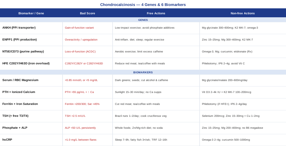
6 Biomarkers to Track If You Have Chondrocalcinosis
Chondrocalcinosis is unusual among joint conditions in that laboratory findings can genuinely shift the treatment picture. Unlike osteoarthritis, which is largely mechanical, CPPD has well-documented metabolic drivers — conditions that show up in blood tests and respond to targeted intervention. The six biomarkers below are chosen because each maps directly to a pathway that feeds calcium pyrophosphate crystal formation or amplifies inflammatory flares. All can be measured with standard lab work, and most cost well under one hundred dollars.
Finding a correctable cause does not guarantee reversal of existing deposits, but in patients with active metabolic drivers, correcting the upstream problem consistently reduces flare frequency and severity — and in some cases, slowly reduces crystal burden over time.
1. Serum Magnesium (and RBC Magnesium)
Why it matters
Magnesium may be the single most underappreciated factor in chondrocalcinosis management. It functions as a direct inhibitor of calcium pyrophosphate crystal nucleation and growth within cartilage tissue. Studies comparing CPPD patients to age-matched controls have found lower magnesium levels in the CPPD group at a frequency that is hard to dismiss as coincidence, and small clinical observations suggest that correcting deficiency can reduce crystal formation over months.
The problem is measurement. Standard serum magnesium reflects only about one percent of total body stores — the rest is intracellular. A patient can present with a result squarely in the "normal" reference range while being genuinely depleted at the cellular level. This gap between apparent and functional magnesium status is one of the most common diagnostic blind spots in metabolic joint disease.
How to measure it
Start with a standard serum magnesium test, available through most labs for $10–30. For a more accurate reading, request an RBC (red blood cell) magnesium test ($30–80), which reflects intracellular stores. A 24-hour urine magnesium excretion test can reveal renal wasting — where the kidneys are excreting magnesium faster than dietary intake can replace it. Target serum magnesium of 0.85–0.95 mmol/L; RBC magnesium above 5.0 mg/dL is generally considered adequate. Retest every three months during active supplementation.
If the score is low: the plan without supplements
Emphasize magnesium-dense whole foods: dark leafy greens (spinach, Swiss chard, beet greens), pumpkin seeds (one of the highest dietary sources at around 150 mg per ounce), black beans, almonds, dark chocolate, and wild-caught fatty fish. Reduce the two biggest magnesium thieves in daily life — alcohol and excess caffeine — both of which increase renal magnesium excretion significantly. Addressing gut health through fermented foods and fiber also improves magnesium absorption, since intestinal inflammation impairs uptake. Cooking in mineral-rich water rather than filtered or distilled water adds a modest but consistent contribution.
If the score is low: the plan with supplements or equipment
Magnesium glycinate or magnesium malate are the preferred forms for systemic repletion — high bioavailability, minimal laxative effect. Start at 200 mg elemental magnesium per day and titrate to 400–600 mg over two to four weeks. Magnesium threonate penetrates the blood-brain barrier more effectively and can be added if cognitive or sleep symptoms accompany the deficiency. Avoid magnesium oxide — it is poorly absorbed and primarily acts as a stool softener. Frequency: daily, continuous use, with RBC magnesium checked every three months. Side effects are mainly dose-dependent loose stools; reduce the dose if this occurs. Transdermal magnesium oil or bath flakes (magnesium chloride dissolved in hot water) is used by some integrative practitioners for patients with gut absorption issues — evidence for transdermal absorption is limited but not absent.
2. Parathyroid Hormone (PTH) and Ionized Calcium
Why it matters
Primary hyperparathyroidism is one of the most well-established metabolic causes of secondary chondrocalcinosis. Elevated parathyroid hormone raises calcium flux into cartilage extracellular matrix, creating a pro-nucleation environment for pyrophosphate crystals. Large case series have documented hyperparathyroidism prevalence in CPPD patients at rates two to three times higher than in the general population.
What makes this particularly important is that hyperparathyroidism is often subclinical for years. Patients feel vaguely unwell, fatigued, or mildly anxious — symptoms routinely attributed to stress, age, or anxiety — while the joints are slowly accumulating calcium deposits in the background. Measuring PTH alongside calcium is one of the highest-yield tests available for someone with chondrocalcinosis of unclear origin.
How to measure it
Order intact PTH ($30–80) alongside total calcium and ionized calcium ($10–30 combined). Always add 25-OH vitamin D at the same time, since vitamin D deficiency drives secondary hyperparathyroidism — a compensatory state that mimics primary disease on paper but requires entirely different management. Optimal intact PTH: 15–55 pg/mL. Total calcium: 8.5–10.2 mg/dL. If both PTH and calcium are simultaneously elevated (rather than PTH elevated with low-normal calcium), refer for parathyroid imaging and endocrinology evaluation. Full panel cost: $50–120.
If the score is abnormal: the plan without supplements
Minimize calcium supplementation in pill form — supplemental calcium has consistently performed worse than dietary calcium in cardiovascular and bone health outcomes, and in a patient with elevated PTH, supplemental calcium can worsen hypercalcemia. Optimize sun exposure: 15–30 minutes of midday sun on arms and legs four to five days per week raises 25-OH D reliably and addresses secondary causes. Maintain robust hydration to reduce renal calcium concentration. If primary hyperparathyroidism is confirmed, parathyroidectomy remains the definitive treatment — and some retrospective data suggests that surgical correction reduces CPPD flare frequency in patients with confirmed hyperparathyroid-driven disease.
If the score is abnormal: the plan with supplements or equipment
Bring 25-OH vitamin D to the 40–60 ng/mL range using vitamin D3 (2,000–4,000 IU/day) paired with vitamin K2 in MK-7 form (100–200 mcg/day). K2 is critical here: it activates matrix Gla protein and osteocalcin, proteins responsible for directing calcium toward bone and away from cartilage and soft tissues. Magnesium (see above) also plays a regulatory role in PTH secretion. These are supportive measures — if primary hyperparathyroidism is confirmed biochemically, medical or surgical management takes priority and these interventions become adjunctive.
3. Ferritin and Iron Saturation Panel
Why it matters
Hereditary hemochromatosis — progressive iron overload driven by HFE gene mutations — is a recognized secondary cause of chondrocalcinosis, with the wrists and second and third metacarpophalangeal joints being the hallmark pattern. Iron deposits in articular cartilage directly inhibit inorganic pyrophosphatase, the enzyme whose job is to clear pyrophosphate before it crystallizes. When that enzyme is suppressed by iron, pyrophosphate accumulates and crystals form.
Clinically, chondrocalcinosis appearing in a patient under 55 — especially affecting the wrists — should be considered hemochromatosis until proven otherwise. Even without full-blown HFE hemochromatosis, elevated ferritin as a standalone finding has been associated with increased CPPD risk, and iron overload secondary to liver disease or repeated transfusions can produce the same articular picture.
How to measure it
Request a complete iron panel: serum ferritin, serum iron, total iron binding capacity (TIBC), and transferrin saturation. Cost: $20–60. If transferrin saturation exceeds 45% with an elevated ferritin (above 200 ng/mL in women, 300 ng/mL in men), proceed to HFE gene testing ($100–300 for the common C282Y and H63D variants). Optimal ferritin: 50–150 ng/mL in men, 20–80 ng/mL in women, ideally below 100 ng/mL in both. Transferrin saturation above 45% is a hard flag regardless of ferritin level.
If the score is elevated: the plan without supplements
Reduce dietary heme iron by limiting red meat and organ meats to once per week or less. Avoid cooking in cast iron pans when reducing iron load. Do not take vitamin C supplements alongside iron-rich meals — ascorbic acid significantly enhances non-heme iron absorption and is contraindicated in iron overload states. Tea, coffee, and dairy products consumed with meals reduce iron absorption through tannin and calcium binding. If HFE homozygous C282Y is confirmed, therapeutic phlebotomy — systematic blood donation every two to three months initially — is the primary treatment and has been shown to reduce joint symptoms and slow articular damage in hemochromatosis arthropathy.
If the score is elevated: the plan with supplements or equipment
IP6 (inositol hexaphosphate) has well-characterized iron-chelating properties and is used in some integrative protocols at 2–4 g/day taken away from food. Evidence is largely in vitro and animal models — human RCT data is limited, so this remains a supportive adjunct, not a primary intervention. Curcumin as a liposomal or phospholipid complex preparation has mild iron-chelating and anti-inflammatory effects. Both should be discussed with a physician before starting in confirmed hemochromatosis, where phlebotomy is the non-negotiable foundation.
4. TSH (Thyroid Stimulating Hormone)
Why it matters
Hypothyroidism — both overt and subclinical — is a recognized but underdiagnosed driver of secondary chondrocalcinosis. The mechanism runs through the same pathway as hemochromatosis: thyroid hormone is required for normal pyrophosphatase activity in cartilage. When thyroid function is depressed, that enzyme underperforms, pyrophosphate accumulates, and crystals form.
What makes this clinically significant is the frequency of subclinical hypothyroidism — elevated TSH with a still-normal free T4 — which often escapes diagnosis and treatment. In the context of CPPD with no other identified cause, even a TSH above 2.5 mIU/L warrants a full thyroid panel and longitudinal monitoring. Autoimmune thyroiditis (Hashimoto's disease) is particularly relevant because its fluctuating hormone levels create episodic enzyme suppression that can correlate with flare frequency.
How to measure it
TSH ($10–30) is the standard first-line test. If TSH is above 2.5 mIU/L, add free T3 and free T4 ($20–40 each) to distinguish subclinical from overt hypothyroidism. Anti-TPO and anti-thyroglobulin antibodies ($30–60 each) identify autoimmune thyroiditis. Order fasting and at the same time of day each test for consistency — TSH varies diurnally by up to 50%. Optimal TSH for treated hypothyroid patients aiming for symptom resolution: 0.5–2.0 mIU/L.
If the score is abnormal: the plan without supplements
Reduce goitrogen exposure: large quantities of raw cruciferous vegetables (broccoli, cabbage, Brussels sprouts) contain compounds that block iodine uptake — cooking deactivates most of these, so cooking your greens rather than eating them raw is a practical first step. Prioritize selenium-rich foods — Brazil nuts (one to two per day is enough, do not exceed this), sardines, and eggs — since selenium is the essential cofactor for T4-to-T3 conversion via the deiodinase enzymes. Adequate iodine through iodized salt or seaweed is necessary for thyroid hormone synthesis, but megadosing iodine is counterproductive and can worsen autoimmune thyroid disease.
If the score is abnormal: the plan with supplements or equipment
Selenium as selenomethionine (200 mcg/day) supports T4/T3 conversion; stay below 400 mcg/day total from all sources to avoid selenosis. Zinc bisglycinate (15–30 mg/day) paired with copper (1–2 mg/day) supports thyroid hormone production — zinc and copper compete for absorption and must be balanced. For confirmed Hashimoto's, a strict gluten-free trial for three to six months is supported by several studies showing reduced anti-TPO antibody titers in genetically susceptible individuals. If subclinical hypothyroidism is confirmed alongside CPPD, a thyroid hormone trial under physician supervision is clinically appropriate and may reduce flare frequency. Side effects of selenium overload include garlic breath, brittle nails, nausea, and hair loss — these are warning signs to reduce dose immediately.
5. Serum Phosphate and Alkaline Phosphatase (ALP)
Why it matters
This biomarker pair points to a less commonly discussed but important driver of CPPD: hypophosphatasia. This is a genetic deficiency of tissue-nonspecific alkaline phosphatase (TNSALP), the enzyme that degrades inorganic pyrophosphate in cartilage and bone. When alkaline phosphatase activity is insufficient — either from a genetic variant or from nutrient deficiencies in cofactors like zinc and magnesium — pyrophosphate is not cleared efficiently and crystals accumulate. CPPD is actually listed as a recognized diagnostic feature of adult-onset hypophosphatasia in clinical guidelines.
The reason this gets missed is that most practitioners only flag values below the laboratory reference range as unusual. But a persistently low-normal ALP — in the 40–55 U/L range — in a CPPD patient deserves closer inspection, particularly when paired with a phosphate level trending low. This is a combination that rarely prompts comment on a standard metabolic panel but can be highly informative in context.
How to measure it
Standard metabolic panels include alkaline phosphatase and serum phosphate at no additional cost. If ALP is consistently below 40–50 U/L in an adult, request plasma pyridoxal-5'-phosphate (PLP, vitamin B6) levels — elevated PLP is a hallmark of hypophosphatasia because the blocked enzyme normally metabolizes it. ALPL gene testing ($150–300) confirms the diagnosis. Optimal ALP: 50–100 U/L in adults. Serum phosphate: 2.5–4.5 mg/dL.
If the score is abnormal: the plan without supplements
Reduce dietary phosphate from ultra-processed foods and sodas containing phosphoric acid — not because dietary phosphate is the main problem, but because excess inorganic phosphate in processed form creates a metabolic load that compounds an already-impaired clearance pathway. Prioritize whole food sources of zinc and magnesium, both cofactors for alkaline phosphatase enzymatic function. Adequate dietary protein is also necessary, as ALP is a protein-dependent metalloenzyme.
If the score is abnormal: the plan with supplements or equipment
Zinc bisglycinate (15–25 mg/day) and magnesium glycinate (200–400 mg/day) support ALP enzymatic activity as direct cofactors. Do not megadose vitamin B6 — if plasma PLP is elevated (as in hypophosphatasia, where B6 cannot be metabolized), additional B6 supplementation is actively counterproductive and potentially toxic. If adult-onset hypophosphatasia is confirmed genetically, the enzyme replacement therapy asfotase alfa (Strensiq) is FDA-approved for severe juvenile and childhood forms; adult cases may require specialist evaluation for eligibility.
6. High-Sensitivity CRP (hsCRP)
Why it matters
High-sensitivity CRP does not cause chondrocalcinosis — but it reliably tracks the inflammatory environment that determines how often and how severely crystals trigger attacks. CPPD flares operate through activation of the NLRP3 inflammasome, a danger-sensing protein complex in innate immune cells. When the background inflammatory burden is high — as reflected by elevated hsCRP — the activation threshold for NLRP3 is lower, meaning smaller crystal sheds produce bigger responses.
Peter Attia, in both his clinical practice and his book Outlive, consistently frames hsCRP as one of the core metabolic age markers, alongside insulin and triglycerides. In CPPD specifically, baseline hsCRP (measured between flares, not during acute episodes) gives a reading of the inflammatory terrain that a patient is living in — and it responds meaningfully to lifestyle intervention.
How to measure it
High-sensitivity CRP is a standard add-on to most metabolic panels, costing $10–30. Critically, measure it at least two weeks after any acute flare — CPPD attacks spike CRP dramatically and distort the baseline picture. Target: below 0.5 mg/L for optimal health; below 1.0 mg/L for acceptable range. Levels above 3.0 mg/L between flares indicate chronic inflammation that warrants active management. Repeat every three to six months while making lifestyle changes to track response.
If the score is elevated: the plan without supplements
Sleep is the highest-leverage intervention most people overlook — insufficient sleep (below seven hours) is one of the most potent drivers of CRP elevation, exceeding the effect of many dietary changes. Eliminate ultra-processed foods, refined carbohydrates, and industrial seed oils (soybean, corn, sunflower), which all drive inflammatory prostaglandin production. Increase fatty fish consumption — salmon, mackerel, sardines — to three or more servings per week for dietary EPA and DHA. Time-restricted eating with a 12–16 hour fasting window consistently reduces inflammatory signaling across multiple pathways. Regular moderate exercise — brisk walking, swimming, cycling, 30–45 minutes five days per week — reduces baseline CRP significantly over 8–12 weeks; avoid excessive endurance training, which acutely spikes CRP and is counterproductive during flares.
If the score is elevated: the plan with supplements or equipment
Omega-3 fatty acids (EPA+DHA combined, 2–4 g/day) represent the most evidence-supported supplement intervention for lowering hsCRP, with multiple RCTs demonstrating consistent reductions at this dose. Curcumin as a phospholipid complex or with piperine (500–1,000 mg curcuminoids twice daily) has demonstrated hsCRP-lowering effects in multiple randomized trials — choose a form with demonstrated bioavailability since standard curcumin powder is poorly absorbed. Berberine (500 mg twice daily with meals) has anti-inflammatory and metabolic effects worth adding in patients with concurrent metabolic syndrome. Cycle curcumin with a one-week break every eight weeks to avoid GI habituation. Note: omega-3 at therapeutic doses above 3 g/day has mild blood-thinning effects — discuss with a physician if on anticoagulants.
With the metabolic picture clarified through these six tests, the next layer of investigation — genetic — helps explain why some people are more vulnerable to these imbalances than others.
4 Genes That May Drive Chondrocalcinosis
Most cases of chondrocalcinosis are sporadic and metabolically driven — the biomarkers above explain the majority. But a meaningful subset of patients, particularly those with onset before age 55, a family history of joint disease, or an unusual distribution of crystal deposits, has a genetic contribution. Understanding which gene is involved changes both the management approach and what family members should be screened for.
The four genes below represent the current state of evidence: one directly causes familial CPPD, two regulate the metabolic pathways that feed crystal formation, and one causes secondary CPPD through iron overload. The distinction matters because the compensation strategies are different for each.
1. ANKH — The Pyrophosphate Transporter Gene
What it does
ANKH encodes a membrane channel protein that transports inorganic pyrophosphate (PPi) from inside the cell to the extracellular matrix. This outward transport is tightly regulated because extracellular PPi is both a promoter of calcium pyrophosphate crystal formation (when too high) and an inhibitor of vascular and soft tissue calcification (when too low). Mutations in ANKH that increase the rate of outward PPi transport — gain-of-function variants — directly raise extracellular pyrophosphate to crystal-forming concentrations in cartilage.
Familial chondrocalcinosis type 2 (CCAL2) is caused by these gain-of-function ANKH mutations. The condition follows autosomal dominant inheritance, meaning one copy of the mutant gene is sufficient to cause disease. Onset is typically in the third or fourth decade of life, earlier than sporadic CPPD. Several distinct ANKH variants have been described in affected kindreds, and some variants appear in sporadic (non-familial) CPPD at a higher frequency than in the general population, suggesting a contribution to risk even in non-Mendelian cases.
If the gene is problematic: the plan without supplements
Reduce the dietary and metabolic substrate that feeds pyrophosphate production. This means limiting processed food sources high in inorganic phosphate (phosphoric acid in sodas, phosphate additives in processed meat and cheese), which increase cellular phosphate load. Maintain consistent hydration to support renal PPi clearance. Regular low-impact exercise — swimming, cycling, walking — preserves cartilage health and joint range of motion without the repetitive mechanical stress that can dislodge crystals and trigger inflammatory flares. Avoid sudden bursts of high-impact activity during periods of inactivity, which is a known precipitant of acute CPPD attacks.
If the gene is problematic: the plan with supplements
Magnesium remains the most evidence-supported intervention downstream of an ANKH defect — it directly inhibits crystal nucleation and growth regardless of the upstream PPi source. Target RBC magnesium in the upper-normal range through magnesium glycinate or malate supplementation (300–600 mg elemental magnesium/day). Vitamin K2 (MK-7, 100–200 mcg/day) supports proper mineralization pathways and may have a role in directing calcium toward bone rather than cartilage. Anti-inflammatory support with omega-3s (2–3 g/day EPA+DHA) helps blunt flare severity by lowering the NLRP3 activation threshold.
2. ENPP1 — The Pyrophosphate Production Gene
What it does
ENPP1 encodes ectonucleotide pyrophosphatase/phosphodiesterase 1, an enzyme that generates inorganic pyrophosphate from extracellular ATP and other nucleotides. It sits at the production end of the PPi balance: ENPP1 makes PPi, ANKH exports it from cells, and tissue-nonspecific alkaline phosphatase (TNSALP) breaks it down. These three together form the regulatory axis for pyrophosphate homeostasis in joint tissue.
Loss-of-function mutations in ENPP1 classically cause generalized arterial calcification by reducing protective PPi levels. However, variants that increase ENPP1 activity — or regulatory changes that upregulate its expression — push the balance toward excess PPi production, contributing to crystal arthropathy. Additionally, the same cartilage cells that produce ENPP1 respond to inflammatory signals by upregulating it, creating a feed-forward loop: crystal-driven inflammation increases ENPP1 activity, which produces more PPi, which produces more crystals.
If the gene is problematic: the plan without supplements
The lifestyle priority is breaking the inflammation-PPi feed-forward loop. Consistent anti-inflammatory dietary patterns — high in omega-3, low in refined carbohydrates, avoiding seed oils — reduce the inflammatory stimulus that upregulates ENPP1. Adequate dietary zinc and magnesium are important cofactors for the downstream enzymes that balance PPi. Regular movement (preventing sedentary cartilage stress) and adequate sleep (preventing systemic inflammation elevation) address the background conditions that amplify ENPP1-mediated PPi production.
If the gene is problematic: the plan with supplements
Zinc bisglycinate (15–25 mg/day) and magnesium glycinate (300–400 mg/day) support alkaline phosphatase activity to help compensate for increased PPi production by enhancing clearance. Vitamin K2 (MK-7, 100–200 mcg/day) activates matrix Gla protein, which modulates the local calcification environment in cartilage and may indirectly reduce crystal nucleation. Colchicine (prescription, typically 0.6 mg once or twice daily) is used medically for CPPD flare prevention and is particularly relevant for patients with a genetically higher PPi production baseline — discuss with a rheumatologist.
3. NT5E (CD73) — The Purine Metabolism Gene
What it does
NT5E encodes CD73, an enzyme that converts extracellular AMP (adenosine monophosphate) to adenosine and inorganic phosphate. Mutations in NT5E cause a rare but well-characterized syndrome called arterial calcifications due to deficiency of CD73 (ACDC), which includes extensive calcification in arteries, periarticular tissues, and joints — with CPPD-like deposits specifically described in affected patients.
The mechanism involves disrupted adenosine signaling: adenosine is anti-inflammatory and modulates ENPP1 activity, so its deficiency amplifies both inflammation and PPi production. The resulting calcification pattern can be extensive and early-onset. While NT5E-related CPPD is rare, it represents an important genetic diagnosis because it is distinct from classic familial CPPD and responds to different management considerations.
If the gene is problematic: the plan without supplements
Support endogenous adenosine production through lifestyle: regular aerobic exercise is one of the most potent physiological stimulators of adenosine signaling in joint tissue, and the long-term anti-inflammatory effects of consistent movement are particularly relevant here. Moderate — not excessive — caffeine consumption is worth considering: methylxanthines like caffeine are adenosine receptor antagonists, and while casual coffee drinking is unlikely to be clinically significant, very high caffeine intake theoretically compounds adenosine deficiency. Prioritize sleep, during which adenosine is one of the primary sleep-pressure signals — adequate sleep cycles support normal adenosine receptor sensitivity.
If the gene is problematic: the plan with supplements
Evidence for direct supplementation in NT5E-related disease is limited and primarily comes from case series and mechanistic studies rather than RCTs. Etidronate, a first-generation bisphosphonate with PPi-mimicking properties, has been used off-label in some ACDC cases to reduce ectopic calcification — this requires specialist involvement and is not a self-directed intervention. Anti-inflammatory foundations — omega-3s, magnesium, curcumin — remain broadly applicable for managing the inflammatory amplification that NT5E deficiency creates. A functional medicine evaluation focused on purine metabolism is worthwhile in patients with early-onset, extensive calcification.
4. HFE (C282Y, H63D) — The Iron Overload Gene
What it does
HFE mutations — primarily C282Y in homozygous form — cause hereditary hemochromatosis, the most common serious single-gene disorder in populations of Northern European ancestry. As described in the ferritin biomarker section above, progressive iron overload deposits iron in articular cartilage where it directly inhibits pyrophosphatase activity and drives PPi accumulation. The resulting CPPD is a form of secondary chondrocalcinosis — the joint disease is real, but the root cause is the iron overload, not an intrinsic joint defect.
Clinically, CPPD affecting wrists and metacarpophalangeal joints in a patient below 55 is the classic presentation that should prompt HFE testing. Compound heterozygosity (C282Y/H63D) carries a lower but still elevated risk compared to C282Y homozygotes. Early diagnosis matters enormously: if iron overload is identified and treated before extensive cartilage damage, phlebotomy can prevent further joint deterioration and reduce ongoing crystal formation.
If the gene is problematic: the plan without supplements
Dietary iron management is the immediate practical step: reduce heme iron from red meat and organ meats, avoid vitamin C supplements with meals, use tea or coffee alongside iron-containing foods to reduce absorption through polyphenol binding. Avoid cooking with cast iron pans. Beyond diet, therapeutic phlebotomy under medical supervision — initially every two to three months until iron indices normalize, then quarterly maintenance — is the established standard of care for HFE hemochromatosis and has demonstrated benefit for joint symptoms in several observational series. Blood donation is a practical, free, and societal-benefit version of this when frequency aligns with donation schedules.
If the gene is problematic: the plan with supplements
IP6 (inositol hexaphosphate, 2–4 g/day away from food) has iron-chelating properties supported by in vitro evidence — consider it adjunctive in borderline cases or as a bridge before phlebotomy frequency is established, not as a replacement. Curcumin as phospholipid complex has mild chelating activity and reduces hepatic and systemic iron-related oxidative stress. Avoid supplemental vitamin C entirely in the context of confirmed iron overload — it dramatically enhances iron absorption and is directly contraindicated. Never start iron chelation supplements without confirming the iron status picture is complete.
The genetic and metabolic layers of chondrocalcinosis share a common thread: pyrophosphate management. The next section approaches this from the broader longevity medicine perspective — looking at how the same metabolic health framework that drives cardiovascular and metabolic disease research maps onto joint protection.
What Outlive by Peter Attia Reveals About Metabolic Joint Health
Peter Attia's Outlive: The Science and Art of Longevity (2023) does not specifically address chondrocalcinosis — but its metabolic health framework maps onto the condition with remarkable precision. Attia, a physician with deep training in oncology, surgery, and applied metabolic science, built the book around the thesis that most chronic disease — including inflammatory and musculoskeletal conditions — is driven by a small number of interacting metabolic dysfunctions that can be measured, tracked, and corrected before clinical disease appears. What follows are ten of the most impactful ideas from the book as they apply to crystal arthropathy.
1. The Reactive Medicine Problem: Treating Disease After It Appears Is Already Too Late
Attia's central argument is that conventional medicine excels at treating late-stage disease but fails at preventing the cellular and metabolic changes that take decades to produce it. CPPD is a perfect example: crystals detected on an X-ray represent years of silent accumulation. The actionable window — when metabolic correction might prevent or reverse deposition — is upstream of the diagnosis, not after it.
2. Metabolic Health Is Not About Weight: It Is About Glucose Disposal, Insulin Sensitivity, and Inflammation
Attia defines metabolic dysfunction through five markers: elevated triglycerides, low HDL, elevated glucose, elevated blood pressure, and increased waist circumference. Importantly, metabolic dysfunction drives systemic inflammatory signaling — directly relevant to CPPD because chronic low-grade inflammation lowers the threshold for NLRP3-mediated crystal attacks. A patient who appears normal-weight but has insulin resistance is metabolically at risk in ways that standard assessments miss.
3. The DEXA Scan and Visceral Fat: The Inflammatory Fat No One Talks About
Visceral adipose tissue — the fat depot surrounding abdominal organs — is metabolically active and produces a continuous stream of pro-inflammatory cytokines including IL-6 and TNF-alpha. Attia advocates for DEXA-based visceral fat quantification as a far more accurate metabolic marker than BMI. Reducing visceral fat through resistance training and time-restricted eating is one of the highest-leverage anti-inflammatory interventions available without a prescription.
4. Zone 2 Exercise: The Most Potent Metabolic Intervention That Is Systematically Underused
Attia makes a strong case that Zone 2 cardio — sustained moderate aerobic exercise at a pace where you can hold a conversation — is uniquely effective at improving mitochondrial efficiency, insulin sensitivity, and metabolic flexibility. For CPPD patients, this matters because improved metabolic health reduces systemic inflammation, while the movement itself maintains joint cartilage health. Swimming and cycling are ideal Zone 2 modalities for those with active joint symptoms.
5. Sleep Is Not Optional: It Is the Single Most Powerful Anti-Inflammatory Tool Available
Attia dedicates a substantial section to sleep quality as a primary driver of health span. Sleep deprivation elevates CRP, IL-6, and cortisol — all of which amplify inflammatory joint responses. For CPPD patients, consistently poor sleep is not a side effect of the condition; in many cases, it is a contributor to flare frequency. Seven to nine hours of quality sleep in a dark, cool room, with consistent sleep-wake timing, should be treated as non-negotiable metabolic maintenance.
6. Muscle Mass Is Protective Against Metabolic Disease in Ways No One Predicted
Attia draws on emerging research showing that skeletal muscle is one of the most important metabolic organs in the body — a primary site of glucose disposal and an anti-inflammatory reservoir of IL-15 and other myokines. Declining muscle mass in aging is one of the key factors that worsens insulin resistance and systemic inflammation. Resistance training twice to three times per week, prioritizing compound movements, protects the metabolic environment that keeps crystal formation in check.
7. ApoB and Lipid Management: The Metric Your Doctor Probably Isn't Measuring
Attia, drawing heavily on Thomas Dayspring and Allan Sniderman's work, argues that ApoB (apolipoprotein B) — the particle count that determines atherogenic risk — is more accurate than LDL-C for metabolic risk. While not directly linked to CPPD, elevated ApoB indicates metabolic dysregulation and vascular inflammation that overlaps with the inflammatory milieu driving crystal arthropathy. If you are testing the biomarkers above, adding ApoB ($20–40) gives a rounder picture of metabolic health.
8. Continuous Glucose Monitoring (CGM): Seeing What Your Fasting Glucose Cannot
Wearing a CGM for two to four weeks reveals postprandial glucose spikes that a fasting glucose test completely misses. Attia uses CGM data to identify foods and patterns that create metabolic stress even in apparently healthy individuals. In CPPD patients, glucose dysregulation is metabolically linked to increased inflammation and altered mineral handling. A CGM trial is a high-information, low-cost intervention that frequently motivates more sustained dietary change than abstract advice.
9. Rapamycin and Longevity: What the Most Studied Longevity Drug Teaches About Inflammation
Attia discusses rapamycin — an mTOR inhibitor — as the most promising longevity intervention in animal models, with emerging human data. Its relevance here is mechanistic: mTOR is a central regulator of the NLRP3 inflammasome that drives CPPD attacks. While rapamycin as a clinical intervention requires medical supervision and is still investigational for longevity purposes, the underlying biology points toward dietary mTOR modulation — time-restricted eating, leucine cycling through varied protein intake — as accessible alternatives.
10. The "Centenarian Decathlon": Designing Backward From the Function You Want at 80
Attia's framework for exercise planning involves identifying the ten physical tasks you want to still be capable of performing at 80–85, then training backward from those goals today. For CPPD patients, this means prioritizing range of motion, grip strength, balance, and lower-limb muscle mass — the physical capacities that CPPD most threatens. Planning exercise not as pain management but as functional longevity investment shifts the relationship with movement in a direction that tends to produce more consistent adherence.
Complementary Approaches With Meaningful Human Evidence
The approaches below were selected specifically for their evidence base in conditions involving joint inflammation, pain management, and musculoskeletal health. None replace medical evaluation or targeted metabolic correction, but several have demonstrated meaningful benefit as adjuncts for the symptom burden of chondrocalcinosis.
Tai Chi
Tai chi is a Chinese movement practice involving slow, deliberate weight-shifting sequences performed in a continuous flow. For joint conditions involving pain, stiffness, and reduced range of motion, it has one of the strongest evidence bases among mind-body modalities — outperforming many pharmacological approaches for quality of life outcomes in arthritis populations. Its relevance for CPPD lies in its low-impact nature (minimal crystal-dislodging mechanical stress), its effects on proprioception (reducing fall risk in patients with unstable joints), and its documented reduction in inflammatory markers including CRP and IL-6 in older adults.
A 2016 systematic review and meta-analysis published in Arthritis Care and Research covering 18 RCTs found that tai chi significantly reduced pain, stiffness, and physical function impairment in osteoarthritis patients, with effects maintained at long-term follow-up. While RCT data specific to CPPD is absent, the mechanism and population overlap are sufficient to support the recommendation.
For practical application: look for a class specifically designed for arthritis or older adults (often labelled "Sun-style tai chi") — 12 weeks of twice-weekly sessions is the minimum studied duration. Chair-adapted versions are available for patients with severe joint limitation. One-hour practice sessions two to three times per week are the most studied format, though even 20-minute daily sessions produce measurable improvement in pain and function over three months.
Low-Level Laser Therapy (Photobiomodulation)
Low-level laser therapy (LLLT) — also called photobiomodulation — delivers specific wavelengths of red or near-infrared light to tissue at non-thermal intensities, stimulating mitochondrial cytochrome c oxidase and triggering anti-inflammatory and tissue-repair cascades. For musculoskeletal conditions including inflammatory arthritis, it has a meaningful body of RCT evidence, with systematic reviews finding consistent benefits in pain reduction and joint mobility.
A 2009 Cochrane systematic review by Brosseau et al. on LLLT for rheumatoid arthritis found statistically significant reductions in pain and morning stiffness, with an effect size considered clinically meaningful. Evidence specific to CPPD is absent, but the anti-inflammatory mechanism — reducing local prostaglandin production and modulating NLRP3 activity — is relevant to any crystal-mediated inflammatory arthropathy.
For application: devices using 630–850 nm wavelengths, applied to affected joints for 60–120 seconds per point at energy densities of 3–6 J/cm², are the most studied parameters. Clinical physiotherapy-grade devices or consumer-grade panels (available for $150–600) can both be used. Treatment frequency of three to five times per week during flare-adjacent periods is typical. LLLT is safe, non-invasive, and has no known drug interactions — contraindications are primarily active cancer over the treatment site and direct application to the eyes.
Mindfulness Meditation and MBSR
Mindfulness-based stress reduction (MBSR) is an eight-week structured program combining breath awareness, body scan meditation, and gentle movement, originally developed by Jon Kabat-Zinn at the University of Massachusetts Medical School. Its relevance to CPPD is twofold: it reduces the psychological burden of chronic pain — which significantly influences pain experience and quality of life — and it has documented effects on reducing systemic inflammatory markers including IL-6 and CRP through vagal tone enhancement and HPA-axis regulation.
A 2017 RCT published in Pain Medicine found that an eight-week MBSR program reduced pain catastrophizing, depression, and self-reported disability in chronic musculoskeletal pain patients, with improvements maintained at six-month follow-up. For CPPD specifically, the fear of unpredictable acute flares creates a chronic anticipatory stress response that MBSR directly addresses.
For practical application: the gold standard is an in-person or live-online eight-week MBSR course (typically $300–600, sometimes covered by insurance). Free-guided versions are available through apps including Insight Timer and Palouse Mindfulness. A daily practice of 20–40 minutes — split between a body scan in the morning and breath awareness before sleep — is the format most associated with inflammatory marker reductions in published studies. Consistency over eight or more weeks matters more than session length.
Microbiome-Directed Therapies
The gut-joint axis is an area of rapid research growth. The gut microbiome modulates systemic inflammation through multiple pathways — short-chain fatty acid production, immune cell education, intestinal permeability, and endotoxin translocation — all of which influence the inflammatory environment that amplifies CPPD flares. Dysbiotic patterns (reduced microbial diversity, overgrowth of gram-negative species) are associated with higher systemic inflammation in multiple inflammatory arthritis conditions.
Research on the microbiome in inflammatory arthritis — primarily RA and psoriatic arthritis — has identified specific microbial patterns associated with disease activity, and interventional studies with Lactobacillus rhamnosus GG and Bifidobacterium longum strains have shown reductions in inflammatory markers in pilot RCTs. Evidence specific to CPPD is entirely absent, but the mechanism — reducing the systemic inflammatory burden that lowers the NLRP3 activation threshold — is directly relevant.
For application: a practical starting protocol includes introducing one to two servings of fermented foods daily (plain yogurt, kefir, kimchi, sauerkraut), increasing dietary fiber to 30–35 g/day from diverse plant sources, and eliminating artificial sweeteners that alter microbial composition. A targeted probiotic supplement (multi-strain including Lactobacillus and Bifidobacterium species, 10–50 billion CFU/day) for eight to twelve weeks can be trialled cautiously. A stool microbiome test ($150–300 through clinical labs) provides a baseline assessment of diversity and can guide more targeted interventions.
Breathing-Based Therapies
Diaphragmatic and slow-paced breathing exercises modulate the autonomic nervous system through vagal afferent signaling — shifting the balance from sympathetic dominance (pro-inflammatory) toward parasympathetic tone (anti-inflammatory). Chronic pain states, including inflammatory arthritis, are typically accompanied by heightened sympathetic activation, which amplifies inflammatory signaling and lowers the threshold for pain perception. Breathing practice addresses both.
A 2018 RCT in Psychoneuroendocrinology found that slow-paced breathing (5–6 breaths per minute, 20 minutes daily for eight weeks) significantly reduced salivary IL-6 and self-reported pain in a chronic inflammatory pain population. Heart rate variability — a proxy for vagal tone — improved in parallel, and the effect correlated with both inflammation reduction and pain reduction.
For application: the most studied protocol is five-second inhale through the nose, five-second exhale through the mouth — approximately 6 breaths per minute — practiced for 15–20 minutes daily, ideally at the same time each morning. The Physiological Sigh (double inhale through the nose followed by a long exhale through the mouth), popularized by Andrew Huberman's work, rapidly reduces acute stress and is useful during pre-flare anxiety or during mild flare onset. Biofeedback devices that display HRV in real time (Polar H10 + HRV4Training app, $90–130) can accelerate learning the correct rhythm and provide motivation through objective improvement tracking.
Conclusion
Chondrocalcinosis is not a fixed destination. In a substantial proportion of patients, it is the visible output of correctable upstream problems — low magnesium, impaired thyroid function, iron overload, hyperparathyroidism, or reduced alkaline phosphatase activity — that most standard workups do not specifically look for. Running the six biomarkers described here, in the context of a clinical history, gives you and your clinician something genuinely actionable: a metabolic profile that explains why crystals are forming and what levers are available to slow or stop the process.
The genetics layer adds precision for patients with early onset, family history, or unusual joint distribution. Understanding whether ANKH, ENPP1, NT5E, or HFE is involved changes both the prognosis and the specific compensation strategies worth pursuing.
The most useful next step is concrete and achievable: bring the biomarker panel to your next appointment — serum magnesium (and RBC magnesium if accessible), intact PTH and ionized calcium, ferritin with iron saturation, TSH with free T3, alkaline phosphatase with phosphate, and high-sensitivity CRP. If any value falls outside the optimal ranges described above, you have a starting point for a targeted conversation. Discuss the findings with a rheumatologist, endocrinologist, or functional medicine physician who is comfortable working across these systems. Better information does not guarantee better outcomes — but it dramatically improves the odds of asking the right questions.
Musculoskeletal: Bone Conditions Joint Conditions
Endocrine & Metabolic: Thyroid Conditions
Autoimmune: Inflammatory Conditions