This article contains AI generated content.
· UpdatedChronic Exertional Compartment Syndrome — 6 Genes And 7 Biomarkers To Track
Introduction
If you are a runner, cyclist, or military athlete who feels a building pressure, burning, or numbness in your lower legs during exercise — symptoms that vanish within minutes of stopping and return every time you push past a certain threshold — you are already familiar with how difficult this experience is to explain. Chronic exertional compartment syndrome (CECS) occupies a frustrating diagnostic gray zone: reproducible, measurable, and yet regularly dismissed or mislabeled as shin splints, stress fractures, or nerve entrapment for months or years before the correct diagnosis is made.
What makes CECS so resistant to standard evaluation is that it hides at rest. Most MRI scans look entirely normal. Routine blood panels return unremarkable. The condition only reveals itself in motion, when pressure inside a muscle compartment rises faster than its surrounding fascia can accommodate, compressing local blood vessels until oxygen delivery falls below the muscle's demand. By the time you sit down, the evidence has largely dissolved.
Generic recommendations — rest more, stretch, try different shoes — fail for most people because CECS has structural and biological roots. Your fascia may be abnormally stiff. Your connective tissue may remodel inefficiently after stress. Your vascular response to rising pressure may be genetically blunted. These are not problems that a new training schedule corrects. Understanding the specific biology underlying your symptoms changes what interventions are actually worth trying.
This article goes deeper than standard advice. It covers seven biomarkers that can give you real, actionable information — from direct compartment pressure measurement to inflammatory, vascular, and structural markers — and what to do when those numbers are abnormal. It also reviews six genes with clear mechanistic relevance to CECS, explaining how each shapes your individual risk profile and what can be done regardless of your genetic starting point. Neither biomarkers nor genetics offer a cure, but together they provide a far more precise map than conventional guidance — and a more targeted path forward.
Summary
This article examines chronic exertional compartment syndrome through two underused lenses: seven measurable biomarkers that go far beyond standard lab work, and six genetic variants that help explain why some athletes develop this condition while others with identical training loads do not. You will find out what intracompartmental pressure, creatine kinase, fascial ultrasound thickness, and inflammatory markers reveal about your specific physiology — and precisely what to do when each is outside the optimal range, both with and without supplementation. The genetics section covers collagen stiffness genes, connective tissue remodeling enzymes, nitric oxide pathway variants, and vascular growth genes, each with a practical action plan. Beyond the core data, this article summarizes the most relevant evidence from sports science and exercise physiology research to challenge the idea that surgery is the only meaningful option — and closes with five evidence-informed complementary approaches for reducing symptoms and improving tissue resilience over time. If you have been told your options are to stop running or to go under the knife, this article was written specifically for you.
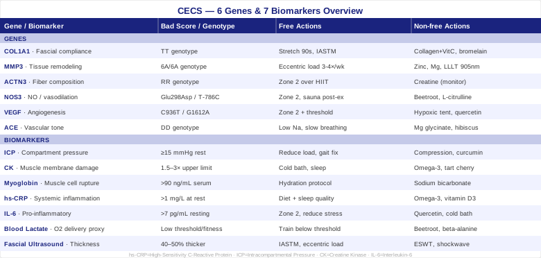
7 Biomarkers That Can Reveal What Standard Testing Misses
Most people with CECS are told their labs look normal. That is typically true — because the wrong labs are ordered. The markers that matter in this condition are not your standard metabolic panel. They require targeted testing, often timed around exercise, and some require specialist equipment or access. Each of the following biomarkers adds a meaningful, distinct piece to your diagnostic or treatment picture. Together they can replace months of guesswork with a working hypothesis your medical team can act on.
1. Intracompartmental Pressure
Intracompartmental pressure (ICP) is the only biomarker that directly confirms the diagnosis of CECS. It measures the hydraulic pressure inside a specific muscle compartment — most commonly the anterior or deep posterior compartment of the lower leg. In healthy individuals, compartment pressure rises moderately during exercise and normalizes quickly with rest. In CECS, pressure spikes abnormally and takes substantially longer to return to baseline.
How to Measure It
A sports medicine physician or orthopedic specialist inserts a needle-tipped pressure sensor into the compartment immediately before and after a standardized exercise protocol — usually running until symptoms appear. The Pedowitz diagnostic criteria are widely used: resting pressure at or above 15 mmHg, one-minute post-exercise pressure at or above 30 mmHg, or five-minute post-exercise pressure at or above 20 mmHg confirms the diagnosis. Cost ranges from $300 to $800 depending on the center. University-affiliated orthopedic departments are the most reliable source for this procedure, as not all sports medicine clinics perform it regularly.
If the Score Is Bad, the Plan Without Supplements
A confirmed elevated ICP is the clearest signal that your fascia cannot accommodate the normal pressure increase of exercise. Conservative first steps include reducing training volume and intensity by 40 to 60 percent, substituting lower-impact activities such as swimming or cycling that generate less compartment loading, and working with a certified running biomechanics specialist to address gait contributors including overpronation, overstriding, and tibial stress. Switching to a lower heel drop shoe may reduce anterior compartment loading. Deep tissue myofascial release applied to the affected compartment 2 to 3 times per week has case series support for modestly reducing post-exercise ICP over 8 to 12 weeks.
If the Score Is Bad, the Plan With Supplements or Equipment
Compression sleeves worn after exercise — not during, where they can acutely worsen compartment dynamics — may assist post-exercise fluid redistribution. Pneumatic compression devices such as NormaTec boots support lymphatic drainage and accelerate the pressure normalization window; use 20 to 30 minutes immediately post-exercise. Curcumin with piperine (500 mg twice daily, 8-week cycles with 4-week breaks) addresses the baseline fascial inflammation component; side effects are minimal at this dose. Topical magnesium applied to the affected compartment daily may support smooth muscle relaxation in vessel walls — evidence is preliminary and this should be seen as an adjunct rather than a primary intervention.
2. Creatine Kinase
Creatine kinase (CK) is released when muscle cell membranes are damaged. In CECS, the brief ischemic episodes that occur during exercise create enough oxidative stress and membrane disruption to elevate CK. Chronically elevated CK after symptomatic exercise confirms that real, cumulative muscle damage is occurring — not just transient pressure discomfort — and that the underlying condition is actively harming tissue with each episode.
How to Measure It
Standard blood draw, collected 12 to 24 hours after a symptomatic exercise session to capture the post-exercise peak. Normal range: 30 to 170 U/L for women, 55 to 370 U/L for men (lab ranges vary slightly). A CECS-related elevation is typically modest — one and a half to three times the upper limit of normal — which distinguishes it from the massive elevations of rhabdomyolysis. Cost: $20 to $60.
If the Score Is Bad, the Plan Without Supplements
Consistently elevated post-exercise CK confirms that repeated ischemic episodes are producing tissue-level damage. The primary intervention is reducing the frequency and intensity of symptomatic exercise sessions. Cold water immersion immediately post-exercise (10 to 15 degrees Celsius, 10 to 15 minutes) has solid evidence for limiting the inflammatory cascade that follows exercise-induced muscle membrane disruption and reducing subsequent CK elevation. Adequate sleep — seven to nine hours — is required for muscle membrane repair and cannot be substituted.
If the Score Is Bad, the Plan With Supplements or Equipment
Omega-3 fatty acids (2 to 3 g EPA plus DHA daily, continuous use) reduce post-exercise membrane inflammation and have controlled study support for lowering exercise-induced CK elevation. Tart cherry extract (480 mg twice daily, or 2 × 30 ml concentrate) has multiple randomized trials showing reduced post-exercise CK; cycle 4 weeks on, 2 weeks off. Vitamin E (400 IU taken 1 to 2 hours before exercise on training days, not as a daily long-term supplement, given potential pro-oxidant effects at sustained high doses) may reduce acute exercise-induced CK — but limit to pre-exercise use only and reassess after 4 weeks.
3. Myoglobin
Myoglobin is a muscle-specific oxygen-binding protein released when muscle cell membranes rupture. Elevated serum or urine myoglobin after symptomatic exercise confirms that muscle fiber damage is occurring at the cellular level, not merely pressure-related discomfort. In severe or frequently repeated CECS episodes, myoglobinuria — brown or tea-colored urine — signals kidney stress and requires prompt medical attention.
How to Measure It
Serum myoglobin is drawn within 1 to 4 hours post-exercise. Urine myoglobin can be detected with a standard dipstick test (available at pharmacies) or a specific laboratory urine myoglobin assay. Serum cost: $30 to $80. Normal serum level: below 90 ng/mL. Any significant urine myoglobin is considered abnormal in this context.
If the Score Is Bad, the Plan Without Supplements
Significant post-exercise myoglobinuria requires an immediate reduction in training load and a conversation with a sports medicine physician about modifying the exercise protocol. Adequate hydration is the most evidence-supported intervention for protecting the kidneys: target pale yellow urine throughout the entire day on exercise days, not just during training. 500 ml of water two hours before exercise, 150 to 250 ml every 20 minutes during, and 500 to 700 ml immediately post-exercise is a practical hydration framework.
If the Score Is Bad, the Plan With Supplements or Equipment
The hydration strategy described above remains central. Sodium bicarbonate (0.2 g per kg body weight, 60 minutes pre-exercise) may buffer the acidic environment that worsens myocyte membrane instability during ischemic episodes; it is an established performance aid used cautiously here for a protective purpose — test tolerance starting at half dose to manage potential GI side effects before using at the full protocol dose. Do not use this routinely without medical guidance if kidney function is a concern.
4. High-Sensitivity C-Reactive Protein
High-sensitivity CRP (hs-CRP) is the most available marker of systemic inflammation. In CECS, chronic low-grade inflammation appears to contribute to fascial fibrosis and reduced tissue compliance — the fundamental mechanical problem of the condition. Peter Attia consistently identifies hs-CRP as one of the most actionable and underused routine biomarkers for athletes, precisely because it captures a chronic state that standard testing ignores.
How to Measure It
Standard blood test drawn at rest, ideally at least three days after any intense exercise (which transiently and normally elevates CRP). Optimal range: below 0.5 mg/L. Concerning range: above 1 mg/L. Cost: $25 to $75.
If the Score Is Bad, the Plan Without Supplements
Chronically elevated hs-CRP above 1 mg/L at rest signals systemic inflammation that is likely worsening fascial health over time. Dietary changes carry the strongest evidence: eliminating ultra-processed foods, refined seed oils, and added sugars for 8 consecutive weeks reliably brings hs-CRP below 1 mg/L in the majority of otherwise healthy athletes. Consistent sleep schedule (fixed bedtime, dark and cool environment, targeting 7 to 9 hours) reduces inflammatory cytokine levels independently of diet. These two changes together represent the highest-leverage intervention with no cost and no side effects.
If the Score Is Bad, the Plan With Supplements or Equipment
Omega-3 fatty acids (2 to 4 g EPA plus DHA daily, ongoing) have the strongest supplement evidence for reducing hs-CRP. Curcumin with piperine (500 mg twice daily, 8-week cycles with 4-week breaks) consistently reduces CRP in randomized trials of inflammatory conditions. Vitamin D3 normalized to a serum 25(OH)D of 40 to 60 ng/mL (typically 2000 to 5000 IU daily depending on baseline) supports connective tissue biology and reduces inflammatory signaling; check levels before dosing. Sauna use (80 to 100°C, 20 minutes, 3 to 4 times per week after exercise) has emerging evidence for meaningfully reducing systemic inflammatory markers in athletes.
5. Interleukin-6
IL-6 is a cytokine with a dual role in exercise physiology. Acutely, it is released by contracting muscles as a beneficial myokine. But chronically elevated resting IL-6 signals a pro-inflammatory state that can promote fibrosis, including the fascial fibrosis that reduces compartment compliance in CECS. Elevated resting IL-6 suggests active pathological remodeling of fascial tissue.
How to Measure It
Serum or plasma IL-6 drawn at rest, at least 48 hours after intense exercise. Reference ranges vary by lab; most consider below 7 pg/mL normal, with below 2 pg/mL optimal in healthy trained athletes. Not offered by all standard lab panels — often available through specialty inflammatory marker panels. Cost: $50 to $120.
If the Score Is Bad, the Plan Without Supplements
Resting IL-6 responds strongly to aerobic fitness improvements. Consistent moderate-intensity training (zone 2, 45 to 60 minutes, four times per week) reduces resting IL-6 by improving insulin sensitivity and reducing visceral fat, the primary non-exercise tissue source of chronic IL-6. Reducing psychological and sleep-debt stressors — both measurable through morning HRV — also lowers resting IL-6 independently.
If the Score Is Bad, the Plan With Supplements or Equipment
Quercetin (500 mg twice daily, 4-week cycles) inhibits IL-6 production through NF-κB pathway modulation; human studies in athletes have shown modest but real reductions in resting inflammatory markers with consistent use. Resveratrol (500 mg daily with a fatty meal) may complement quercetin's anti-inflammatory action through overlapping but distinct pathways. Cold water immersion (3 to 4 times per week, 10 to 12 degrees Celsius, 10 minutes) acutely blunts the post-exercise IL-6 spike and, with consistent use, appears to reduce the baseline resting IL-6 state over time.
6. Blood Lactate
In CECS, rising intracompartmental pressure reduces local blood flow, forcing muscles into anaerobic metabolism at lower exercise intensities than their aerobic fitness level would otherwise require. Blood lactate will climb at unusually low power outputs or running speeds, providing an indirect window into how severely compartment pressure is compromising local oxygen delivery. A staged lactate threshold test can reveal whether your threshold is abnormally depressed relative to your overall cardiovascular fitness.
How to Measure It
Finger-prick blood samples taken every 3 to 5 minutes during a staged exercise protocol (incrementally increasing speed or power). Requires a portable lactate analyzer such as Lactate Scout or Lactate Plus, available at sports performance labs or purchasable for $100 to $300. Professional staged testing at a sports physiology lab: $100 to $250. A well-conditioned athlete should not see meaningful lactate accumulation below 60 to 70 percent of VO2 max; a depressed threshold relative to fitness level is a functional fingerprint of CECS-related perfusion compromise.
If the Score Is Bad, the Plan Without Supplements
Identifying the specific exercise intensity at which compartment pressure begins limiting oxygen delivery and training below that threshold is the foundational intervention. This is not simply reducing effort arbitrarily — it means building aerobic base at an intensity the affected compartment can tolerate. Zone 2 heart rate training (roughly conversational pace), monitored with a heart rate chest strap and HRV device, provides the framework. Systematic progression within the tolerable range over 12 to 16 weeks can shift the threshold upward as vascular adaptation occurs.
If the Score Is Bad, the Plan With Supplements or Equipment
Dietary nitrate from beetroot juice (400 to 600 mg nitrate, 2 to 3 hours pre-exercise) enhances mitochondrial efficiency and reduces the oxygen cost of submaximal exercise — meaning muscles do more work per unit of available blood flow, partially compensating for impaired perfusion. This is one of the most mechanistically well-matched interventions for CECS among all available supplements. Beta-alanine (3.2 to 6.4 g daily in divided doses to avoid paresthesia) raises intramuscular carnosine and buffers the hydrogen ion accumulation that accelerates fatigue perception; cycle 8 weeks on, 4 weeks off. L-Citrulline (6 g, 60 minutes pre-exercise) supports nitric oxide availability for vessel dilation during the exercise bout.
7. Fascial Thickness on Ultrasound
Research has consistently found that the fascia enclosing the anterior compartment in CECS patients is significantly thicker and less compliant than in healthy controls — with mean measurements approximately 40 to 50 percent greater in symptomatic individuals in comparative studies. Musculoskeletal ultrasound measurement of fascial thickness is non-invasive, does not require exercise provocation, and can confirm one of the key structural abnormalities underlying the condition. It also provides a measurable baseline against which to track changes over time.
How to Measure It
Musculoskeletal ultrasound performed by a radiologist or a sports medicine physician with ultrasound competence. Measurements taken at rest and during passive and active dorsiflexion provide the most clinically useful data about tissue behavior across loading states. Cost: $150 to $400 depending on the clinic and whether bilateral comparison is included. Studies examining anterior compartment fascia in CECS patients are available via PubMed search for "chronic exertional compartment syndrome ultrasound fascia thickness."
If the Score Is Bad, the Plan Without Supplements
Thickened, fibrotic fascia does not reverse rapidly, but it does respond to sustained mechanical intervention applied consistently over months. Deep tissue manipulation by a therapist specifically trained in Instrument-Assisted Soft Tissue Mobilization (IASTM) or myofascial release — not standard Swedish massage — applied directly to the compartment fascia 2 to 3 times per week over 8 to 12 weeks has case series support for improving tissue compliance. Eccentric loading programs targeting the muscle-tendon-fascia interface stimulate matrix metalloproteinase (MMP) activity, which initiates remodeling of fibrotic collagen; single-leg heel drops with progressive load, performed daily, represent a practical starting point.
If the Score Is Bad, the Plan With Supplements or Equipment
Extracorporeal shockwave therapy (ESWT) applied to the anterior compartment has emerging evidence for stimulating fascial remodeling through local growth factor recruitment; 4 to 6 sessions weekly or biweekly through a physiotherapy clinic with shockwave equipment. Photobiomodulation at 808 to 980 nm wavelength applied to the affected compartment reduces fibrotic signaling markers in connective tissue studies and may modestly support fascial remodeling over 4 to 8 weeks of regular use. Collagen peptides (10 to 15 g daily with 50 to 100 mg vitamin C, taken 30 to 60 minutes before exercise or before a stretching session) provide building blocks and co-factors for adaptive collagen remodeling; use continuously for 3 to 6 months before evaluating structural change.
Understanding why your fascia and vasculature respond the way they do requires looking one level deeper, into the genetic variants that shape these biological systems before training ever begins.
6 Genes That May Shape Your Risk and Response
Genetics research specific to CECS remains in its early stages — no large-scale genome-wide association study has been published for this condition. However, several well-characterized genes governing connective tissue biology, muscle fiber composition, and vascular regulation are mechanistically central to CECS pathophysiology. If you have access to genetic testing (23andMe, raw data analysis via Genetic Genie, or clinical genomics panels), these are the variants worth examining. If you do not, the intervention logic based on phenotype — how your body actually responds — is still applicable.
1. COL1A1 — Fascial Compliance and Collagen Architecture
What it does and why it matters: COL1A1 encodes the alpha-1 chain of type I collagen, the dominant structural protein in tendons, ligaments, and fascia. The Sp1 binding site polymorphism (rs1800012, T to C substitution) alters how collagen fibers are organized and cross-linked, influencing tissue extensibility. Individuals with the TT genotype tend toward stiffer, less extensible collagen structures. In CECS, a fascia that cannot expand under rising compartment pressure is the core mechanical problem — stiffer collagen variants worsen this limitation. The clinical relevance of this gene has been established in Achilles tendon and ligament injury research; its application to compartment fascia stiffness follows mechanistically.
If the Gene Is Bad, the Plan Without Supplements
Prioritize long-duration, low-intensity fascial stretching of the affected compartment (60 to 90 second holds, at or just below the discomfort threshold), performed 2 to 3 times daily rather than once. This specifically targets viscoelastic creep in collagen fibers — a property that responds to duration of load, not intensity. IASTM applied to the fascia 2 to 3 times per week mechanically disrupts pathological cross-links. Consistent yoga-based fascial work with holds exceeding 60 seconds (yin yoga format) 4 to 5 times per week over 8 to 12 weeks has demonstrated measurable improvements in tissue compliance in athletic populations.
If the Gene Is Bad, the Plan With Supplements or Equipment
Collagen peptides (15 g with 50 to 100 mg vitamin C, 30 to 60 minutes before a stretching or loading session) are the most evidence-supported supplement for collagen tissue remodeling; the pre-loading timing takes advantage of elevated amino acid availability during the mechanical stimulus window. Bromelain (500 mg twice daily between meals, cycle 4 weeks on, 2 weeks off) may reduce pathological collagen cross-linking. Vitamin C (500 to 1000 mg daily as a baseline, increasing to 1500 mg on exercise days) is a required co-factor for prolyl hydroxylase, the enzyme central to collagen synthesis and repair; without it, collagen supplementation has limited upstream effect.
2. MMP3 — Fascial Remodeling Capacity
What it does and why it matters: MMP3 encodes matrix metalloproteinase 3, an enzyme that degrades and remodels extracellular matrix components including collagen, fibronectin, and proteoglycans. It plays a central role in connective tissue turnover after repeated mechanical stress. The 5A/6A promoter polymorphism (rs3025058) strongly affects expression levels: the 5A/5A genotype produces higher MMP3 activity and faster remodeling capacity; the 6A/6A genotype is associated with lower enzyme expression and slower, less efficient connective tissue turnover. For CECS sufferers, the 6A/6A genotype may mean that microtrauma from repeated pressure episodes accumulates progressively rather than resolving between sessions, driving the fascial fibrosis that worsens compartment compliance over time.
If the Gene Is Bad, the Plan Without Supplements
Mechanical loading is the primary physiological driver of MMP3 expression. Progressive eccentric loading programs targeting the affected compartment — single-leg heel drops, tibialis anterior eccentrics — stimulate MMP3 upregulation through mechanotransduction pathways. Frequency should be 3 to 4 times per week; underdosing by exercising too infrequently fails to achieve the mechanical signal needed for remodeling. This is a 12-plus week intervention before structural changes become detectable on imaging.
If the Gene Is Bad, the Plan With Supplements or Equipment
Zinc (15 to 25 mg daily with food, not exceeding 40 mg to avoid copper displacement; cycle 4 weeks on, 2 weeks off) is a required catalytic co-factor for MMP3 activity — deficiency impairs enzymatic function regardless of genetic expression. Magnesium glycinate (300 to 400 mg daily, ongoing) supports multiple MMP co-factor pathways. LLLT at 905 nm wavelength applied to the affected compartment area has cellular evidence for upregulating MMP activity in fibrotic tissue; use 3 times per week, 10 to 15 minutes per session.
3. ACTN3 — Muscle Fiber Composition and Peak Pressure Generation
What it does and why it matters: ACTN3 encodes alpha-actinin-3, a structural protein found exclusively in fast-twitch (type II) muscle fibers. The R577X polymorphism (rs1815739) introduces a premature stop codon; individuals with the XX genotype produce no functional alpha-actinin-3 protein. RR genotype individuals have stronger fast-twitch fiber characteristics; XX individuals trend toward slow-twitch fiber dominance with different contraction mechanics and fatigue profiles. Fiber type composition influences the degree of intracompartmental pressure generated during exercise — explosive, forceful contractions using fast-twitch fibers generate higher peak compartment pressures than sustained slow-twitch contractions. RR individuals participating in sports requiring repeated explosive bursts may be generating higher absolute pressure spikes per exercise bout.
If the Gene Is Bad, the Plan Without Supplements
Modifying training modality to reduce the proportion of high-intensity interval work and explosive strength movements that preferentially recruit fast-twitch fibers — replacing a portion with zone 2 aerobic work — may reduce peak compartment pressure generation without sacrificing overall training stimulus. Strength training in higher repetition ranges (15 to 20 reps per set) provides loading without the maximal force production that peaks compartment pressure. This is not eliminating performance training — it is recalibrating the ratio of training types for connective tissue tolerance.
If the Gene Is Bad, the Plan With Supplements or Equipment
Creatine monohydrate (3 to 5 g daily, no cycling required) supports fast-twitch fiber function but increases intramuscular water retention and may transiently raise resting compartment pressure when first introduced — monitor for symptom changes in the first 2 to 3 weeks of use. HMB (beta-hydroxy beta-methylbutyrate, 3 g daily) may support muscle fiber integrity with less water retention than creatine; evidence for CECS specifically is absent, but the mechanistic rationale for avoiding exacerbation while maintaining tissue support is reasonable.
4. NOS3 — Nitric Oxide Production and Vascular Response
What it does and why it matters: NOS3 encodes endothelial nitric oxide synthase (eNOS), the enzyme that produces nitric oxide in the walls of blood vessels. Nitric oxide is the primary signaling molecule for vasodilation — it relaxes the smooth muscle surrounding arterioles, enabling blood vessels to expand and accommodate increased flow during exercise. The Glu298Asp polymorphism (rs1799983) and the T-786C promoter variant both reduce eNOS activity and expression, resulting in lower nitric oxide production and a blunted vasodilatory response to exercise intensity. Gary Brecka has discussed NOS3 variants extensively in the context of vascular and recovery performance, identifying it as one of the most impactful circulation-related genetic variants. In CECS, where rising compartment pressure is already mechanically compressing local vessels, a genetically impaired ability to dilate those same vessels compounds the oxygen delivery problem significantly.
If the Gene Is Bad, the Plan Without Supplements
Consistent aerobic training is the most validated method for upregulating eNOS expression through increased shear stress on vessel walls. Zone 2 training (60 to 70 percent of maximum heart rate, 45 to 60 minutes per session, 4 to 5 times per week) drives the mechanical shear stimulus that chronically increases eNOS mRNA transcription over weeks. Heat exposure through sauna (80 to 100°C, 20 minutes post-exercise, 3 to 4 times per week) provides an additive thermal shear stimulus that further upregulates eNOS independently of training volume.
If the Gene Is Bad, the Plan With Supplements or Equipment
Dietary nitrate from beetroot juice or concentrate (400 to 600 mg nitrate, 2 to 3 hours pre-exercise) provides an eNOS-independent route to nitric oxide through the nitrate-nitrite-NO reduction pathway — this is particularly valuable for NOS3 poor responders, who cannot rely fully on enzymatic NO production. L-Citrulline (3 to 6 g daily, or 6 to 8 g as a pre-exercise dose) is preferred over L-arginine for sustaining plasma arginine availability for eNOS; daily use, with 4-week breaks every 3 months. Pycnogenol derived from French maritime pine bark (150 to 200 mg daily, continuous use) has human trials demonstrating increased eNOS expression and improved peripheral circulation in vascular conditions; GI mild upset is the most commonly reported side effect at this dose.
5. VEGF — Angiogenic Adaptation to Training
What it does and why it matters: VEGF (vascular endothelial growth factor) drives the formation of new blood vessels (angiogenesis) and governs how effectively muscle vasculature adapts to the demands of training. Polymorphisms in the VEGF gene, including C936T (rs3025039) and the G1612A variant, reduce circulating VEGF levels and blunt angiogenic capacity. Lower VEGF expression means fewer new capillaries form per unit of training stimulus, meaning that the vascular adaptation that would normally partially compensate for CECS-related perfusion deficits proceeds more slowly and less completely. This keeps the symptomatic intensity threshold lower for longer than in athletes with efficient angiogenic signaling.
If the Gene Is Bad, the Plan Without Supplements
Hypoxic training stimuli — whether through altitude camps, altitude simulation tents, or high-altitude residence — powerfully upregulate VEGF expression by activating hypoxia-inducible factor 1-alpha (HIF-1α) regardless of genetic baseline. Practically, consistent zone 2 training remains foundational, but adding 2 to 3 weekly threshold-level sessions (if compartment symptoms permit) provides a stronger VEGF induction signal than zone 2 alone. Intermittent fasting (16:8 protocol, not extending beyond 20 hours) also upregulates HIF-1α and VEGF signaling modestly through metabolic stress pathways.
If the Gene Is Bad, the Plan With Supplements or Equipment
Altitude simulation equipment (hypoxic tents at 2500 to 3500 m simulated altitude for 8+ hours nightly) is the most direct equipment-based approach for forcing VEGF upregulation; this requires financial investment but is used by high-level endurance athletes for exactly this purpose. Iron status (measured as ferritin — check before any supplementation) is critical for HIF-1α protein stability, which drives VEGF transcription; iron deficiency without anemia can significantly impair this pathway. Quercetin (500 mg twice daily, 4-week cycles) has been shown in controlled trials in athletes to increase VEGF mRNA expression and improve capillary density markers, making it one of the more mechanistically interesting options for genetically blunted angiogenic responders.
6. ACE — Vascular Tone and Ischemic Sensitivity
What it does and why it matters: The ACE gene encodes angiotensin-converting enzyme, a central regulator of blood pressure, vascular tone, and fluid balance. The insertion/deletion polymorphism (I/D, a 287 base-pair Alu sequence) is one of the most studied functional variants in exercise physiology. The DD genotype produces the highest ACE activity, the II genotype the lowest, with ID intermediate. High ACE activity promotes angiotensin II-mediated vasoconstriction and is associated with faster oxidative stress accumulation in exercising muscle. In CECS, where rising compartment pressure is already mechanically compressing blood vessels, the superimposed vasoconstrictor drive of high ACE activity in DD individuals worsens the effective perfusion deficit and may intensify symptom severity at any given compartment pressure level.
If the Gene Is Bad, the Plan Without Supplements
Reduce dietary sodium below 2 g per day to lower the substrate driving renin-angiotensin-aldosterone system activation. Consistent aerobic training progressively downregulates both sympathetic nervous system tone and tissue ACE activity over months — this is not a short-term effect. Slow breathing practices (4-second inhale, 6 to 8 second exhale, 10 minutes before and after training) reduce sympathetic vasoconstrictor drive at the nervous system level and can be adopted immediately with no cost.
If the Gene Is Bad, the Plan With Supplements or Equipment
Foods containing natural ACE-inhibiting peptides — fermented dairy products such as cultured yogurt or kefir (casein hydrolysates), sardines (fish peptides), and garlic (allicin metabolites) — have modest but documented ACE-inhibiting effects in human studies and can be incorporated daily without risk. Magnesium glycinate (300 to 400 mg daily) supports vasodilation through the calcium-magnesium vascular competition pathway and is broadly safe as an ongoing supplement. Hibiscus tea (2 to 3 cups daily, consistent use) has multiple randomized controlled trials demonstrating ACE-inhibiting and blood-pressure-lowering effects comparable to low-dose pharmaceutical ACE inhibitors in mild hypertension populations — making it an unusually well-supported food-based intervention for the DD genotype.
With both the measurement framework and the genetic foundation in place, the next dimension worth examining is the broader exercise science context — particularly how inflammation biology, fascial neuroscience, and vascular adaptation have been translated into practical protocols for athletes.
What Exercise Science and Inflammation Research Offer Athletes with CECS
The Huberman Lab podcast episodes covering exercise, inflammation, and recovery — particularly those featuring research on connective tissue adaptation, zone 2 physiology, and post-exercise protocols — contain a dense concentration of mechanistically relevant information for CECS. While no episode addresses the condition directly, the underlying biology of fascial inflammation, vascular adaptation, and sympathetic modulation maps precisely onto the mechanisms driving CECS. The following ten insights represent the most impactful and actionable content from this body of work.
1. Zone 2 Training Is the Most Potent Anti-Inflammatory Exercise Mode
Moderate-intensity aerobic exercise at roughly 65 to 70 percent of maximum heart rate — a conversational pace where nasal breathing is sustainable — consistently reduces resting IL-6, TNF-alpha, and CRP over weeks to months. High-intensity training without a sufficient zone 2 base tends to keep resting inflammatory markers chronically elevated rather than reducing them.
2. Sleep Outranks Every Anti-Inflammatory Supplement
Below seven hours of sleep per night, IL-6 and CRP rise measurably within days. Chronic sleep restriction produces the same systemic inflammatory profile as metabolic syndrome. No supplement adequately compensates for this. For athletes doing tissue remodeling work — particularly those targeting fascial compliance and MMP-driven remodeling — adequate sleep is not optional.
3. The Post-Exercise Inflammatory Window Is Where Interventions Matter Most
The 60 minutes following intense exercise are when most inflammatory cascades initiate. Cold water immersion within this window blunts the CK rise and IL-6 spike meaningfully. Conversely, NSAID use in this same window suppresses the inflammatory signal required for positive structural adaptation — habitual post-exercise NSAID use in CECS can paradoxically slow the tissue remodeling that might gradually improve compartment compliance.
4. Omega-3s Resolve Inflammation Rather Than Just Suppressing It
The mechanism of omega-3 anti-inflammatory action extends well beyond COX-2 inhibition. EPA and DHA generate specialized pro-resolving mediators (SPMs) — resolvins, protectins, maresins — that actively clear inflammatory debris and close the inflammatory response rather than simply dampening it. This is a meaningful distinction for chronic conditions involving fibrosis.
5. Cyclic Sighing Reduces Sympathetic Tone Faster Than Any Other Breathing Technique
A double inhale through the nose followed by a full slow exhale through the mouth, repeated 5 times, drops physiological arousal faster than box breathing, standard deep breathing, or meditation in controlled comparisons. For CECS sufferers with an ACE DD background or NOS3 poor-responder genotype, reducing sympathetic vasoconstrictor drive before exercise with this technique is a zero-cost, immediate intervention.
6. Heat Exposure Compounds Vascular Adaptation Beyond What Exercise Achieves Alone
Sauna use at 80 to 100°C for 20 minutes after aerobic exercise increases VEGF, heat shock proteins, and eNOS expression additively beyond exercise alone. For individuals with genetically blunted angiogenic or nitric oxide responses, combining heat exposure with aerobic training partially compensates for the genetic shortfall in adaptive capacity.
7. Fascia Is Neurologically Active in Ways That Affect Pain Perception
Fascia contains mechanoreceptors and nociceptors that communicate directly with the central nervous system. Chronic pain in CECS may involve a component of central sensitization of fascial pain fibers, not purely mechanical pressure. Slow, sustained stretching held for 60 to 90 seconds calms fascial mechanoreceptors in a way that rapid or bouncing stretches do not — providing a neurological benefit distinct from the mechanical compliance benefit.
8. Morning Light Exposure Entrains the Anti-Inflammatory Cortisol Rhythm
Ten to thirty minutes of outdoor light exposure within the first hour of waking anchors the circadian cortisol rhythm that governs daily anti-inflammatory signaling patterns. Disrupted cortisol rhythms — common in shift workers and people with irregular schedules — produce a chronically elevated evening inflammatory baseline. This is correctable with consistent morning light exposure at no cost.
9. Post-Exercise Carbohydrate Timing Protects Connective Tissue Synthesis
Consuming 30 to 60 g of carbohydrates within 30 minutes after training suppresses the post-exercise cortisol spike that follows training-induced stress. Chronic cortisol elevation impairs collagen synthesis and promotes inflammatory fascial signaling. For athletes actively working to remodel compartment fascia, carbohydrate timing around exercise sessions matters more than total daily carbohydrate intake.
10. Non-Sleep Deep Rest Accelerates Neurological and Vascular Recovery Between Sessions
Ten to twenty minutes of guided non-sleep deep rest (NSDR — a systematic body-scan relaxation protocol) performed immediately after training significantly accelerates neurological recovery and reduces the sustained sympathetic drive that keeps peripheral vasoconstriction elevated between exercise sessions. For CECS athletes managing reduced training loads, NSDR provides a recovery stimulus that does not require additional physical loading and is directly relevant to the vascular component of the condition.
Complementary Approaches Supported by Clinical Evidence
Beyond biomarker testing and genetic insight, several evidence-informed modalities have meaningful mechanistic and clinical relevance for CECS — either for symptom reduction, fascial tissue improvement, or vascular adaptation support. The following five have the most applicable evidence base from the approved modalities.
Massage Therapy and Myofascial Release
Massage therapy — specifically deep transverse friction massage and myofascial release applied to the anterior or posterior lower leg compartments — addresses one of the most direct mechanical problems in CECS: restricted fascial mobility and the limited sliding capacity between muscle layers and the muscle-fascia interface. Regular therapeutic work at this level can reduce the biomechanical strain on the fascia and, over time, improve tissue behavior under loading.
Case series documented in sports medicine and bodywork literature have reported symptom improvement in CECS patients following structured myofascial release protocols, including measurable reductions in post-exercise compartment pressure. Evidence remains at case series level rather than randomized controlled trial, which means findings should be interpreted cautiously and this approach should complement, not replace, formal medical evaluation.
Work with a therapist specifically trained in IASTM or myofascial release for lower limb sports injuries — this is distinct from general massage. Two to three sessions per week for 8 to 12 weeks, targeting the specific affected compartments, is the protocol most commonly reported in case literature. Add daily self-directed foam rolling between sessions to maintain inter-session tissue mobility. Expect 2 to 4 weeks before noticing any functional change.
Low-Level Laser Therapy and Photobiomodulation
Photobiomodulation at wavelengths of 808 to 980 nm penetrates the skin and fascia, stimulating mitochondrial cytochrome c oxidase activity and reducing local oxidative stress, inflammatory cytokine production, and fibrotic signaling — including TGF-beta1 and collagen III overexpression, which are drivers of pathological fascial thickening. For a condition where fascial fibrosis is a central structural abnormality, these mechanisms are directly relevant even in the absence of CECS-specific trials.
Systematic reviews examining LLLT in musculoskeletal conditions support its anti-inflammatory and tissue-repair effects across a range of connective tissue pathologies. The 2019 review in Photobiomodulation, Photomedicine, and Laser Surgery examined musculoskeletal applications comprehensively. Randomized CECS-specific trials do not yet exist; the evidence base is therefore mechanistic and extrapolated, which should be communicated clearly to any treating clinician.
Seek a physiotherapy or sports medicine clinic with a class IV therapeutic laser (10 to 60 W, 808 to 980 nm) for the most efficient tissue dosing. Treatment to the affected compartment in 4 to 6 sessions over 2 to 3 weeks, followed by reassessment. Home-use panel devices (lower wattage) require longer sessions of 15 to 20 minutes per site to reach comparable total energy doses. Avoid use during acute symptomatic exacerbations; position this as inter-episode tissue management.
Breathing-Based Therapies
Diaphragmatic breathing and CO2 tolerance training directly reduce sympathetic nervous system activation, translating into lower baseline vascular tone and reduced sympathetically driven vasoconstriction in the peripheral vessels of the lower limbs. For CECS athletes — particularly those with NOS3 or ACE variants that already compromise local vasodilation — reducing sympathetic vasoconstrictor drive through breath training may meaningfully shift the symptomatic threshold during exercise.
Nasal breathing during submaximal exercise — the Buteyko-derived approach of maintaining nasal-only breathing throughout zone 2 training sessions — reduces sympathetic arousal and improves CO2 tolerance, which carries its own vasodilatory signaling. Small controlled studies in exercise physiology have demonstrated improved peripheral oxygen saturation under nasal versus oral breathing conditions at matched intensities. Evidence for CECS specifically is absent, but the vascular mechanism is applicable.
Commit to nasal-only breathing during all zone 2 training. Initially this requires a significant pace reduction — most athletes need 4 to 8 weeks before the pace at which nasal breathing becomes unsustainable rises back toward pre-change levels. Add 5 minutes of slow diaphragmatic breathing (4-second inhale, 6 to 8 second exhale) before and after each session as a sympathetic downregulation practice.
Biofeedback
Biofeedback — particularly heart rate variability (HRV) biofeedback using resonance frequency breathing — trains voluntary parasympathetic activation and reduces the chronic sympathetic drive that contributes to peripheral vasoconstriction in exercising limbs. For CECS athletes with documented vascular gene variants, HRV biofeedback provides a systematic, measurable approach to shifting autonomic balance over weeks.
Controlled studies in applied psychophysiology have documented significant improvements in peripheral circulation and reduced pain thresholds following 6-week HRV biofeedback protocols in individuals with exercise-induced vascular symptoms. Evidence for CECS specifically does not yet exist, and the mechanistic bridge — while plausible and well-reasoned — requires individual testing to confirm relevance for each person.
HRV biofeedback apps paired with a chest strap sensor (Polar H10 is the most validated hardware for this application; software options include HeartMath Inner Balance and Elite HRV) allow self-directed training at a one-time hardware cost of $50 to $150. Practice 15 to 20 minutes of resonance frequency breathing — typically approximately 6 breaths per minute, though the exact rate is individually calibrated — daily for 6 to 8 weeks before assessing impact on exercise symptoms.
Progressive Muscle Relaxation
Progressive muscle relaxation (PMR) systematically trains voluntary muscular relaxation by cycling through deliberate tension and release of sequential muscle groups. While it does not directly address the structural components of CECS, some athletes maintain chronically elevated baseline muscle tension that increases resting compartment pressure and reduces the safety margin before symptoms are triggered. PMR addresses this habitual tension pattern over weeks of consistent practice.
PMR has strong evidence as an adjunct in chronic pain management guidelines, consistently demonstrating reductions in pain perception and improvements in quality of life for musculoskeletal pain conditions. Its direct application to CECS has not been studied; its relevance is as a sympathetic tone modulator and muscle tension reducer, which may marginally raise the threshold at which compartment pressure becomes symptomatic. Athletes who notice symptom worsening during high-stress periods may find this particularly applicable.
Ten to fifteen minutes of full-body PMR before sleep — systematically tensing each muscle group for 5 seconds, then releasing fully for 30 seconds, moving from feet to head — is the standard protocol. Guided audio recordings are freely available on major podcast and audio platforms. Consistent use over 4 to 6 weeks is needed before meaningful changes in habitual baseline muscle tension become apparent. Side effects: none.
Conclusion
Chronic exertional compartment syndrome is not a mystery, even if it has long been treated like one. The diagnostic and biological tools to understand it exist and are accessible when you know what to ask for — from direct compartment pressure measurement and fascial ultrasound to inflammatory, vascular, and genetic markers that help explain individual variation in risk and symptom severity.
The practical starting point is not to pursue everything simultaneously. Begin with the two or three biomarkers most relevant to your specific history: ICP confirmation if you lack a formal diagnosis, hs-CRP if chronic inflammation seems likely given your lifestyle, and fascial ultrasound if you want a structural baseline to measure against. Layer one dietary change, one targeted supplement with genuine evidence, and one recovery practice — and give each intervention 8 to 12 weeks before drawing conclusions.
Bring this level of specificity to a sports medicine physician or orthopedic specialist with real CECS experience. If your current provider has not discussed fascial compliance, vascular contributions, or conservative management options in this kind of detail, a second opinion from a university-affiliated sports medicine center is a reasonable and often productive step. You have more options than surgery or continued suffering — use them with precision.
Musculoskeletal: Muscle Conditions Sports Injuries
Cardiovascular: Vascular Conditions
Autoimmune: Inflammatory Conditions Connective Tissue Conditions