This article contains AI generated content.
· UpdatedChronic Knee Effusion: 5 Genes And 7 Biomarkers To Track
Introduction
Chronic knee effusion — that persistent, stubborn joint swelling that keeps returning no matter what you try — puts people in a loop that feels almost designed to frustrate. You drain it, it refills. You rest, it stiffens and worsens. You exercise carefully, and it flares again. For most people, the management conversation stops at cortisone injections, compression, and a vague instruction to "reduce inflammation," with little clarity on what is actually sustaining the problem.
The fundamental issue is that knee effusion is not one condition with one cause. It can be the downstream result of cartilage breakdown, autoimmune-driven synovitis, crystal deposition, metabolic inflammation, or structural vulnerability — and each of these root mechanisms requires a meaningfully different response. Generic advice to eat better and move more is not wrong, but it is far too broad to help when you need to understand which specific biological process is keeping your knee swollen week after week.
This article takes a more targeted approach. By measuring specific biomarkers — several of which are available through standard blood draws — you can identify whether your effusion is primarily inflammatory, cartilage-mediated, crystal-driven, or metabolically fueled. The genetics section goes one layer deeper: certain gene variants predispose some people to higher baseline inflammation, faster cartilage degradation, or impaired tissue repair, and knowing your genetic profile can help you prioritize the interventions most likely to matter for your specific biology.
None of what follows is a cure. But better information creates better decisions, and better decisions, applied consistently over months, produce measurable biological change. That is a realistic and grounded kind of hope — the only kind worth pursuing.
Summary
This article identifies 7 blood biomarkers — hsCRP, IL-6, COMP, MMP-3, uric acid, TNF-alpha, and vitamin D — that can pinpoint the exact biological mechanism driving your chronic knee effusion. For each one, you will find how to test it affordably, what the results actually mean clinically, and specific protocols (with and without supplements) to correct a bad score. The genetics section covers 5 gene variants (GDF5, IL-6 rs1800795, MMP-3 5A/6A, COL11A1, and VEGF) that explain why some people are far more prone to persistent joint swelling than others — along with concrete compensation plans for each unfavorable genotype. Beyond labs and genetics, the article distills the 10 most joint-relevant insights from Peter Attia's Outlive and reviews four complementary approaches — low-level laser therapy, tai chi, massage therapy, and mindfulness — each with the clinical evidence and specific protocols needed to apply them realistically. If you have been managing your knee by trial and error, this is where the guesswork ends.
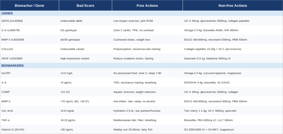
7 Biomarkers That Reveal What Is Driving Your Chronic Knee Effusion
Most physicians will order a basic inflammatory panel or aspirate the knee for visual inspection when effusion is large. What rarely gets requested — outside of sports medicine or longevity-focused practices — is a targeted panel that maps the actual biological mechanisms sustaining fluid accumulation. These seven markers can distinguish between a primarily inflammatory process, active cartilage destruction, crystal-related synovitis, or a correctable systemic deficiency. Getting this distinction right can save months of pursuing the wrong interventions.
Biomarker 1: High-Sensitivity C-Reactive Protein (hsCRP)
Why it matters: hsCRP is the most widely available and affordable gateway into inflammatory biology. In chronic knee effusion, a persistently elevated hsCRP confirms that systemic inflammation is actively sustaining synovial irritation, not just that you had a bad week. Peter Attia consistently positions hsCRP as a foundational biomarker for anyone tracking long-term health, and for joint conditions it is the natural first measurement.
What it reveals: Values above 1.0 mg/L suggest low-grade systemic inflammation. Above 3.0 mg/L indicates moderate inflammatory activity that may be directly driving synovial fluid overproduction. Values above 10 mg/L warrant investigation for acute infection or reactive arthritis before attributing findings to chronic inflammation.
How to measure it: Ordered through any standard blood lab — but specifically request high-sensitivity CRP. Regular CRP is calibrated for acute illness and misses chronic low-grade elevations. Cost: $10–$30 without insurance.
If hsCRP is elevated — without supplements
The most impactful non-supplement intervention is dietary. Eliminating ultra-processed foods, refined seed oils, and added sugar for 6–8 consecutive weeks produces reliable hsCRP reductions in clinical studies. Zone 2 aerobic exercise — sustained conversational-pace effort, 45–60 minutes, 4–5 times per week on a bike or in a pool to protect the knee — lowers basal hsCRP measurably over 8–12 weeks. Sleep quality is one of the most underused levers: seven to eight hours with consistent timing directly reduces next-day inflammatory signaling. Cold water immersion (14–15°C, 10–15 minutes, 3–4 times per week) also shows anti-inflammatory effects across multiple small trials.
If hsCRP is elevated — with supplements or equipment
Omega-3 fatty acids (EPA+DHA) at 2–4g/day with a fat-containing meal are the most evidence-supported supplement for hsCRP reduction across dozens of RCTs. No cycling required; use continuously. Side effects: fishy aftertaste, mild GI upset — take with the largest meal of the day. Curcumin with piperine (500–1000mg curcumin with 5–10mg piperine, twice daily) has shown meaningful hsCRP reduction in joint-specific RCTs. Cycling protocol: 8 weeks on, 2 weeks off as a precaution. Side effects: occasional GI discomfort in sensitive individuals. Magnesium glycinate (300–400mg at night) shows modest hsCRP benefit and additionally improves sleep quality, creating a dual effect. Use continuously. Equipment: an infrared sauna (20–30 minutes, 3–4 times per week) demonstrates CRP-lowering effects in cardiovascular studies and is a reasonable joint-health adjunct when direct knee loading is limited.
Biomarker 2: Interleukin-6 (IL-6)
Why it matters: IL-6 is a pro-inflammatory cytokine found in elevated concentrations directly within the synovial fluid of knees with chronic effusion. It drives synoviocyte proliferation, promotes fluid overproduction, and activates downstream cartilage-degrading enzymes. Unlike hsCRP — which is a downstream response marker — IL-6 is closer to the actual mechanism. Thomas Dayspring has described IL-6 as an upstream driver that hsCRP sometimes lags by days, making it more timely for tracking joint inflammation actively.
What it reveals: Serum IL-6 above 5 pg/mL in the context of chronic knee effusion points toward an inflammatory or autoimmune driver rather than purely mechanical wear. High IL-6 also promotes osteoclast activity, which can accelerate subchondral bone changes over time — a reason to address it early, not just manage symptoms.
How to measure it: Available through Quest Diagnostics, LabCorp, and similar specialty labs. Cost: $40–$80. IL-6 fluctuates relatively quickly with illness, stress, and exercise, so it is best interpreted alongside hsCRP trends and the clinical picture rather than in isolation.
If IL-6 is elevated — without supplements
Time-restricted eating (a 14:10 or 16:8 feeding window) consistently reduces circulating IL-6 in metabolic studies. Reducing visceral adiposity — even 5–10% body weight loss in overweight individuals — can dramatically lower IL-6 because adipose tissue is itself a significant source of this cytokine. Resistance training 2–3 times per week has a paradoxical anti-inflammatory effect: while IL-6 rises acutely during muscle contraction, sustained training progressively lowers basal levels. Chronic psychological stress elevates IL-6 through HPA axis activation; a structured daily breathing practice (4-7-8 breathing, 10 minutes daily) meaningfully dampens this neuroendocrine response.
If IL-6 is elevated — with supplements or equipment
EPA/DHA at 3–4g/day suppresses IL-6 production through prostaglandin pathway modulation — use as described for hsCRP. Boswellia serrata (AKBA-standardized extract, 100–250mg twice daily) has specific IL-6 inhibiting properties through NF-κB pathway inhibition, supported by human studies in joint inflammation. Cycling: 12 weeks on, 4 weeks off. Side effects: rare GI discomfort. Vitamin D3 + K2: correcting vitamin D deficiency (see Biomarker 7) significantly reduces IL-6 — this connection makes vitamin D one of the highest-leverage interventions in the list. Red and near-infrared light therapy (660nm and 850nm, 10–20 minutes directly over the knee, daily) shows cytokine-modulating effects that are increasingly supported by controlled trials.
Biomarker 3: COMP (Cartilage Oligomeric Matrix Protein)
Why it matters: COMP is released into the bloodstream when cartilage is damaged or undergoing accelerated structural turnover. In chronic knee effusion linked to early or established osteoarthritis, serum COMP rises before imaging changes are visible. It is one of the most specific available markers of actual cartilage matrix breakdown — distinct from general inflammatory signaling.
What it reveals: Elevated serum COMP (above approximately 12 U/L, though reference ranges vary by lab) suggests active cartilage degradation. In a knee with persistent effusion, high COMP indicates that tissue breakdown products are actively irritating the synovium and driving fluid accumulation — a different mechanistic target than purely cytokine-driven inflammation, requiring a different intervention priority.
How to measure it: Available through ARUP Laboratories and select LabCorp panels. Cost: $80–$150. Less commonly ordered in routine care; you may need to request it specifically through a sports medicine or rheumatology provider, or a longevity-focused clinician.
If COMP is elevated — without supplements
Aquatic exercise (pool walking, water cycling, 30–45 minutes, 4–5 times per week) reduces cartilage mechanical stress while maintaining muscular support — a critical distinction from land-based exercise when COMP is high. Avoiding prolonged high-impact activity during flares protects the cartilage surface from additional breakdown. Proprioception and quadriceps strengthening exercises reduce aberrant joint mechanics that accelerate COMP release over time. Even a 5–7% body weight reduction in overweight individuals measurably lowers per-step compressive forces on knee cartilage.
If COMP is elevated — with supplements or equipment
Undenatured type II collagen (UC-II) at exactly 40mg daily (not higher — the immune tolerance mechanism is dose-specific) has demonstrated in multiple RCTs the ability to reduce cartilage biomarker deterioration in knee OA patients. Use continuously for a minimum of 3 months before reassessing. Side effects: minimal; occasional mild GI effects. Glucosamine sulfate (1500mg/day) combined with UC-II provides complementary cartilage matrix support. Hydrolyzed collagen peptides (15g/day, taken 30–60 minutes before exercise with vitamin C) have shown in controlled trials to increase cartilage collagen synthesis markers when timed peri-exercise. Continuous passive motion (CPM) devices, available for home rental in post-surgical contexts, can be used prophylactically to improve synovial fluid circulation and cartilage nutrition without compressive loading.
Biomarker 4: MMP-3 (Matrix Metalloproteinase-3)
Why it matters: MMP-3, also called stromelysin-1, is an enzyme that degrades multiple components of the joint extracellular matrix including collagen, fibronectin, and proteoglycans. Its elevation in blood and synovial fluid is closely associated with both inflammatory arthritis — particularly rheumatoid arthritis — and progressive joint destruction. In chronic knee effusion of unclear origin, MMP-3 is one of the most useful markers for distinguishing aggressive inflammatory joint disease from lower-grade mechanical wear.
What it reveals: Serum MMP-3 above 70 ng/mL in men or above 45 ng/mL in women is considered elevated in most reference ranges (values vary by lab). High MMP-3 combined with persistent effusion suggests aggressive synovial inflammation and warrants specialist evaluation to rule out rheumatoid or psoriatic arthritis before pursuing self-directed protocols alone.
How to measure it: Available through ARUP, Quest, and specialty rheumatology labs. Cost: $80–$120. Often ordered alongside ANA, rheumatoid factor, and anti-CCP panels when inflammatory arthritis is suspected.
If MMP-3 is elevated — without supplements
Reducing systemic inflammation through the dietary approach described for hsCRP is the primary lever. Avoiding joint overload and repetitive trauma is critical since mechanical stress directly upregulates MMP-3 expression in synoviocytes at the cellular level. Sleep quality matters specifically here: nocturnal inflammatory signaling drives MMP-3 in ways that 7–8 hours of quality sleep reliably dampens. Eliminating alcohol — even moderate intake — meaningfully lowers MMP-related joint tissue degradation in regular drinkers, though this effect is rarely discussed in clinical consultations.
If MMP-3 is elevated — with supplements or equipment
Green tea extract (EGCG) at 400–600mg/day with food has specific MMP-inhibiting properties demonstrated in human synovial cell studies — this is among the most targeted supplement choices for this specific marker. Cycling: 8 weeks on, 2 weeks off. Side effects: caffeine sensitivity; at very high doses, potential hepatic strain — stay within the recommended range and avoid combining with other concentrated stimulants. Resveratrol (500mg/day with a fat-containing meal) shows MMP-3 downregulation in joint tissue studies. Use for 12 weeks then reassess. Side effects: mild GI discomfort; check with your physician if on anticoagulants. Photobiomodulation at 830nm applied directly to the knee has been shown in controlled research to reduce MMP activity in joint tissue — a physical modality option for those preferring to minimize supplements.
Biomarker 5: Serum Uric Acid
Why it matters: Uric acid crystal deposition within the knee joint is a far more common and underdiagnosed driver of chronic or recurrent effusion than most people realize. Even without a classic acute gout attack — the sudden, severe flare most people associate with the condition — chronic low-grade crystal-induced synovitis can produce persistent joint swelling that clinically mimics other inflammatory arthritis forms, leading to years of misdirected treatment.
What it reveals: Serum uric acid above 6.8 mg/dL represents the saturation point at which monosodium urate crystals can precipitate into joint tissue. Values above 7 mg/dL in someone with chronic knee effusion warrant synovial fluid analysis under polarized light microscopy to confirm or exclude crystal arthropathy as a driver. This is one of the most actionable findings in the list.
How to measure it: Included in standard metabolic panels ordered by any primary care physician. Cost: essentially free within a routine panel ($10–$20 as a standalone). This is the most accessible biomarker in this article and should be among the first checked.
If uric acid is elevated — without supplements
Hydration is the most underutilized lever: targeting 2.5–3 liters of water daily lowers serum uric acid through dilution and increased renal excretion — straightforward and immediate. Reducing dietary purines from organ meats, anchovies, sardines, and — critically — beer shows consistent results. Dietary fructose is the most strongly linked dietary driver of hyperuricemia; eliminating liquid fructose (fruit juice, high-fructose corn syrup, sodas) alone can reduce uric acid by 1–2 mg/dL in regular consumers. Regular consumption of whole cherries (one cup daily) or 8oz of tart cherry juice has demonstrated reductions in uric acid levels and gout attack frequency across multiple clinical trials.
If uric acid is elevated — with supplements or equipment
Tart cherry extract (1000–1500mg/day, standardized extract) is the most evidence-backed supplement for uric acid reduction short of pharmaceuticals. Use continuously; no cycling protocol needed. Side effects: minimal. Vitamin C (500–1000mg/day) acts as a mild uricosuric agent by promoting renal uric acid excretion, confirmed in multiple clinical studies. Quercetin (500mg twice daily) inhibits xanthine oxidase — the same enzyme targeted by allopurinol — and shows uric acid-lowering effects in human trials. Cycling: 8 weeks on, 2 weeks off. Side effects: rare GI symptoms. Important note: if uric acid is persistently above 8 mg/dL with documented joint involvement, discuss pharmaceutical urate-lowering therapy with your physician — supplements alone may be insufficient for this threshold.
Biomarker 6: TNF-alpha (Tumor Necrosis Factor-alpha)
Why it matters: TNF-alpha is a master inflammatory cytokine found in elevated concentrations within the synovial fluid and blood of patients with persistent knee effusion and inflammatory arthritis. It drives synovial membrane hyperplasia, promotes further IL-6 release, activates MMP enzymes, and broadly sustains the inflammatory cycle that keeps the joint producing excess fluid. Understanding TNF-alpha levels is especially valuable when effusion persists despite addressing hsCRP and IL-6, or when an autoimmune cause is under evaluation.
What it reveals: Serum TNF-alpha above 8–10 pg/mL (reference ranges vary by lab and assay) in the context of chronic effusion suggests an active and potentially aggressive inflammatory process. It is also useful for tracking treatment response in diagnosed inflammatory arthritis over time.
How to measure it: Available through ARUP and LabCorp specialty panels. Cost: $80–$150. Levels fluctuate with acute illness and require careful sample handling; interpret in the context of trends and alongside IL-6 and CRP data rather than as a single isolated value.
If TNF-alpha is elevated — without supplements
A Mediterranean-pattern diet rich in polyphenols and diverse plant fiber reliably reduces basal TNF-alpha through multiple mechanisms. Regular moderate aerobic exercise produces anti-inflammatory myokines from skeletal muscle that directly suppress TNF-alpha production between sessions. Prebiotic fiber intake (10–15g/day from diverse plant sources) and fermented foods reduce gut-derived TNF-alpha by supporting a less pro-inflammatory microbiome composition. Chronic psychological stress elevates TNF-alpha via the sympathetic nervous system; even a basic, consistent breathing practice (10 minutes of diaphragmatic breathing daily) measurably dampens this pathway.
If TNF-alpha is elevated — with supplements or equipment
Boswellia serrata (AKBA form), as noted for IL-6, also reduces TNF-alpha through NF-κB pathway inhibition and is one of the most versatile supplements in this list for multi-cytokine activity. Palmitoylethanolamide (PEA) (600mg twice daily) has emerging evidence for TNF-alpha modulation and joint inflammation with an excellent safety profile. Minimum assessment period: 12 weeks. Side effects: minimal as reported in available studies. Low-level laser therapy applied to the knee at 904nm has direct evidence for reducing TNF-alpha in joint tissue and combines well with dietary strategies for this biomarker specifically.
Biomarker 7: 25-OH Vitamin D
Why it matters: Vitamin D deficiency is one of the most prevalent and most easily correctable factors across chronic inflammatory joint conditions, yet it is frequently overlooked in the context of knee effusion. Vitamin D receptors are expressed directly on synoviocytes and immune cells within the joint. Deficiency amplifies the inflammatory cascade driving every cytokine in this list — IL-6, TNF-alpha, CRP — while simultaneously impairing the regulatory immune pathways that should be constraining synovial inflammation.
What it reveals: Levels below 30 ng/mL indicate deficiency. Researchers including Rhonda Patrick consider 40–60 ng/mL as the functional optimum for joint and immune function. Values below 20 ng/mL are associated with significantly higher risk of inflammatory joint disease, impaired repair capacity, and poor response to other therapeutic interventions. Correcting deficiency is one of the most reliably impactful and low-risk interventions available across this entire biomarker list.
How to measure it: A standard 25-hydroxyvitamin D blood test available through any physician or direct-to-consumer lab. Cost: $30–$60. Test before supplementing and recheck every 3–4 months during active repletion to avoid overshooting.
If vitamin D is deficient — without supplements
Midday sun exposure (10–30 minutes with significant skin surface exposed, 4–5 days per week) is the most physiologically natural repletion method, though effectiveness varies considerably with skin tone, geographic latitude, and season. Dietary sources — fatty fish, egg yolks, fortified dairy — contribute modest amounts that are unlikely to fully correct deficiency without sun or supplement support. Weight loss in overweight individuals can independently raise 25-OH vitamin D levels because adipose tissue sequesters the vitamin, reducing its circulating bioavailability.
If vitamin D is deficient — with supplements or equipment
Vitamin D3 (2000–5000 IU/day, taken with a fat-containing meal) is the standard repletion approach. Always combine with Vitamin K2-MK7 (100–200mcg/day) to ensure proper calcium directional routing and avoid arterial calcification during prolonged D3 supplementation. Use continuously and retest every 3–4 months, adjusting dose to reach the 40–60 ng/mL target. Side effects: toxicity is rare and requires sustained very high doses (above 10,000 IU/day without monitoring) over extended periods. Magnesium glycinate (300–400mg/day) is a required cofactor for vitamin D activation and conversion — many people supplementing D3 without adequate magnesium see a blunted response in blood levels.
Tracking these seven biomarkers as a panel gives you a working map of your knee's inflammatory landscape. The next dimension worth exploring — especially if lab results are repeatedly borderline or if you want to understand your long-term susceptibility — is genetic.
5 Gene Variants That Shape Your Risk for Chronic Knee Effusion
Genetic variants do not predetermine outcomes, but they do establish the biological terrain on which your lifestyle, diet, and environment operate. For chronic knee effusion, several well-studied variants alter inflammatory response intensity, cartilage repair capacity, blood vessel growth within the synovium, and structural collagen quality. Consumer genomics platforms such as 23andMe, combined with interpretation tools, make this information increasingly accessible — and when combined with biomarker data, it allows for meaningful prioritization of interventions.
Gene 1: GDF5 (Growth Differentiation Factor 5)
What it affects: GDF5 is the most robustly replicated genetic risk factor for osteoarthritis identified through genome-wide association studies. The key variant (rs143383, in the 5' untranslated region of GDF5) reduces gene expression, impairing cartilage and joint tissue formation during development and repair. Lower GDF5 activity means reduced chondrocyte signaling and thinner cartilage over time — a biological foundation for wear-driven effusion that begins earlier and progresses more readily.
Evidence level: Strong human evidence, replicated across multiple Asian and European cohort studies. This variant is among the most validated genetic associations in musculoskeletal medicine. Miyamoto et al. (2007) in Nature Genetics established this association across two independent populations.
If GDF5 is unfavorable — without supplements
Since GDF5 governs cartilage repair capacity, protecting cartilage from unnecessary mechanical stress becomes higher-priority than in the general population. Prioritize low-impact exercise — swimming and cycling — over high-impact activities like running or jumping. Daily dynamic joint mobility work (10–15 minutes of controlled range-of-motion movements) maintains cartilage nutrition through synovial fluid circulation. Quadriceps and hip abductor strengthening reduces per-step compressive forces and partially compensates for the structural vulnerability this variant creates.
If GDF5 is unfavorable — with supplements or equipment
Undenatured type II collagen (UC-II) at 40mg daily is the most cartilage-specific supplement for genetically predisposed individuals — the oral tolerance mechanism it triggers directly supports the immune-cartilage interface. Use for a minimum of 3–6 months before assessing response. Glucosamine sulfate (1500mg/day) combined with chondroitin sulfate (1200mg/day) showed meaningful benefit in a subset of moderate-to-severe knee OA patients in the large GAIT trial — those with GDF5 variants may represent the responder subgroup most likely to benefit from this combination. Hydrolyzed collagen peptides (15g, peri-exercise with vitamin C) provide matrix building blocks that are more critical for individuals with impaired GDF5-dependent repair.
Gene 2: IL-6 Promoter Variant (rs1800795)
What it affects: The G allele of this common IL-6 promoter polymorphism is associated with higher baseline IL-6 transcription. Carriers of the GG genotype may mount significantly stronger synovial inflammatory responses to the same mechanical insult or joint injury compared to CC carriers. Gary Brecka, who has made genetic interpretation for health optimization a central focus of his work, specifically identifies high-IL-6 gene variants as priority targets for omega-3 and anti-inflammatory lifestyle protocols.
Evidence level: Moderate-to-strong human evidence; replicated in inflammatory arthritis, post-injury recovery, and exercise physiology populations.
If IL-6 rs1800795 is unfavorable — without supplements
The anti-inflammatory lifestyle interventions described in the biomarker section apply with greater urgency for GG carriers. Zone 2 aerobic exercise and time-restricted eating become particularly high-priority since both reliably lower IL-6 over weeks. Avoiding prolonged joint overload and repetitive microtrauma is more consequential for this genotype than for others. The baseline inflammatory ceiling is higher, which means that the same environmental insult creates more synovial response — protective habits become non-negotiable rather than optional.
If IL-6 rs1800795 is unfavorable — with supplements or equipment
Omega-3 (EPA+DHA) at 3–4g/day is a priority supplement for GG carriers — it reliably downregulates IL-6 expression through multiple prostaglandin and NF-κB pathways. Boswellia serrata (AKBA) is particularly appropriate for this genotype given its direct IL-6 inhibition. Near-infrared light therapy applied daily to the knee (850nm, 15 minutes) offers a non-supplement cytokine modulation option well suited to high-IL-6 individuals. The combination of omega-3, boswellia, and photobiomodulation represents a reasonable multi-target approach for GG carriers managing chronic effusion.
Gene 3: MMP-3 5A/6A Polymorphism (rs3025058)
What it affects: The 5A allele of MMP-3 is associated with higher MMP-3 transcription, meaning faster enzymatic degradation of joint matrix components in response to inflammatory stimuli. Carriers of the 5A/5A genotype may break down cartilage collagen and proteoglycans more aggressively than 6A/6A carriers when exposed to the same level of joint inflammation. Ali Torkamani, genomics researcher at Scripps Research, has highlighted MMP gene variants as high-priority actionable targets in musculoskeletal genetic profiles.
Evidence level: Moderate human evidence; replicated in rheumatoid arthritis, knee OA progression, and post-surgical outcome studies.
If MMP-3 5A/5A is unfavorable — without supplements
Mechanical joint protection becomes more consequential for 5A/5A carriers: appropriate footwear with cushioning and support, using walking poles on uneven terrain to reduce knee loading, and avoiding prolonged kneeling or deep squatting without controlled return. Weight normalization is particularly impactful for this genotype because adipose-derived cytokines further upregulate MMP-3 expression — the two drivers amplify each other in overweight 5A carriers.
If MMP-3 5A/5A is unfavorable — with supplements or equipment
EGCG (green tea extract) at 400–600mg/day is the most targeted supplement for MMP-3 inhibition and warrants prioritization in 5A/5A individuals. Resveratrol (500mg/day) provides complementary MMP downregulation through a different molecular pathway. Photobiomodulation at 830nm (15 minutes on the knee, 5 times per week) has direct evidence for MMP activity reduction in joint tissue and combines well with the dietary approach for this genotype. Cycling EGCG as described (8 weeks on, 2 weeks off) is the most prudent protocol.
Gene 4: COL11A1 (Collagen Type XI Alpha 1)
What it affects: COL11A1 encodes a structural collagen found specifically in cartilage where it provides tensile strength and maintains proper fibril organization. Variants associated with reduced COL11A1 function produce cartilage that is mechanically inferior — more prone to surface fissuring, internal breakdown, and the release of degradation products that irritate the synovium and trigger persistent effusion. This gene has been implicated in early-onset OA and in anterior cruciate ligament injury susceptibility studies.
Evidence level: Moderate human evidence, particularly in early-onset osteoarthritis and joint laxity research populations.
If COL11A1 is unfavorable — without supplements
Proprioception and neuromuscular control training become especially important when structural collagen quality is genetically compromised. Balance exercises on unstable surfaces (wobble boards, single-leg work), progressive agility training, and explicit neuromuscular patterning during movement protect joints with weaker intrinsic collagen from the injury patterns that trigger or worsen effusion. A whole-food collagen-rich dietary pattern (bone broth, slow-cooked cuts, gelatin-rich preparations) provides structural amino acid building blocks for endogenous collagen synthesis.
If COL11A1 is unfavorable — with supplements or equipment
Hydrolyzed collagen peptides (15–20g/day taken 30–60 minutes before exercise with Vitamin C at 250–1000mg) have shown in multiple RCTs to increase cartilage collagen synthesis markers specifically when timed peri-exercise. This timing is critical — not just any time of day. Use continuously; the evidence supports sustained use over 3–6 months. Vitamin C is a required enzymatic cofactor for collagen crosslinking (prolyl hydroxylase activity) and becomes especially important in COL11A1 variant carriers. Side effects at these doses: very rare for either compound.
Gene 5: VEGF (Vascular Endothelial Growth Factor) — rs2010963
What it affects: VEGF promotes new blood vessel formation — a process that sounds beneficial but is actually a primary driver of inflammatory joint disease when it occurs in excess within the synovium. Pathological synovial angiogenesis feeds immune cell infiltration, raises joint temperature and fluid production, and creates the structural basis for chronic effusion that self-sustains even after the initial inflammatory trigger has resolved. Variants at rs2010963 associated with higher VEGF expression amplify this response.
Evidence level: Moderate human evidence, primarily from rheumatoid arthritis association studies; emerging data in knee OA populations.
If VEGF rs2010963 is unfavorable — without supplements
Reducing systemic oxidative stress — through dietary antioxidant density, sleep quality, and avoiding excessive training load — dampens VEGF upregulation. Avoiding prolonged joint hypoxia, which occurs predictably with extended inactivity and poor local circulation, removes a primary stimulus for pathological synovial angiogenesis. Intermittent fasting has shown VEGF-modulating effects through AMPK and mTOR signaling in experimental models, with plausible translation to human joint biology.
If VEGF rs2010963 is unfavorable — with supplements or equipment
Quercetin (500–1000mg/day) inhibits VEGF activity through HIF-1α pathway modulation — one of the more specific supplement mechanisms for this gene variant. Berberine (500mg twice daily with meals) inhibits VEGF signaling through AMPK activation and brings additional anti-inflammatory benefits. Cycling protocol: 8 weeks on, 4 weeks off, given its effects on hepatic enzyme handling with longer-term use. Side effects: GI discomfort, especially in the first 1–2 weeks — start with one dose per day and titrate up. Note: berberine should not be combined with metformin or certain antibiotics without physician guidance, as interactions are clinically significant.
With both biomarker data and genetic context in hand, it is worth stepping back to look at the broader framework connecting all of these variables — and no resource does this more comprehensively than Peter Attia's approach to longevity medicine.
"Outlive" by Peter Attia: The Joint Health Framework Hidden Inside a Longevity Book
Published in 2023, Outlive: The Science and Art of Longevity by Peter Attia, MD, is one of the most densely referenced health books of the past decade. While not written specifically about joint disease, its central framework — personalized biomarker tracking, early intervention, and treating root causes of biological aging rather than managing downstream symptoms — maps directly onto the strategy needed for chronic knee effusion. These are the 10 most impactful ideas from the book, applied specifically to the joint health context.
1. The Centenarian Decathlon: Train Backwards From Who You Want to Be at 85
Attia asks readers to identify the physical activities they want to perform in their 80s and train deliberately toward them starting now. For someone managing chronic knee effusion, this reframes the problem: treatment is not just symptom suppression — it is joint preservation as an investment in future capability. Every decision about managing effusion today has a compounding return decades from now.
2. Zone 2 Cardio Is the Most Accessible Anti-Inflammatory Intervention
Attia devotes extensive discussion to zone 2 exercise (sustained conversational-pace effort at roughly 60–70% of maximum heart rate). For chronic knee effusion specifically, zone 2 on a bike or in water is uniquely suited: it lowers basal inflammatory cytokines, improves mitochondrial efficiency, reduces metabolic contributors to IL-6 and TNF-alpha overproduction, and carries minimal joint loading. There is no supplement on this list that reliably produces the same systemic anti-inflammatory effect as consistent zone 2 training over 12 weeks.
3. Metabolic Inflammation and Joint Inflammation Share the Same Upstream Drivers
Attia identifies ApoB and insulin resistance as primary cardiovascular risk drivers — but the deeper insight is that metabolic inflammation driven by visceral adiposity, glucose dysregulation, and dyslipidemia elevates the identical cytokines (IL-6, TNF-alpha, CRP) that directly sustain synovial inflammation. Fixing metabolic health often means partially fixing joint health at the same time, without a single knee-specific intervention.
4. Muscle Is Both a Longevity Organ and a Joint Protector
Attia makes the case for treating muscle mass as a vital sign that predicts future functional capacity. For the knee specifically, quadriceps and hip abductor strength are the primary shock absorbers and load distributors for the joint. Sarcopenia dramatically increases compressive cartilage forces per step, accelerates degradation, and amplifies effusion risk. Resistance training 3 days per week is a form of joint preservation, not an optional fitness choice.
5. Glucose Spikes Damage Collagen in Ways Standard Testing Misses
Advanced glycation end-products (AGEs) form when blood glucose is chronically elevated, damaging collagen in cartilage and other connective tissues just as they damage arterial walls and the eye's lens. Standard HbA1c misses post-meal glucose spikes that contribute to this glycation. Attia recommends continuous glucose monitoring (CGM) to identify individual spike patterns. For knee patients, reducing post-meal glucose spikes is directly actionable through meal composition, walking after eating, and dietary pattern changes.
6. Sleep Deprivation Is One of the Fastest Paths to Elevated Inflammatory Markers
Attia devotes a full chapter to sleep as biological infrastructure. During sleep, regulatory immune balance (Treg cell activity) is restored and inflammatory waste clearance is maximized. Even a single night of poor sleep measurably elevates CRP, IL-6, and TNF-alpha the following day. For anyone managing chronic joint inflammation, seven to eight hours of quality sleep is not a wellness aspiration — it is a clinical necessity that directly impacts every biomarker in this article.
7. Protein Timing and Distribution Matter for Tissue Repair
Attia recommends 1.6–2.2g of protein per kilogram of body weight distributed evenly across meals, with emphasis on leucine-rich sources that trigger muscle protein synthesis. For joint health, this same approach supports synovial tissue repair, collagen synthesis, and immune cell function. Insufficient protein intake during periods of active joint inflammation — when the body needs to remodel damaged tissue — measurably slows the repair process.
8. Gut Permeability Feeds Systemic Inflammation That Reaches the Joint
Attia discusses lipopolysaccharides (LPS) from gut bacteria that, when they leak into circulation due to impaired intestinal barrier integrity, trigger exactly the cytokine responses that sustain synovial inflammation. Dietary fiber diversity (targeting 30+ different plant species per week), fermented foods, and reducing reliance on chronic NSAID use (which directly damages the gut barrier) are joint-relevant strategies that work upstream of every marker in the biomarker section.
9. Tracking Trends Over Time Matters More Than Single Values
One of Attia's most practically useful insights: a single lab result is context-free; a trend across six months tells a biological story. This applies directly to joint biomarkers — an hsCRP of 2.5 once may be noise; a pattern of 2.5, 2.9, 3.4 over six months indicates progressive inflammation. Track your biomarkers in a simple spreadsheet dated to each blood draw and look for directional changes rather than isolated data points.
10. Intervening Early Wins Decisively Over Treating Advanced Disease
Attia's central clinical thesis is that medicine intervenes far too late — after visible imaging damage, after metabolic disease is established, after the structural window for reversal has partially closed. For chronic knee effusion, this means acting on rising biomarkers before MRI confirms cartilage loss, addressing metabolic dysfunction before it compounds joint inflammation, and building muscular reserve before sarcopenia becomes severe. The interventions in this article are most effective precisely when applied early, before the joint's structural landscape has fully deteriorated.
Complementary Approaches With Clinical Evidence for Chronic Knee Effusion
The biomarker and genetics strategies above address the root drivers. The following approaches address the lived experience of chronic knee effusion — pain, stiffness, functional limitation, and the accumulated stress of managing a condition that does not resolve quickly. Each is selected on the basis of meaningful human clinical evidence in knee conditions specifically.
Low-Level Laser Therapy (Photobiomodulation)
Low-level laser therapy (LLLT), also called photobiomodulation (PBM), uses red and near-infrared light at low power density to modulate cellular function in target tissue without producing thermal damage. For chronic knee effusion, its relevance lies in two converging effects: direct reduction of inflammatory cytokines including TNF-alpha and IL-6 in joint tissue, and promotion of mitochondrial activity in damaged cells, supporting tissue repair without mechanical loading. It is one of the few physical modalities with documented effects on the specific biomarkers identified earlier in this article.
A systematic review of LLLT for knee osteoarthritis published in Photomedicine and Laser Surgery (Hegedus et al., 2009) found significant reductions in pain and functional improvements with knee-directed LLLT. A broader analysis of 22 randomized trials in musculoskeletal conditions found moderate-to-strong evidence for pain and inflammatory marker reduction when wavelengths of 780–860nm and 904nm pulsed are applied at energy densities of 4–8 J/cm² over target joint structures. Continuous wave 808–830nm and pulsed 904nm lasers show the strongest tissue-penetrating and anti-inflammatory profiles for deep joint structures.
For realistic home application, consumer-grade near-infrared panels (660nm + 850nm combination devices from established manufacturers) can be positioned 5–15cm from the knee for 10–20 minutes daily, five days per week. Professional clinical LLLT (typically 8–12 sessions of 10 minutes each with calibrated devices) is the recommended starting point before committing to home equipment, as clinical devices offer more precise energy delivery. Expect results to emerge within 4–8 weeks of consistent use.
Tai Chi
Tai chi is a slow, flowing mind-body movement practice combining controlled weight shifting, postural alignment, deep breathing, and mental focus. For chronic knee effusion, it occupies a unique niche: low-impact and gentle enough to avoid provocative joint loading during an active effusion period, yet weight-bearing enough to maintain quadriceps engagement and stimulate synovial fluid circulation. Its simultaneous effects on proprioception, balance, stress reduction, and gentle strengthening address multiple drivers of knee instability and effusion persistence at once.
A landmark randomized controlled trial published in Annals of Internal Medicine (Wang et al., 2016) compared tai chi to standard physical therapy in 204 patients with symptomatic knee osteoarthritis over 52 weeks. Both groups achieved equivalent pain and functional improvements, with tai chi producing additional benefits in depression scores and quality of life measures — and no increase in adverse events. A meta-analysis of 15 RCTs confirmed consistent tai chi benefits on pain and physical function in knee OA patients, with effect sizes comparable to conventional supervised exercise programs.
For chronic knee effusion specifically, begin with Yang-style tai chi fundamentals — the most studied style — at 20–30 minutes per session, three days per week. Keep initial knee flexion gentle (no deeper than 90 degrees) and progress slowly based on the knee's response. Seated tai chi variations are available and appropriate during acute effusion episodes when standing weight-bearing is uncomfortable. In-person classes or structured online programs from trained instructors are both reasonable starting points; 8–12 weeks of consistent practice is the minimum meaningful trial period.
Massage Therapy
Manual massage of the musculature and fascia surrounding a chronically swollen knee — primarily the quadriceps, hamstrings, iliotibial band, and calf — addresses a driver of persistent effusion that is rarely discussed in clinical visits: impaired lymphatic drainage, restricted fascial tissue, and muscular guarding patterns that reduce the joint's ability to reabsorb fluid through its normal mechanisms. While massage cannot directly reduce intra-articular inflammatory cytokines, improving peripheral circulation and releasing tissue tension around the joint creates the mechanical conditions for more effective self-regulation.
A focused RCT published in Archives of Internal Medicine (Perlman et al., 2012) demonstrated that 60-minute Swedish massage sessions weekly for 8 weeks produced significant reductions in pain and improvements in physical function in patients with knee OA compared to controls. Separately, manual lymphatic drainage (MLD) applied to the lower extremity has demonstrated effectiveness for post-surgical knee swelling reduction — the same lymphatic pathways are relevant in chronic non-surgical effusion, though direct RCT evidence in this specific population is more limited.
Practically, seek a licensed massage therapist with orthopedic or sports massage experience. Sessions of 45–60 minutes focused on quadriceps, hamstrings, IT band, and calf — with gentle effleurage near but not directly compressing the swollen joint — are the appropriate protocol. Frequency: weekly during active effusion periods, monthly for ongoing maintenance. Self-massage adjuncts — foam rolling the quadriceps and a lacrosse ball for the calf and peroneal region — can be used daily as a low-cost complement ($20–$40 in equipment).
Mindfulness Meditation / MBSR
Mindfulness-Based Stress Reduction (MBSR), the structured 8-week clinical program developed by Jon Kabat-Zinn at the University of Massachusetts Medical School, is increasingly recognized not merely as a coping strategy but as a direct modulator of inflammatory biology. Chronic psychological stress activates the HPA axis and sympathetic nervous system, both of which drive pro-inflammatory cytokine production — including the same IL-6 and TNF-alpha that sustain synovial inflammation. For patients who observe that stress reliably precedes or worsens their knee flares, MBSR addresses a root mechanism rather than a perception.
A study published in Brain, Behavior, and Immunity (Creswell et al., 2012) demonstrated that MBSR training reduced circulating IL-6 by approximately 30% in stressed older adults compared to waitlist controls. Multiple clinical studies in chronic musculoskeletal pain populations have confirmed that MBSR reduces pain catastrophizing, improves pain tolerance, decreases analgesic use, and improves quality of life. While direct RCT evidence in chronic knee effusion is limited, the mechanistic rationale is strong and the risk-to-benefit profile is favorable.
The standard MBSR course — available through hospital-based programs, mindfulness centers, and increasingly through validated online formats — includes guided body scan meditation, sitting meditation, and mindful movement, with 45 minutes of daily home practice as the target. For beginners, starting with 15–20 minutes using a structured app (Insight Timer, Waking Up, or Calm) and progressing gradually is realistic. The biological effects on cytokine levels require a consistent 6–8 weeks of daily practice to emerge; results from a single session are primarily subjective. This is a low-cost, zero-risk adjunct to every other strategy in this article.
Conclusion
Chronic knee effusion is almost never a single-cause problem, and it reliably resists single-solution approaches. The seven biomarkers explored here — hsCRP, IL-6, COMP, MMP-3, uric acid, TNF-alpha, and vitamin D — illuminate different dimensions of what may be sustaining your knee's persistent fluid accumulation. The five gene variants add a layer of predisposition context that explains why identical exposures produce very different outcomes in different people, and why some individuals need to prioritize cartilage protection while others need to focus more aggressively on inflammatory cytokine control.
The most practical next step is not to pursue all of this simultaneously. Begin with the most accessible and actionable biomarkers: hsCRP, uric acid, and vitamin D can be measured in a routine blood draw for under $60 in most health systems. If any of those return clearly elevated results, you have an immediately actionable and specific target. Bring persistent or unusual findings to a rheumatologist or sports medicine physician who engages with precision diagnostics. The goal is a smaller, better-targeted set of consistent actions — not a perfect genetic score and a supplement cabinet, but a clear, honest understanding of your individual drivers and steady progress toward correcting them.
Musculoskeletal: Joint Conditions
Autoimmune: Inflammatory Conditions Connective Tissue Conditions