This article contains AI generated content.
· UpdatedChronic Proliferative Synovitis — 5 Genes And 7 Biomarkers To Track
Introduction
Chronic proliferative synovitis sits in a frustrating diagnostic middle ground. The joint lining thickens, fills with immune cells, grows new blood vessels it should not have, and progressively damages cartilage and bone — yet the condition is routinely described to patients in the vaguest possible terms: "joint inflammation," "early arthritis," or simply "synovitis." That kind of framing does not give you much to work with.
What makes this particularly difficult is that the biology driving it is not uniform. In one person, the dominant mechanism is cytokine-driven inflammation; in another, it is fibroblast hyperproliferation; in a third, it is angiogenesis and pannus formation. Generic advice — reduce stress, eat better, take an anti-inflammatory — touches none of those mechanisms specifically. Without knowing which pathways are most active in your case, any intervention is essentially a guess.
The right biomarkers change that. Seven specific measurable indicators can tell you how inflamed your synovium is, whether joint destruction has started, whether your gut is feeding the process, and whether angiogenesis is driving the proliferative component. Genetics, meanwhile, can tell you which pathways you are structurally prone to over-activating — giving you a longer-term picture of where to focus preventive effort.
Neither replaces a rheumatologist's assessment, and nothing in this article should be read as a cure claim. But better information genuinely leads to better conversations with your doctor, smarter choices about testing, and a clearer sense of which lifestyle and supplement interventions are actually worth pursuing in your specific case. What follows covers both angles in depth, plus a section on the gut-joint connection that most standard rheumatology appointments never mention, and five complementary approaches with meaningful clinical backing.
Summary
This article identifies seven biomarkers — hsCRP, IL-6, anti-CCP antibodies, rheumatoid factor, MMP-3, calprotectin, and VEGF — that collectively reveal whether your synovial inflammation is active, whether structural damage has begun, and whether the gut-immune axis is involved. For each one, you will find testing costs, target ranges, and concrete plans for when results come back abnormal, both with and without supplements. The genetics section then covers five key gene variants — HLA-DRB1, PTPN22, STAT4, IL6R, and TRAF1 — with the same practical two-track approach. After that, a section on Tom O'Bryan's research into gut permeability and autoimmune joint disease surfaces ten findings most standard protocols ignore entirely. Five complementary approaches close it out, including the full Autoimmune Protocol, photobiomodulation, tai chi, mindfulness-based stress reduction, and microbiome-directed strategies — each selected for having real human clinical evidence behind it, not just theoretical plausibility.
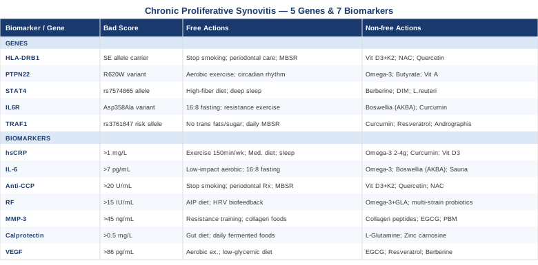
7 Biomarkers to Track in Chronic Proliferative Synovitis
The synovium does not break down silently. When it becomes hyperplastic, the body generates measurable signals in the blood, and sometimes in the synovial fluid itself. The seven markers below provide the clearest available window into what is happening at the tissue level — which processes are dominant, how aggressive the trajectory is, and whether structural damage is underway. Some are standard on routine inflammatory panels; others require a specific request or a referral to a specialty lab.
1. hsCRP — The First Signal Worth Watching
Why it matters
High-sensitivity C-reactive protein is produced by the liver in response to interleukin-6 and TNF-α, both of which are elevated in active synovitis. It is the most widely used systemic inflammation marker and correlates reasonably well with disease activity in inflammatory arthropathies. The advantage of hsCRP over standard CRP is its sensitivity at lower concentrations, which makes it more useful for monitoring subtle changes over time rather than only catching obvious flares. Multiple studies in inflammatory joint disease have found elevated baseline CRP to be associated with faster radiographic progression and worse functional outcomes over 2–5 year follow-ups.
Optimal range: below 1.0 mg/L. Borderline: 1–3 mg/L. Above 3 mg/L indicates active inflammation. In severe flares, values can exceed 50 mg/L.
How to measure it
Standard blood draw, available at any standard lab. Cost: $15–40 out of pocket, often covered by insurance when ordered for arthritis workup. No fasting required. Retest every 6–8 weeks when monitoring response to treatment or lifestyle changes. Request hsCRP specifically — standard CRP panels in routine chemistry are often less sensitive.
If the score is bad, the plan without supplements
- Aerobic exercise: 150 minutes per week of moderate-intensity activity — brisk walking, cycling, swimming — consistently lowers hsCRP. A Cochrane review in rheumatoid arthritis patients confirmed significant CRP reductions with regular aerobic exercise and no meaningful increase in disease activity during the intervention period. - Mediterranean-style diet: High in oily fish, extra-virgin olive oil, legumes, and colorful vegetables. A randomized controlled trial in RA patients showed significant hsCRP reductions after 12 weeks compared to a standard Western diet. The effect is driven by reduced IL-6, TNF-α, and oxidized LDL, all of which feed CRP production. - Sleep optimization: Less than six hours of sleep per night elevates CRP by 25–40% in chronic inflammatory conditions. Consistent 7–9 hour sleep windows, with fixed sleep and wake times, should be treated as a non-negotiable baseline before adding any other intervention. - Weight management: Adipose tissue, particularly visceral fat, is an inflammatory organ that actively secretes IL-6 and TNF-α. Even a 5–10% reduction in body weight can reduce hsCRP by 15–30% in overweight individuals with synovitis.
If the score is bad, the plan with supplements or equipment
- Omega-3 fatty acids (EPA + DHA): 2–4 g/day of combined EPA and DHA is the most evidence-backed supplement for CRP reduction. Multiple RCTs in RA and inflammatory arthritis show reduced CRP, joint tenderness, and morning stiffness duration. Best taken with meals to reduce GI effects. Long-term continuous use is safe; doses above 3 g/day may have a mild blood-thinning effect, which requires attention if you are on anticoagulants. - Curcumin: Standard curcumin has poor bioavailability. Use a phospholipid complex (Meriva), BCM-95, or piperine-enhanced form. Dose: 500–1000 mg/day. Studies show modest but consistent reductions in CRP and IL-6 across 8–12-week trials. Safe for continuous use; rare GI sensitivity in some individuals; avoid in gallbladder disease. - Vitamin D3 with K2: Low vitamin D (below 30 ng/mL) is independently associated with higher CRP and greater synovial inflammation severity. Supplementing to optimize serum 25(OH)D (target: 50–70 ng/mL) typically requires 2000–5000 IU of D3 daily, paired with 100–200 mcg MK-7 form K2 to direct calcium appropriately. Retest 25(OH)D after 3 months and adjust accordingly. - Red light therapy / photobiomodulation: Devices using 630–850 nm wavelengths applied to affected joints reduce cellular inflammation through cytochrome c oxidase activation and reduction of NF-κB signaling. Typical protocol: 10–20 minute sessions, 4–5 times per week. See the complementary section for full clinical detail.
2. IL-6 — The Master Driver of Synovial Proliferation
Why it matters
Interleukin-6 is arguably the most central cytokine in chronic proliferative synovitis. It drives synovial fibroblast proliferation, promotes angiogenesis in the joint lining, stimulates osteoclast activity (meaning bone erosion), and is the primary upstream trigger for CRP production. Elevated serum IL-6 in synovitis predicts a more aggressive disease trajectory and correlates with radiographic joint damage at follow-up. The clinical success of tocilizumab — a drug that specifically blocks the IL-6 receptor — underscores how load-bearing this cytokine is in the disease mechanism.
Normal range: below 7 pg/mL in most lab reference intervals. Elevated IL-6 warrants attention even when CRP is borderline, particularly if joint symptoms persist.
How to measure it
Serum or plasma IL-6 via ELISA. Cost: $50–150, depending on the lab. Less standardized than CRP — values can vary between laboratories, so retesting should always use the same lab. This is not on most routine inflammatory panels, so you will likely need to specifically request it, most easily through a rheumatologist or integrative medicine physician.
If the score is bad, the plan without supplements
- Regular aerobic exercise: Acute exercise transiently elevates muscle-derived IL-6, but chronic regular exercise reduces baseline IL-6 production from adipose tissue and immune cells — the opposite effect. For inflamed joints, low-impact options like cycling, elliptical, or aquatic exercise are preferable. Target: 30–45 minutes, 4–5 days per week. - Time-restricted eating: A 16:8 intermittent fasting pattern reduces IL-6 in metabolic syndrome patients, likely through mTOR pathway normalization and reduced adipose-derived cytokine production. The effect on joint-specific IL-6 is less studied but mechanistically sound. - Cold water immersion: Cold hormesis reduces post-exercise IL-6 and systemic inflammatory markers through norepinephrine release and anti-inflammatory sympathetic signaling. Protocol: 10–15°C water, 10–15 minutes, 3 times per week. Contraindicated in Raynaud's phenomenon or cardiovascular instability.
If the score is bad, the plan with supplements or equipment
- Omega-3 (EPA/DHA): EPA specifically inhibits the phospholipase A2 pathway, reducing upstream precursors to IL-6. Same protocol as for hsCRP. The effect on IL-6 itself is modest but consistent across meta-analyses. - Boswellia serrata (AKBA-standardized extract): 100–250 mg of standardized AKBA twice daily. RCTs in inflammatory joint disease show meaningful reductions in IL-6, inflammatory scores, and joint function. Cycle: 8–12 weeks on, 2–4 weeks off. Side effects: rare GI discomfort; take with food. - Ginger extract: 500–2000 mg/day of standardized extract. Human studies show modest IL-6 and TNF-α reductions with consistent use over 6–12 weeks. Side effects are primarily GI at high doses. - Sauna therapy (traditional or infrared): Repeated sauna sessions (20–30 minutes, 3–4 times per week) have shown reductions in IL-6 and inflammatory markers in arthritis patients in Finnish clinical studies, possibly through heat shock protein activation and sympathetic regulation. Start with lower temperatures and shorter durations if new to sauna.
3. Anti-CCP Antibodies — The Autoimmune Fingerprint
Why it matters
Anti-cyclic citrullinated peptide antibodies are among the most specific biomarkers available for rheumatoid arthritis-associated synovitis. They are detectable in up to 70% of RA patients, and critically, they often appear 5–10 years before clinical symptoms emerge — making them among the earliest warning signals the body produces. High titers are associated with more aggressive, erosive disease and predict worse structural outcomes over time. Unlike rheumatoid factor, anti-CCP carries approximately 95% specificity for RA, meaning a strongly positive result is highly informative rather than merely suggestive. The foundational research establishing the clinical validity of this test was published by Schellekens and colleagues in Arthritis & Rheumatism (2000), and assays have since been refined through second and third-generation versions that improve sensitivity without sacrificing specificity.
Normal: below 20 U/mL. Borderline: 20–40 U/mL. High: above 40 U/mL. Very high titers (above 100 U/mL) carry the most significant prognostic weight.
How to measure it
Standard blood draw, second or third-generation ELISA. Cost: $60–120. Often ordered alongside rheumatoid factor for initial RA/synovitis screening. Retesting is rarely necessary once the level is established — the test identifies autoimmune predisposition, which does not change rapidly. It can be useful to retest after 6–12 months of intensive intervention to evaluate whether titers are trending down.
If the score is bad, the plan without supplements
Anti-CCP positivity reflects an immune dysregulation that is harder to reverse through lifestyle alone than nonspecific inflammation, but meaningful modulation is possible: - Smoking cessation: Cigarette smoking is the single strongest modifiable environmental factor for anti-CCP positivity. Smoking triggers citrullination of lung proteins and primes the autoimmune response that generates ACPA. HLA-DRB1 shared epitope carriers who smoke have multiplicatively elevated risk. Cessation can modestly reduce titers over 1–2 years. - Periodontal disease treatment: Porphyromonas gingivalis, the primary periodontal pathogen, produces its own peptidylarginine deiminase enzyme (PPAD) that generates citrullinated proteins, fueling ACPA generation. Small controlled trials have shown modest reductions in anti-CCP and RA disease activity scores following aggressive periodontal treatment. This is an underappreciated and inexpensive intervention. - MBSR (mindfulness-based stress reduction): Chronic psychological stress amplifies Th17 immune responses, which feed the citrullination cascade. Eight-week MBSR programs have shown measurable reductions in inflammatory activity and autoimmune markers in RA patients. See the complementary section for the protocol.
If the score is bad, the plan with supplements or equipment
- Vitamin D3 with K2: Vitamin D has direct immunomodulatory effects on Th17/Treg balance, and several studies show inverse associations between serum 25(OH)D and anti-CCP titers. Target 25(OH)D: 50–70 ng/mL. Typical dose: 3000–5000 IU D3 + 100–200 mcg MK-7 form K2 daily. - Quercetin: 500–1000 mg/day. Quercetin inhibits peptidylarginine deiminase (PAD) enzymes — the exact enzymes that generate citrullinated proteins and drive ACPA production. Animal and cell data are compelling; human data remains early-stage but mechanistically sound. Safe for continuous use; occasional mild GI upset. - N-acetylcysteine (NAC): 600–1200 mg/day. Reduces oxidative stress that amplifies PAD enzyme activity. Pilot data in RA suggests reduced oxidative markers. Cycle: continuous use is safe; some practitioners recommend a 1-month break every 3–4 months to prevent glutathione pathway tolerance.
4. Rheumatoid Factor — Context, Not Conclusion
Why it matters
Rheumatoid factor is an autoantibody directed against the Fc portion of IgG. It is less diagnostically specific than anti-CCP — it can be positive in infections, other autoimmune conditions, and even in healthy elderly individuals — but when elevated alongside anti-CCP, it significantly raises diagnostic confidence for RA-associated synovitis. High-titer RF is associated with extra-articular manifestations (nodules, vasculitis, lung involvement) and a more aggressive joint disease course. Tracking it alongside anti-CCP over time also provides a rough measure of immune activation. If both normalize together following intervention, that is a meaningful signal.
Normal: below 14–15 IU/mL in most lab reference ranges. Clinically significant: consistently above 60–80 IU/mL in the context of joint symptoms.
How to measure it
Standard blood draw. Cost: $20–50, routinely included in RA screening panels. The same lifestyle interventions that reduce anti-CCP and systemic inflammation also tend to lower RF over time, though the response is usually slower and less dramatic than changes in CRP.
If the score is bad, the plan without supplements
- Autoimmune protocol diet (AIP): Systematically removing potential immune triggers — grains, legumes, dairy, nightshades, eggs, seed oils — for 30–90 days reduces exposure to molecular mimicry targets and lowers gut-driven immune activation that can sustain RF production. See the complementary section for the full Sarah Ballantyne protocol. - Structured stress management: RF production correlates with Th2 immune skewing, which is amplified by dysregulated cortisol. HRV biofeedback training — 20 minutes daily targeting slow resonance breathing at approximately 5–6 breaths per minute — has shown parasympathetic restoration and reduced inflammatory immune markers in RA patients.
If the score is bad, the plan with supplements or equipment
- Omega-3 with GLA (gamma-linolenic acid): The combination of EPA/DHA (2–3 g/day) with GLA from evening primrose or borage oil (500–1000 mg/day) shows synergistic anti-inflammatory effects with modest RF-lowering in RA trial data. Cycle: continuous use is safe long-term. - Multi-strain probiotics: RF elevation is partly sustained by gut dysbiosis and increased intestinal permeability, which delivers bacterial antigens to a primed immune system. Lactobacillus casei and Bifidobacterium longum strains have shown immunomodulatory effects with modest RF reductions in small RA trials. Dose: 20–50 billion CFU/day. Rotate strains every 3–4 months to sustain microbiome diversity.
5. MMP-3 — The Early Warning Signal for Joint Destruction
Why it matters
Matrix metalloproteinase-3 (stromelysin-1) is produced primarily by synovial fibroblasts when they shift into an aggressive, tissue-remodeling state. It degrades collagen, proteoglycans, and extracellular matrix components in the joint — the structural material that keeps cartilage intact. Elevated serum MMP-3 is among the earliest indicators that synovitis is progressing beyond inflammation toward structural damage. Its clinical importance lies in what standard panels miss: MMP-3 can be significantly elevated even when CRP and ESR are within normal range, making it a valuable catch for subclinical but structurally destructive disease activity.
Normal range: approximately 3–45 ng/mL in males, 2–28 ng/mL in females. Post-menopausal women may have higher baseline values, requiring age-adjusted interpretation.
How to measure it
Blood test via ELISA. Cost: $80–200; less commonly available at standard labs and usually ordered through specialty or functional medicine labs. Worth requesting specifically when CRP and ESR are normal or borderline but joint symptoms persist — this is the scenario where MMP-3 most often catches what other markers miss. Most useful for tracking structural risk over time rather than as a one-time diagnostic.
If the score is bad, the plan without supplements
- Resistance training: Mechanical loading of bones and cartilage through resistance exercise stimulates the production of tissue inhibitors of metalloproteinases (TIMPs), which directly counteract MMP-3 degradative activity. Even light resistance work — bodyweight exercises, resistance bands, water resistance — 2–3 times per week within joint-safe ranges shows this effect. Progressive loading as inflammation allows. - Collagen-rich whole foods: Regular consumption of bone broth, gelatin, slow-cooked meats, and similar collagen-containing foods supplies hydroxyproline, glycine, and proline — the building blocks for extracellular matrix repair. This does not reduce MMP-3 production directly but offsets its degradative effect.
If the score is bad, the plan with supplements or equipment
- Hydrolyzed collagen peptides or undenatured type II collagen: 10 g/day of hydrolyzed collagen (types I and III) or 40 mg/day of undenatured type II collagen. Multiple RCTs show reductions in joint pain, MMP activity markers, and improved cartilage biomarkers. Continuous use is safe and well-tolerated. - Green tea extract (EGCG): 400–800 mg/day of EGCG (epigallocatechin gallate). Inhibits MMP-1, MMP-3, and MMP-13 in synovial cells in both cell and human studies, while also reducing IL-1β-stimulated MMP production. Take with food to reduce GI sensitivity. Cycle: 12–16 weeks on, 4 weeks off at higher doses due to theoretical hepatic load. - Low-dose doxycycline (prescription only): This antibiotic has well-established MMP-inhibiting properties independent of its antimicrobial action. At 20 mg twice daily — a dose used clinically for periodontal disease — it reduces MMP-3 and MMP-8 activity without significant antibiotic effects. Studied in RA with modest positive results. Requires physician oversight. - Photobiomodulation: Emerging evidence suggests reduced MMP expression in synovial tissue following low-level laser or near-infrared light therapy applied to affected joints. Protocol: 10–20 minutes, 3–5 sessions per week over 4–8 weeks. See the complementary section for clinical trial detail.
6. Calprotectin — Reading the Gut-Joint Signal
Why it matters
Calprotectin is a calcium-binding protein released by activated neutrophils at sites of inflammation. While fecal calprotectin is most commonly associated with IBD, serum calprotectin is increasingly recognized as a sensitive marker of inflammatory arthropathy activity. Studies in psoriatic arthritis and RA have found serum calprotectin to correlate with disease activity independently of CRP — and to be particularly useful when CRP is borderline or discordant with the clinical picture. Beyond its role as a direct inflammation marker, calprotectin elevation also reflects the gut-joint inflammatory axis: intestinal permeability and gut dysbiosis drive neutrophil activation that shows up in calprotectin levels, making it a proxy for whether the gut is fueling the joint disease.
Reference range: serum calprotectin below 0.5 mg/L in healthy individuals (lab-dependent). Fecal calprotectin normal range: below 50 mcg/g stool for adults.
How to measure it
Fecal calprotectin: stool test, cost $50–120, widely available at standard labs. Serum calprotectin is less standardized and mostly available through specialty labs. Either test provides meaningful information about neutrophil-mediated inflammation. Fecal calprotectin is the more accessible starting point and provides the clearest view of gut-joint inflammatory connection.
If the score is bad, the plan without supplements
- Gut-targeted dietary changes: Elevated calprotectin strongly implicates gut barrier involvement. A 4–6 week elimination of ultra-processed foods, refined sugars, alcohol, and known food sensitivities — combined with daily fermented foods (plain kefir, kimchi, sauerkraut, water kefir) — reduces intestinal neutrophil activation and downstream calprotectin levels in multiple observational studies. - Fasting-mimicking protocols: Short-term caloric restriction — 5-day cycles every 1–3 months, following the ProLon-style protocol developed by Valter Longo at USC — has demonstrated reductions in gut inflammatory markers and autoimmune signaling in clinical studies. The mechanism involves gut mucosa regeneration during the refeeding phase.
If the score is bad, the plan with supplements or equipment
- L-glutamine: 5–10 g/day. Gut epithelial cells rely heavily on glutamine for energy and maintenance of tight junction integrity. Supplementation reduces intestinal permeability markers and downstream inflammatory signaling. Cycle: 3 months on, 1 month off. Avoid in epilepsy (glutamine can cross-convert to glutamate). - Zinc carnosine: 75–150 mg/day. Among the best-studied compounds for gut barrier repair, with RCT evidence for reduced intestinal permeability markers. Take with meals to avoid nausea. - Spore-based probiotics: Bacillus coagulans and Bacillus subtilis strains have shown reductions in gut permeability markers and systemic inflammatory signaling in patients with inflammatory joint conditions. Dose: 2–4 billion CFU/day. Continuous use is well-tolerated.
7. VEGF — The Angiogenesis Signal That Makes Synovitis Proliferative
Why it matters
What distinguishes proliferative synovitis from simple inflammation is the abnormal growth of new blood vessels in the synovial tissue — a process called angiogenesis. VEGF (vascular endothelial growth factor) is the primary driver of this process, and elevated serum VEGF reflects active synovial neovascularization. Higher VEGF correlates with thicker, more vascular pannus tissue, more aggressive immune cell delivery to the joint, and greater structural damage. Critically, VEGF also creates a self-sustaining loop: more blood vessels mean more inflammatory cell infiltration, which produces more VEGF. Tracking VEGF specifically tells you whether the proliferative — not just the inflammatory — component of the disease is being addressed by your current approach.
Normal range: approximately 31–86 pg/mL, though this is highly lab-dependent. Compare values to your lab's specific reference interval.
How to measure it
Serum VEGF via ELISA. Cost: $100–200. Not included in standard inflammatory panels — requires a specific request, most easily made through a rheumatologist or functional medicine physician. Most useful for monitoring treatment response over 3–6-month intervals rather than as a one-time diagnostic. It is the one biomarker on this list most specific to the proliferative character of the condition.
If the score is bad, the plan without supplements
- Regular moderate aerobic exercise: Endurance exercise normalizes HIF-1α signaling — the upstream regulator of VEGF — reducing pathological angiogenesis while preserving physiological blood vessel function. Target: 30–45 minutes, 4–5 days per week, low-impact formats for joint protection. - Low-glycemic, anti-angiogenic diet: Insulin and IGF-1 are major upstream VEGF activators through the PI3K/Akt/mTOR pathway. Significantly reducing refined carbohydrates and sugar lowers this drive. Foods with anti-angiogenic evidence include cruciferous vegetables (sulforaphane), green tea, berries, dark chocolate, and turmeric — a diet worth building around regardless of other factors.
If the score is bad, the plan with supplements or equipment
- EGCG (green tea extract): 400–600 mg/day. Among the best-studied natural VEGF modulators. Multiple in vitro and in vivo studies confirm anti-angiogenic activity and VEGF suppression. As above: take with food, cycle at high doses. - Resveratrol: 250–500 mg/day of high-quality trans-resveratrol or pterostilbene. Inhibits VEGF expression and NF-κB-driven angiogenesis via SIRT1 activation. Cycle: 3 months on, 1 month off. Side effects minimal; avoid combining with anticoagulants without medical supervision. - Berberine: 500 mg twice daily with meals. Reduces VEGF expression through AMPK activation and direct inhibition of HIF-1α transcription. Also improves the metabolic parameters (insulin sensitivity, lipid profile) that independently feed VEGF overproduction. Cycle: 12 weeks on, 4 weeks off. Side effects: initial loose stools in some people; do not combine with blood sugar medications without physician supervision.
Genetics and Epigenetics: 5 Gene Variants Shaping Your Synovitis
Biomarkers tell you what is happening now. Genetics tells you why you are structurally predisposed to it — and which pathways to watch most carefully over time. The five gene variants below are among the most replicated findings in the RA and inflammatory synovitis literature. None of them are destiny, but each creates a tendency that can be worked with once you know it is there.
1. HLA-DRB1 — The Shared Epitope
What the gene does
HLA-DRB1 encodes a class II MHC receptor expressed on antigen-presenting cells. Specific alleles — DRB1*0401, *0404, *0101, and several others — share a five-amino-acid sequence in the peptide-binding groove known as the shared epitope (SE). This configuration presents citrullinated peptides to T cells with unusual efficiency, which is why carriers of two SE alleles face up to tenfold the population baseline risk for seropositive RA. The SE hypothesis was established by Gregersen and colleagues in a landmark 1987 paper in Arthritis & Rheumatism and has been replicated in hundreds of subsequent studies. Since the SE works by presenting citrullinated proteins, interventions that reduce protein citrullination directly target the gene's mechanism.
If the gene is bad, the plan without supplements
- Smoking cessation: The most impactful modifiable trigger. Smoking induces citrullination of lung proteins through neutrophil PAD4 activation, and SE carriers who smoke face multiplicatively elevated risk. Cessation reduces this risk substantially over 2–5 years. - Aggressive periodontal care: Porphyromonas gingivalis produces PPAD, a bacterial citrullinating enzyme that generates the exact kinds of peptides that SE molecules present to T cells. Regular dental cleaning, treating any active gum disease, and maintaining periodontal health is a specific, evidence-linked intervention for SE carriers. - Stress reduction: Cortisol-driven immune dysregulation amplifies antigen presentation efficiency in HLA-SE individuals. Structured mindfulness practice or HRV biofeedback dampens this amplification.
If the gene is bad, the plan with supplements or equipment
- Vitamin D3: Modulates HLA class II expression and reduces aggressive antigen presentation. Target 25(OH)D: 50–70 ng/mL. Typical dose: 3000–5000 IU/day with K2. - NAC (N-acetylcysteine): Reduces PAD enzyme activity through oxidative stress reduction. 600–1200 mg/day, continuous. Brief 1-month breaks every 3–4 months. - Quercetin: Direct PAD inhibitor in early studies. 500–1000 mg/day. Evidence is preliminary in humans but mechanistically specific to the SE pathway.
2. PTPN22 — The Overactive T Cell
What the gene does
PTPN22 encodes lymphoid tyrosine phosphatase (LYP), which normally acts as a brake on T cell receptor signaling. The R620W variant (rs2476601) reduces this brake function, producing hyperactive T cells more prone to recognizing self-antigens. Present in approximately 10% of Europeans, it is among the strongest non-HLA risk factors for RA, and also elevates risk for type 1 diabetes, lupus, and Hashimoto's thyroiditis — suggesting a systemic immune regulation defect. Carriers who develop synovitis tend to have more aggressive T cell-driven disease activity, particularly in the synovial pannus.
If the gene is bad, the plan without supplements
- Regular aerobic exercise: Consistently expands T regulatory cell (Treg) populations and restores tolerance mechanisms. 30–45 minutes, 4–5 days per week, low-impact formats. - Circadian alignment: T cell hyperactivity follows circadian dysregulation. Morning light exposure within 30 minutes of waking, avoidance of blue light after 9 pm, and sleeping before midnight support appropriate immune tone and Treg function. Research from Satchin Panda's group at the Salk Institute links circadian disruption to amplified T cell responses. - Cold water exposure: Progressive cold shower protocols — starting at 30 seconds cold and extending to 2–3 minutes over weeks — activate anti-inflammatory pathways through norepinephrine release and expand Treg populations.
If the gene is bad, the plan with supplements or equipment
- Omega-3 (EPA/DHA): EPA and DHA support Treg generation from naive T cells through PPAR-γ and FoxP3 signaling. 2–4 g/day, continuous. - Butyrate or resistant starch: Gut-derived short-chain fatty acids — particularly butyrate — are essential for Treg expansion in the intestinal immune compartment. Sodium butyrate: 600–1200 mg/day. Dietary resistant starch: 15–30 g/day from green banana, cooked and cooled potato, or raw oats. - Vitamin A: Retinoic acid signaling is essential for Treg differentiation and mucosal immune tolerance. Adequate intake from dietary sources (liver, eggs, orange and yellow vegetables) is first-line. Supplemental mixed carotenoids (15 mg/day) as a safe adjunct. High-dose preformed retinol should only be used under supervision.
3. STAT4 — Amplifier of Th17 Responses
What the gene does
STAT4 encodes a transcription factor activated by IL-12 and IL-23, which in turn drive the differentiation of Th1 and Th17 immune cells. The rs7574865 risk allele amplifies Th17 responses — a cell population directly implicated in synovial proliferation, neutrophil recruitment, and osteoclast-mediated bone erosion. IL-17, the primary product of Th17 cells, is elevated in synovial fluid of patients with active proliferative synovitis, and STAT4 risk allele carriers show amplified IL-17 production in response to inflammatory triggers. This variant also increases susceptibility to lupus and psoriatic arthritis.
If the gene is bad, the plan without supplements
- High-fiber, Mediterranean or AIP diet: Short-chain fatty acids from fermented dietary fiber suppress Th17 differentiation through FoxP3 upregulation. High-fiber diets — and the AIP protocol in particular — reduce the gut-derived IL-17 stimulation that STAT4 amplifies. - Sleep depth optimization: Slow-wave sleep suppresses IL-12 signaling and Th17 output. Strategies: bedroom temperature at 65–68°F, complete darkness, avoidance of alcohol within 3 hours of sleep (alcohol specifically fragments slow-wave sleep).
If the gene is bad, the plan with supplements or equipment
- Berberine: Inhibits IL-17 production and STAT4 downstream signaling through AMPK activation. 500 mg twice daily with meals, 12 weeks on, 4 weeks off. - DIM (diindolylmethane): Cruciferous vegetable derivative that shifts Th17/Treg balance toward tolerance through aryl hydrocarbon receptor signaling. 200–400 mg/day. Caution: alters estrogen metabolism, relevant if on hormonal therapies. - Lactobacillus reuteri DSM 17938: This specific probiotic strain has the most consistent evidence for inhibiting Th17 differentiation at the gut-immune interface. 1–5 billion CFU/day, continuous.
4. IL6R — Wired for IL-6 Hypersensitivity
What the gene does
Variants in the IL6R gene affect IL-6 receptor expression and downstream signaling. The Asp358Ala variant (rs2228145) influences soluble IL-6 receptor production and alters the balance between classical IL-6 signaling (pro-inflammatory) and trans-signaling through the soluble receptor. Individuals without protective variants may be particularly sensitive to IL-6-driven synovitis, and paradoxically, this variant also predicts better response to tocilizumab. Understanding your IL6R status gives clinical context for why IL-6-targeted approaches — including lifestyle interventions — may be more or less effective in your case.
If the gene is bad, the plan without supplements
The approach mirrors the IL-6 biomarker plan above, with additional emphasis on: - 16:8 intermittent fasting: Reduces IL-6 most significantly in individuals with functional IL-6 receptor pathway amplification, likely through mTOR-STAT3 normalization. - Resistance exercise for adipose IL-6 reduction: Low-load, high-repetition resistance work is effective for reducing adipose-derived IL-6 without the joint stress of heavy loading.
If the gene is bad, the plan with supplements or equipment
- Boswellia serrata (AKBA): 100–250 mg/day. Targets the downstream consequences of IL-6R over-signaling through NF-κB and 5-LOX inhibition. Cycle: 8–12 weeks on, 2–4 weeks off. - Curcumin (high-bioavailability form): Directly inhibits STAT3 activation — a primary mediator of IL-6R downstream inflammatory effects. 500–1000 mg/day of Meriva or BCM-95 formulation.
5. TRAF1 — NF-κB on a Hair Trigger
What the gene does
TRAF1 (TNF receptor-associated factor 1) mediates downstream signaling from TNF-α and IL-1β through the NF-κB pathway. The rs3761847 variant in the TRAF1-C5 locus is among the most replicated genetic associations in RA genome-wide association studies. Risk allele carriers show amplified NF-κB activation in response to pro-inflammatory stimuli — meaning the same environmental trigger produces a larger and more sustained synoviocyte response compared to non-carriers. This variant predicts both disease susceptibility and progression from early non-erosive to erosive joint disease, making it one of the most prognostically important genetic findings in proliferative synovitis.
If the gene is bad, the plan without supplements
- Eliminating dietary NF-κB activators: Trans fats, excess omega-6 oils (soybean, corn, sunflower), refined sugar, and advanced glycation end products (AGEs from charred, fried, or ultra-processed foods) are the primary dietary NF-κB activators. Eliminating these is first-line and achievable without supplementation. - Formal meditation practice: Chronic psychological stress activates NF-κB through glucocorticoid receptor resistance in immune cells. Eight-week MBSR programs have produced direct reductions in NF-κB DNA-binding activity in peripheral blood cells in clinical studies. Forty-five minutes of daily formal practice is the protocol with the strongest evidence.
If the gene is bad, the plan with supplements or equipment
- Curcumin (high-bioavailability): The most evidence-supported natural NF-κB inhibitor. 1000–1500 mg/day, with meals. - Resveratrol: SIRT1 activation deacetylates the p65 subunit of NF-κB, preventing its nuclear translocation. 250–500 mg/day of trans-resveratrol. Cycle: 3 months on, 1 month off. - Andrographis paniculata: 300–600 mg/day of standardized extract (andrographolide 10%). Shows direct NF-κB inhibition in human synoviocyte studies and in small clinical trials in RA. Cycle: 8–12 weeks on, 4 weeks off. Avoid during pregnancy or if planning conception.
The Gut-Joint Connection: 10 Things Tom O'Bryan's Research Reveals
Tom O'Bryan is a clinician and researcher who has spent decades synthesizing the evidence on intestinal permeability, molecular mimicry, and autoimmune disease — particularly as they relate to inflammatory conditions like RA and synovitis. His book The Autoimmune Fix draws on hundreds of peer-reviewed studies to make a case that standard rheumatology protocols treat the downstream fire without addressing the upstream fuel. These are the ten most clinically impactful ideas from that body of work.
1. Intestinal hyperpermeability precedes the joint disease
In multiple studies, measurable gut barrier dysfunction appears years before clinical autoimmune joint disease is diagnosed. Bacterial peptides and undigested food proteins cross the compromised barrier, trigger systemic immune activation, and are sometimes structurally similar enough to joint proteins to produce cross-reactive antibodies. The joint becomes a target of immune activity that started in the gut.
2. The 3-stage progression: latent, silent, active
O'Bryan frames autoimmune disease as a progression through three stages: latent (genetic predisposition), silent (measurable immune activation without symptoms), and active (clinical disease). Biomarkers like anti-CCP can detect the silent stage years early. Intervening at the silent stage — when gut permeability is measurable but the joint is not yet damaged — is dramatically more effective than treating established disease.
3. Gluten and molecular mimicry
Certain gliadin peptides from wheat share structural sequences with joint proteins including type II collagen, fibrinogen, and calreticulin. Immune cells sensitized to gliadin in the gut can produce antibodies that cross-react with these joint proteins. This mechanism is particularly relevant for HLA-DRB1 SE carriers, whose antigen-presenting cells are especially efficient at presenting both gliadin and citrullinated joint proteins.
4. Zonulin — the measurable gateway
Zonulin is the protein that regulates tight junction opening in the gut epithelium. Elevated serum zonulin is a direct measure of intestinal permeability and is testable through functional labs. O'Bryan cites research from Alessio Fasano's group at Harvard showing that elevated zonulin is significantly more prevalent in autoimmune patients than healthy controls — and that reducing it through dietary and supplement intervention can reduce downstream immune activation.
5. Not all gluten-sensitive people have celiac disease
Non-celiac gluten sensitivity (NCGS) produces measurable immune activation — including elevated anti-gliadin antibodies and intestinal permeability — without the villous atrophy of celiac disease. Standard testing misses NCGS entirely. O'Bryan's clinical work and the broader NCGS research suggest that inflammatory joint disease patients with elevated fecal calprotectin and unexplained immune activation should be tested for anti-gliadin IgA and IgG before concluding gut health is not a factor.
6. The microbiome shifts years before diagnosis
Specific microbiome signatures — reduced Faecalibacterium prausnitzii, elevated Prevotella copri, and reduced overall diversity — have been identified in pre-RA individuals before clinical onset. Prevotella copri specifically has been linked to Th17 activation and joint inflammation. The microbiome shift is not a consequence of the joint disease; in many cases it precedes and possibly drives it.
7. Environmental toxins amplify gut-immune dysregulation
Pesticide residues, heavy metals (mercury, lead), and plastic-derived compounds (BPA, phthalates) impair gut barrier integrity and dysregulate immune signaling. O'Bryan links chronic low-level toxin exposure to the kind of gut-immune breakdown that eventually surfaces as autoimmune joint disease, making environmental toxin reduction — filtered water, organic produce where feasible, reduced plastic contact with food — a meaningful but often overlooked component of the protocol.
8. The 90-day dietary intervention window
O'Bryan's clinical framework involves a sustained dietary elimination and gut repair protocol of at least 90 days, based on the biology of immune memory cell turnover and gut epithelial regeneration. Shorter trials of 2–4 weeks are typically insufficient to produce measurable changes in autoimmune markers. The commitment to a full quarter is what distinguishes effective trials from ones that are abandoned before results are possible.
9. Testing before assuming
O'Bryan consistently advocates for testing gut permeability (fecal calprotectin, zonulin, lactulose-mannitol ratio), food reactivity (anti-gliadin, anti-casein, IgA/IgG food panels), and microbiome composition before assuming which dietary changes are necessary. Assumptions about food sensitivities without testing often result in unnecessary restriction or missed relevant triggers.
10. Sleep is gut repair, not just rest
During deep slow-wave sleep, the gut epithelium undergoes its primary repair cycle — tight junction protein synthesis, microbial balance restoration, and mucosal immune regulation all peak during non-REM stages. O'Bryan cites research showing that sleep disruption increases intestinal permeability and elevates anti-gliadin antibodies within days. Sleep hygiene is not a soft recommendation in this framework; it is a structural requirement for gut barrier maintenance and downstream autoimmune resolution.
Complementary and Alternative Approaches
The following five approaches each have meaningful human clinical evidence for inflammatory joint conditions. They are not replacements for medical care or the biomarker-guided strategies above, but they address real mechanisms and are worth considering as part of a comprehensive approach.
The Autoimmune Protocol (AIP) — Sarah Ballantyne
The Autoimmune Protocol is a structured dietary and lifestyle framework developed by Dr. Sarah Ballantyne, a research scientist who applied it to her own autoimmune disease and subsequently reviewed the underlying science exhaustively. It removes foods that are known to aggravate gut permeability or trigger immune reactivity — grains, legumes, dairy, eggs, nightshades, nuts, seeds, seed oils, alcohol, and NSAIDs — while emphasizing nutrient density, sleep, stress management, and gentle movement. The protocol has a specific reintroduction phase that allows systematic identification of individual triggers.
A 2017 pilot study published in Inflammatory Bowel Diseases found significant reductions in inflammatory markers and symptom scores in IBD patients following a 6-week AIP diet, and similar protocols have shown measurable immunological changes in other autoimmune conditions. The evidence base in RA-specific synovitis remains smaller but mechanistically coherent — the gut-immune pathway it targets is directly implicated in the condition.
For chronic proliferative synovitis, the practical starting point is a strict 60–90 day elimination phase, followed by structured reintroduction of one food category every 5–7 days while monitoring joint symptoms and biomarkers. Ballantyne's book The Paleo Approach provides the most thorough clinical rationale and practical protocol, and her website (Autoimmune Wellness) includes a detailed guide. The approach requires significant dietary commitment but generates personalized data about food-joint symptom relationships that no laboratory test can produce.
Tai Chi
Tai chi is a low-impact mind-body movement practice originating in Chinese martial arts, characterized by slow, continuous movements, weight-shifting, and coordinated breathing. For chronic joint inflammation, it is particularly well suited because it improves joint proprioception, muscle strength, and flexibility without the compressive loading of conventional exercise that can worsen active synovitis. The meditative component also downregulates sympathetic nervous system tone, which directly reduces inflammatory cytokine production.
A 2013 Cochrane review on tai chi for rheumatoid arthritis identified consistent evidence of improved physical function, reduced pain, and improved patient-reported quality of life, with no adverse effects on disease activity. A 2020 meta-analysis in Complementary Therapies in Medicine examining tai chi in inflammatory arthropathies found significant reductions in pain scores, DAS28 (disease activity), and self-reported disability across 11 RCTs.
For someone with active chronic proliferative synovitis, a beginner tai chi program of 3–4 sessions per week, each 30–45 minutes, represents an accessible starting point. Many community centers and online programs offer Yang-style beginners' sequences specifically adapted for people with limited mobility. Consistency over 12 weeks is where functional and inflammatory benefits become measurable.
Low-Level Laser Therapy / Photobiomodulation
Photobiomodulation (PBM) uses non-thermal red and near-infrared light (typically 630–905 nm) to stimulate mitochondrial cytochrome c oxidase, reduce reactive oxygen species, and suppress NF-κB-driven inflammatory signaling in tissue. In synovitis, this translates to reduced local cytokine production, reduced MMP activity, and modest improvements in synovial tissue histology in studies where biopsies were taken pre and post treatment.
A systematic review published in Lasers in Medical Science examining low-level laser therapy in RA found consistent reductions in pain, morning stiffness, and hand grip disability, with effects most pronounced over 4-week courses of treatment. The Cochrane review on LLLT for RA (2005, updated reviews since) found moderate evidence of short-term benefit in pain and functional outcomes. Evidence is strongest for hand and wrist joint involvement, with less data on larger joints.
Practically, photobiomodulation can be applied at home using consumer-grade devices (660–850 nm panels or targeted handheld devices). Protocol for joint application: 10–20 minutes per session, applied directly to affected joints, 4–5 times per week for a minimum of 4 weeks before evaluating response. Devices should provide at least 20–60 mW/cm² of irradiance. The treatment has an excellent safety profile and no significant side effects at standard parameters.
Mindfulness-Based Stress Reduction (MBSR)
MBSR, developed by Jon Kabat-Zinn at the University of Massachusetts Medical School, is an 8-week program of structured mindfulness meditation practice — body scan, sitting meditation, gentle yoga — with 45 minutes of daily home practice. Its relevance to chronic proliferative synovitis is not simply pain psychology: stress-driven cortisol dysregulation amplifies NF-κB activity in synoviocytes, amplifies Th17 differentiation, and reduces Treg function — all mechanistically relevant to the disease biology.
An RCT published in Annals of the Rheumatic Diseases found that RA patients following an 8-week MBSR program showed significant reductions in disease activity scores, CRP, and IL-6 compared to a control group, with effects maintained at 6-month follow-up. A separate clinical study in inflammatory arthropathy patients showed reduced cortisol awakening response — a marker of HPA axis dysregulation that directly feeds synovial inflammation — following the same 8-week protocol.
The practical approach requires commitment to the full 8-week curriculum, which is available through hospital programs, dedicated apps (Insight Timer includes free MBSR-guided courses), and Kabat-Zinn's original books. The benefits appear to build with consistent practice — casual or intermittent use does not produce the same measurable outcomes as the structured protocol.
Microbiome-Directed Therapies
The gut-joint axis in proliferative synovitis is not theoretical — it is increasingly mechanistic and measurable. Specific dysbiotic signatures, particularly elevated Prevotella copri and reduced butyrate-producing species, have been consistently identified in RA patients before and after diagnosis. Microbiome-directed approaches aim to shift these populations toward configurations associated with reduced systemic immune activation.
A 2022 trial in Annals of the Rheumatic Diseases examined a high-fiber, plant-centered dietary intervention in early RA patients and found significant shifts in microbiome composition alongside reductions in joint inflammation markers, suggesting that targeted dietary fiber intake directly modifies the gut-joint inflammatory axis. Separately, RCTs of specific probiotic strains — particularly Lactobacillus casei and Lactobacillus rhamnosus — have shown modest but significant reductions in DAS28 scores and inflammatory markers in RA patients over 8–12 weeks.
Practically, a microbiome-directed approach involves three simultaneous strategies: removing microbiome-disruptive inputs (refined sugar, alcohol, antibiotics unless necessary, processed food emulsifiers like carrageenan and polysorbate-80); actively feeding butyrate-producing bacteria through diverse dietary fibers (inulin, pectin, resistant starch, beta-glucan from oats); and reseeding with targeted probiotic strains. A comprehensive stool analysis (GI-MAP or similar) provides a baseline before intervention and a 3–6 month follow-up measurement to track progress.
Conclusion
Chronic proliferative synovitis is a condition with real, measurable drivers — and that means it has real, measurable targets. Tracking the seven biomarkers in this article gives you a live picture of which inflammatory mechanisms are most active, whether structural damage is beginning, and whether the gut is feeding the process. The genetics section adds the longer view: which pathways you are inherently prone to over-activating and where preventive effort will be most efficiently placed. Neither set of insights is a cure, but both are significantly more actionable than generic inflammation advice.
The next smart step is to identify which biomarkers you have not yet measured and to discuss testing with your rheumatologist or GP. If anti-CCP and hsCRP are the only values you have, MMP-3, calprotectin, IL-6, and VEGF may be worth adding to the next panel. If your genetics are available through a consumer test, reviewing the five variants covered here with a functional medicine practitioner can clarify which lifestyle interventions deserve the most consistent effort. Start with the one or two levers most clearly implicated in your case, track the relevant biomarker over 8–12 weeks, and adjust from there.
Musculoskeletal: Joint Conditions
Autoimmune: Inflammatory Conditions Connective Tissue Conditions