This article contains AI generated content.
· UpdatedCommon Variable Immunodeficiency and Arthritis: 7 Biomarkers and 6 Genes to Track
Introduction
If you live with common variable immunodeficiency and joint pain, you already know the confusion that comes with it. One week brings a respiratory infection, the next brings swollen knees or aching wrists, and too often the two problems get treated by two different specialists who never quite connect them. The link between CVID and arthritis is real, documented, and mechanistically coherent — yet it frequently falls through the cracks of standard care.
Generic arthritis advice rarely applies here. CVID-associated arthritis is typically seronegative — the standard antibody markers that define rheumatoid arthritis often come back negative — which can delay appropriate diagnosis and treatment by months or years. The biology driving joint inflammation in CVID differs meaningfully from classic autoimmune arthritis: it is driven by compensatory innate immune hyperactivation, cytokine dysregulation, and in some cases specific genetic defects in immune regulatory pathways, not simply by misguided antibody production.
This article takes a more targeted approach. Rather than broad advice about managing inflammation, it focuses on the specific biological signals you can actually measure and act on — including the seven most clinically useful biomarkers for tracking CVID-related arthritis and the six genetic variants that explain why some CVID patients develop inflammatory complications while others do not.
Better information does not guarantee better outcomes, but it substantially improves the quality of decisions made in consultation with your care team. The biomarker section gives you a practical monitoring framework with measurement guidance, interpretation context, and action plans for each value. The genetics section explains what specific gene variants mean for your joint inflammation risk and what targeted interventions may help. Later sections offer a genuinely useful summary of what immunology literature reveals about the immune-inflammation connection, and which complementary approaches carry enough human clinical evidence to be worth discussing with your clinician.
Summary
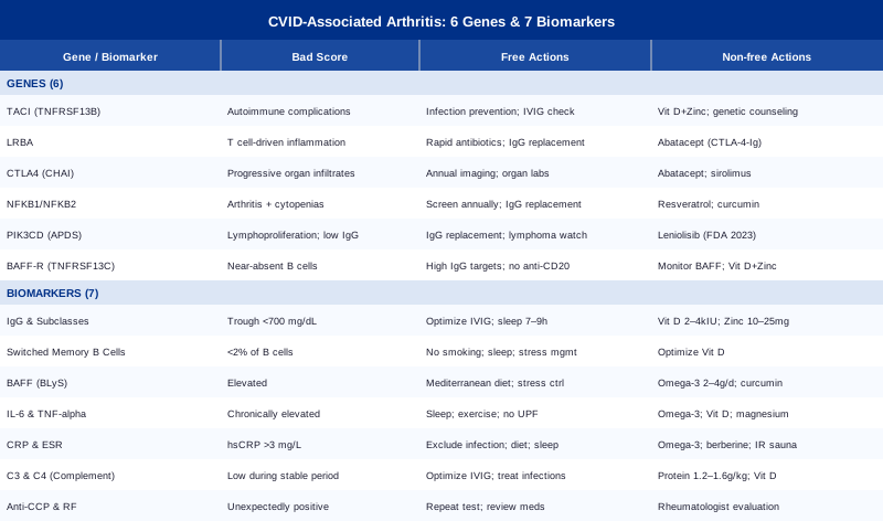
This article examines CVID-associated arthritis from the angle that is most actionable: measurable biomarkers and underlying genetic variants. The 7 biomarkers include serum IgG and subclasses, switched memory B cells, BAFF, IL-6 and TNF-alpha, CRP and ESR, complement proteins C3 and C4, and anti-CCP with rheumatoid factor — each covered with measurement costs, what abnormal values reveal, and specific plans to act on them with and without supplements. The 6 genes — TNFRSF13B (TACI), LRBA, CTLA4, NFKB1/NFKB2, PIK3CD, and BAFF-R — explain the mechanistic differences between CVID patients who develop inflammatory arthritis and those who do not, with targeted strategies for each. A deep dive into Philipp Dettmer's book Immune provides ten non-obvious insights about the immune-inflammation relationship that challenge the "weak immune system" framing most patients encounter. The final section covers five evidence-supported complementary approaches: mindfulness-based stress reduction, the Autoimmune Protocol, tai chi, massage therapy, and microbiome-directed strategies — each with a specific protocol and the human evidence behind it.
7 Biomarkers to Track in CVID-Associated Arthritis
Understanding your biomarker profile in CVID goes well beyond a routine blood count. The immune defect underlying CVID creates a cascade of measurable downstream effects — in immunoglobulin levels, B cell populations, inflammatory signaling, and complement activity. Tracking these signals over time gives you and your care team a far clearer picture of disease activity, treatment adequacy, and arthritis risk than clinical symptoms alone. Here are the seven most clinically useful markers to monitor.
Biomarker 1: Serum IgG and IgG Subclasses
Why it matters: Immunoglobulin G (IgG) is the central diagnostic marker in CVID, and its level directly correlates with infection risk, treatment adequacy, and systemic inflammatory burden. When IgG replacement therapy is suboptimally dosed, low-grade uncontrolled infections or immune dysregulation can sustain articular inflammation. IgG subclasses add precision: IgG2 deficiency is associated with encapsulated bacterial infections and specific B cell maturation defects that may contribute to inflammatory joint complications, while isolated IgG4 elevation can suggest a separate IgG4-related inflammatory process.
What it may reveal: Persistently low IgG despite treatment suggests under-replacement or accelerated catabolism. Isolated subclass deficiencies can explain clinical patterns that total IgG alone misses. In the context of arthritis, suboptimal IgG levels may indicate that immune dysregulation is insufficiently controlled, allowing inflammatory cascades to continue without interruption.
How to measure it: Standard serum immunoglobulin panels are available at any clinical laboratory. Total IgG costs approximately $50–$150. IgG subclass testing (IgG1, IgG2, IgG3, IgG4) adds another $100–$250 and is typically ordered by immunologists. Most CVID patients on IVIG or SCIG therapy should target a trough IgG above 700–800 mg/dL before the next infusion, though individual optimal levels vary and some patients require higher targets to control inflammatory complications. For detailed context, see the StatPearls overview of Common Variable Immunodeficiency.
If the score is low — plan without supplements: Work with your immunologist to review IVIG or SCIG dosing and infusion intervals. Track trough IgG levels at every visit — measured just before the next infusion. Prioritize infection prevention: vaccinations for household contacts, hand hygiene protocols, and avoidance of crowded spaces during peak respiratory illness season. Prioritize sleep of seven to nine hours, as this is when immunoglobulin catabolism slows and immune memory consolidates. Regular moderate-intensity exercise (30 minutes, four to five days per week) has modest evidence for supporting immune function without further stressing a compromised immune system.
If the score is low — plan with supplements or equipment: Vitamin D at 2000–4000 IU daily (targeting serum 25-OH vitamin D of 40–60 ng/mL) supports immune regulatory gene expression — verify baseline before supplementing. Zinc at 10–25 mg elemental daily supports antibody production pathways; avoid higher doses without monitoring, as excess zinc depletes copper over time. Selenium at 100–200 mcg daily provides cofactors for immune enzymes; cycle every three months to prevent accumulation, and do not exceed 400 mcg daily. For home management, subcutaneous immunoglobulin (SCIG) devices allow smaller, more frequent self-administration that maintains more stable IgG troughs than monthly IV infusions — discuss feasibility with your immunologist.
Biomarker 2: Switched Memory B Cells (CD27+IgM−IgD−)
Why it matters: Switched memory B cells are mature B cells that have completed class-switch recombination. In healthy individuals they constitute roughly 4–14% of circulating B cells. In CVID they are often severely reduced or absent, and their absence is now considered one of the most diagnostically reliable markers for CVID — more specific than immunoglobulin levels alone. Crucially, very low switched memory B cells correlate strongly with the severe, complication-prone disease phenotype that includes splenomegaly, granulomatous disease, and inflammatory arthritis.
What it may reveal: When switched memory B cells fall below 2% of total B cells, this places a patient in the highest-risk CVID subgroup for non-infectious complications. The absence of these cells signals a fundamental maturation block that predicts more aggressive inflammatory disease. This biomarker also explains why CVID arthritis differs mechanistically from rheumatoid arthritis: it is not driven by aberrant antibody production but by the compensatory dysregulation of T cells and innate immune pathways that occurs when B cell maturation fails.
How to measure it: Flow cytometry is required to quantify B cell subsets. This is a specialty test, not available in standard blood panels. Costs range from $200–$500 and are best ordered through academic medical centers or specialized immunology laboratories. Proper sample handling is critical — results are affected by delays between blood draw and processing. Request results as both percentage of total lymphocytes and absolute cell counts for the most useful interpretation.
If the score is low — plan without supplements: There is currently no proven dietary or lifestyle intervention that reverses the switched memory B cell deficit in CVID — this is a structural immune defect. What you can do is prevent additional immune suppression: eliminate smoking (which profoundly impairs B cell development), control chronic stress, and optimize sleep consistently. These measures do not fix the primary defect but prevent compounding it. Very low switched memory B cells often justify higher IgG dosing targets — discuss with your immunologist.
If the score is low — plan with supplements or equipment: No supplement directly raises switched memory B cells in CVID. Optimizing vitamin D supports regulatory T cell balance, which may partially compensate for absent B cell regulation. Avoid high-dose biotin supplementation (above 300 mcg daily) in the days before flow cytometry testing — it can interfere with certain assay formats and produce falsely abnormal results. For monitoring frequency, specialist lab testing every six to twelve months is the current standard; home lymphocyte testing is not yet reliable enough for clinical decisions in this context.
Biomarker 3: BAFF (B-Cell Activating Factor)
Why it matters: BAFF (also called BLyS) is a cytokine produced primarily by myeloid and stromal cells that acts as a survival signal for B cells. In CVID, BAFF levels are often paradoxically elevated: the body detects depleted peripheral B cells and upregulates BAFF production as compensation. This chronic BAFF elevation does not correct the B cell maturation defect but creates downstream consequences — including partial activation of autoreactive B cell clones that would normally be eliminated — and these consequences may contribute to inflammatory and autoimmune manifestations including joint disease.
What it may reveal: Elevated BAFF in CVID patients correlates with higher rates of autoimmune complications, granulomatous disease, and interstitial lung disease. In the arthritis context, elevated BAFF may indicate that the inflammatory component has a B cell-activating driver that could respond to BAFF-targeted therapeutic approaches. A BAFF level within the normal range may in contrast suggest a less inflammatory disease course, though this interpretation requires validation in the full clinical context.
How to measure it: BAFF is measured by ELISA on serum. This is not a routine clinical test and requires a specialty laboratory. Costs range from $150–$300. It is most informative when measured as part of a comprehensive immunological workup rather than in isolation, and when measured at baseline during clinical stability rather than during acute illness.
If the score is elevated — plan without supplements: Address all modifiable drivers of immune activation: treat infections promptly and completely, review whether medications are contributing to immune dysregulation, and apply anti-inflammatory lifestyle practices consistently. Chronic sleep deprivation and psychological stress both upregulate pro-inflammatory cytokines that amplify BAFF signaling. A Mediterranean-pattern diet — emphasizing olive oil, fatty fish, legumes, vegetables, and minimal refined foods — has the broadest evidence base for reducing systemic inflammatory burden over the long term.
If the score is elevated — plan with supplements or equipment: Omega-3 fatty acids at 2–4 g combined EPA and DHA daily (from fish oil or algal sources) have evidence for downregulating inflammatory signaling pathways relevant to BAFF. Take with food to reduce GI side effects; reassess after three months with repeat inflammatory markers. Curcumin at 500–1000 mg of a phospholipid-complexed or piperine-enhanced formulation twice daily has preclinical and early clinical evidence for inhibiting inflammatory cytokine pathways — use with caution if taking anticoagulants. In clinical settings, belimumab (a biologic targeting BAFF/BLyS) is being studied in certain CVID complications, though currently primarily in research settings — discuss with a specialist if inflammatory complications persist despite standard management.
Biomarker 4: IL-6 and TNF-alpha
Why it matters: Interleukin-6 (IL-6) and tumor necrosis factor-alpha (TNF-alpha) are two of the most important pro-inflammatory cytokines driving joint inflammation in CVID-related arthritis. TNF-alpha directly promotes synovitis and cartilage degradation; IL-6 drives acute phase responses, bone resorption, fatigue, and the fever that often accompanies joint flares. These cytokines are now targets for biologic therapies in various inflammatory arthritis conditions, and measuring them helps determine whether the inflammatory burden is high enough to warrant such approaches in the CVID context.
What it may reveal: Chronically elevated IL-6 suggests ongoing, inadequately controlled inflammation likely causing progressive joint damage even when symptoms appear controlled. Elevated TNF-alpha — particularly alongside elevated IL-1beta — points to innate immune pathway hyperactivation, which is mechanistically relevant in CVID since innate immunity is often compensatorily overactivated when adaptive antibody-mediated immunity fails.
How to measure it: Serum cytokine levels are measured by ELISA or multiplex bead-based assays. Individual cytokine tests cost $100–$200 each; multiplex panels measuring ten or more cytokines simultaneously cost $300–$600 and provide more contextual information. Timing matters significantly — cytokines fluctuate with infections, sleep quality, and recent exercise. Measure during a clinically stable period, not during acute illness or within the first week of starting a new medication.
If the score is elevated — plan without supplements: Remove inflammatory inputs systematically: eliminate ultra-processed foods and refined sugars, which acutely elevate IL-6. Address sleep quality rigorously — even a single night of poor sleep measurably raises TNF-alpha in healthy individuals. Regular moderate-intensity exercise has a net anti-inflammatory effect through transient IL-6 release from exercising muscles, which paradoxically triggers downstream anti-inflammatory responses. Sustained psychological stress is not optional to manage here — cortisol dysregulation from chronic stress amplifies TNF-alpha production through glucocorticoid resistance in immune cells.
If the score is elevated — plan with supplements or equipment: Omega-3 fatty acids at 2–4 g EPA and DHA daily have the strongest supplement evidence for reducing both IL-6 and TNF-alpha — multiple randomized controlled trials in inflammatory arthritis show meaningful reductions. Vitamin D optimization (targeting 40–60 ng/mL) has immunomodulatory effects on cytokine production through vitamin D response elements in immune cell genes. Magnesium glycinate or malate at 200–400 mg daily (taken at night) may reduce IL-6 in individuals with magnesium deficiency, which is common in people using proton pump inhibitors frequently prescribed in CVID with enteropathy. In clinical settings, anti-TNF biologics or anti-IL-6R agents are used for refractory inflammatory arthritis — discuss eligibility and safety with your rheumatologist, noting that CVID immunology adds complexity to biologic decisions.
Biomarker 5: CRP and ESR
Why it matters: C-reactive protein (CRP) and erythrocyte sedimentation rate (ESR) are the workhorses of inflammation monitoring — inexpensive, widely available, and useful for tracking change over time. High-sensitivity CRP (hsCRP) is the preferred version for low-grade systemic inflammation and is the marker most consistently cited by clinicians like Peter Attia for systemic inflammatory burden assessment. In CVID-associated arthritis, these markers help gauge whether anti-inflammatory interventions are working and whether arthritis is in active flare or relative remission.
What it may reveal: Persistently elevated hsCRP above 3 mg/L suggests ongoing systemic inflammation. In CVID, this may reflect an active infection requiring immediate attention, or the non-infectious inflammatory component of the disease. ESR is less specific but can be elevated in settings of elevated immunoglobulins — interpret ESR in the context of the full immunological picture rather than in isolation. Serial measurements over three to six months are far more informative than any single value.
How to measure it: Both tests are available at any clinical laboratory. hsCRP costs $20–$50 and is more sensitive than standard CRP for low-grade inflammation. ESR costs $15–$30. Test ideally in a fasted state in the morning for maximal reproducibility. For monitoring arthritis activity over time, measure at the same time of day and under similar conditions each time.
If the score is elevated — plan without supplements: First exclude active infection — in CVID, elevated CRP may indicate an unrecognized or under-treated bacterial or fungal infection that requires direct treatment before any anti-inflammatory strategy makes sense. Once infection is excluded, focus on anti-inflammatory diet, sleep optimization (seven to nine hours with consistent timing), stress management, and regular moderate exercise. Time-restricted eating within a 10–12 hour window has emerging evidence for reducing hsCRP through metabolic and circadian pathways.
If the score is elevated — plan with supplements or equipment: Beyond omega-3s and vitamin D already described, berberine at 500 mg twice daily with meals reduces inflammatory markers through AMPK pathway activation — but check for interactions with antibiotics (common in CVID) and any medications metabolized by CYP3A4. Infrared sauna sessions of 15–20 minutes at 140–160°F, three to four times per week, have evidence from small RCTs for reducing hsCRP through heat shock protein activation and improved vascular endothelial function; begin with lower temperatures and shorter sessions, and avoid sessions when acutely unwell.
Biomarker 6: Complement Proteins C3 and C4
Why it matters: The complement system is an ancient arm of innate immunity bridging pathogen defense and inflammatory signaling. In CVID, C3 and C4 can be low for several reasons: consumption during chronic infections, secondary deficiency from immune complex formation, or a co-occurring primary partial complement deficiency. In CVID-associated arthritis, complement activation contributes to synovial inflammation through deposition of complement fragments in joint tissues. Monitoring C3 and C4 helps distinguish inflammatory mechanisms and can reveal whether certain infections — particularly encapsulated bacteria — are driving joint disease.
What it may reveal: Low C3 during stable periods may indicate ongoing immune complex formation or a co-existing complement deficiency. Isolated low C4 with normal C3 can suggest a C4A null allele, associated with systemic lupus erythematosus-like manifestations. Persistently low complement alongside elevated inflammatory markers points toward active immune complex deposition — a pattern that may require more aggressive management than standard CVID protocols.
How to measure it: C3 and C4 are measured by nephelometry on standard blood samples at $25–$50 each, often included in comprehensive rheumatology or autoimmune panels. For a fuller complement assessment, CH50 (total hemolytic complement activity) can be added at similar cost. Measure during clinical stability to establish a true baseline — complement is consumed acutely during infection, making acute-illness measurements difficult to interpret.
If the score is low — plan without supplements: Work with your immunologist to determine whether low complement reflects consumption from active infection or primary production deficiency. Optimizing IVIG therapy is the primary intervention — adequate IgG levels reduce the infectious burden that consumes complement. Treat infections promptly and fully; maintain mucosal barriers through nasal saline irrigation; ensure prophylactic antibiotic coverage is appropriate for your infection history.
If the score is low — plan with supplements or equipment: No supplement directly raises C3 or C4 levels — these are liver-synthesized proteins. Adequate dietary protein (1.2–1.6 g/kg/day) supports complement protein synthesis. Vitamin D deficiency impairs expression of complement regulatory proteins; correcting to optimal levels is a reasonable supporting measure. For patients with a confirmed primary complement deficiency co-existing with CVID, specialist evaluation before any immunosuppressive therapy is essential, as complement deficiency significantly alters infection risk calculations.
Biomarker 7: Anti-CCP Antibodies and Rheumatoid Factor
Why it matters: This biomarker occupies a unique position in CVID arthritis management: anti-cyclic citrullinated peptide antibodies (anti-CCP) and rheumatoid factor (RF) are typically negative in CVID-associated arthritis, and that negativity is clinically meaningful. Because CVID patients cannot produce normal antibodies, they usually cannot generate the autoantibodies that define seropositive rheumatoid arthritis. Testing these markers serves two functions: confirming the seronegative nature of the arthritis (supporting a CVID-driven mechanism over a separate RA process) and catching the rare possibility of a coincident seropositive condition emerging over time.
What it may reveal: A positive anti-CCP in a CVID patient is genuinely unusual and warrants careful interpretation — it may suggest an overlap with RA, a medication effect, or a testing artifact. Persistently negative results alongside symmetrical inflammatory joint disease reinforce the CVID-arthritis diagnosis and direct management toward strategies that differ from standard RA protocols: IVIG optimization, targeted treatment for Mycoplasma hominis and Ureaplasma urealyticum (which cause a characteristic infectious oligoarticular arthritis specifically in hypogammaglobulinemic patients), and immunomodulatory approaches rather than DMARDs as the first-line strategy.
How to measure it: Anti-CCP (second-generation, anti-CCP2, preferred for specificity) costs $50–$150. RF costs $20–$50. Both are available at any standard clinical laboratory. Test at diagnosis and recheck annually or when the arthritis pattern changes, to detect any diagnostic shift over time.
If anti-CCP or RF are unexpectedly positive — plan: Request confirmatory repeat testing. Discuss with your rheumatologist whether a seropositive inflammatory arthritis is developing independently of CVID. Review current medications — certain biologics can induce autoantibodies. If positivity is confirmed, treatment strategy may need to incorporate RA-specific approaches alongside continued CVID management.
If negative (the expected finding) — plan without supplements: Seronegative CVID arthritis responds best to addressing root immune dysregulation: optimize IVIG dosing, screen for and treat mycoplasma and ureaplasma organisms with appropriate antibiotics (doxycycline for six to twelve weeks is often used), and reduce overall inflammatory burden through lifestyle. Joint aspiration and culture should be considered in any patient with monoarticular or oligoarticular flares to exclude these organisms.
If negative — plan with supplements or equipment: Anti-inflammatory strategies targeting innate immune pathways are most relevant. Boswellic acids (AKBA-enriched Boswellia serrata extract at 100–250 mg AKBA daily) have clinical evidence for reducing synovial inflammation in arthritis, with reasonable safety profiles — use in eight to twelve week cycles with four-week breaks. Low-level laser therapy devices for joint pain (650–904 nm wavelength, 1–4 J/cm², three sessions per week) have meta-analytic support for pain reduction across various arthritis types; evidence is moderate quality but the approach is accessible and low-risk when used on stable, non-infected joints.
With a clear picture of what these biomarkers can tell you, it is worth understanding the genetic blueprint that explains why CVID-associated arthritis develops in the first place — and why some patients face far more inflammatory complications than others.
The Genetic Architecture Behind CVID and Joint Inflammation
Only 10–15% of CVID cases have a clearly identified single-gene cause, but as next-generation sequencing becomes more accessible and less expensive, that figure is rising steadily. For patients with CVID-associated arthritis, identifying the underlying genetic variant can genuinely change treatment decisions — from medication selection to monitoring intensity and family screening. Here are the six most clinically relevant genetic players.
Gene 1: TNFRSF13B (TACI)
TNFRSF13B encodes the TACI receptor (Transmembrane Activator and CAML Interactor), which receives survival and differentiation signals from both BAFF and APRIL cytokines. Heterozygous mutations — particularly C104R and A181E — are found in approximately 8–10% of CVID patients, making TACI the most commonly identified genetic cause. These mutations impair B cell class switching and IgA production and are associated with higher rates of autoimmune manifestations including arthritis. Importantly, the same mutations occur in roughly 1% of healthy individuals, confirming they act as risk factors requiring additional hits rather than as complete disease determinants.
If the gene variant is present — plan without supplements: Impaired class switching means reduced diversity in the antibodies your immune system can generate, leaving you more vulnerable to certain bacterial organisms. Prioritize infection prevention systematically — vaccinations for household contacts, prompt antibiotic treatment at the first sign of bacterial infection, and prophylactic antibiotics as directed by your immunologist. For arthritis specifically, seronegative strategies described in biomarker 7 apply directly. Regular monitoring every three to six months for emerging complications (enteropathy, splenomegaly, lymphoproliferative disease) is warranted given the higher complication risk profile associated with TACI variants.
If the gene variant is present — plan with supplements or equipment: IVIG or SCIG replacement remains the cornerstone intervention, and BAFF pathway monitoring (biomarker 3) is particularly relevant given TACI's central role in BAFF and APRIL signaling. Optimizing vitamin D and zinc as described under biomarker 1 supports residual B cell function. There is currently no targeted small molecule therapy that corrects TACI dysfunction directly — management remains supportive and replacement-based. Genetic counseling is reasonable given the autosomal pattern of inheritance and variable penetrance.
Gene 2: LRBA (Lipopolysaccharide-Responsive Beige-Like Anchor Protein)
LRBA deficiency is one of the most clinically important monogenic causes of CVID-like disease because it is specifically treatable with a targeted biologic. LRBA protein is required for recycling CTLA-4 from intracellular vesicles back to the cell surface. When LRBA is absent, CTLA-4 degrades rather than recycling — resulting in chronically reduced CTLA-4 expression on regulatory T cells. Without adequate CTLA-4 on T regulatory cells, these cells cannot properly suppress inflammatory effector T cells, producing a syndrome of simultaneous immune deficiency and immune dysregulation: recurrent infections and autoimmune complications occurring together. Inflammatory bowel disease, autoimmune cytopenias, granulomatous disease, and inflammatory arthritis are all documented manifestations. LRBA deficiency should be suspected in any CVID patient presenting with multiple autoimmune features alongside hypogammaglobulinemia.
If the gene variant is present — plan without supplements: Conventional infection-focused management is insufficient here because LRBA deficiency generates T cell-driven inflammatory disease that progresses independently of infection control. Managing the T cell hyperactivation component is essential: this typically requires specific immunosuppressive agents (corticosteroids, sirolimus, or mycophenolate depending on the specialist's approach) alongside IgG replacement. For inflammatory arthritis specifically, flares often coincide with infections that trigger immune activation — rapid antibiotic treatment at the first sign of bacterial infection is therefore both infection management and arthritis prevention.
If the gene variant is present — plan with supplements or equipment: Abatacept (a CTLA-4-Ig fusion protein) has demonstrated substantial efficacy in LRBA deficiency — it compensates for the deficient CTLA-4 signaling that LRBA loss creates. Case series and registry data show dramatic reductions in autoimmune manifestations including arthritis, enteropathy, and cytopenias. This is a prescription biologic requiring specialist prescription and regular monitoring. If genetic testing identifies LRBA deficiency in a patient with multiple autoimmune features, this shifts treatment from IgG replacement alone to CTLA-4 pathway supplementation — a clinically meaningful distinction worth pursuing with genetic testing if your presentation fits.
Gene 3: CTLA4 Haploinsufficiency
CTLA4 haploinsufficiency results from heterozygous loss-of-function mutations in the gene encoding CTLA-4 itself. The resulting syndrome — CTLA-4 haploinsufficiency with autoimmune infiltration (CHAI) — is phenotypically very similar to LRBA deficiency because both converge on the same endpoint: reduced CTLA-4 function on T regulatory cells and loss of immune self-regulation. Patients present with the same combination of immune deficiency (low B cells, hypogammaglobulinemia) and immune dysregulation (autoimmune cytopenias, inflammatory bowel disease, organ infiltrates, arthritis). This gene is now increasingly identified through next-generation sequencing panels for primary immunodeficiency.
If the gene variant is present — plan without supplements: Monitoring is more intensive than for typical CVID — inflammatory organ infiltration in CTLA4 haploinsufficiency can be progressive and clinically silent in early stages. Annual imaging and regular laboratory assessment of organ function are warranted. Arthritis in this context reflects active immune cell infiltration of the synovium and does not respond predictably to standard anti-inflammatory approaches without addressing the underlying T regulatory cell dysfunction.
If the gene variant is present — plan with supplements or equipment: Abatacept has emerging evidence for efficacy in CTLA4 haploinsufficiency, paralleling its demonstrated benefit in LRBA deficiency. Sirolimus (mTOR inhibitor) has also been used to modulate immune dysregulation in this condition. Genetic counseling is essential — CTLA4 haploinsufficiency is autosomal dominant, with a 50% transmission probability to each child. Anti-inflammatory lifestyle measures including omega-3s, anti-inflammatory diet, and sleep optimization support medical management but cannot substitute for it in this genetically driven condition.
Gene 4: NFKB1 and NFKB2
The NF-kB signaling pathway is a master regulator of both immune cell development and inflammatory gene expression. Heterozygous loss-of-function mutations in NFKB1 account for approximately 5–8% of monogenic CVID cases and are recognized as the most common monogenic cause in European population studies. Mutations in NFKB2 are less common but clinically distinct — often presenting with pituitary insufficiency alongside immune deficiency. The NF-kB pathway drives transcription of inflammatory cytokines including IL-6, TNF-alpha, and IL-1beta, the same cytokines driving arthritis. This creates a notable paradox: the same pathway defect that impairs B cell development for antibody production also predisposes to dysregulated innate inflammatory responses that damage joints.
If the gene variant is present — plan without supplements: NFKB1 CVID tends to have a milder overall clinical course in some series, but inflammatory complication rates are higher than typical CVID. Inflammatory arthritis and autoimmune complications (thyroid disease, cytopenias) should be actively screened for at annual follow-up visits rather than awaiting symptoms. Standard IgG replacement is the treatment foundation. For NFKB2 patients, pituitary function testing (thyroid, adrenal, and gonadal axes) is warranted even in the absence of overt symptoms, given the documented association with hypopituitarism.
If the gene variant is present — plan with supplements or equipment: The NF-kB pathway can be modestly modulated by several evidence-supported supplements. Resveratrol at 150–500 mg of a high-absorption formulation (micronized or combined with quercetin) daily inhibits NF-kB through SIRT1 activation in multiple preclinical and early clinical studies. Curcumin at 500–1000 mg of a piperine-enhanced formulation twice daily directly inhibits NF-kB nuclear translocation. Cycle resveratrol eight weeks on and four weeks off, and monitor liver enzymes if using high daily doses of curcumin long-term. Neither supplement replaces medical management but may modestly reduce the inflammatory component of disease in a mechanistically coherent way.
Gene 5: PIK3CD (PI3K-delta)
PIK3CD encodes the catalytic subunit of PI3-kinase delta, a signaling enzyme critical for B and T cell receptor signal transduction. Gain-of-function mutations cause Activated PI3K Delta Syndrome (APDS), a primary immunodeficiency that overlaps substantially with CVID: recurrent sinopulmonary infections, low IgG, lymphoproliferation, and a higher rate of inflammatory complications. Loss-of-function variants in PIK3CD alternatively present with a CVID-like phenotype by impairing the B cell activation signals required for antibody production. Distinguishing these requires genetic sequencing.
If the gain-of-function variant is present (APDS) — plan with or without supplements: Leniolisib (Joenja), a selective PI3K-delta inhibitor, received FDA approval in 2023 for APDS in patients aged 12 and older. It directly corrects the overactive PI3K-delta signaling, improving lymphocyte function and potentially reducing the inflammatory complications including joint disease that result from the dysregulated T cell activation in APDS. Discuss eligibility with a physician experienced in primary immunodeficiency — this is now a targeted, disease-modifying therapy for this specific genetic variant.
If the loss-of-function variant is present — plan: Management parallels general CVID with IgG replacement. Because PI3K-delta is central to B cell activation, B cell counts and function may be more severely reduced, potentially requiring higher IgG dosing targets. Monitor for lymphoma risk, which is elevated particularly with gain-of-function variants and requires specialist-guided long-term surveillance.
Gene 6: TNFRSF13C (BAFF-R)
TNFRSF13C encodes the BAFF receptor (BAFF-R), which is the primary survival signal receptor for transitional and mature B cells. Homozygous loss-of-function mutations cause near-complete absence of mature B cells — the most severe end of the CVID spectrum, with virtually no peripheral B cells and profound hypogammaglobulinemia. Heterozygous mutations have more variable clinical penetrance. BAFF-R deficiency creates a direct biological bridge between biomarker 3 (BAFF) and its genetic cause: elevated circulating BAFF in BAFF-R deficiency reflects the accumulation of BAFF that has no receptor to bind, explaining the seeming paradox of high BAFF with absent B cell responses.
If the gene variant is present — plan without supplements: Given very low B cell numbers in homozygous BAFF-R deficiency, infection prevention is critical and IgG replacement dosing targets should be on the higher end. Avoid medications that further deplete B cells — anti-CD20 agents (rituximab and biosimilars) are contraindicated without very careful specialist oversight. Consider annual cardiovascular screening, as emerging data suggests cardiac inflammatory infiltrates in some patients with severe B cell lymphopenia.
If the gene variant is present — plan with supplements or equipment: Higher IgG dosing targets are typically needed and can be guided by monitoring serum BAFF levels — a falling BAFF may indicate that replacement therapy is adequately reducing the compensatory BAFF upregulation that occurs in response to B cell depletion. The same foundational supplements (vitamin D, zinc, omega-3s) apply as for general CVID. Gene therapy for BAFF-R deficiency is in early-stage research and not yet clinically available; clinical trial enrollment through academic immunodeficiency centers may be worth exploring for severely affected patients.
Understanding these genetic mechanisms helps reframe the entire immune system — not just as a defense system, but as a finely tuned regulatory network. The following section translates this into a broader framework that can shift how you think about daily decisions that affect inflammatory burden.
Ten Things the Book "Immune" Reveals That Most Patients Are Never Told
Immune: A Journey into the Mysterious System That Keeps You Alive (2021, Avery) by Philipp Dettmer draws from hundreds of peer-reviewed studies to challenge the simplistic "strong vs. weak immune system" framing that dominates popular health culture. For anyone managing CVID with inflammatory complications, the book's core thesis is genuinely useful: the immune system's greatest threat is not only failure to defend, but failure to regulate. Here are the ten most impactful insights.
1. The Immune System Is an Ecosystem, Not a Single Defense Force
Dettmer explains that the immune system consists of dozens of distinct cell types with specialized roles operating in layers. CVID is often described loosely as a "weak immune system," but this misrepresents the biology. CVID specifically impairs the adaptive humoral layer — antibody production — while leaving, and often hyperactivating, the innate immune layer. This mismatch between a deficient adaptive system and an overactive innate system is precisely the biology behind CVID-arthritis: innate immune cells amplify joint inflammation without the regulatory restraint that a functioning adaptive system would provide.
2. Innate Immunity Is Fast, Powerful, and Destructive by Design
The innate immune system responds within minutes using broad pattern recognition rather than pathogen-specific targeting. In CVID, when specific antibodies are absent, the innate system compensates — but its responses are inflammatory by nature. Dettmer describes how this produces bystander tissue damage in locations including joint synovium, even in the absence of a specific pathogen in the joint. This is the biological basis for the "sterile" inflammatory arthritis that can occur in CVID without any identifiable infectious trigger and without the autoantibodies typical of RA.
3. Inflammation Is a Deliberate Tool — The Problem Is When It Stays On
One of the book's most reframing insights: inflammation is not a malfunction — it is a programmed, purposeful response. The problem in CVID-arthritis is that the signals responsible for switching inflammation off are missing or impaired. CTLA-4 on regulatory T cells is one of the primary "off" signals for T cell activation. Without it (as in LRBA deficiency and CTLA4 haploinsufficiency), inflammation persists far beyond its functional usefulness, damaging tissues it was originally intended to protect.
4. B Cells Do Much More Than Produce Antibodies
Several chapters in the book focus on B cell biology beyond antibody production. B cells present antigens to T cells, provide co-stimulatory signals that regulate T cell activation, and secrete cytokines that calibrate both pro- and anti-inflammatory responses. In CVID, the loss of mature switched memory B cells therefore impairs not just antibody production but the entire B cell-mediated immune regulatory network. This explains why very low switched memory B cells predict inflammatory complications — the regulatory function is lost alongside the effector function.
5. Complement Connects the Ancient and the Modern Immune System
The complement cascade is elegantly described as the bridge between innate and adaptive immunity. When adaptive immunity fails to generate antibodies that direct complement precisely toward specific targets, complement activation becomes non-specific and can deposit fragments in uninvited tissues — including joint synovium. This mechanistic connection makes monitoring C3 and C4 clinically relevant rather than just academically interesting for CVID patients with arthritis.
6. Sleep Is the Immune System's Maintenance Window
During sleep, coordinated immune maintenance occurs: inflammatory cytokines produced during the day are metabolized, T regulatory cells regenerate, and immune memory is consolidated. For CVID patients with arthritis, chronic sleep disruption creates a compounding cycle — inadequate immune regulation perpetuates inflammation, which disturbs sleep, which further undermines regulation. Even one night of poor sleep measurably elevates IL-6 and TNF-alpha in healthy individuals; in CVID, where these cytokines are already elevated, poor sleep amplifies an already compromised baseline.
7. The Gut Microbiome Is Where Most Immune Cells Live
Approximately 70% of immune cells reside in gut-associated lymphoid tissue. The microbiome actively trains the immune system, calibrating inflammatory thresholds and supporting regulatory T cell populations. CVID patients frequently have profound gut dysbiosis due to recurrent antibiotic use and the absence of secretory IgA — which normally shapes and stabilizes microbiome composition. This dysbiosis directly activates intestinal innate immune cells, contributing to elevated systemic inflammatory markers and potentially amplifying joint inflammation.
8. Chronic Stress Creates Both Immunosuppression and Inflammation Simultaneously
This apparent paradox is clearly explained: acute stress suppresses some immune responses (useful for short-term threat responses), but chronic stress creates glucocorticoid resistance in immune cells — they stop responding to cortisol's anti-inflammatory signals while continuing to receive cortisol's B cell-suppressing effects. For CVID patients already dealing with B cell dysfunction, chronic stress compounds both problems: further suppressing antibody production while removing the cortisol-mediated check on inflammatory cytokine production.
9. Nutrition Provides Cofactors, Not Cures
One of the book's most grounded positions: no single nutrient "boosts" immunity in the way popular culture suggests. What nutrition provides are cofactors — zinc for antibody folding and T cell function, selenium for glutathione peroxidase, vitamin D for gene expression in immune regulatory cells, vitamin C for neutrophil bactericidal capacity. Deficiencies create genuine impairments; correction restores function. The supplement recommendations throughout this article are grounded in exactly this principle: correct documented deficiencies, do not oversupplement in the hope of amplifying an already dysregulated system.
10. Regulatory Failure Is as Dangerous as Effector Failure
The book's final major insight: the most dangerous immune failures are not only failures to attack, but failures to stop attacking. Autoimmune disease, cytokine storm, and inflammatory organ damage are all failures of regulation. In CVID, the cascade from reduced switched memory B cells to impaired T regulatory function (through BAFF, LRBA, and CTLA4 pathways) represents exactly this regulatory failure. The practical implication: prioritizing regulatory strategies — sleep, stress reduction, anti-inflammatory diet, microbiome support — is primary care for inflammatory complications in CVID, not an optional afterthought.
The evidence for complementary and lifestyle-based approaches in CVID specifically is limited by the rarity of the condition, but the mechanistic biology connects sufficiently to well-studied modalities that the following approaches deserve consideration.
Complementary Approaches with Meaningful Clinical Evidence
Mindfulness-Based Stress Reduction (MBSR)
Mindfulness-based stress reduction is an eight-week structured program combining seated meditation, body scanning, and gentle movement, developed by Jon Kabat-Zinn at the University of Massachusetts Medical School. Its relevance to CVID-related arthritis lies directly in the glucocorticoid resistance mechanism described above: MBSR has been shown in multiple studies to restore cortisol sensitivity in immune cells and reduce the paradox where chronic stress simultaneously amplifies both immune deficiency and inflammation. For inflammatory arthritis specifically, MBSR reduces perceived pain severity, morning stiffness, and fatigue, and several trials show measurable reductions in pro-inflammatory cytokines including IL-6.
A randomized controlled trial in patients with rheumatoid arthritis demonstrated that MBSR participants showed significant improvements in disease activity scores and psychological well-being compared to control groups at six-month follow-up. A subsequent meta-analysis of mindfulness interventions across inflammatory conditions found consistent moderate-effect improvements in pain, fatigue, and quality of life. Direct CVID data are absent, but the stress-immune pathway mechanism applies regardless of the specific immune condition.
To apply MBSR for CVID-arthritis, begin with a formal eight-week course — available at low or no cost through online platforms such as Palouse Mindfulness. Commit to 30–45 minutes of daily practice during the course, with the body scan (practiced in the evening) being the most relevant technique for pain management. After completing the formal course, maintain 15–20 minutes of daily practice — consistency matters far more than session length. Apps can supplement but should not replace formal structured learning initially.
The Autoimmune Protocol (AIP)
The Autoimmune Protocol developed by Dr. Sarah Ballantyne, PhD, is a dietary and lifestyle framework designed to reduce immune-mediated inflammation through gut microbiome restoration, high-nutrient density eating, and systematic elimination of dietary antigens that may trigger or maintain immune reactivity in susceptible individuals. While CVID is a primary immunodeficiency rather than a classic autoimmune disease, its inflammatory complications — including arthritis — share the same gut-immune pathway dysregulation that the AIP is designed to address. The microbiome component is especially relevant given the documented microbiome disruption in CVID from recurrent antibiotic use and secretory IgA deficiency.
A pilot study by Konijeti et al. demonstrated significant endoscopic and symptomatic improvement in Crohn's disease patients following the AIP dietary protocol, with measurable reductions in inflammatory biomarkers. The broader AIP framework extends well beyond food: it includes sleep optimization, circadian rhythm alignment, stress management, and light exposure regulation. The dietary elimination phase removes grains, legumes, dairy, eggs, nightshades, nuts, seeds, and processed foods while emphasizing organ meats, wild-caught fish, fermented foods, and diverse vegetables at every meal.
For CVID-arthritis, implement AIP as a structured 30-day elimination phase followed by systematic reintroduction to identify personal triggers. Given the immunological complexity of CVID, introduce changes gradually and monitor for any increase in infectious susceptibility during the dietary transition. Avoid starting the AIP during active infections or immediately after beginning new medications. Working with a registered dietitian experienced in both AIP and primary immunodeficiency is strongly recommended to ensure nutritional completeness, particularly for fat-soluble vitamins and minerals that can decrease during aggressive elimination phases.
Tai Chi
Tai chi is a mind-body practice combining slow, deliberate movement sequences with controlled breathing and meditative focus. Its relevance to CVID-arthritis is twofold: it improves joint range of motion, strength, and pain through gentle mechanical loading; and it reduces inflammatory cytokines through the same stress-regulation pathway activated by MBSR. Unlike vigorous exercise that can transiently spike inflammatory markers — which matters more in an already dysregulated immune system — tai chi's gentle intensity has a net anti-inflammatory profile appropriate for CVID patients.
A meta-analysis analyzing 21 randomized controlled trials of tai chi in rheumatoid and osteoarthritis found consistent, statistically significant improvements in pain scores, physical function, and disease activity measures. Several of the included trials used Yang-style 24-form tai chi at 45–60 minutes per session, two to five times per week, over eight to twelve weeks. A 2020 systematic review in Rheumatology further confirmed benefits for inflammatory arthritis, with some studies showing reduced ESR and CRP after twelve weeks of regular practice.
For CVID patients, begin with beginner-level instruction three times per week for 30 minutes, either in-person or through structured video guidance (the Yang 24-form is the most widely studied and taught). Practice on stable, non-slip surfaces; wear supportive footwear to protect joints and reduce fall risk. Avoid outdoor practice during respiratory infection season, and adapt postures to current joint range of motion rather than forcing positions. Results typically emerge after eight to twelve weeks of consistent practice, with continued improvement over six to twelve months.
Massage Therapy
Massage therapy for inflammatory arthritis works through several complementary mechanisms: reducing muscular tension and guarding around affected joints, improving local circulation and lymphatic drainage, activating parasympathetic nervous system responses that suppress pro-inflammatory signaling, and providing pain relief through gate control mechanisms in the peripheral nervous system. For CVID-arthritis specifically, massage addresses the symptomatic joint pain and stiffness independently of the underlying immune mechanism, providing functional benefit while other interventions address root causes.
A systematic review in the Journal of General Internal Medicine found moderate evidence that therapeutic massage reduces pain and improves function across musculoskeletal conditions including inflammatory arthritis. Swedish massage and myofascial release techniques showed the most consistent benefits in included studies. A randomized trial by Field et al. found that twice-weekly moderate-pressure massage over four weeks significantly reduced pain and morning stiffness in rheumatoid arthritis patients compared to progressive relaxation controls.
For CVID patients, monthly professional massage combined with daily self-massage of accessible joints using warm oil offers a practical protocol. Communicate clearly with your therapist about which joints are currently inflamed — avoid deep tissue pressure directly over acutely inflamed joints, and request moderate pressure with active symptom feedback throughout. If sessions produce systemic flushing, unusual fatigue, or feel immunologically activating, reduce frequency and pressure. Foam rolling for larger muscle groups and warm paraffin wax baths for hands and feet provide accessible daily options between professional sessions.
Microbiome-Directed Therapies
Microbiome-directed therapy encompasses dietary strategies (fermented foods, diverse prebiotic fibers), targeted probiotic supplementation, and in research settings, fecal microbiota transplantation. In CVID, the gut microbiome is consistently disrupted — reduced species diversity, depleted short-chain fatty acid-producing bacteria, and overgrowth of inflammatory taxa — largely because secretory IgA, which normally shapes and stabilizes microbiome composition, is absent or severely reduced in CVID. This dysbiosis directly activates intestinal innate immune cells that signal systemically to amplify inflammatory responses, potentially contributing to arthritis flares through the gut-joint immune axis.
A study published in JCI Insight documented significant microbiome alterations in CVID patients compared to immunocompetent controls and found correlations between specific taxa depletions and inflammatory complication rates. While fecal microbiota transplantation is still investigational for primary immunodeficiency, dietary prebiotic strategies have strong safety profiles and biologically coherent mechanisms. Clinical trials of microbiome restoration in CVID are ongoing, reflecting the growing recognition of the gut-immune connection in this condition.
The most practical implementation: consume 30 or more distinct plant species per week (diversity drives microbiome diversity directly), include two to three daily servings of fermented foods (unsweetened kefir, kimchi, or sauerkraut if tolerated), and consider supplementation with well-characterized strains such as Lactobacillus rhamnosus GG and Bifidobacterium longum at 5–20 billion CFU daily with food. In CVID patients, these commensal-species probiotics are generally considered safe — but discuss with your immunologist before starting, particularly if you have a central venous line, are severely immunosuppressed, or have active GI disease. Prebiotic fiber including inulin, fructooligosaccharides (FOS), and resistant starch feeds the SCFA-producing bacteria that regulate systemic inflammation — begin at low doses to minimize GI discomfort and build gradually over two to three weeks.
Conclusion
Common variable immunodeficiency with arthritis sits at the intersection of immune deficiency and immune dysregulation, and managing it effectively requires understanding both. The seven biomarkers described here provide a practical monitoring framework: IgG levels and subclasses track treatment adequacy, switched memory B cells reveal disease severity and complication risk, BAFF and cytokine levels reflect the active inflammatory state, and CRP, complement proteins, and seronegative arthritis markers together guide therapeutic decisions and diagnosis. The six genetic variants — TACI, LRBA, CTLA4, NFKB1/NFKB2, PIK3CD, and BAFF-R — explain the mechanisms that distinguish patients with aggressive inflammatory complications from those with less inflammatory disease courses, and for several of these variants, targeted therapies now exist that can change outcomes significantly.
The strategies in this article are tools to help you participate more meaningfully in your own care — not alternatives to it. Review your existing labs against the biomarker framework, identify what you have not yet measured, and bring specific questions to your immunologist and rheumatologist. If multiple inflammatory complications are present alongside CVID, advocate for genetic testing — it may reveal a variant that changes your entire treatment approach. For the lifestyle and complementary strategies, start with what is most accessible: sleep quality, omega-3 supplementation, and consistent stress management. Each carries genuine mechanistic rationale and a low risk profile. Small, well-targeted steps are where the real progress accumulates.
Respiratory Digestive Autoimmune
Musculoskeletal: Joint Conditions
Autoimmune: Inflammatory Conditions