This article contains AI generated content.
· UpdatedCongenital Contractural Arachnodactyly - 5 Genes And 6 Biomarkers To Track
Introduction
Living with congenital contractural arachnodactyly — or CCA, sometimes called Beals syndrome — means navigating a condition that most physicians have never seen in clinical practice. The joint contractures, the elongated limbs, the curved spine, the unusually shaped ears: each of these features has a precise biological origin, rooted in a single gene and an entire signaling cascade that affects how your body builds and maintains its connective tissue framework. If you have been told simply to "do physical therapy and come back in six months," that advice is not wrong — but it misses most of the picture.
Generic guidance for rare connective tissue disorders is almost always borrowed from more common conditions. What CCA actually demands is an understanding of why the connective tissue behaves the way it does, which cells are most affected, and which biological markers can tell you whether the underlying processes are accelerating or stabilizing over time. That kind of precision changes what you track, what you supplement, and what questions you bring to your specialist.
This article takes a deeper approach, informed by current genetics and biomarker research. It does not promise a cure — CCA is a genetic condition and the mutation in FBN2 does not disappear. But understanding which genes are at work, which downstream pathways they disrupt, and which measurable signals reflect that disruption gives you real tools for smarter monitoring and better daily decisions.
Two main directions are covered here. The first maps the five most relevant genes, including the primary causal gene and the pathway genes that determine how severe or manageable the condition becomes — and for each one, it outlines what you can do, with or without supplementation. The second direction identifies the six most practical biomarkers to track over time, with measurement methods, cost ranges, and specific intervention plans. Together, they offer a more complete map than most people with CCA have ever received.
Summary
This article covers five key genes directly relevant to congenital contractural arachnodactyly: FBN2 (the primary causative gene), FBN1 (its closely related partner in the extracellular matrix), TGFBR1/TGFBR2 (the TGF-β receptors whose signaling goes unchecked when fibrillin-2 is defective), MTHFR (a methylation gene that quietly amplifies or dampens every inflammatory and tissue-repair process in your body), and VDR (the vitamin D receptor gene that governs muscle function, inflammation, and skeletal integrity — all directly relevant to CCA complications). For each gene, you will find a practical intervention plan, with and without supplements.
The six biomarkers section covers TGF-β1 serum levels (the pathway marker most directly tied to fibrillinopathy biology), echocardiographic aortic root measurements (the critical cardiac screen), scoliosis Cobb angle tracking, 25-OH vitamin D (cheap, actionable, and almost always suboptimal in CCA patients), high-sensitivity CRP (the inflammation signal that tells you whether tissue stress is ongoing), and CTX-1/P1NP (collagen turnover markers that reveal whether connective tissue is breaking down faster than it rebuilds).
Beyond genes and biomarkers, the article also summarizes landmark research from the fibrillinopathy field that genuinely changed how specialists think about treating these conditions — including how a blood pressure drug normally used for hypertension opened a new chapter in connective tissue disease management. Complementary approaches with real clinical relevance for CCA round out the picture.
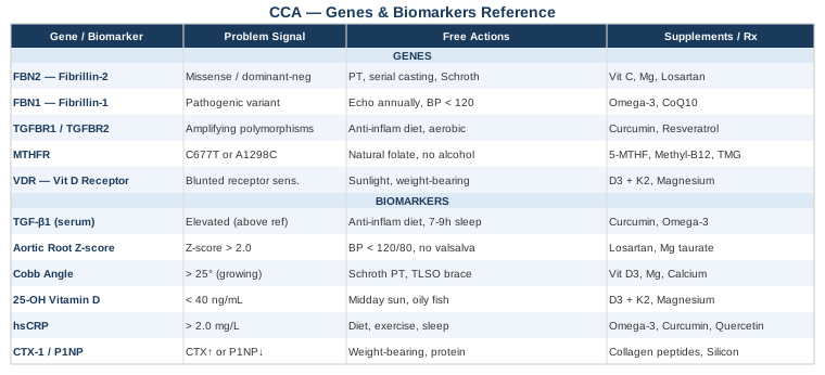
Your Genetic Blueprint: What Five Key Genes Reveal About CCA
Understanding the genetics of CCA is not about accepting a fate — it is about understanding the exact biological mechanisms causing your symptoms so that you can address them with precision. Most people with CCA carry a heterozygous FBN2 mutation, meaning one working copy of the gene remains. That single working copy still produces fibrillin-2, and research increasingly shows that the downstream consequences of the mutation — particularly in TGF-β signaling — can be meaningfully influenced by lifestyle, nutrition, and in some cases, targeted therapies. Here is what the five most relevant genes tell you.
Gene 1: FBN2 (Fibrillin-2) — The Root of the Condition
What FBN2 does: Fibrillin-2 is a large glycoprotein that forms microfibrils in the extracellular matrix — the structural scaffolding that surrounds and supports cells throughout the body. It is especially important during fetal development, when it guides the formation of joints, ligaments, and the architectural shape of the skeleton. FBN2 (NCBI Gene) encodes this protein, and mutations in it — over 90% of which are missense mutations that produce a structurally abnormal protein — cause CCA through a dominant-negative mechanism: the defective fibrillin-2 interferes with the function of the normal copy.
What a bad FBN2 gene affects: Contractures of large joints (elbows, knees, fingers, hips), arachnodactyly, kyphoscoliosis, crumpled ear helix, and occasionally mitral valve prolapse. Crucially, FBN2 normally sequesters TGF-β (transforming growth factor-beta) in the extracellular matrix, preventing excessive signaling. When FBN2 is dysfunctional, TGF-β is released in larger quantities, driving aberrant tissue remodeling and inflammation.
If the FBN2 Gene Is Mutated: The Plan Without Supplements
The foundation of managing an FBN2 mutation without supplementation is structured physical therapy, ideally started early and maintained consistently throughout life. Serial casting or splinting during infancy and childhood can significantly reduce the severity of joint contractures; this window of opportunity should not be missed. Range-of-motion exercises performed daily — targeting particularly the elbows and knees — prevent progression and often allow meaningful functional improvement, since CCA contractures tend to improve with age rather than worsen (unlike many other connective tissue conditions).
Postural training and core strengthening reduce kyphoscoliosis progression by reducing mechanical load asymmetry on the spine. A scoliosis-specific exercise program such as the Schroth method has evidence for slowing Cobb angle progression in adolescent scoliotic curves. Monitoring echocardiographically every one to two years is essential, as is avoiding contact sports and high-impact activities that stress the aortic root.
If the FBN2 Gene Is Mutated: The Plan With Supplements and Equipment
Vitamin C (1,000–2,000 mg/day, divided doses): Ascorbic acid is a cofactor for prolyl hydroxylase and lysyl hydroxylase, the enzymes that cross-link collagen and elastin. Supporting these enzymes does not fix FBN2, but it ensures that the connective tissue the body can build is as structurally sound as possible. No cycling needed; take consistently with meals to reduce GI irritation.
Magnesium glycinate or threonate (300–400 mg/day): Magnesium regulates hundreds of enzymatic processes including those governing muscle contraction and relaxation. Muscle hypoplasia and persistent tension around contractured joints are common in CCA; adequate magnesium supports muscle function and reduces cramp frequency. Evening dosing is well tolerated. No cycling needed.
Losartan (prescription only, 25–100 mg/day): This is the most mechanistically relevant pharmacological option. Originally an angiotensin II receptor blocker used for hypertension, losartan was discovered — largely through work in Marfan syndrome, which shares TGF-β dysregulation with CCA — to significantly reduce TGF-β signaling. In the Marfan context, it slowed aortic root dilation. For CCA, the evidence is extrapolated from case reports and the biological rationale rather than randomized trials. Discuss with your cardiologist or geneticist whether the benefit-risk profile is appropriate for your specific presentation. Not for use in pregnancy.
Orthotic equipment: Dynamic splints worn at night (particularly for elbow contractures) maintain and gradually improve joint range of motion through sustained, low-load stretch. These are available through specialized orthotists and have a meaningful evidence base in joint contracture management.
Gene 2: FBN1 (Fibrillin-1) — The Parallel Architecture
What FBN1 does: Fibrillin-1 is FBN2's close structural homolog and is expressed in the same extracellular matrix compartments, though with a different temporal pattern — FBN2 dominates in fetal development while FBN1 becomes the predominant fibrillin postnatally. FBN1 (NCBI Gene) mutations cause Marfan syndrome, which has significant clinical overlap with CCA, including aortic involvement and skeletal features. In the context of CCA, FBN1 variants act as modifiers: individuals with CCA who also carry sub-threshold FBN1 variants may experience more pronounced cardiovascular or skeletal manifestations.
What a suboptimal FBN1 status affects: Aortic root diameter, lens position, systemic connective tissue integrity. Unlike FBN2, a pathogenic FBN1 variant in someone with CCA raises the cardiovascular monitoring threshold significantly.
If FBN1 Carries Variants: The Plan Without Supplements
Annual or biannual echocardiography becomes mandatory rather than optional. Blood pressure management is critical — maintain systolic below 120 mmHg through dietary sodium restriction and aerobic conditioning. Avoid isometric exercise (heavy weightlifting, straining) which transiently elevates aortic wall stress. This is not permanent exclusion from strength training; it means progressing weight loads conservatively under guidance.
If FBN1 Carries Variants: The Plan With Supplements
Omega-3 fatty acids (2–4 g/day EPA+DHA): Reduces vascular inflammation and improves endothelial function. Multiple meta-analyses support cardiovascular benefit at these doses. Take with food to minimize fish aftertaste. No cycling required.
Coenzyme Q10 (100–200 mg/day, ubiquinol form): Supports mitochondrial function in vascular smooth muscle cells. Particularly relevant if the individual is on statin therapy (which depletes CoQ10). Take with a fat-containing meal to improve absorption.
Gene 3: TGFBR1 and TGFBR2 — The Dysregulated Signaling Pathway
What these genes do: TGFBR1 and TGFBR2 encode the receptor proteins that TGF-β binds to, initiating its downstream effects on cell behavior, tissue remodeling, and inflammation. In a healthy connective tissue, fibrillin-2 keeps TGF-β sequestered until it is needed; when FBN2 is defective, more free TGF-β binds these receptors, amplifying signals that drive fibrosis, inflammation, and abnormal tissue growth. Pathogenic mutations in these genes directly cause Loeys-Dietz syndrome; but common polymorphisms in TGFBR1 and TGFBR2 act as modifiers in CCA, influencing how aggressively TGF-β signaling runs when fibrillin-2 scaffolding is compromised.
If the TGF-β Receptors Show Amplifying Variants: The Plan Without Supplements
The lifestyle approach to excess TGF-β signaling centers on reducing inflammatory input. Anti-inflammatory diet — emphasizing oily fish, olive oil, cruciferous vegetables, and minimizing ultra-processed foods and refined carbohydrates — has been shown to reduce circulating TGF-β1 in multiple intervention studies. Caloric restriction and avoidance of obesity reduce TGF-β pathway activation, since adipose tissue is a significant source of inflammatory cytokines that amplify this pathway.
Aerobic exercise (moderate intensity, 150+ minutes per week) reduces systemic inflammation through multiple mechanisms including reduction in visceral fat and improvement in endothelial nitric oxide synthesis.
If the TGF-β Receptors Show Amplifying Variants: The Plan With Supplements
Curcumin with piperine (500–1,000 mg/day of curcumin, 5–10 mg piperine for absorption): Curcumin is among the best-studied natural TGF-β inhibitors. Human studies support reductions in serum TGF-β1 at these doses. Take with a fat-containing meal. Cycling (8 weeks on, 2 weeks off) is reasonable to prevent adaptation.
Resveratrol (250–500 mg/day): Activates SIRT1 and modulates TGF-β signaling. Evidence is primarily from in-vitro and animal studies, with some positive human data in cardiovascular contexts. Take with the main meal; cycling 12 weeks on, 4 weeks off is standard practice. Avoid high doses during pregnancy or with anticoagulants.
Gene 4: MTHFR — The Methylation Modifier
What MTHFR does: Methylenetetrahydrofolate reductase converts folate into the active form the body uses for methylation — a chemical process that controls gene expression, DNA repair, neurotransmitter production, and inflammation regulation. Two common variants — C677T and A1298C — reduce enzyme efficiency by 30–70%, depending on whether one or both copies of the gene are affected. Popularized by researchers including Gary Brecka, who has built significant public awareness around this gene's downstream effects, MTHFR variants are found in roughly 40–60% of the general population.
Why it matters in CCA: Poor methylation amplifies systemic inflammation (by elevating homocysteine), impairs collagen cross-linking (via reduced methylation of collagen-modifying enzymes), and can reduce the expression of compensatory extracellular matrix proteins. In a condition already characterized by structurally compromised extracellular matrix, a concurrent methylation deficit adds a second layer of fragility.
If MTHFR Is Suboptimal: The Plan Without Supplements
Increase dietary intake of natural folate — leafy greens, legumes, avocado — rather than relying on synthetic folic acid, which requires the same enzyme that is deficient to convert it to the active form. Reduce alcohol consumption, which depletes folate. Ensure adequate B12 intake from animal sources (or fortified foods). Homocysteine levels above 10 µmol/L are worth addressing.
If MTHFR Is Suboptimal: The Plan With Supplements
5-MTHF (active methylfolate, 400–800 mcg/day): This is the pre-converted form that bypasses the MTHFR enzyme entirely. No cycling required. Choose a reputable brand that uses the [6S]-5-MTHF form. Start low to avoid the "overmethylation" symptoms (anxiety, irritability) some people report at higher doses.
Methylcobalamin B12 (500–1,000 mcg/day, sublingual for best absorption): Supports the methionine cycle downstream of MTHFR. Works synergistically with 5-MTHF. No cycling required.
TMG (trimethylglycine, 500–1,000 mg/day): Provides an alternative methyl donor that can bypass the MTHFR bottleneck. Particularly relevant if homocysteine is elevated. Take with breakfast. Side effects are rare at standard doses; some individuals notice loose stool at higher doses.
Gene 5: VDR (Vitamin D Receptor) — The Overlooked Modifier
What VDR does: The vitamin D receptor gene determines how effectively your cells respond to vitamin D signaling. Common polymorphisms — FokI (rs2228570), BsmI (rs1544410), ApaI (rs7975232), and TaqI (rs731236) — affect receptor sensitivity by 10–50%, meaning that even with adequate circulating vitamin D levels, cellular response may be blunted.
Why it matters in CCA: Vitamin D signaling directly governs muscle fiber development and function (highly relevant given CCA-associated muscle hypoplasia), inflammatory cytokine production, bone mineral density (relevant to scoliosis severity and osteoporosis risk), and immune regulation. Several studies link poor VDR function to worse scoliosis outcomes in adolescents — a key concern in CCA management.
If VDR Carries Suboptimal Variants: The Plan Without Supplements
Sunlight exposure: 15–30 minutes of midday sun on arms and legs (without sunscreen during this window) provides direct vitamin D synthesis and also drives photobiomodulation effects independent of vitamin D. Consistency matters more than duration.
Weight-bearing exercise: Mechanical loading stimulates VDR expression in osteoblasts and muscle cells, partially compensating for receptor sensitivity deficits. Daily walking, swimming, and resistance training (within safe limits for the individual) support this.
If VDR Carries Suboptimal Variants: The Plan With Supplements
Vitamin D3 (2,000–5,000 IU/day, titrated to blood level): The goal is a 25-OH vitamin D serum level of 50–80 ng/mL (125–200 nmol/L). With suboptimal VDR variants, target the higher end of this range. Always pair D3 with K2 to direct calcium appropriately.
Vitamin K2 (MK-7 form, 100–200 mcg/day): Activates matrix Gla protein (MGP), which prevents inappropriate calcium deposition in soft tissues — particularly relevant in a condition where extracellular matrix is already structurally compromised.
Magnesium (300–400 mg/day): Required for vitamin D conversion enzymes. Most people are insufficient; deficiency blunts response to D3 supplementation regardless of dose.
With suboptimal VDR variants, retest 25-OH vitamin D levels every 3 months when adjusting supplementation, then every 6 months once stable.
Six Biomarkers Worth Tracking Over Time
Genetics tells you about predisposition; biomarkers tell you what is actually happening right now. For CCA, the six markers below provide the clearest window into the processes most likely to drive complications or signal stabilization. Tracking them over time transforms clinical appointments from reactive problem-solving into proactive management.
Biomarker 1: TGF-β1 (Transforming Growth Factor-Beta 1)
Why it matters: TGF-β1 is the master signaling molecule of the fibrillinopathy pathway. Elevated serum TGF-β1 indicates that the downstream consequences of defective fibrillin-2 — abnormal tissue remodeling, fibrosis, joint stiffness — are actively progressing. It is the most mechanistically direct biomarker available for CCA outside of genetic testing itself.
How to Measure It
TGF-β1 can be measured in serum or plasma through most reference labs. The test is typically ordered by a specialist (cardiologist, geneticist, or rheumatologist) and costs approximately $60–$150 USD through standard labs. Specialty functional medicine labs may offer it directly to consumers at similar pricing. Note that sample handling matters — platelet activation during processing can artificially elevate results, so use a lab with reliable protocols.
If TGF-β1 Is Elevated: The Plan Without Supplements
Anti-inflammatory dietary pattern, moderate aerobic exercise, blood pressure optimization (losartan, if prescribed), and avoidance of high-intensity isometric training. Sleep quality directly influences TGF-β regulation — targeting 7–9 hours of high-quality sleep reduces systemic inflammatory tone.
If TGF-β1 Is Elevated: The Plan With Supplements
Curcumin (500–1,000 mg with piperine), omega-3 fatty acids (2–4 g EPA+DHA), and vitamin D optimization as described above. Cycling curcumin in 8-week blocks. Retest TGF-β1 at 3 months after intervention to confirm directional change.
Biomarker 2: Echocardiographic Aortic Root Z-Score
Why it matters: Cardiac involvement in CCA most commonly manifests as mitral valve prolapse and, less frequently, aortic root dilation. When present, aortic root dilation carries risk of dissection — a rare but life-threatening complication. The aortic root Z-score (aortic diameter corrected for body surface area) provides a standardized measure that can be tracked longitudinally regardless of the child's or adult's growth status.
How to Measure It
Transthoracic echocardiography, performed by a cardiologist or cardiac sonographer. Standard echocardiogram costs range from $500–$2,500 depending on facility and insurance. For most CCA patients without known cardiac involvement, annual echocardiography is recommended; those with documented mitral valve prolapse or borderline aortic measurements warrant 6-month intervals.
If the Z-Score Is Elevated (>2.0): The Plan Without Supplements
Blood pressure control is the single most important modifiable factor. Target resting blood pressure below 120/80 mmHg. Restrict aerobic exercise intensity to moderate zones (able to hold a conversation). Avoid valsalva-type exertion entirely. Work with a cardiologist to establish sport and activity clearance.
If the Z-Score Is Elevated: The Plan With Supplements
Losartan (prescription, 25–100 mg/day): Reduces TGF-β signaling and has been shown to slow or stabilize aortic root dilation in fibrillinopathy patients in multiple studies. The pediatric Marfan losartan trials provide the primary evidence base. Discuss with a cardiologist.
Magnesium taurate (300–400 mg/day): The taurate form specifically supports vascular smooth muscle relaxation. Some cardiologists include it as adjunctive support. No cycling required.
Biomarker 3: Scoliosis Cobb Angle
Why it matters: Kyphoscoliosis is one of the most clinically significant complications of CCA, with potential for progression during adolescent growth spurts and, in severe cases, respiratory compromise. The Cobb angle — the standard measurement of spinal curvature from X-ray — is the primary monitoring tool. Curves above 25° in growing patients typically trigger bracing consideration; curves above 45–50° are often referred for surgical evaluation.
How to Measure It
Standing full-spine X-ray with Cobb angle measurement, typically done annually during growth phases and every 1–2 years in adults. Cost: $100–$500 depending on facility. Low-dose EOS imaging systems are increasingly available and significantly reduce radiation exposure compared to conventional X-ray — worth specifically requesting, especially in children.
If the Cobb Angle Is Progressing: The Plan Without Supplements
Schroth method physical therapy: A scoliosis-specific exercise program with the strongest evidence base among conservative approaches. In randomized trials, it reduces Cobb angle progression by a mean of 5–7 degrees compared to observation alone. Requires training with a certified Schroth therapist, with home practice daily.
Thoracolumbosacral orthosis (TLSO brace): Prescribed by an orthopedic surgeon for curves between 25–45° in skeletally immature patients. Worn 16–23 hours/day. Evidence supports reduction in progression to surgical range.
If the Cobb Angle Is Progressing: The Plan With Supplements
Vitamin D3 optimization (50–80 ng/mL target), magnesium, and calcium from dietary sources — these support bone density but will not independently halt curve progression. They create the best possible bone-quality foundation for whatever structural interventions are applied.
Biomarker 4: 25-OH Vitamin D
Why it matters: Vitamin D deficiency is extraordinarily common in people with musculoskeletal conditions, partly due to reduced outdoor activity and partly due to underlying absorption issues. In CCA specifically, vitamin D status directly affects muscle fiber composition (poor status worsens the muscle hypoplasia already present), bone density (relevant to fracture risk and scoliosis management), and systemic inflammatory tone. Recommended by virtually every practitioner who works longitudinally with chronic musculoskeletal conditions, from Peter Attia to functional medicine specialists.
How to Measure It
Standard serum 25-hydroxyvitamin D test, available at any primary care physician's office. Cost: $30–$80 without insurance, often covered with chronic condition codes. Retest 3 months after any supplement adjustment.
If 25-OH Vitamin D Is Below 40 ng/mL: The Plan Without Supplements
Daily midday sun exposure (15–30 minutes, arms and legs exposed), prioritizing oily fish (salmon, sardines, mackerel) 3–4 times per week, and egg yolks regularly. For most people in northern latitudes or with limited sun exposure, dietary and sunlight sources are insufficient to correct deficiency — supplementation is nearly always required.
If 25-OH Vitamin D Is Below 40 ng/mL: The Plan With Supplements
Vitamin D3 (2,000–5,000 IU/day) paired with K2 (100–200 mcg MK-7) and magnesium (300 mg). Target the 50–80 ng/mL range; retest at 3 months. Those with VDR variants (Gene 5 above) may need 5,000–8,000 IU to achieve this range. No cycling needed at maintenance doses.
Biomarker 5: High-Sensitivity C-Reactive Protein (hsCRP)
Why it matters: CRP is the most widely available and affordable inflammation marker available. In connective tissue conditions, elevated hsCRP signals ongoing tissue stress, immune activation around damaged joints, or systemic inflammatory load from other sources (poor diet, sleep deprivation, visceral fat). Peter Attia consistently lists hsCRP among his essential cardiovascular and longevity markers, and for CCA it has particular relevance as a proxy for TGF-β pathway activity — the two often move together.
How to Measure It
Included in standard comprehensive metabolic panels or ordered specifically as "hsCRP." Cost: $10–$40. Available at any lab. Target: below 1.0 mg/L; values above 3.0 mg/L indicate significant inflammatory burden requiring intervention.
If hsCRP Is Above 2.0 mg/L: The Plan Without Supplements
Anti-inflammatory dietary overhaul, sleep optimization (7–9 hours), moderate daily aerobic exercise, dental health (untreated gum disease is a major driver of elevated CRP), and elimination of hidden food sensitivities (particularly gluten in susceptible individuals). Address sleep apnea if present, as it powerfully drives systemic inflammation.
If hsCRP Is Above 2.0 mg/L: The Plan With Supplements
Omega-3 fatty acids (3–4 g EPA+DHA/day): Among the most evidence-supported anti-inflammatory supplements. Multiple meta-analyses confirm CRP reduction. No cycling required.
Curcumin with piperine (500–1,000 mg/day): Systematic reviews support CRP reduction. Cycle 8 weeks on, 2 weeks off.
Quercetin (500–1,000 mg/day): Flavonoid with anti-inflammatory and antioxidant activity. Human studies show modest CRP reduction. Take with food; cycle similarly to curcumin.
Biomarker 6: CTX-1 and P1NP (Collagen Turnover Markers)
Why it matters: CTX-1 (C-terminal telopeptide of type 1 collagen) measures collagen breakdown; P1NP (procollagen type 1 N-terminal propeptide) measures collagen synthesis. The ratio between these two markers reveals whether connective tissue is in a net catabolic (breaking down) or anabolic (building up) state. In CCA, where the structural integrity of connective tissue is already compromised by defective fibrillin-2, chronic collagen catabolism — often driven by inflammation, poor nutrition, or immobility — compounds the underlying structural deficit. Tracking these markers over time reveals whether your current intervention plan is actually working at the tissue level.
How to Measure It
CTX-1 is best measured in fasting morning serum; P1NP can be measured at any time. Both are available through specialty labs and some academic medical centers. Cost: $80–$200 each. Endocrinologists and rheumatologists are familiar with these tests in the context of osteoporosis, and can order them in the context of CCA monitoring.
If CTX-1 Is Elevated or P1NP Is Low: The Plan Without Supplements
Weight-bearing exercise (the most powerful stimulus for collagen synthesis), adequate dietary protein (1.6–2.0 g/kg/day), and sleep optimization (most collagen synthesis occurs during slow-wave sleep). Reduce immobility and excessive sedentary time — even brief walking breaks during a sedentary day reduce CTX-1 compared to prolonged sitting.
If CTX-1 Is Elevated or P1NP Is Low: The Plan With Supplements
Collagen peptides (10–20 g/day, hydrolyzed type 1/3 collagen): Human studies show increased P1NP and reduced CTX-1 with collagen peptide supplementation, particularly when taken 30–60 minutes before exercise. The effect appears larger when combined with vitamin C (200–500 mg co-administered).
Silicon (from orthosilicic acid, 10–25 mg/day): A cofactor for collagen cross-linking enzymes. Orthosilicic acid (choline-stabilized form) is the bioavailable form. Evidence from double-blind trials supports improvement in connective tissue markers. No cycling required.
Landmark Research That Changed How This Condition Is Understood
For most of the 20th century, the connective tissue damage seen in fibrillinopathies — including CCA — was explained purely mechanically: defective scaffolding simply could not bear load, so vessels, joints, and bones deteriorated from structural weakness. The management implication was bracing, surgical repair, and activity restriction. That model was not wrong, but it was profoundly incomplete.
The Discovery That Changed Everything: TGF-β as the Driver
The paradigm shift came largely from the laboratory of Dr. Hal Dietz at Johns Hopkins University School of Medicine, working primarily on Marfan syndrome but with direct implications for CCA. Dietz and colleagues demonstrated in landmark mouse model studies — and subsequently in human data — that excess TGF-β signaling, not purely mechanical failure, drives the progressive tissue pathology in fibrillinopathies. The microfibrils built from fibrillin-1 and fibrillin-2 do not simply hold tissues together mechanically; they store and regulate TGF-β, releasing it in controlled amounts to guide repair and remodeling. When the fibrillin is defective, TGF-β floods tissues continuously, driving fibrosis, abnormal smooth muscle behavior, and progressive joint and vascular pathology. (Related research on PubMed)
Ten Key Insights From This Research
1. The mutation causes loss of TGF-β regulation, not just structural weakness. The mechanical deficit is real, but the signaling dysregulation is equally important — and more amenable to pharmacological intervention.
2. Losartan's benefit in fibrillinopathies is independent of its blood pressure effect. It works primarily by reducing angiotensin-II-driven TGF-β amplification — a discovery that opened a new treatment avenue and validated the TGF-β hypothesis.
3. TGF-β dysregulation can be measured in serum. This is not just an academic finding — it means that patients can monitor pathway activity over time and assess whether interventions are working.
4. Early intervention matters more than late intervention. The TGF-β-driven remodeling begins prenatally (which is why contractures are present at birth), but the postnatal period offers a window to reduce ongoing pathway activation before irreversible fibrosis accumulates.
5. The contractures in CCA are partly fibrotic, not purely mechanical. This changes the logic of treatment — anti-fibrotic approaches (including TGF-β modulation) may benefit joint mobility in ways that pure mechanical stretching cannot.
6. Muscle hypoplasia in CCA is also partly TGF-β-driven. Excess TGF-β inhibits satellite cell activation and muscle fiber development. This is why physical therapy alone may have a ceiling in some patients.
7. Diet and inflammation directly modify TGF-β pathway activity. This means lifestyle interventions are not just supportive — they are mechanistically relevant to the core biology of the condition.
8. The ear abnormalities (crumpled helix) in CCA may partially correct over time. This reflects the postnatal dominance of FBN1 over FBN2 — as the body's extracellular matrix shifts toward adult configuration, some developmental features normalize. The biology is more dynamic than the diagnosis suggests.
9. Aortic involvement in CCA is less severe on average than in Marfan syndrome, but it requires the same monitoring vigilance, because the cases where it does progress can do so rapidly once the threshold is crossed.
10. The same TGF-β pathway insight that led to losartan trials has opened investigation into other agents — including doxycycline (an MMP inhibitor) and newer angiotensin pathway drugs — which are currently under study and represent the next generation of connective tissue disease management.
Complementary Approaches With Real Clinical Relevance
The strategies below are not replacements for medical management, genetic counseling, or physical therapy. They are complementary tools with genuine human evidence that can be integrated alongside conventional care for CCA.
Yoga (Adapted Practice)
Yoga, in its adapted therapeutic forms, offers a structured approach to improving joint range of motion, postural alignment, and body awareness — three areas directly relevant to CCA. The gentle sustained stretching characteristic of yin yoga and therapeutic yoga is consistent with the low-load, long-duration stretching that physical therapists prescribe for joint contractures. Unlike aggressive stretching, yoga-based protocols incorporate breath synchronization and parasympathetic activation, which reduces muscle guarding around contracted joints.
A randomized trial published in Spine (Fishman et al.) studied yoga-based stretching in adolescent idiopathic scoliosis and found significant improvements in Cobb angle with a side-plank pose practiced asymmetrically — suggesting that targeted single-sided yoga postures can influence scoliosis curves measurably. While this trial was in idiopathic scoliosis, the mechanical principle applies to CCA-associated kyphoscoliosis. (Related studies on PubMed)
For CCA, the practical application involves working with a certified yoga therapist (C-IAYT credential) who can design a practice respecting existing contractures, avoiding hypermobile range of motion in unaffected joints, and progressively extending work on the most limited joints. Start with 2–3 sessions per week, 30–45 minutes each. Avoid heated yoga rooms and breath-holding practices (both increase vascular stress). The focus should be on controlled, sustained stretching with complete muscular relaxation — not on achieving the full expression of any pose.
Massage Therapy
Massage therapy is relevant to CCA primarily through its effects on muscle hypoplasia, fascial restriction around contracted joints, and pain management. Connective tissue massage (CTM) and myofascial release work on the fascia — the fibrillin-containing connective tissue wrapping muscles and joints — rather than purely on muscle bulk, making it mechanistically relevant to a condition affecting extracellular matrix architecture.
A systematic review by Bervoets et al. (2015) found that massage therapy produced statistically significant improvements in pain, range of motion, and functional outcomes in musculoskeletal conditions with chronic joint restriction. While CCA-specific trials do not exist, the biological rationale and the evidence in analogous conditions (arthrogryposis, joint contractures from other causes) support its use as an adjunct. (Related research on PubMed)
In practical terms, deep tissue massage around the elbow, knee, and finger contractures should be performed by a therapist familiar with connective tissue disorders — not generic sports massage. Sessions of 45–60 minutes, 1–2 times per week during active rehabilitation phases, transitioning to monthly maintenance. Important caution: avoid aggressive massage near hypermobile segments or over areas of active inflammation. Communicate openly with the therapist about which joints are contractured versus which are unaffected.
Breathing-Based Therapies
Kyphoscoliosis in CCA can reduce thoracic cage compliance and limit respiratory expansion. When the Cobb angle reaches significant degrees, breathing mechanics are compromised even before symptoms appear — a subclinical process that reduces oxygen delivery to tissues and increases respiratory muscle fatigue. Breathing-based therapies address this directly and are among the few complementary approaches with a clear mechanical rationale in this specific anatomical context.
The Buteyko method and controlled breathing protocols (particularly those emphasizing nasal breathing and diaphragmatic activation) have been studied in conditions involving restricted chest expansion, including scoliosis and chest wall deformities. Studies in adolescent scoliosis have demonstrated improvements in forced vital capacity (FVC) and peak expiratory flow when breathing exercises were added to standard physical therapy — a finding with direct implications for CCA-related kyphoscoliosis.
For CCA patients, a practical protocol involves 10–15 minutes of daily diaphragmatic breathing practice (lying supine over a small bolster to open the anterior chest), combined with incentive spirometry (a simple device available for $10–$30 that provides biofeedback on inhalation volume). Work with a respiratory physiotherapist annually to monitor pulmonary function tests — particularly FVC and FEV1 — as early indicators of thoracic compromise. Avoid breath-holding techniques (e.g., Wim Hof) without specialist guidance, as these transiently increase intrathoracic pressure in ways that may stress already-compromised thoracic architecture.
Biofeedback
Biofeedback teaches individuals to consciously regulate physiological responses — muscle tension, heart rate, skin conductance — by receiving real-time data about these processes. For CCA, its primary value lies in neuromuscular re-education around contracted joints and in managing the pain and anxiety that often accompany chronic musculoskeletal conditions in childhood and adolescence.
Surface electromyography (sEMG) biofeedback has been used to retrain muscle activation patterns around scoliotic spines and contracted limb joints, helping patients learn to recruit underused muscles and reduce compensatory tension in overloaded ones. A review in Archives of Physical Medicine and Rehabilitation supported sEMG biofeedback as a useful adjunct to physical therapy for joint contractures and movement rehabilitation. (Related research on PubMed)
For CCA, biofeedback sessions are typically conducted by a certified biofeedback therapist (BCIA credential) or integrated into physical therapy. Eight to twelve sessions is a standard course, with the goal of developing skills that transfer to home practice. Heart rate variability (HRV) biofeedback — which trains the autonomic nervous system through specific breathing patterns — also reduces systemic inflammatory markers including CRP, making it relevant beyond just movement rehabilitation. HRV biofeedback devices for home use are available at $100–$300 and can be used independently once the basic technique is learned.
Progressive Muscle Relaxation
Progressive muscle relaxation (PMR) involves systematically tensing and releasing muscle groups throughout the body in sequence, developing body awareness and reducing baseline muscle tension. For CCA, where muscle hypoplasia and joint contractures create persistent tension-compensation patterns throughout the kinetic chain, PMR provides a low-risk, no-equipment tool for reducing that chronic muscular load.
Clinical evidence for PMR in musculoskeletal pain is well-established — a meta-analysis published in JAMA Internal Medicine found significant reductions in pain intensity and improved physical function in participants using mind-body relaxation techniques including PMR. In CCA specifically, the most relevant application is reducing the secondary muscle guarding that develops around contracted joints over years, which adds to functional limitation beyond the contracture itself.
A standard PMR practice takes 15–20 minutes, performed daily before sleep. Begin at the feet and work systematically upward, tensing each muscle group for 5–7 seconds and releasing for 20–30 seconds. For joints that cannot achieve full range of motion, apply gentle isometric tension rather than full contraction. Apps and guided recordings are widely available for free and require no specialist support once the technique is learned. PMR pairs particularly well with the breathing-based therapies described above, and many people find combining both into a 25–30 minute evening routine creates meaningful cumulative benefits.
Conclusion
Congenital contractural arachnodactyly is a condition with a precise genetic origin — but its day-to-day impact is shaped by an entire cascade of downstream processes, many of which are measurable, addressable, and modifiable. The core message of this article is that understanding which genes are relevant (FBN2 first, then the modifier genes that determine severity) and which biomarkers give you the clearest signal (TGF-β1, echocardiographic parameters, Cobb angle, vitamin D, hsCRP, and collagen turnover markers) transforms a diagnosis from a static label into a dynamic picture you can actually navigate.
You do not need to act on everything at once. The most useful next step is usually the simplest: get the biomarkers you have not yet tested, share this information with your medical team, and ask specifically about TGF-β monitoring and whether losartan assessment is appropriate for your presentation. Better information does not guarantee better outcomes — but it meaningfully improves the odds of making decisions that move things in the right direction.
Musculoskeletal: Bone Conditions Joint Conditions Muscle Conditions Spine Conditions
Cardiovascular: Heart Conditions
Autoimmune: Connective Tissue Conditions