This article contains AI generated content.
· UpdatedCongenital Knee Dislocation — 5 Genes And 6 Biomarkers To Track
Introduction
Living with the effects of congenital knee dislocation — whether you are a parent of a child who received this diagnosis, an adult treated in infancy, or a clinician trying to provide better guidance — means navigating a condition rare enough to fall through the cracks of mainstream orthopedic literature. Most general advice about knee health is written for degenerative or traumatic cases. It rarely accounts for the connective tissue specifics, developmental joint geometry differences, or the heritable biology that shapes congenital presentations from the very beginning.
What makes this condition more complex than it appears is that the visible problem — the dislocated joint — is typically corrected early in life through casting, traction, or surgery. What remains less visible is the downstream biology: the subtle inflammatory load, the connective tissue fragility, the cartilage that may wear differently because the joint was structurally abnormal during critical developmental windows. These are not inevitable outcomes. But they are real risks, and understanding them requires looking beyond standard imaging and routine clinical visits.
This article takes a more specific approach. It focuses on two layers of information that are underused in routine care: measurable biomarkers that can reflect cartilage health, inflammation, and nutrient status in real time, and genetic variants that influence connective tissue integrity, joint laxity, and structural resilience. Neither replaces clinical judgment. Both can sharpen it considerably.
Better information does not automatically produce better outcomes — but it enables better decisions. The biomarker section gives you a practical framework for tracking what is happening inside the joint and the surrounding tissue. The genetics section explains which inherited variants may have contributed to the condition and how they can be partially addressed. Beyond those two pillars, you will also find the most actionable research on joint mobility and a look at which complementary physical approaches have genuine clinical backing. Each section ends with concrete guidance, not just theory.
Summary
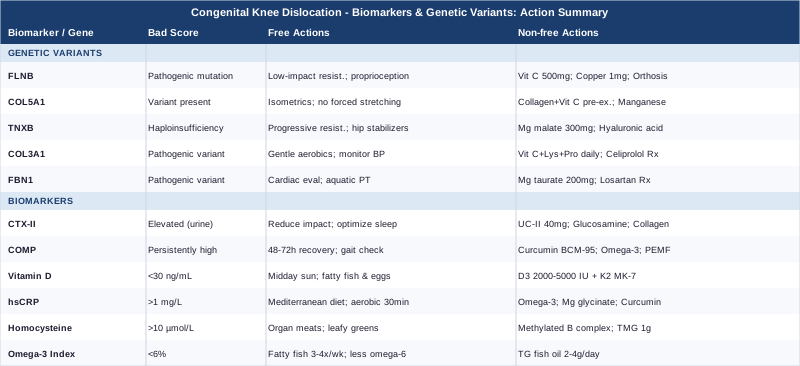
This article covers six key biomarkers — CTX-II, COMP, Vitamin D, hsCRP, Homocysteine, and the Omega-3 Index — that are rarely ordered in routine follow-up care for congenital knee dislocation but can reveal a great deal about cartilage degradation speed, systemic inflammation, and nutritional deficits. It also examines five genetic variants (FLNB, COL5A1, TNXB, COL3A1, FBN1) directly linked to connective tissue fragility and joint structural vulnerability, with targeted action plans for each — both with and without supplementation. You will also find a detailed breakdown of the most impactful research-backed principles for joint mobility and tissue repair, drawn from current neuroscience and exercise physiology. Finally, five complementary approaches with real clinical evidence — including photobiomodulation, therapeutic yoga, and biofeedback — are reviewed for practical, cautious application. Every section connects to what you can actually do, not just what the science says in the abstract.
6 Biomarkers to Monitor Joint Health in Congenital Knee Dislocation
People treated for congenital knee dislocation often leave follow-up care with a structurally corrected joint and no framework for monitoring tissue health over time. Standard check-ups focus on alignment and range of motion — both important — but miss several blood and urine markers that can reveal early cartilage breakdown, chronic low-grade inflammation, and nutritional gaps that accelerate joint degeneration. The six biomarkers below are not exotic research tools. Most are available through standard labs or direct-to-consumer testing, and together they build a picture of joint health from the inside out.
Biomarker 1: CTX-II (C-Terminal Telopeptide of Type II Collagen)
Why it matters: CTX-II is a degradation fragment of type II collagen — the primary structural protein in articular cartilage. When cartilage is breaking down faster than it is being repaired, CTX-II concentrations rise in urine. In people with a history of congenital knee dislocation, joint geometry may have been subtly altered even after successful reduction, creating uneven load distribution that accelerates cartilage turnover over years and decades. Elevated CTX-II can flag this process long before pain or imaging changes appear.
What it may reveal: Elevated CTX-II correlates with future cartilage loss and increased osteoarthritis risk. It has been studied extensively in post-traumatic and early-onset osteoarthritis populations, which share biological parallels with structurally corrected congenital knee cases. Research by Lohmander and colleagues at Lund University established CTX-II as one of the most predictive urinary markers of cartilage breakdown in active joint conditions.
How to Measure CTX-II
CTX-II is measured from a fasting, second-morning urine sample, corrected for creatinine concentration. It is not universally available at every standard commercial lab, but specialty labs focused on joint health and rheumatology biomarkers carry it. Direct-to-consumer services in some countries also offer it. Cost range: $60–$150 depending on the provider. Repeat testing every 6–12 months establishes the trend that matters far more than any single data point.
If CTX-II Is Elevated: Plan Without Supplements
Reduce repetitive high-impact loading on the knee — running on hard surfaces, stair-heavy training, and loaded deep squats are the primary culprits. Prioritize aquatic exercise or cycling to maintain strength with substantially lower joint stress. Ensure adequate sleep (7–9 hours per night), since growth hormone released during slow-wave sleep is the primary driver of nocturnal cartilage repair. Optimize body weight if applicable: research consistently shows that each 1 kg reduction in body mass reduces compressive knee joint load by approximately 4 kg during normal walking.
If CTX-II Is Elevated: Plan With Supplements or Equipment
Undenatured type II collagen (UC-II, 40 mg/day on an empty stomach): Evidence from multiple randomized trials supports UC-II's ability to reduce joint pain and support cartilage integrity through oral tolerance mechanisms — a distinct pathway from structural collagen supplementation. Evaluate effects at 90 days. No established cycling needed. Side effects are rare.
Glucosamine sulfate (1500 mg/day with food): The NIH-funded GAIT trial (Clegg et al., NEJM 2006) found modest but meaningful effects for moderate-to-severe knee osteoarthritis. Supports cartilage matrix preservation over the long term. Side effects: occasional mild GI discomfort; rare interactions with warfarin.
Hydrolyzed collagen peptides (10–15 g/day with vitamin C, 50 mg): Supports systemic collagen synthesis. Taking it 30–45 minutes before joint-loading exercise appears to preferentially direct collagen building blocks toward connective tissue, based on research from the Australian Institute of Sport. No cycling needed; minimal side effects.
Knee offloading brace: For people with confirmed malalignment or uneven cartilage wear, an evidence-based varus/valgus unloading brace redistributes mechanical stress and reduces cartilage breakdown independent of any nutritional approach.
Biomarker 2: COMP (Cartilage Oligomeric Matrix Protein)
Why it matters: COMP is a non-collagen protein found in cartilage and joint capsule tissue. Serum COMP is one of the earliest-rising biomarkers of cartilage stress and damage — it increases with abnormal mechanical loading and inflammatory joint conditions. Unlike CTX-II, which reflects breakdown end-products, COMP reflects both active stress and matrix remodeling, making it a useful companion marker when monitored as a trend.
What it may reveal: In studies of children with skeletal dysplasias and joint conditions, elevated serum COMP has been associated with accelerated joint deterioration. For adults who had congenital knee dislocation corrected in infancy, a rising COMP trend may indicate that the joint is under excess stress even in the complete absence of symptoms. Research by Saxne and Heinegård at Lund University established COMP as a clinically relevant marker of cartilage turnover and joint stress, now referenced in major rheumatology guidelines.
How to Measure COMP
COMP is measured from fasting blood (serum). It is available through academic medical centers and specialty rheumatology labs. Cost: approximately $80–$200. A single measurement has limited interpretive value; the trend over 6–12 months is the meaningful signal. Order it alongside CTX-II if possible — the combination distinguishes active breakdown from mechanical stress responses.
If COMP Is Elevated: Plan Without Supplements
Modify training load: COMP rises acutely after exercise and typically normalizes within 24–48 hours. Persistently elevated levels suggest the joint is not recovering adequately between sessions. Introduce structured recovery periods of 48–72 hours between sessions involving significant knee loading. Work with a physical therapist to assess for compensatory gait patterns — especially hip drop, foot overpronation, or knee valgus under load — that generate asymmetric joint stress independent of overall activity volume.
If COMP Is Elevated: Plan With Supplements or Equipment
Curcumin (high-bioavailability form such as BCM-95 or Meriva, 500–1000 mg/day with a fat-containing meal): Multiple randomized controlled trials have shown that well-absorbed curcumin formulations reduce inflammatory joint markers including prostaglandins and metalloproteinases that drive cartilage degradation. Suggested cycling: 8 weeks on, 2 weeks off. Side effects: mild GI effects at high doses; avoid concurrent use with strong anticoagulants.
Omega-3 fatty acids (EPA+DHA, 2–4 g/day total): Reduces prostaglandin-E2-driven cartilage degradation and synovial inflammation. No cycling needed. Side effects: mild fishy taste (use enteric-coated form), very mild anticoagulant effect at doses above 3 g/day.
Pulsed electromagnetic field (PEMF) therapy device: 20–30 minutes over the knee joint daily. Clinical trials in knee osteoarthritis populations show PEMF reduces pain and inflammatory biomarkers. Home devices in the $100–$600 range are available for personal use and offer a non-pharmacological complement to supplement protocols.
Biomarker 3: Vitamin D (25-Hydroxyvitamin D)
Why it matters: Vitamin D is far more than a bone-mineral nutrient. It plays a direct regulatory role in musculoskeletal development, skeletal muscle function, and immune modulation. Low vitamin D status during developmental windows has been linked to connective tissue laxity, impaired bone mineralization, and reduced periarticular muscle strength — all of which matter in congenital knee dislocation both acutely and long-term.
What it may reveal: Levels below 30 ng/mL (75 nmol/L) — which affect a substantial portion of the general population year-round — correlate with weaker quadriceps and hamstrings, reduced proprioception, and higher systemic inflammatory load. Peter Attia, in his work on longevity medicine, consistently emphasizes that the functional target for musculoskeletal health is 40–60 ng/mL, not merely the laboratory "sufficient" cutoff of 20 ng/mL.
How to Measure Vitamin D
25-OH vitamin D is a standard blood test available at virtually every laboratory and often covered by health insurance. Cost: $30–$80. Test twice per year — once in late summer and once in late winter — to capture the seasonal variation that affects most people in northern latitudes. The late-winter value is typically the most clinically relevant because it reflects the lowest annual point.
If Vitamin D Is Low: Plan Without Supplements
20–30 minutes of midday sun exposure (UV index above 3) on arms and legs, without sunscreen, three to four times per week provides meaningful vitamin D synthesis when the season permits. Increase dietary sources: fatty fish (salmon, mackerel, sardines), egg yolks, and beef liver are the most concentrated food sources. Reduce factors that impair conversion: obesity (adipose tissue sequesters vitamin D), liver dysfunction, and certain medications including antiepileptics and corticosteroids.
If Vitamin D Is Low: Plan With Supplements or Equipment
Vitamin D3 (2000–5000 IU/day with a fat-containing meal): Pair with vitamin K2 (MK-7, 100–200 mcg/day) to direct calcium toward bone matrix and away from soft tissue. No cycling needed. Monitor blood levels every 6 months to calibrate dose; toxicity requires chronically elevated levels well above standard supplementation ranges. Retest at 90 days after initiating supplementation and adjust.
Biomarker 4: hsCRP (High-Sensitivity C-Reactive Protein)
Why it matters: hsCRP is the most accessible and widely used clinical measure of systemic low-grade inflammation. In joint conditions, chronic inflammation — even at sub-symptomatic levels — drives matrix metalloproteinase (MMP) activation, which degrades cartilage extracellular matrix and disrupts the structural integrity of the joint capsule. People with structural joint abnormalities, including those corrected early, are more susceptible to inflammatory cycles initiated by mechanical stress and perpetuated by poor metabolic health.
What it may reveal: Values above 1 mg/L signal elevated tissue degradation risk. Values above 3 mg/L indicate clinically significant chronic inflammation. Thomas Dayspring, a leading lipidologist in preventive medicine, consistently identifies hsCRP as one of the few affordable inflammatory markers with strong predictive value across both cardiovascular and musculoskeletal outcomes. Optimal range for long-term tissue preservation: below 0.5 mg/L.
How to Measure hsCRP
Standard fasting blood test. Cost: $15–$50 at standard commercial labs. Avoid testing during acute illness — any active infection or injury will temporarily spike the result. For baseline measurement and monitoring, test every 6 months, or every 3 months when actively intervening to lower inflammation.
If hsCRP Is Elevated: Plan Without Supplements
Address the most common modifiable drivers: poor sleep quality, excess visceral adiposity, chronic psychological stress, high consumption of ultra-processed foods, and sedentary behavior. A Mediterranean-style whole-food diet — emphasizing olive oil, fatty fish, legumes, colorful vegetables, and minimizing refined carbohydrates and seed oils high in linoleic acid — consistently lowers hsCRP in intervention studies. Aerobic exercise performed at moderate intensity (30–45 minutes, 4–5 times per week) is one of the most reliably anti-inflammatory interventions available and requires no prescription.
If hsCRP Is Elevated: Plan With Supplements or Equipment
Omega-3 (EPA+DHA, 2–4 g/day): Consistently and significantly lowers hsCRP across multiple intervention trials. No cycling needed. The benefit is dose-dependent and takes 8–12 weeks to fully manifest.
Magnesium glycinate or malate (300–400 mg/day at night): Magnesium deficiency — common in Western diets — correlates directly with elevated CRP. The nighttime dosing supports sleep quality and muscle relaxation simultaneously. Side effects: softened stools at higher doses; reduce dose if this occurs.
High-dose curcumin (BCM-95 or Meriva, 500–1000 mg/day): See COMP section above for full protocol. Curcumin's NF-κB inhibitory effect is among its most validated anti-inflammatory mechanisms, and it directly lowers hsCRP in clinical trials.
Biomarker 5: Homocysteine
Why it matters: Homocysteine is a sulfur-containing amino acid that, when elevated, impairs collagen cross-linking — the molecular process that gives connective tissue its tensile strength and resilience under load. This is particularly significant in congenital knee dislocation, where connective tissue quality is often a contributing factor to the original structural vulnerability. Elevated homocysteine also drives oxidative stress and compromises vascular integrity around joint structures, reducing nutrient and oxygen delivery to cartilage.
What it may reveal: Optimal levels are below 7–8 µmol/L. Values above 10 µmol/L measurably impair collagen quality and integrity. Elevated homocysteine is most commonly driven by insufficient B12, folate, or B6 — all affected by dietary quality, the MTHFR gene variants discussed in the next section, and gut absorption capacity. Addressing elevated homocysteine directly supports the connective tissue biology at the core of congenital knee dislocation.
How to Measure Homocysteine
Fasting blood test. Cost: $30–$80 at standard commercial labs. Always test alongside a B-vitamin status panel (serum B12, red cell folate, pyridoxal-5-phosphate B6) to identify the specific driver. Retest at 60–90 days after initiating intervention to verify response.
If Homocysteine Is Elevated: Plan Without Supplements
Increase dietary sources of B vitamins: organ meats (liver is the single most concentrated source), eggs, dark leafy greens, and legumes. Reduce alcohol intake — even moderate habitual alcohol depletes folate and B6 through multiple pathways. Address gut health issues that impair B12 absorption, particularly low stomach acid (common in people over 40 and those on proton pump inhibitors) and small intestinal dysbiosis.
If Homocysteine Is Elevated: Plan With Supplements or Equipment
Methylated B complex (methylfolate 400–800 mcg/day + methylcobalamin B12 500–1000 mcg/day + pyridoxal-5-phosphate B6 25–50 mg/day): Use methylated forms, particularly if MTHFR variants are present (see genetics section). Non-methylated forms require enzymatic conversion that is impaired in these variants. No cycling needed. Start at lower doses if you are sensitive to methyl donors (some people experience mild anxiety or irritability with high-dose methylfolate).
Trimethylglycine/TMG (500–1000 mg/day): A methyl donor that directly converts homocysteine to methionine via the BHMT pathway, independent of folate/B12. Useful when B-vitamin supplementation alone does not normalize levels. Side effects: mild GI effects in some; avoid in pregnancy without clinical guidance.
Biomarker 6: Omega-3 Index
Why it matters: The Omega-3 Index measures the percentage of EPA and DHA in red blood cell membranes — a reliable reflection of long-term omega-3 status over the preceding 3–4 months. This is directly relevant for joint health: EPA and DHA are the precursors to pro-resolving lipid mediators (resolvins, protectins, maresins) that actively resolve inflammation in synovial tissue and cartilage. A low Omega-3 Index means the biological resolution machinery is running at reduced capacity, keeping the joint in a chronically inflamed state even without an acute trigger.
What it may reveal: An optimal Omega-3 Index is above 8%. Most people eating a typical Western diet have an index between 4–6%, which corresponds to significantly higher inflammatory load and slower tissue repair. Peter Attia consistently identifies the Omega-3 Index as one of the highest-yield blood markers for both cardiovascular and musculoskeletal longevity — and it is inexpensive to test.
How to Measure the Omega-3 Index
Dried blood spot test from a finger-prick sample. Home collection kits are available from several direct-to-consumer lab services. Cost: $50–$90. No fasting required. Retest every 4–6 months when actively adjusting fish or supplement intake, to verify the index is moving toward the target range.
If the Omega-3 Index Is Low: Plan Without Supplements
Eat fatty fish (salmon, sardines, mackerel, anchovies, herring) at least 3–4 times per week. Reduce competing linoleic acid (omega-6) from soybean, sunflower, and corn oils, as high dietary omega-6 competes with EPA and DHA incorporation into cell membranes. For those who do not eat fish, algae-derived EPA+DHA provides a plant-based source with equivalent bioavailability.
If the Omega-3 Index Is Low: Plan With Supplements or Equipment
Triglyceride-form fish oil or algae-based omega-3 (2–4 g/day total EPA+DHA with a fat-containing meal): The triglyceride form absorbs approximately 70% better than the ethyl ester form common in cheaper products. No cycling needed — the benefit is continuous. Side effects: mild fishy aftertaste (enteric-coated form eliminates this); at doses above 4 g/day, monitor if on anticoagulant medication. Retest the index after 4–6 months of consistent supplementation to confirm improvement.
The biomarker framework above gives you a clear, actionable picture of what is happening at the tissue level. Understanding the genetic layer that may have shaped the original vulnerability adds a further dimension — and often points to the same biological pathways, confirming where targeted support is most justified.
The Genetic Side: 5 Variants Worth Knowing About
Congenital knee dislocation is not caused by a single gene in most cases, but it is rarely random either. Several genetic variants affect connective tissue architecture, collagen quality, and joint laxity in ways that make structural dislocations more likely — and that continue to influence tissue health long after the joint is anatomically corrected. Understanding your genetic profile in these pathways is not about assigning blame or predicting outcomes. It is about knowing which biological systems deserve targeted support, and why standard recommendations may not be enough.
Consumer genetic testing platforms such as 23andMe or AncestryDNA provide raw SNP data that can be interpreted through specialized tools. Researchers such as Ali Torkamani (Scripps Research Institute) and practitioners like Gary Brecka emphasize that genetic variants are not fixed sentences — they are tendencies that can be meaningfully modified by nutrition, targeted supplementation, and lifestyle. The five variants below are among the most relevant to congenital knee dislocation and connective tissue vulnerability.
Gene 1: FLNB (Filamin B)
What it does: Filamin B is a cytoskeletal actin-binding protein essential for skeletal morphogenesis and joint formation. Pathogenic mutations in FLNB cause a spectrum of skeletal dysplasias including Larsen syndrome, atelosteogenesis type I and III, and spondylocarpotarsal synostosis — all of which feature multiple congenital joint dislocations, including of the knee. Even subclinical FLNB variants may reduce joint capsule and ligament integrity without producing a recognized syndrome.
Evidence base: Bicknell et al. (2007, Nature Genetics) identified dominant and biallelic FLNB mutations as causal across multiple skeletal dysplasia syndromes featuring congenital dislocations. This remains one of the most direct established genetic links to congenital knee dislocation in the published literature. If family history suggests a pattern of joint instability across generations, FLNB sequencing through clinical genetics is worth discussing with a specialist.
If the FLNB Variant Is Present: Plan Without Supplements
Prioritize low-impact resistance-based exercise that builds periarticular muscle strength without placing shear stress on the joint capsule: water resistance exercises, leg press (not hack squat or leg extension), and hip abductor work. Avoid high-velocity sport activities during growth phases in children; introduce them gradually with professional physiotherapy guidance as the musculature matures. Implement structured joint proprioception training — balance board work, single-leg stance progressions, and perturbation training — as a non-negotiable component of weekly exercise. Proprioceptive compensation is the primary mechanism by which the nervous system protects a structurally vulnerable joint.
If the FLNB Variant Is Present: Plan With Supplements or Equipment
Vitamin C (ascorbic acid, 500–1000 mg/day): Essential cofactor in prolyl and lysyl hydroxylation — the steps that give individual collagen chains the stability to form their triple-helix structure. This is not optional for connective tissue with structural compromise. No cycling needed. Well tolerated; loose stools may occur at doses above 2000 mg/day.
Copper bisglycinate (1–2 mg/day): Required for lysyl oxidase activity, the enzyme responsible for cross-linking collagen fibers and elastin. Deficiency — more common than recognized — produces structurally weak connective tissue even when collagen synthesis is adequate. Recheck copper status via serum ceruloplasmin periodically; excessive supplementation is harmful.
Proprioceptive orthosis or ankle-foot brace during high-demand activity: Provides both mechanical support and external sensory feedback to the joint position system — particularly useful during the neuromuscular retraining phase.
Gene 2: COL5A1 (Collagen Type V Alpha 1 Chain)
What it does: COL5A1 encodes the alpha-1 chain of type V collagen, which regulates the diameter and organization of type I collagen fibrils in tendons, ligaments, and joint capsules. Mutations in COL5A1 are primarily associated with the classical and hypermobile forms of Ehlers-Danlos Syndrome (EDS). Even the well-studied rs12722 SNP — a relatively common variant — has been reproducibly associated with elevated tendon and ligament injury risk in athletic populations, reflecting a broader role for COL5A1 variants in joint structural integrity.
Evidence base: Multiple studies have linked COL5A1 variants to connective tissue injury risk in human cohorts. Research published in Annals of Human Genetics and the American Journal of Human Genetics confirms COL5A1 mutations as causative in classical EDS, with the shared biological mechanism — impaired collagen fibril diameter regulation — directly affecting joint capsule and ligament resistance to mechanical stress.
If COL5A1 Is Affected: Plan Without Supplements
Avoid aggressive joint manipulation, forced stretching beyond comfortable range, or habitual "popping" of the joint — already-lax ligaments do not benefit from further elongation and each cycle carries micro-injury risk. Focus isometric and closed-chain exercises: wall sits, terminal knee extensions, step-ups, and leg press movements generate quadriceps strength without the anterior shear forces of open-chain leg extensions. Use therapeutic tape (Kinesio or rigid athletic taping) around the knee during sports to provide external capsule support without circulatory restriction.
If COL5A1 Is Affected: Plan With Supplements or Equipment
Hydrolyzed collagen peptides (15 g) + vitamin C (50 mg), taken 30–45 minutes before exercise: Research from the Australian Institute of Sport demonstrates that this specific pre-exercise timing significantly increases collagen synthesis in tendons and ligaments compared to post-exercise or fasting-state supplementation. The mechanism involves peak amino acid availability during the period of exercise-induced collagen turnover signaling. No cycling needed; minimal side effects.
Manganese (5–10 mg/day): Required for glycosaminoglycan synthesis and collagen cross-linking. Manganese is rarely tested and frequently low in people with high-sugar diets. Standard supplementation doses are safe; avoid high doses (above 20 mg/day) for extended periods.
Compression sleeve (graduated knee sleeve during weight-bearing activity): Not a substitute for strength training, but provides proprioceptive input, thermal support, and mild circumferential stability that reduces cumulative micro-stress to the capsule.
Gene 3: TNXB (Tenascin X)
What it does: Tenascin X is an extracellular matrix glycoprotein that regulates collagen fibril organization and the elastic properties of connective tissue. TNXB deficiency — caused by homozygous loss-of-function mutations — produces a recessive form of EDS characterized by joint hypermobility, skin hyperextensibility, and spontaneous joint dislocations. Haploinsufficiency (a single functional copy) produces a milder but clinically significant hypermobility phenotype that can predispose to instability and structural dislocations during development.
Evidence base: Schalkwijk et al. (2001, Nature Genetics) identified TNXB deficiency as the cause of a distinct autosomal recessive form of EDS, establishing tenascin X as a critical organizer of connective tissue architecture. Subsequent studies confirmed haploinsufficiency effects, making TNXB one of the few EDS-associated genes with a clear dosage-sensitive phenotype.
If TNXB Is Affected: Plan Without Supplements
Muscle strength is the primary compensatory mechanism for the ligamentous laxity produced by TNXB insufficiency — structured progressive resistance training is not optional. Design programs around hip and knee stabilizers, with special attention to the gluteus medius, VMO, and deep hip external rotators, which stabilize the knee joint indirectly through alignment. Work with a physiotherapist experienced in hypermobility conditions; they understand how to load hypermobile tissue without triggering the micro-injury cascade that naive training programs cause. Shorter, more frequent sessions outperform long exhausting ones — fatigue dramatically increases injury risk in hypermobile joints.
If TNXB Is Affected: Plan With Supplements or Equipment
Magnesium malate (300–400 mg/day at night): Reduces muscle fatigue, cramping, and post-exertional soreness that are disproportionately common in hypermobility conditions — likely related to the higher muscular work required to compensate for ligamentous laxity. Supports mitochondrial ATP production in connective tissue fibroblasts.
Oral hyaluronic acid (80–200 mg/day): May support synovial fluid viscosity and joint lubrication. The evidence base is early but mechanistically sound — hyaluronic acid is the primary structural component of synovial fluid and is reduced in chronically stressed joints. Well tolerated; no cycling needed.
Gene 4: COL3A1 (Collagen Type III Alpha 1 Chain)
What it does: Type III collagen is a major structural component of blood vessel walls, skin, and the joint capsule. Pathogenic mutations in COL3A1 cause vascular Ehlers-Danlos syndrome (vEDS) — a serious condition with life-threatening vascular complications. However, subclinical or milder COL3A1 variants are more common and affect joint capsule quality and vascular integrity around the joint without reaching diagnostic vEDS threshold. In congenital knee dislocation, where capsule integrity is often a primary contributing factor, variants in this gene can amplify underlying structural vulnerability.
Evidence base: COL3A1 variants have been documented across the connective tissue disorder spectrum. Pepin et al. and subsequent work by the International EDS Consortium have characterized the full range of COL3A1 pathogenic and subclinical variant effects on connective tissue strength and vascular fragility.
If COL3A1 Is Affected: Plan Without Supplements
For confirmed pathogenic variants (vEDS end of the spectrum): avoid contact sports and high-fall-risk activities; the vascular risk is the primary clinical concern. For subclinical variants affecting structural collagen quality: focus on regular, gentle aerobic activity (walking, swimming, cycling) that maintains vascular compliance and periarticular circulation without high-impact stress. Monitor blood pressure routinely — vascular connective tissue fragility is significantly worsened by sustained hypertension, and even mildly elevated pressure accelerates COL3A1-related tissue stress.
If COL3A1 Is Affected: Plan With Supplements or Equipment
Vitamin C (500 mg) + L-lysine (500 mg) + L-proline (500 mg) daily: These three nutrients are direct rate-limiting cofactors in type III collagen synthesis. This combination is frequently used in evidence-informed connective tissue support protocols. Well tolerated; no cycling needed.
Celiprolol (prescription only, beta-1 antagonist): For confirmed pathogenic COL3A1 mutations, celiprolol is the only pharmacological intervention with RCT evidence for reducing vascular events in vEDS (Ong et al., European Heart Journal, 2010). This is entirely outside any self-management approach — document this genetic finding and bring it to a geneticist or vascular specialist for evaluation.
Gene 5: FBN1 (Fibrillin-1)
What it does: Fibrillin-1 forms the structural scaffold for elastin-containing microfibrils throughout connective tissue in every organ. FBN1 mutations cause Marfan syndrome and MASS phenotype — both characterized by extreme joint hypermobility, skeletal overgrowth, and a documented higher incidence of congenital joint dislocations including of the knee. Milder FBN1 variants that do not meet diagnostic criteria for Marfan syndrome may still contribute to ligamentous laxity and structural joint vulnerability through reduced microfibril network integrity.
Evidence base: FBN1 is one of the most extensively studied connective tissue genes. Dietz, Pyeritz, and colleagues characterized its pathogenic spectrum in landmark publications in Nature and Nature Genetics throughout the 1990s. Joint manifestations — including congenital dislocation — are documented across multiple large case series in the Marfan syndrome literature.
If FBN1 Is Affected: Plan Without Supplements
Cardiac evaluation is mandatory before intensive exercise programs if FBN1 pathogenic variants are confirmed — aortic root dilation is the primary risk in Marfan syndrome, and it is exercise-sensitive. For joint management: aquatic physical therapy is ideal — it provides resistance without impact loading and eliminates fall risk. Avoid heavy isometric exercises with breath-holding (Valsalva maneuver) — these transiently spike blood pressure and stress the aorta and connective tissue in the presence of FBN1 pathogenic variants.
If FBN1 Is Affected: Plan With Supplements or Equipment
Magnesium taurate (200–400 mg/day): Supports cardiovascular smooth muscle function and has mild vasodilatory properties relevant to FBN1-associated vascular considerations. Taurate form preferred for cardiovascular tissue affinity.
Losartan (prescription ARB, angiotensin receptor blocker): In confirmed Marfan syndrome, losartan has Level I evidence from randomized trials for slowing aortic root dilation by blocking TGF-beta signaling — a pathway that is directly dysregulated by abnormal fibrillin-1 microfibrils. This is a medical decision and not a supplement. If FBN1 pathogenic variants are confirmed through clinical genetic testing, document the finding and discuss losartan candidacy with a cardiologist or clinical geneticist. Several studies, including those by Brooke and Dietz at Johns Hopkins, confirmed the TGF-beta / FBN1 mechanism underlying the therapeutic effect.
With the genetic and biomarker picture now established, there is one additional source of evidence-grounded guidance that brings many of these ideas together in a practical, accessible form.
Ten Things Neuroscience Research on Joint Mobility Should Have Told You Earlier
The Huberman Lab podcast, hosted by Stanford neuroscientist Dr. Andrew Huberman, has produced some of the most research-dense and practically applicable content on musculoskeletal function available outside a clinical textbook. Several episodes — notably his deep dive into flexibility, stretching science, and proprioceptive training — synthesize dozens of peer-reviewed studies into actionable protocols. The ten most impactful insights from that body of work, for someone managing congenital knee dislocation and its long-term consequences, are:
1. Stretching Works Primarily by Changing Neural Tolerance, Not Tissue Length
The primary adaptation from consistent stretching is neurological — the nervous system becomes progressively more tolerant of the stretched position — rather than a permanent mechanical lengthening of the tissue. For people with hypermobile joints, this reframes the goal: the priority is not more passive flexibility, but building strength at end range so that increased mobility becomes functional rather than destabilizing.
2. Proprioception Is Highly Trainable
The nervous system learns joint position through repeated, precise sensory feedback. In knees that have been dislocated, the proprioceptive map — the brain's sense of where the joint is in space — may be degraded. Research cited by Huberman confirms that balance training, single-leg stance progressions, and perturbation training (unexpected surface disruptions) rebuild proprioceptive accuracy measurably over 6–12 weeks, directly reducing re-injury risk.
3. Frequency Outperforms Duration for Flexibility Gains
Multiple studies show that 5 minutes of daily mobility work produces greater long-term range of motion improvements than 30 minutes once per week. Total weekly volume matters, but daily exposure matters more. For post-CKD knee rehabilitation, short daily sessions (5–10 minutes of controlled range-of-motion work) compound into significant structural adaptations over months.
4. Static Stretching Before Strength Training Reduces Force Output
Pre-exercise static stretching of the quadriceps can reduce force output by 5–8% for up to 30 minutes after the stretch. For people rehabilitating a knee through progressive resistance training, this matters: use dynamic warm-up (leg swings, controlled knee circles, walking lunges) before loading, and save static stretching for the post-workout cooldown.
5. Resistance Training Beneficially Stiffens Connective Tissue
Progressive resistance training increases tendon stiffness — which sounds counterproductive in a hypermobility context but is functionally protective. Stiffer tendons transmit muscular force more efficiently, reduce abnormal joint excursion at end range, and lower the risk of capsule micro-injury under load. This is why muscle strength is consistently the most reliable protective factor in hypermobile joint conditions.
6. Sleep Is When Joint Tissue Repairs
The majority of collagen synthesis and cartilage anabolic activity occurs during slow-wave sleep, when growth hormone is secreted in its largest daily pulse. Chronic sleep deprivation — consistently below 7 hours — measurably reduces cartilage repair rates. This is one of the most modifiable variables in long-term joint health and one of the least discussed in standard clinical guidance.
7. Heat Improves Extensibility; Cold Reduces It
Warm tissue is significantly more extensible and less injury-prone than cold tissue. For morning exercise, post-sleep stiffness, or cold-climate training, a thorough warm-up before knee loading is not optional — it is structural protection. For people with post-CKD knees where joint geometry may be subtly asymmetric, cold-stiff tissue under load is a meaningful injury risk.
8. Cold Exposure After Exercise Has Timing Constraints
Cold water immersion reduces delayed-onset muscle soreness and several inflammatory markers. However, if applied immediately after resistance training, it blunts the hypertrophic adaptation — the very mechanism building the muscle that protects the joint. The practical recommendation: time cold exposure at least 6 hours after resistance exercise, or use it on non-training recovery days.
9. Breathing Directly Modulates Muscle Tone and Pain Perception
Slow, extended-exhale breathing activates the parasympathetic nervous system, which reduces muscle tone and pain sensitization via the periaqueductal gray — a key pain modulation center. Huberman recommends physiological sighs (double inhale through the nose, full extended exhale through the mouth) and extended-exhale breathing patterns before and during stretching and rehabilitation sessions to measurably reduce protective muscle guarding.
10. Consistency Is the Only Law That Compounds
The most replicated finding across flexibility, proprioception, and joint rehabilitation research: small, daily, consistent inputs accumulate into significant structural and neurological adaptations over months to years. No single session is transformative; no single missed session is catastrophic. The clinical principle is continuity — which is precisely what is hardest to maintain and most consistently predictive of long-term outcomes.
Complementary Approaches With Real Clinical Evidence
The following modalities have meaningful evidence in human populations for joint pain, cartilage health, neuromuscular function, or connective tissue management — conditions that directly overlap with the long-term challenges of congenital knee dislocation. Each is offered here as a complement to, not a substitute for, medical care.
Low-Level Laser Therapy (Photobiomodulation)
Photobiomodulation uses specific wavelengths of red and near-infrared light (typically 630–850 nm) to stimulate mitochondrial cytochrome c oxidase, increase intracellular ATP production, reduce inflammatory mediators, and promote tissue repair. In the context of congenital knee dislocation, where the primary long-term concern is cartilage preservation and chronic low-grade synovial inflammation, LLLT is one of the better-evidenced non-pharmacological physical modalities currently available.
A systematic review and meta-analysis published in Physical Therapy in Sport (2016) found that LLLT significantly reduced pain and improved function in patients with knee osteoarthritis — a condition that shares the same cartilage-degradation biology that post-CKD knees are at elevated risk for. The World Association for Laser Therapy (WALT) has published dosage guidelines for musculoskeletal applications based on accumulated RCT data. Evidence is strongest for pain reduction and modest for structural cartilage effects; neither should be overstated.
Practical protocol: Clinical-grade devices offer higher power densities; consider a supervised course of 10–12 sessions with a physiotherapist trained in LLLT to assess individual response before investing in home equipment. Home devices in the 630–850 nm range are available at $100–$600. Apply directly over the joint for 10–20 minutes, 3–5 times per week. Begin at 3x/week for the first month and monitor for skin sensitivity or mild transient warmth. No systemic side effects at standard clinical doses.
Yoga (Therapeutic)
Therapeutic yoga — distinguished from dynamic or power yoga by its emphasis on supported, controlled postures and alignment — offers several specific benefits for post-CKD knee management: targeted strengthening of the vastus medialis oblique (VMO) and hip stabilizers, improved proprioception, and a documented neurological calming effect that reduces central pain sensitization over time.
A randomized controlled trial by Saper et al. (Annals of Internal Medicine, 2017) found that a structured yoga program significantly reduced pain and improved physical function in patients with chronic musculoskeletal knee pain compared to standard care. While not specific to congenital knee dislocation (given its rarity), the shared mechanism — strengthening periarticular stabilizers and improving patellar tracking — makes the evidence highly relevant. Saper et al. (2017) documented significant function gains over a 12-week yoga intervention in a randomized design.
Practical protocol: Begin with Iyengar yoga or restorative yoga — both use props (blocks, bolsters, straps) to modify postures for structural limitations. Avoid deep squats, Virasana (hero pose), or any posture placing the knee in extreme flexion under body weight until structural stability is well established. 45–60 minute sessions twice per week, building toward three times per week over 8–12 weeks. Always work with an instructor who explicitly understands joint hypermobility or structural knee limitations.
Biofeedback
Electromyographic (EMG) biofeedback uses surface electrodes to provide real-time visual or auditory information about the activation pattern and timing of specific muscles. In knee rehabilitation, it is most commonly used to restore vastus medialis oblique (VMO) activation — frequently impaired in knees with a history of dislocation, structural abnormality, or prolonged immobilization. When the VMO is chronically underactive, the patella tracks laterally under load, generating asymmetric cartilage wear and persistent anterior knee pain.
A systematic review in Clinical Rehabilitation found that EMG biofeedback significantly improved VMO activation timing and reduced patellofemoral pain in patients with knee dysplasia and tracking disorders. The mechanism — closing the sensory loop between intended and actual muscle activation — accelerates neuromuscular re-education beyond what unassisted exercise alone achieves in the early rehabilitation phases. Evidence is particularly strong for post-surgical and structurally altered knees.
Practical protocol: Introduce biofeedback under physiotherapy supervision for the initial 8–12 sessions over 6–8 weeks. The electrode is placed over the VMO (medial distal quadriceps) during loaded exercises — step-ups, mini squats, terminal knee extensions — and provides real-time feedback to correct activation timing. Wearable home biofeedback devices are increasingly available for continuing practice between sessions. The clinical goal is to transfer improved activation from conscious, feedback-dependent performance to automatic neuromuscular control under real activity demands.
Massage Therapy
People living with the long-term effects of congenital knee dislocation frequently develop secondary compensatory muscle patterns: overactive iliotibial band and lateral quadriceps, tight hip flexors, and asymmetric hamstring tension in the treated leg. These patterns generate secondary pain and alter gait mechanics in ways that accelerate knee degeneration over years — independent of the original structural issue. Soft tissue massage targeting the periarticular muscles addresses these compensation patterns directly.
A Cochrane review on massage therapy for musculoskeletal conditions found moderate evidence for short-term pain reduction and improved physical function across lower extremity joint conditions. Evidence specific to post-CKD populations does not exist given the condition's rarity, but the mechanism — reducing hypertonic compensatory musculature that generates abnormal joint loading — is well-supported across the broader musculoskeletal literature. Massage does not repair cartilage but reliably reduces one of the primary drivers of secondary joint degeneration.
Practical protocol: Monthly deep tissue massage targeting the quadriceps, hamstrings, iliotibial band, and gastrocnemius-soleus complex. Between sessions, self-myofascial release with a foam roller (3–5 minutes per muscle group, 3–4 times per week) maintains results between professional sessions. Focus particular attention on the lateral quadriceps and IT band, which become hyperactive in knees with altered tracking. Avoid direct deep pressure over the joint line if active inflammation or effusion is present.
Breathing-Based Therapies
Controlled breathing sits at the intersection of pain management, autonomic nervous system regulation, and physical rehabilitation. Extended-exhale breathing activates the vagal brake — the parasympathetic nervous system's mechanism for downregulating the stress response — which reduces both pain sensitization and systemic inflammatory signaling. For people with a chronic structural condition like post-CKD, the neurological burden of persistent joint uncertainty can amplify pain perception beyond what the tissue state alone would predict — a phenomenon called central sensitization.
A 2020 review in Frontiers in Human Neuroscience documented evidence for breathing interventions across musculoskeletal pain conditions, showing consistent reductions in pain intensity, catastrophizing, and cortisol-mediated inflammatory load across multiple controlled protocols. The mechanism includes activation of the nucleus tractus solitarius and modulation of the periaqueductal gray — key nodes in the brain's descending pain control system.
Practical protocol: The most accessible and evidence-aligned entry point is extended-exhale breathing: inhale for 4 counts through the nose, exhale for 6–8 counts through the mouth. Practice 5 minutes before sleep and 5 minutes before physiotherapy or exercise sessions. Box breathing (4-4-4-4) is an alternative for those who find extended exhale difficult. Daily practice over 4–6 weeks produces measurable reductions in resting pain and cortisol. No equipment needed; no side effects; no contraindications.
Conclusion
Congenital knee dislocation is most often treated as a structural problem solved by early intervention. The biology that made the joint vulnerable at birth does not disappear after casting or surgery — it continues to shape cartilage health, inflammatory load, and connective tissue resilience across a lifetime. The six biomarkers covered here give you a way to monitor that biology in real time, with clear action plans for each abnormal result. The five genetic variants give you a framework for understanding which connective tissue pathways may need targeted support — and why generic recommendations often fall short for this specific population.
The next smart step is not to act on all of this simultaneously — it is to begin where the yield is highest and the access is easiest. Order an hsCRP and 25-OH vitamin D test this month; these two alone can reveal meaningful, actionable information at low cost. If joint symptoms are present, ask about CTX-II and COMP. If family history suggests syndromic features or recurrence across generations, a clinical genetics consultation to assess FLNB, COL5A1, or FBN1 is a reasonable and increasingly accessible request. Each piece of information narrows the field of what is actually worth doing — and that is how better decisions get made.
Musculoskeletal: Bone Conditions Joint Conditions Tendon & Ligament Conditions
Autoimmune: Inflammatory Conditions Connective Tissue Conditions