This article contains AI generated content.
· UpdatedCongenital Ligamentous Laxity: 6 Genes And 7 Biomarkers To Track
Introduction
Living with congenital ligamentous laxity means dealing with joints that move beyond their intended range — and the chronic pain, fatigue, and instability that often follow. Most people with this condition have heard the same advice for years: strengthen your muscles, do more physical therapy, work on stability. That guidance is not wrong, but it is incomplete, and for many people it is simply not enough to change the trajectory.
The reason generic physical therapy often plateaus is that it does not account for the biological drivers underneath the surface. Congenital ligamentous laxity is not purely a structural problem — it is a metabolic and genetic one. The collagen and elastin that form your ligaments depend on a specific biochemical environment to synthesize, cross-link, and hold together properly. When that environment is disrupted, no amount of resistance bands will fully compensate.
What has become clearer in recent years is that specific measurable variables — both in blood and in genetics — can reveal which part of that biochemical chain is failing. Whether it is a copper-dependent enzyme that cross-links collagen, a methylation defect that elevates homocysteine, or a collagen gene variant that reduces fiber quality, these signals are detectable and, in many cases, modifiable.
This article takes two complementary approaches to giving you that clarity. The first is a set of seven biomarkers you can test through standard or functional medicine labs, each tied directly to the biology of connective tissue. The second is a review of six key genes that genetic testing can reveal, with practical guidance on what each variant may mean and what you can do about it. Beyond that, a science-grounded book summary and evidence-backed complementary approaches round out the picture. Better data leads to better decisions — and for a condition this complex, that matters.
Summary
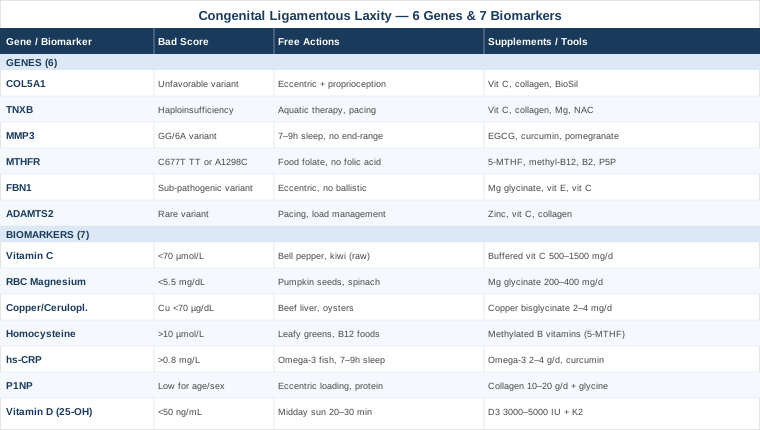
This article covers 7 measurable biomarkers tied directly to collagen synthesis and ligament integrity — from copper and ceruloplasmin to homocysteine and P1NP — along with 6 genes including COL5A1, TNXB, MTHFR, and FBN1 that influence how your connective tissue is built and maintained. For each biomarker and gene, you will find specific protocols: what to do without supplements, what to take with full dosing and cycling guidance, and which side effects to watch for. You will also find a summary of a book that directly challenges the medical establishment's handling of hypermobility — and four evidence-based complementary approaches most clinicians never mention. If you have been told to "just strengthen" your way out of this condition, what follows may fundamentally change how you think about it.
7 Biomarkers That Reveal What Is Happening in Your Connective Tissue
The ligaments holding your joints together are not passive cables — they are living tissue, constantly being synthesized, cross-linked, and remodeled. That process depends on specific nutrients, enzymes, and signaling molecules. When any of them fall out of range, the system weakens — not catastrophically and all at once, but gradually, in ways that show up as pain, instability, and fatigue.
The seven biomarkers below are chosen specifically for their connection to connective tissue biology. Each one is measurable, interpretable, and linked to concrete intervention options. Taken together, they give you a working picture of whether your body has the raw materials and biochemical conditions it needs to maintain ligament integrity.
Biomarker 1: Serum Vitamin C (Ascorbic Acid)
Why it matters. Vitamin C is not optional for collagen — it is mandatory. The enzymes that hydroxylate proline and lysine residues (prolyl and lysyl hydroxylase) require ascorbic acid as a direct co-factor. Without adequate hydroxylation, collagen chains cannot form proper triple-helix structures, and the resulting fibers are mechanically inferior. Research published in Nutrients (2017) documented the essential role of vitamin C in collagen biosynthesis and structural skin integrity. For ligamentous tissue, the same biochemistry applies — the demand is constant, and the margin for deficiency is narrow.
How to measure it. A serum ascorbic acid test is available through most labs and typically costs $30–$60. Optimal functional range: 70–120 µmol/L. Standard lab ranges are wider, but for connective tissue support, staying in the upper third of the reference range is preferable. Plasma levels do not fully reflect tissue saturation, so clinical context matters.
If the score is low, the plan without supplements
Prioritize high-density dietary sources: one red bell pepper provides approximately 150 mg of ascorbic acid; fresh guava, kiwi, strawberries, and broccoli are also strong contributors. These foods also deliver bioflavonoids that may enhance collagen fiber stabilization. Cooking destroys ascorbic acid, so raw or lightly steamed preparation is preferable. A consistent whole-food approach can raise serum levels by 20–30 µmol/L within 4–6 weeks.
If the score is low, the plan with supplements or equipment
Buffered vitamin C (calcium or magnesium ascorbate): 500–1500 mg/day, divided into two doses to improve retention. Liposomal vitamin C at 500–1000 mg/day offers better bioavailability and less gastrointestinal impact for sensitive individuals. No cycling is necessary — vitamin C is water-soluble and not stored. Side effects: doses above 2000 mg/day can cause loose stools and, with sustained very high intake, may increase oxalate load in people prone to kidney stones. For connective tissue support, 1000 mg/day in divided doses is a reasonable target for most people.
Biomarker 2: RBC Magnesium
Why it matters. Standard serum magnesium is a poor indicator of total body status — the body maintains serum levels in a tight range by pulling magnesium from cells and bone, masking intracellular deficiency. Red blood cell (RBC) magnesium is a meaningfully better proxy. Magnesium is involved in hundreds of enzymatic reactions, including those governing energy production in stabilizing muscles, regulation of the autonomic nervous system (directly relevant in hypermobility-associated POTS), and neuromuscular signaling that affects joint proprioception.
How to measure it. RBC magnesium is available through specialty and functional medicine labs including SpectraCell, Quest, and Genova Diagnostics. Cost: $50–$100. Optimal range: 5.5–7.0 mg/dL. If your doctor has already tested "magnesium" and it came back normal, it was almost certainly serum magnesium — not RBC.
If the score is low, the plan without supplements
Emphasize magnesium-dense whole foods: pumpkin seeds are among the highest per ounce, followed by dark chocolate (70%+), spinach, Swiss chard, almonds, and black beans. Reducing alcohol and excess refined sugar also meaningfully reduces urinary magnesium excretion. Expect 8–12 weeks of consistent dietary change before RBC levels shift.
If the score is low, the plan with supplements or equipment
Magnesium glycinate: 200–400 mg elemental magnesium/day, taken in the evening — it also supports sleep quality as a secondary benefit. Magnesium L-threonate is an alternative if neurological symptoms or cognitive fog are prominent. Side effects: doses above 400 mg/day can cause loose stools, especially with oxide or citrate forms. Glycinate is the most GI-friendly option. Cycling: continuous use at physiological doses is appropriate for deficient individuals; no cycling protocol is required.
Biomarker 3: Copper and Ceruloplasmin
Why it matters. Copper is the essential co-factor for lysyl oxidase, the enzyme that catalyzes cross-linking of collagen and elastin fibers into mechanically robust networks. Without adequate lysyl oxidase activity, collagen fibers form but fail to interconnect — structurally analogous to rope strands that are not twisted together. The clinical parallel is instructive: Menkes disease, a genetic disorder of copper transport, produces severe connective tissue laxity, joint hypermobility, and skin fragility as direct consequences of copper enzyme deficiency. Ceruloplasmin, the primary copper-carrying protein in blood, is a more stable indicator of functional copper status than serum copper measured alone.
How to measure it. Order both serum copper and ceruloplasmin simultaneously. Combined cost: $50–$80. Optimal serum copper: 70–140 µg/dL. Ceruloplasmin: 20–35 mg/dL. The copper-to-zinc ratio (ideally between 0.7 and 1.0) is worth calculating — high-dose zinc supplementation, extremely common in wellness protocols, competitively depletes copper through shared intestinal transporters.
If the score is low, the plan without supplements
Beef liver is the single richest dietary source of copper, providing 4–12 mg per 3 oz serving. Oysters are the densest source per ounce. Cocoa powder, cashews, and sesame seeds offer meaningful plant-based amounts. If high-dose zinc supplements are currently in use, reducing or pausing them before retesting is often sufficient to restore copper balance.
If the score is low, the plan with supplements or equipment
Copper bisglycinate: 2–4 mg/day. Never supplement copper in isolation — maintain an appropriate zinc-to-copper ratio (approximately 8:1, meaning 16–32 mg zinc alongside 2–4 mg copper). Cycling: copper accumulates; a one-week break monthly or quarterly serum copper reassessment is prudent. Side effects: excess copper causes nausea, liver toxicity, and neurological symptoms. Do not exceed 4 mg supplemental copper per day without medical supervision. Adequacy, not excess, is the target here.
Biomarker 4: Homocysteine
Why it matters. Homocysteine is an amino acid produced during methionine metabolism. When it accumulates — due to genetic variants like MTHFR, B vitamin insufficiency, or poor diet — it directly inhibits lysyl oxidase activity and disrupts normal collagen cross-linking. Research has linked elevated homocysteine to connective tissue fragility, higher fracture risk, and markers consistent with reduced tensile strength in ligamentous tissue. This is one of the most underappreciated mechanisms in the hypermobility literature.
Peter Attia emphasizes keeping homocysteine below 10 µmol/L as part of a longevity-oriented panel. For individuals with ligamentous laxity, this threshold is especially relevant since the collagen machinery is already operating under genetic pressure from other directions.
How to measure it. Standard serum homocysteine, $30–$60. Below 10 µmol/L is optimal; above 15 µmol/L is a clear red flag for connective tissue, cardiovascular, and neurological risk simultaneously.
If the score is high, the plan without supplements
Increase dietary folate (dark leafy greens, lentils, asparagus), B12 (meat, eggs, dairy, shellfish), and B6 (poultry, fish, potatoes). Reducing alcohol is important — it impairs B vitamin absorption and disrupts homocysteine clearance. Consistent dietary improvement can lower homocysteine by 3–5 µmol/L within 8–12 weeks. Avoiding synthetic folic acid (common in fortified foods and standard B-complex supplements) is advisable for individuals with MTHFR variants — see the genetics section below.
If the score is high, the plan with supplements or equipment
Methylated B vitamins: 5-MTHF (methylfolate) 400–800 mcg/day + methylcobalamin 1000–2000 mcg/day + pyridoxal-5-phosphate (activated B6) 25–50 mg/day. These active forms bypass potential MTHFR enzyme limitations. Riboflavin (B2): 25–50 mg/day supports the MTHFR reaction independently and is often overlooked. Cycling: reassess homocysteine at 3–6 months and adjust dose accordingly. Side effects: in people with MTHFR TT genotype beginning high-dose methylfolate, occasional overmethylation symptoms (anxiety, irritability, palpitations) can occur — start at 200–400 mcg and increase gradually.
Biomarker 5: hs-CRP
Why it matters. High-sensitivity C-reactive protein (hs-CRP) is the most accessible marker of systemic low-grade inflammation. In connective tissue, chronic inflammation upregulates matrix metalloproteinases (MMPs) — particularly MMP-1, MMP-3, and MMP-9 — which degrade collagen in ligaments and joints. Even mild elevations in hs-CRP (1–3 mg/L) can accelerate ligament matrix turnover in a net-negative direction, meaning the body breaks down more collagen than it rebuilds. For someone already working against a genetic predisposition to laxity, this biochemical environment is particularly costly.
Peter Attia recommends targeting hs-CRP below 0.8 mg/L. For individuals with ligamentous laxity, staying well under 1.0 mg/L is a practical and meaningful goal.
How to measure it. Standard hs-CRP blood test, $20–$40, available through virtually any laboratory. Test during a stable, illness-free period — hs-CRP is nonspecific and elevates acutely with any infection or injury, which would confound interpretation.
If the score is high, the plan without supplements
Dietary quality is the highest-leverage intervention: reduce refined carbohydrates, seed oils, and ultra-processed foods, and prioritize omega-3 rich fish (salmon, sardines, mackerel), olive oil, and colorful vegetables. Sleep quality has a significant independent effect on hs-CRP — inadequate sleep (under 7 hours) reliably elevates it. Regular moderate exercise also reduces chronic inflammation, while overtraining does the opposite — the dose matters.
If the score is high, the plan with supplements or equipment
Omega-3 fatty acids (EPA + DHA): 2–4 g/day of combined EPA/DHA — the most robustly evidence-backed anti-inflammatory supplement available. Phospholipid-bound curcumin (Meriva or BCM-95 formulations): 500–1000 mg/day — standard curcumin has near-zero bioavailability; only phospholipid-complexed or piperine-enhanced forms reach meaningful tissue concentrations. Cycling curcumin: 8–12 weeks on, 4 weeks off, particularly without regular liver enzyme monitoring. Side effects: curcumin can inhibit CYP3A4 enzymes, affecting the metabolism of some medications; omega-3s at high doses can modestly increase bleeding time — relevant if surgery is planned.
Biomarker 6: P1NP — Your Direct Collagen Synthesis Marker
Why it matters. Procollagen type I N-terminal propeptide (P1NP) is the most direct available marker of type I collagen synthesis in the body. When collagen is actively being built, P1NP rises; when synthesis is suppressed or insufficient, P1NP falls. Type I collagen is the dominant structural protein in ligaments, tendons, and bone. Tracking P1NP gives you a real-time signal of whether your body is in a net-positive collagen synthesis state — something no other standard biomarker panel directly captures.
While P1NP is most commonly used in osteoporosis monitoring as a bone formation marker, its relevance to ligamentous tissue is logical and underutilized in clinical practice for this population.
How to measure it. Serum P1NP is often ordered as part of a bone turnover panel. Cost: $70–$150 depending on the laboratory. Reference ranges vary by age and sex. For connective tissue context: higher P1NP within the normal range suggests active anabolic collagen synthesis; very low P1NP may indicate insufficient substrate or biological drive to rebuild. Test fasted in the morning for best consistency.
If the score is low, the plan without supplements
Load-bearing exercise is the most potent natural stimulator of collagen synthesis. Eccentric loading of tendons and ligaments — slow-descent squats, eccentric heel raises, controlled resistance band exercises — triggers a meaningful collagen synthesis response. Timing matters: a brief high-load mechanical stimulus before consuming collagen building-block nutrients enhances the response (this is supported by research from Keith Baar's lab at UC Davis). Adequate dietary protein — 1.6–2.0 g per kg of body weight — provides the amino acid substrate P1NP reflects.
If the score is low, the plan with supplements or equipment
Hydrolyzed collagen peptides: 10–20 g/day, taken 30–60 minutes before exercise or alongside a vitamin C source to support hydroxylation. The combination of collagen peptides plus vitamin C before mechanical loading has been specifically studied for ligament tissue and shows promise for upregulating collagen synthesis. Glycine: 3–5 g/day — glycine is the rate-limiting amino acid in collagen synthesis and is chronically underrepresented in modern protein intake. Side effects: hydrolyzed collagen and glycine are both exceptionally well-tolerated; glycine at 3–5 g may improve sleep quality as an additional benefit. No cycling required.
Biomarker 7: 25-OH Vitamin D
Why it matters. Vitamin D receptors are expressed in connective tissue cells — fibroblasts, tenocytes, and chondrocytes — where active vitamin D influences gene expression involved in extracellular matrix synthesis and remodeling. Low vitamin D is consistently associated with higher rates of musculoskeletal pain, joint instability, and impaired tissue healing. For individuals already managing ligamentous laxity, this association compounds an existing structural vulnerability. There is also emerging evidence linking vitamin D status to autonomic regulation, directly relevant for those with hypermobility-associated dysautonomia.
How to measure it. 25-OH vitamin D blood test, $30–$60. Optimal functional range for musculoskeletal health: 50–80 ng/mL, following Peter Attia's framework. The conventional clinical "sufficient" threshold of 30 ng/mL is too conservative for connective tissue optimization.
If the score is low, the plan without supplements
Midday sun exposure (arms and legs, without sunscreen) for 20–30 minutes can generate 10,000–20,000 IU endogenously in fair-skinned individuals — far more than any food source. Fatty fish (salmon, mackerel, herring), pastured egg yolks, and beef liver provide smaller but meaningful dietary amounts. In northern latitudes during winter months, sun alone is rarely sufficient to maintain optimal levels regardless of dietary effort.
If the score is low, the plan with supplements or equipment
Vitamin D3: 3000–5000 IU/day as a starting point; this is fat-soluble, so take with the largest meal. Always pair with vitamin K2 (MK-7 form): 100–200 mcg/day — K2 directs calcium appropriately and reduces soft tissue calcification risk associated with higher vitamin D levels. Retest at 90 days to confirm you are in the 50–80 ng/mL target range; some individuals require 7,000–10,000 IU/day depending on absorption and genetic factors. Side effects: vitamin D toxicity is rare under 10,000 IU/day but is possible with months of unsupervised high-dose supplementation; pairing with K2 and periodic retesting reduces practical risk. No cycling required — supplementation is typically continuous, especially through winter.
With seven clear biochemical targets identified, the question that naturally follows is why some individuals are more vulnerable to these imbalances in the first place. That answer lives in the genome — and the next section maps the most clinically relevant genetic factors.
What Your Genes May Reveal About Ligament Laxity
Genetic testing for connective tissue health is not yet a precise clinical tool — most variants identified to date represent probabilistic risk factors, not deterministic diagnoses. That said, several specific genes have accumulated enough human evidence to be clinically meaningful. Understanding your predispositions can explain why standard interventions sometimes fall short — and can direct more targeted biochemical support toward the specific weak points.
Ali Torkamani at Scripps Research has championed the use of polygenic risk scores in clinical care, arguing that aggregating many modest genetic effects produces more actionable signal than individual variants considered in isolation. Gary Brecka's approach, which has gained significant traction in applied genomics, focuses particularly on methylation pathway variants — especially MTHFR — as high-leverage points that affect multiple downstream biological processes including connective tissue integrity through homocysteine-mediated mechanisms.
The six genes below represent the most clinically tractable variants for connective tissue laxity. Consumer genetic data (23andMe, Ancestry) can be interpreted through third-party tools like Genetic Genie or SelfDecode, while clinical genetic panels ordered through a medical geneticist or specialist provide more comprehensive coverage.
Gene 1: COL5A1 — The Type V Collagen Regulator
What it does. COL5A1 encodes the alpha-1 chain of type V collagen, a minor but critically important fibril-forming collagen that regulates the diameter and organizational architecture of type I collagen fibers in tendons and ligaments. Without proper type V collagen template function, type I fibers form irregularly — larger in diameter and less structured, producing mechanically weaker tissue.
The rs12722 polymorphism (C to T variant) in COL5A1's 3' UTR has been studied extensively in sports injury research. The TT genotype is associated with reduced tendon and ligament mechanical integrity and significantly higher ACL injury rates in athletes. Classical Ehlers-Danlos Syndrome (caused by pathogenic COL5A1 or COL5A2 mutations) produces frank joint hypermobility and skin hyperextensibility — the clinical endpoint of a spectrum that rs12722 may represent at a more subtle level.
If the gene is unfavorable, the plan without supplements
Eccentric and isometric resistance training is the most important structural intervention — these loading patterns stimulate COL5A1 expression in tendons and ligaments. Proprioception-focused exercises (single-leg balance, unstable surface work, perturbation training) are especially relevant because type V collagen loss also impairs sensory mechanoreceptors in connective tissue. Avoid excessive joint end-range loading — particularly into hypermobile ranges — as this compounds fiber disorganization over time.
If the gene is unfavorable, the plan with supplements or equipment
Vitamin C (1000 mg/day) supports all collagen hydroxylation steps. Hydrolyzed collagen peptides (10–15 g/day) provide hydroxyproline-rich peptides shown to stimulate collagen gene expression in fibroblasts. Choline-stabilized orthosilicic acid (BioSil): 5–10 mg/day — silicon in this bioavailable form has published human data showing stimulation of type I and type V collagen synthesis in connective tissue cells. Cycling: collagen peptides can be continuous; orthosilicic acid is typically cycled 12 weeks on, 4 weeks off. Side effects: both collagen peptides and orthosilicic acid at these doses are very well tolerated.
Gene 2: TNXB — Tenascin-X and Matrix Architecture
What it does. Tenascin-X (encoded by TNXB) is an extracellular matrix glycoprotein that organizes collagen fibers and regulates their spacing and lateral adhesion. It is essential for proper ligament architecture. TNXB haploinsufficiency — where one copy of the gene is non-functional — produces a well-documented connective tissue disorder clinically indistinguishable from hypermobile EDS: generalized joint hypermobility, skin hyperextensibility, and easy bruising, without the characteristic scarring features of classical EDS. TNXB variants are found in a subset of patients originally classified as hEDS, making it one of the few genetically explained causes of the hypermobility-dominant phenotype.
If the gene is unfavorable, the plan without supplements
Aquatic therapy and pool-based exercise reduce joint loading while building neuromuscular control — particularly valuable when collagen organization is globally impaired. Pacing strategies are critical: TNXB haploinsufficiency is associated with fatigue and post-exertional symptom flares. Stabilization bracing (custom orthotics, prophylactic joint taping) reduces accumulated connective tissue stress during daily activity.
If the gene is unfavorable, the plan with supplements or equipment
A broad connective tissue support stack is the most practical approach: Vitamin C (1000–2000 mg/day), hydrolyzed collagen peptides (15–20 g/day), magnesium glycinate (300–400 mg/day), and antioxidants targeting oxidative damage to extracellular matrix proteins — alpha-lipoic acid (300–600 mg/day) or N-acetylcysteine (NAC) (600–1200 mg/day). Cycling NAC: 8–12 weeks on, 4-week break, due to potential glutathione feedback effects. Side effects: NAC can cause nausea at higher doses; alpha-lipoic acid may modestly lower blood glucose — relevant for individuals on diabetes medications.
Gene 3: MMP3 — The Matrix Breakdown Regulator
What it does. MMP3 (stromelysin-1) is an enzyme that degrades multiple extracellular matrix proteins including fibronectin, laminin, and collagen types II, III, IV, IX, and X. It also activates several other matrix metalloproteinases, amplifying its effect. The rs679620 polymorphism influences MMP3 expression level — the GG genotype, as well as the 6A/6A promoter variant, is associated with higher MMP3 activity, meaning faster collagen breakdown. In the context of ligamentous laxity, an elevated MMP3 activity genotype accelerates matrix degradation and directly opposes any collagen synthesis effort.
If the gene is unfavorable, the plan without supplements
Sleep is one of the most potent, underutilized regulators of MMP activity — inadequate sleep elevates systemic MMP levels measurably and consistently. Consistent 7–9 hours, with attention to sleep architecture (avoid alcohol before bed; optimize room temperature to 65–68°F), is the highest-leverage non-supplemental intervention available. Avoiding repetitive joint end-range loading also reduces local MMP3 induction within connective tissue itself.
If the gene is unfavorable, the plan with supplements or equipment
Several natural compounds selectively down-regulate MMP3 expression. EGCG (from green tea or supplement): 400–800 mg/day — multiple in vitro and early human studies document reduced MMP-3 and MMP-9 activity. Cycling: 6–8 weeks on, 2–4 weeks off given liver considerations at higher doses. Phospholipid curcumin: 500–1000 mg/day inhibits NFkB-driven MMP transcription. Pomegranate extract (punicalagin/ellagic acid): 500 mg/day — emerging evidence for MMP inhibition through antioxidant pathway modulation. Side effects: EGCG above 800 mg/day can raise liver enzymes in sensitive individuals; liver function monitoring is advisable with long-term use.
Gene 4: MTHFR — The Methylation Gateway
What it does. MTHFR does not encode a structural protein — it is a metabolic enzyme that converts dietary folate into the active form (5-methyltetrahydrofolate) needed for the methionine cycle. Its relevance to connective tissue runs through homocysteine: impaired MTHFR function reduces methyl-folate production, elevates homocysteine, and that elevated homocysteine directly inhibits lysyl oxidase — the same collagen cross-linking enzyme implicated in the copper section above.
This is the mechanism Gary Brecka has made central to his genomics framework: MTHFR variants are not just cardiovascular risk factors — they compromise the enzymatic infrastructure that holds connective tissue together. The C677T variant in homozygous TT form reduces MTHFR activity by up to 70%; A1298C has milder effects. Compound heterozygosity (one copy of each variant) also meaningfully impairs function. Testing the homocysteine biomarker above directly quantifies the downstream consequence.
If the gene is unfavorable, the plan without supplements
Maximize dietary folate from natural food sources — dark leafy greens, lentils, asparagus, and liver — rather than synthetic folic acid. Critically: for TT individuals, folic acid (the synthetic form found in fortified foods and many standard B-complex supplements) can block folate receptors and worsen the functional picture. Label reading and switching to whole-food folate sources is the non-supplement path.
If the gene is unfavorable, the plan with supplements or equipment
L-methylfolate (5-MTHF): 400–1000 mcg/day — this bypasses the defective MTHFR enzyme entirely by providing the active end-product directly. Methylcobalamin (B12): 1000–2000 mcg/day sublingual. Riboflavin (B2): 25–50 mg/day — riboflavin is MTHFR's enzymatic co-factor and can partially rescue function even with TT variants. Pyridoxal-5-phosphate (activated B6): 25–50 mg/day. Cycling: reassess homocysteine at 3–6 months and adjust dose. Side effects: some TT individuals experience overmethylation symptoms (anxiety, irritability, heart palpitations) when starting methylfolate — begin at 200–400 mcg and titrate up slowly.
Gene 5: FBN1 — Fibrillin and the Elastic Scaffold
What it does. FBN1 encodes fibrillin-1, the structural glycoprotein forming microfibrils in the extracellular matrix. These microfibrils serve as scaffolding for elastin assembly and contribute the elastic recoil properties of connective tissue. Pathogenic FBN1 mutations cause Marfan syndrome. However, the spectrum of FBN1 variants extends well beyond classic Marfan — hypermobile individuals with marfanoid features (tall stature, long limbs, pectus deformity, arachnodactyly) frequently carry sub-pathogenic FBN1 variants that impair fibrillin-1 function without meeting full Marfan diagnostic criteria.
A key downstream consequence: FBN1 dysfunction impairs sequestration of TGF-β (transforming growth factor beta) in the matrix, causing excessive TGF-β signaling — a driver of aberrant tissue remodeling and fragility.
If the gene is unfavorable, the plan without supplements
Avoiding high-impact and ballistic loading (plyometrics, contact sports) is prudent, especially when marfanoid features are present — for confirmed pathogenic FBN1 variants, cardiac evaluation (echocardiography for aortic root dimensions) is medically necessary. Eccentric, controlled resistance training builds tensile strength in connective tissue without the joint-loading spikes that compound fibrillin-related laxity.
If the gene is unfavorable, the plan with supplements or equipment
Magnesium glycinate or taurate: 300–400 mg/day — animal research in Marfan model mice has shown magnesium supplementation reduces aortic pathology by modulating TGF-β signaling; human extrapolation is preliminary but biologically grounded. Mixed tocopherols (vitamin E): 200 IU/day, targeting oxidative damage to fibrillin microfibrils. Vitamin C: 1000 mg/day as a baseline antioxidant and collagen co-factor. Note: individuals with confirmed pathogenic FBN1 variants should be under cardiological and genetics medical supervision — supplement protocols here are adjunctive, never primary. Side effects: high-dose alpha-tocopherol alone above 400 IU/day may increase bleeding tendency; mixed tocopherols at 200 IU/day are safer.
Gene 6: ADAMTS2 — Procollagen Processing Quality
What it does. ADAMTS2 is a procollagen N-proteinase — it cleaves the N-terminal propeptide from type I, II, and III procollagen, a required step to convert precursor molecules into mature, mechanically functional collagen fibers. Variants or insufficiency of ADAMTS2 result in dermatosparaxis-type EDS in severe form, but more subtle ADAMTS2 variants are identifiable in broader hypermobility presentations. Impaired ADAMTS2 function leaves incompletely processed procollagen units in collagen fibers, reducing their mechanical quality and increasing tissue laxity over time.
Zinc is the essential metal co-factor for ADAMTS2 activity — suboptimal zinc status (which is widespread in Western diets) can amplify the functional consequences of any ADAMTS2 variant.
If the gene is unfavorable, the plan without supplements
Pacing and load management are the primary non-supplemental tools. When collagen processing quality is compromised, the cumulative effect of tissue micro-trauma without adequate recovery is disproportionately damaging. Scheduled rest days, activity modification, and avoiding overuse of symptomatic joints reduce the burden on an impaired matrix remodeling system.
If the gene is unfavorable, the plan with supplements or equipment
Zinc bisglycinate: 15–30 mg/day with food (best absorbed away from high-phytate foods like grains and legumes). Always maintain zinc-to-copper balance — add 2 mg copper bisglycinate if using zinc consistently. Vitamin C (1000 mg/day) and collagen peptides (10–15 g/day) provide the substrate support. Cycling: at 30 mg zinc/day, quarterly copper testing is advisable. Side effects: zinc above 40 mg/day suppresses copper absorption and causes nausea on an empty stomach — always take with food and monitor copper status.
The genetic and biochemical picture now has considerable depth. What follows draws both of these frameworks together through a lens that most physicians still have not encountered — and that directly challenges how hypermobility is categorized and managed clinically.
A Book That Reframes Everything: Key Insights from Disjointed
Disjointed: Navigating the Diagnosis and Management of Hypermobile Ehlers-Danlos Syndrome and Hypermobility Spectrum Disorders by Diana Jovin (2020) assembles contributions from over 30 international EDS specialists and constitutes one of the most comprehensive, evidence-referenced resources on hypermobility available in print. It is not a self-help book — it is a clinical and patient reference that systematically challenges several assumptions that still define standard medical practice.
1. Hypermobility Is Rarely Benign
The label "benign joint hypermobility syndrome" — the older clinical term — vastly underestimates the condition's systemic impact. The book documents with extensive citations that pain, fatigue, proprioceptive deficits, dysautonomia, and gastrointestinal dysmotility are documented comorbidities with real biological mechanisms, not psychosomatic phenomena. Renaming does not change the burden, but accurate characterization changes treatment.
2. The Genetics Are Polygenic, Not Single-Gene
Disjointed reviews the evidence that hEDS is almost certainly a polygenic condition — no single gene explains most cases. This is why genetic panels for "EDS genes" frequently return negative results in hypermobile patients. Many genes, each contributing modest effects, collectively produce the phenotype. This aligns precisely with polygenic risk score approaches from researchers like Ali Torkamani and explains why standard genetic testing underwhelms clinically.
3. Mast Cells Create a Connective Tissue Degradation Loop
The book details the emerging science linking mast cell activation syndrome (MCAS) with hypermobility. Mast cells embedded in connective tissue release proteases and histamine that degrade extracellular matrix — potentially explaining why many hypermobile individuals notice worsening laxity during allergic, stress, or dietary reactivity episodes. This loop (lax tissue → mast cell activation → matrix degradation → more laxity) is a vicious cycle, and one rarely discussed in standard orthopedic or rheumatological care.
4. Proprioception Is Neurologically Impaired, Not Just Structurally Deficient
Joint position sense is objectively reduced in hypermobile individuals — measured consistently in controlled studies. This is a neurological deficit, not merely a structural one. Treatment that ignores proprioceptive retraining addresses only half the problem. Research summarized in the book shows that sensory motor training produces better functional outcomes than strengthening alone.
5. Dysautonomia Is a Core Feature
POTS and related autonomic dysfunctions appear in a significant proportion of hypermobile individuals. Lax connective tissue in blood vessel walls impairs normal venous return, and the autonomic nervous system compensates through tachycardia and altered vasoconstriction. Interventions addressing vascular tone — sodium loading, compression garments, graded exercise reconditioning, and hydration management — are as clinically important as joint-specific management.
6. Physical Therapy Approach Must Fundamentally Shift
The book makes the case that therapists treating hypermobile patients must move away from flexibility protocols (counterproductive) toward stability, co-contraction, and proprioception-first programs. Therapeutic priorities include deep stabilizing muscles (multifidus, transversus abdominis), sensory motor integration exercises, and closed kinetic chain movements performed in joint-neutral positions. Open kinetic chain exercises that load joint end-ranges should be minimized or removed.
7. Fatigue Has a Biochemical and Mechanical Explanation
The mechanical inefficiency of hypermobile joints — requiring more continuous muscle activation just to maintain basic stability — creates a persistent energetic drain. This is compounded by mitochondrial dysfunction, dysautonomia, and disordered sleep common in chronic pain. The book draws on ME/CFS pacing strategies as a framework for energy management, arguing these translate directly to hypermobility-related fatigue with good clinical rationale.
8. GI Dysfunction Is Structural, Not Anxiety-Based
Gut connective tissue shares the same structural vulnerabilities as joint connective tissue. Hypermobile individuals have documented higher rates of GERD, gastroparesis, irritable bowel, SIBO, and pelvic floor dysfunction — not because they are anxious, but because the extracellular matrix holding intestinal smooth muscle, mesenteric ligaments, and sphincter structures together is the same impaired matrix affecting joints. Treating GI symptoms without addressing the connective tissue context misses a major driver.
9. Central Sensitization Compounds the Pain Picture
Chronic pain in hypermobility develops central sensitization components over time — the nervous system itself becomes hyperreactive. Disjointed documents this and argues for a combined biological-neurological approach: tissue-level biochemical interventions (as outlined in this article) alongside pain neuroscience education, graded exposure therapy, and appropriate psychological support. Neither the biological nor the neurological dimension alone is sufficient.
10. Knowing the 2017 Diagnostic Criteria Shortens the Diagnostic Odyssey
On average, hypermobile EDS patients see 7–10 specialists before receiving an accurate diagnosis. The book quantifies this delay and its consequences: years of inappropriate treatment and accumulated damage from unrecognized instability. Presenting biomarker data, genetic information, and a working knowledge of the 2017 EDS International Classification (Malfait et al.) to a knowledgeable physician compresses this process significantly.
These frameworks — biochemical, genetic, and now systems-level — converge on the same core insight: the body's connective tissue infrastructure responds to the conditions you create for it. The following approaches offer additional levers that align with this biology.
Complementary Approaches With Meaningful Evidence
Four modalities from the evidence-reviewed list have sufficient condition-relevant human support to include here. Evidence quality varies — the strongest applies specifically to hypermobility-related features; others are drawn from closely adjacent conditions sharing the same biological substrate. None replace the biochemical and genetic strategies above; all are best used as adjuncts.
Yoga — Stability Practice, Not Flexibility Practice
Yoga for ligamentous laxity is counterintuitive on the surface — hypermobile individuals often gravitate toward it because they are naturally good at it, but standard yoga repeatedly loads joints through end-range positions that worsen laxity over time. The clinically relevant application is hypermobility-aware yoga: emphasis on isometric holds, joint stacking rather than joint opening, and conscious co-activation of stabilizing muscles rather than passive mobility exploration.
A clinical framework published in the Journal of Bodywork and Movement Therapies has examined modified yoga protocols for hypermobile EDS populations, emphasizing proprioceptive awareness, shortened lever arms, and stability-first sequencing. Large randomized trials specific to ligamentous laxity remain limited, but the broader proprioceptive training literature consistently supports the mechanism: repetitive sensorimotor engagement in joint-neutral positions reconstructs position sense and improves functional stability over time.
In practice: seek teachers certified in therapeutic or Iyengar yoga, or explicitly trained with hypermobility populations. Avoid Bikram and hot yoga (heat increases ligament laxity). Yin yoga — passive, long-hold deep stretches — is actively contraindicated. Focus on slow, isometric sequences using props that prevent end-range loading. 20–30 minutes, 3–4 sessions per week, emphasizing consistency over volume.
Biofeedback — Training the Proprioceptive Deficit Directly
Biofeedback directly targets the proprioceptive deficit documented in hypermobile individuals — the measurable reduction in joint position sense that drives instability, compensatory muscle overuse, and injury risk. By providing real-time sensory information about body position, muscle activation patterns, or balance that the nervous system is not generating reliably on its own, biofeedback creates a training stimulus for sensorimotor recalibration.
Balance and postural biofeedback using force plates and pressure-sensitive insoles has been studied in hypermobility-related conditions, with documented improvements in postural stability and self-reported joint stability scores. Proprioceptive training intervention studies in hypermobile populations have demonstrated significant improvements in joint position sense and functional stability after 8 weeks of biofeedback-enhanced exercise protocols.
Practical application: begin with simple balance board training (wobble boards, BOSU balls), which provides low-cost proprioceptive biofeedback accessible at home. Clinical electromyographic (EMG) biofeedback to guide muscle activation patterns around specific unstable joints is available through physiotherapists and rehabilitation clinics. Sessions of 20–30 minutes, 2–3 times per week for an initial 8–12 week course, constitute a standard protocol before reassessment.
Breathing-Based Therapies — The Autonomic Angle
The connection between breathing and ligamentous laxity runs through the autonomic nervous system. Dysautonomia and POTS — documented comorbidities in hypermobile populations — involve dysregulated sympathetic-parasympathetic balance that worsens under physical and metabolic stress. Diaphragmatic and slow-paced breathing techniques directly modulate vagal tone and heart rate variability (HRV), which are measurable indices of autonomic stability.
Slow paced breathing at approximately 5–6 breath cycles per minute has been shown in multiple clinical trials to significantly increase HRV and reduce sympathetic dominance. In hypermobility, where vessel wall laxity contributes to orthostatic symptoms, improving autonomic regulation through breathing addresses a genuine biological mechanism — not merely a relaxation response. The physiological effect is real and reproducible.
Practice: Box breathing (4-4-4-4: inhale 4 seconds, hold 4, exhale 4, hold 4) or resonance frequency breathing (5–6 cycles/minute) practiced daily for 10–20 minutes. Morning sessions during the lying-to-standing transition are particularly relevant for POTS-related symptoms. Consistency over 6–8 weeks produces measurable improvements in HRV and orthostatic tolerance. A biofeedback-paired HRV monitor (Polar H10 chest strap with a compatible HRV app) makes the protocol more precise and trackable.
Photobiomodulation (Low-Level Laser Therapy)
Photobiomodulation (PBM) applies red and near-infrared light to tissue, stimulating cytochrome c oxidase in mitochondria, increasing local ATP production, reducing oxidative stress, and promoting tissue repair. In connective tissue, this translates to measurable increases in fibroblast proliferation and collagen synthesis. For individuals with ligamentous laxity, PBM may support the connective tissue rebuilding process when applied over affected joints — particularly in combination with the biochemical substrates described in the biomarker section.
A randomized controlled trial published in Lasers in Medical Science found that low-level laser therapy applied to knee tendons significantly increased collagen synthesis markers and reduced tendinopathy pain scores compared to sham treatment over 8 weeks. Studies specific to ligamentous laxity are limited, but the biological mechanism — photon absorption driving fibroblast and collagen activity — is well-established in connective tissue.
Practical application: devices delivering 630–850 nm wavelengths at 10–50 mW/cm² are appropriate for soft tissue work. Sessions of 10–20 minutes over affected joint areas, 3–4 times per week, for an initial 4–8 week period, constitute a reasonable protocol. Home-use PBM panels (Joovv, Mito Red Light) cover larger surface areas than most clinical devices and are available for personal use. Combining PBM with post-application light eccentric loading may offer a synergistic collagen synthesis benefit. Note: avoid application to areas of active infection, open wounds, or directly over the thyroid.
Conclusion
Congenital ligamentous laxity is a biological condition, not a fixed outcome. The genetics and biomarkers covered here do not define your ceiling — they define your starting point and help identify which specific mechanisms need targeted support. Tracking seven key biomarkers (vitamin C, RBC magnesium, copper and ceruloplasmin, homocysteine, hs-CRP, P1NP, and 25-OH vitamin D) gives you a measurable picture of whether your connective tissue is getting what it needs. Understanding the six genetic factors (COL5A1, TNXB, MMP3, MTHFR, FBN1, ADAMTS2) tells you where to direct that support most precisely.
The practical next step is not to implement everything at once. Start with two or three biomarkers — homocysteine and 25-OH vitamin D are widely available, inexpensive, and highly actionable. If you already have consumer genetic data, reviewing your MTHFR status first is logical, since its downstream effect on connective tissue is the most directly modifiable with a targeted B vitamin protocol. From there, layer in the additional biomarkers and genetic considerations methodically.
If you have not yet received a formal evaluation for hypermobile EDS or a hypermobility spectrum disorder, the 2017 clinical diagnostic criteria (Tinkle et al.) provide a clear framework — and presenting them alongside your biomarker data to a knowledgeable physician shortens the diagnostic process considerably. Better information creates better decisions. The biology is complex, but it is not unapproachable.