This article contains AI generated content.
· UpdatedCongenital Myopathy Genes Biomarkers — 9 Genes And 6 Biomarkers To Track
Introduction
Congenital myopathies are a group of inherited muscle disorders that typically present at or near birth, often as low muscle tone, generalized weakness, and — in many subtypes — early and disproportionate respiratory difficulties. The term covers a diverse family of conditions, each tied to a specific genetic mutation and a specific pattern of structural change visible in muscle tissue under a microscope. What unites them is that they are genetic at their core, often chronic, and deeply individual in the way they affect daily life. Two people with the same diagnostic label can have dramatically different severities, risks, and needs based on which gene is affected and which variant of that gene they carry.
If you are living with one of these diagnoses — or caring for someone who is — you have likely encountered advice that is either too broad to be useful ("exercise gently," "stay active") or too bleak to be actionable ("this is genetic, management is supportive"). Neither framing serves you well. Both miss a layer of specificity that has become available in the past decade through advances in genetic sequencing and neuromuscular biomarker science. The difference between knowing you have "congenital myopathy" and knowing you carry an RYR1 mutation with malignant hyperthermia susceptibility, for example, is not academic — it is the difference between being safe and being in danger during a routine surgical procedure.
What has changed significantly is the precision of available information. We now know which genes account for the majority of cases, which mutations carry the highest risk for respiratory failure or cardiac involvement, and which combinations of biomarkers provide the earliest warning before a complication escalates. At the same time, a growing body of exercise neuroscience — much of it not written specifically for rare muscle disease — contains principles directly relevant to preserving function and quality of life in this population.
This article takes a structured approach through the most useful of these tools. First, it covers six biomarkers that give the clearest real-time picture of disease activity and complication risk, with specific guidance on what to do when values are abnormal. Then it maps nine key genes currently responsible for the majority of congenital myopathy diagnoses, explaining what each one means practically and what evidence-supported strategies exist with or without supplements. Additional sections synthesize insights from exercise neuroscience and examine the complementary approaches with the strongest human clinical evidence for neuromuscular conditions. The premise throughout is straightforward: better, more specific information leads to better decisions.
Summary
This article covers six measurable biomarkers — including respiratory muscle tests, creatine kinase, cardiac peptides, lactate, myoglobin, and aldolase — and explains what each reveals about disease activity, how to measure it affordably, and what to do if results are abnormal, with and without supplements. The genetics section maps nine key genes (RYR1, NEB, ACTA1, TPM2, TPM3, MTM1, DNM2, SELENON, and MYH7), explaining what each means for daily risk management and which compensatory strategies have the strongest evidence behind them. Beyond the core clinical content, the article also presents ten actionable insights from exercise neuroscience, summarizes the complementary approaches with the most credible human evidence (breathing therapies, biofeedback, photobiomodulation, mindfulness, and massage therapy), and closes with a direct call to action. The central message: knowing your specific gene variant and tracking a small, well-chosen set of biomarkers can shift care from reactive crisis response to proactive, informed self-management.
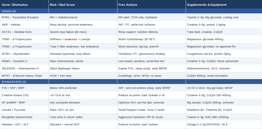
Moving from the overview to practical action, the biomarker section below is where the most immediate clinical value lives — these are measurements you can initiate today, with a specialist referral or a standard lab order.
6 Biomarkers to Track if You Have Congenital Myopathy
Not all laboratory tests are equally useful in congenital myopathy. Because these are structural and genetic conditions rather than primarily inflammatory or degenerative in the muscular dystrophy sense, standard "muscle enzyme panels" can be misleading — creatine kinase can be entirely normal in several subtypes, a fact that has contributed to delayed diagnoses in people who had real weakness but unremarkable blood work. The six biomarkers that follow are chosen deliberately: each either directly reflects muscle fiber integrity, flags the most serious known complications, or provides early warning before symptoms worsen to the point where options narrow.
Biomarker 1: Forced Vital Capacity (FVC) and Respiratory Muscle Pressure Testing
Why it matters: Respiratory failure is the leading cause of death across many congenital myopathy subtypes, including those driven by SELENON, NEB, and RYR1 mutations. The alarming aspect is that respiratory muscle weakness often progresses silently — patients may notice only mild breathlessness or fatigue without recognizing that their reserve has dropped close to the threshold where nighttime hypoventilation begins. By the time obvious daytime symptoms appear, the situation can be advanced.
What it may reveal: Forced vital capacity (FVC) measures how much air you can exhale after a full breath. Maximal inspiratory pressure (MIP) and maximal expiratory pressure (MEP) test the strength of the breathing muscles directly. Together, they distinguish reduced lung volume from reduced muscle force. A supine FVC significantly lower than upright FVC — a drop of more than 10% between positions — indicates diaphragm weakness specifically, a warning sign that can be missed entirely without this comparison. FVC below 50% of predicted significantly increases the risk of sleep-disordered breathing. Below 30%, the risk of acute respiratory failure is high.
How to measure it: Spirometry with MIP, MEP, and supine FVC is performed in a pulmonary function laboratory by a respiratory therapist or pulmonologist. The test takes approximately 30 minutes and is painless. Cost: $50–$150 with standard insurance coverage in the US; usually covered for documented neuromuscular conditions. Annual testing is recommended as the minimum frequency, increasing to every six months once FVC falls below 60% of predicted. Published neuromuscular guidelines consistently rank this as the highest-priority surveillance test in congenital myopathy care.
If the score is bad — the plan without supplements: Inspiratory muscle training (IMT) using a threshold resistance trainer — available commercially for $35–$50 — trains the inspiratory muscles against a calibrated load. Research in neuromuscular disease populations supports improvements in MIP of 15–25% over 8–12 weeks of consistent use (20–30 minutes daily, 5 days per week at 30% of current MIP). Pacing activities to avoid respiratory fatigue, sleeping in a semi-recumbent position, and learning to recognize early signs of nocturnal hypoventilation (morning headaches, excessive daytime sleepiness, difficulty concentrating) are essential behavioral steps. Non-invasive ventilation (NIV) with bilevel positive airway pressure (BiPAP) is the evidence-based standard of care once FVC drops below 50% or symptoms appear; early initiation consistently improves outcomes.
If the score is bad — the plan with supplements or equipment: Vitamin D3 (2000–4000 IU daily) with vitamin K2 (100–200 mcg) supports respiratory muscle contractility alongside all skeletal muscle function — deficiency is associated with greater neuromuscular weakness across multiple patient populations. Magnesium glycinate (200–400 mg daily) supports muscle relaxation and may reduce respiratory muscle spasm in susceptible individuals. The most impactful piece of equipment is a BiPAP device, used nightly, which dramatically reduces the workload on respiratory muscles during sleep and consistently improves daytime energy and cognitive clarity. A home pulse oximeter ($20–$40) used nightly allows independent monitoring of oxygen saturation and catches silent drops before daytime symptoms develop. For IMT, cycle 6 weeks of progressive training followed by 2 weeks at a maintenance level.
Biomarker 2: Creatine Kinase (CK)
Why it matters: Creatine kinase is the standard indicator of muscle fiber damage — it is released into the bloodstream when muscle cells are injured or break down. In muscular dystrophies, CK is often 10–100 times the upper limit of normal. In congenital myopathies, the pattern is different and more subtle: CK can be normal, mildly elevated (2–5 times normal), or — particularly in RYR1 carriers during physical or thermal stress — acutely and significantly elevated. The subtlety is the problem; a normal CK does not mean nothing is happening.
What it may reveal: A persistently elevated resting CK suggests ongoing muscle fiber damage, which may indicate that exercise or activity levels are exceeding what the structurally compromised muscle can safely recover from. An acute spike — especially in an RYR1 mutation carrier — can be the first signal of rhabdomyolysis, which requires urgent medical attention. Serial measurements over time allow correlation with activity level, illness, temperature exposure, and medication changes, building a personalized map of what drives damage in that individual.
How to measure it: Standard serum CK is a routine blood test available at any clinical laboratory. Cost: $20–$60 without insurance; often included in broader metabolic panels at minimal additional cost. Always measure CK in a rested state — no intense exercise for at least 48 hours before the draw — to establish a reliable baseline. CK naturally varies by sex, ethnicity, and total muscle mass, so interpreting results in isolation without context is a common source of confusion. Research on CK in congenital myopathy diagnosis highlights this variability.
If the score is bad — the plan without supplements: A persistently elevated CK (more than 3 times the upper limit of normal at rest, in the absence of acute illness) warrants a careful review of activity type and volume. High-impact and eccentric exercise — descending stairs, lowering weights, downhill walking — is disproportionately damaging to structurally compromised muscle fibers and should be minimized or replaced with concentric and low-impact alternatives (cycling, swimming, aquatic resistance training). Hydration is critical, with a daily target of 2–3 liters of water adjusted for body size, since dehydration concentrates myoglobin in the kidneys and multiplies rhabdomyolysis risk significantly.
If the score is bad — the plan with supplements or equipment: Creatine monohydrate (3–5 g daily) has the strongest evidence base of any supplement for improving available muscle function in myopathic conditions. It increases phosphocreatine availability in muscle cells, improves ATP regeneration, and has been shown to reduce exercise-induced CK elevation in several trials. Cycling protocol: 12 weeks on, 4 weeks off, monitoring CK at the start and midpoint of each cycle. Coenzyme Q10 (200–400 mg daily, taken with a fat-containing meal for absorption) supports mitochondrial ATP production and may reduce oxidative damage to muscle cell membranes. Avoid NSAIDs acutely during elevated CK episodes — they reduce renal clearance of myoglobin and compound kidney injury risk.
Biomarker 3: NT-proBNP and BNP (Cardiac Biomarkers)
Why it matters: Cardiac involvement in congenital myopathy is subtype-specific but real and often undermonitored. Mutations in MYH7 and some TTN variants directly affect cardiac muscle fibers and can cause cardiomyopathy. Beyond direct cardiac gene involvement, any congenital myopathy with chronic respiratory insufficiency imposes secondary stress on the right ventricle through chronically elevated pulmonary pressure — a condition called cor pulmonale. N-terminal pro-B-type natriuretic peptide (NT-proBNP) is released by the ventricular walls in response to pressure and volume overload, making it a sensitive early signal of cardiac stress.
What it may reveal: Elevated NT-proBNP can indicate early dilated or hypertrophic cardiomyopathy, right heart strain from chronic nocturnal hypoventilation, or developing fluid retention from reduced cardiac output. Identifying this elevation before overt symptoms appear — before ankle edema, before exertional dyspnea, before orthopnea — allows cardiologists to intervene with medications or NIV titration before irreversible structural changes occur. Even mild but persistent elevation warrants a cardiac echo.
How to measure it: NT-proBNP is a standard serum test. Cost: $30–$100 without insurance; commonly covered in neuromuscular workups and cardiac screenings. It should be measured at initial evaluation and then annually in patients with subtypes known to involve cardiac risk, or any time respiratory function declines noticeably. A cardiologist with neuromuscular disease experience is the appropriate specialist for full interpretation, including echocardiography correlation.
If the score is bad — the plan without supplements: The primary intervention for NT-proBNP elevation driven by respiratory causes is optimizing nighttime ventilation — ensuring NIV settings are appropriately calibrated and that nocturnal oxygen saturation is not dropping below 94%. Sodium restriction (under 2 g daily) reduces cardiac preload. Adapted low-intensity aerobic activity (30 minutes of gentle cycling or water-based movement, 3–5 days per week, adjusted for individual capacity) supports cardiac conditioning without imposing excessive demand. Daily weight monitoring flags early fluid retention before it becomes symptomatic.
If the score is bad — the plan with supplements or equipment: Magnesium taurate (200–400 mg daily) supports cardiac muscle function and rhythm regulation. CoQ10 at higher doses (400 mg daily) has specific evidence in cardiomyopathy management; the Q-SYMBIO trial demonstrated improvements in cardiac outcomes with CoQ10 in heart failure populations. A pulse oximeter used nightly ($20–$40) is the most cost-effective piece of monitoring equipment for detecting the silent nocturnal desaturations that most commonly drive NT-proBNP elevation in neuromuscular patients. If cardiomyopathy is confirmed, prescribed medications (ACE inhibitors or beta-blockers, depending on type) are the primary intervention; supplements serve as adjuncts, not replacements.
Biomarker 4: Lactate and Pyruvate (and the Lactate-to-Pyruvate Ratio)
Why it matters: Several congenital myopathy subtypes involve secondary mitochondrial dysfunction — particularly those driven by RYR1 and SELENON mutations, where calcium dysregulation chronically impairs mitochondrial activity. When mitochondria are underperforming, cells shift toward anaerobic energy production, generating lactate as a byproduct. Elevated resting lactate — or a lactate-to-pyruvate ratio above 20:1 — reflects this metabolic imbalance and identifies a potentially addressable component of muscle dysfunction.
What it may reveal: In the context of a known congenital myopathy, elevated lactate points to mitochondrial involvement that may respond to targeted nutritional and supplementation strategies. A lactate stress test, measuring lactate at rest, during graded exercise, and in recovery, provides a more detailed map of where the metabolic bottleneck lies and how quickly the system recovers. This information directly guides exercise prescription — specifically, identifying the intensity threshold above which lactate accumulates rather than clears.
How to measure it: Resting plasma lactate and pyruvate are run on the same draw. Cost: $50–$150 for the pair. The blood draw requires specific handling — no tourniquet stasis, immediate placement on ice — to avoid artifactual elevation that is common with poor technique. Academic neuromuscular centers and specialist metabolic labs reliably manage this protocol; routine community laboratories may not. Always request the lactate-to-pyruvate ratio calculation alongside the individual values.
If the score is bad — the plan without supplements: Avoiding prolonged fasting, which stresses mitochondrial metabolism. Small, frequent meals that include adequate carbohydrates and fat — the mitochondria's primary substrates — throughout the day. For exercise: targeting moderate intensity (the conversational zone, roughly 60–70% of maximum heart rate), which stimulates mitochondrial biogenesis without driving anaerobic metabolism. Sustained moderate activity for 20–30 minutes, 3–4 days per week, is likely more beneficial than shorter high-intensity efforts for this specific metabolic phenotype.
If the score is bad — the plan with supplements or equipment: Riboflavin (vitamin B2, 100–400 mg daily) is a cofactor for mitochondrial electron transport complexes I and II and is used in mitochondrial myopathy protocols where lactate elevation is present. Thiamine (vitamin B1, 100–300 mg daily) is essential for pyruvate dehydrogenase function and can normalize the lactate-to-pyruvate ratio when functional deficiency is present. Acetyl-L-carnitine (1000–2000 mg daily, taken in the morning) improves fatty acid transport into mitochondria. CoQ10 as ubiquinol (200–600 mg daily for better bioavailability than standard CoQ10) directly supports the mitochondrial electron transport chain at Complex III. These are commonly combined in mitochondrial disease protocols; in congenital myopathy with secondary mitochondrial involvement, the evidence is mechanistically extrapolated but the rationale is well-grounded.
Biomarker 5: Serum and Urine Myoglobin
Why it matters: Myoglobin is an oxygen-carrying protein stored inside muscle fibers. When muscle fibers are massively damaged — rhabdomyolysis — myoglobin floods into the bloodstream and then the urine, where it can precipitate and cause acute kidney injury. This is a medical emergency. Patients with RYR1 mutations are at particularly high risk: their calcium handling is dysregulated in ways that can be triggered into uncontrolled muscle breakdown by volatile anesthetic agents, extreme physical exertion, or sustained heat exposure. But rhabdomyolysis risk is not limited to this gene.
What it may reveal: Cola-colored or red-brown urine is the most visible warning sign of myoglobinuria and requires immediate emergency care. Before this point, serum myoglobin elevation alongside rising CK provides early warning during an episode in progress. In a non-emergency context, tracking myoglobin after specific activities or environmental exposures helps identify personal triggers — building an individualized avoidance map that reduces risk over time.
How to measure it: Serum myoglobin is a standard laboratory test. Cost: $30–$80. Urine myoglobin is typically checked during acute presentations. For routine monitoring in high-risk patients — particularly RYR1 carriers — a urine dipstick test after any unusually intense exercise provides a low-cost first screen: a positive blood result on dipstick without visible red blood cells on microscopy strongly suggests myoglobinuria. Home dipsticks cost $15–$30 per pack and require no specialist access.
If the score is bad — the plan without supplements: If acute rhabdomyolysis is suspected, the immediate response is aggressive oral hydration (1–2 liters of water quickly, then continued high-volume intake) and cessation of the triggering activity. Hospital presentation for intravenous hydration and kidney function monitoring is required for any confirmed episode. Long-term: every RYR1 carrier should carry a written medical alert indicating their mutation status and malignant hyperthermia susceptibility, inform all healthcare providers (particularly surgeons and anesthesiologists) before any procedure involving sedation, and confirm that dantrolene is available at the facility. Total intravenous anesthesia (TIVA) with propofol is the safe alternative to volatile agents. The published anesthesia management literature for RYR1 carriers should be shared with any treating anesthesiologist in advance.
If the score is bad — the plan with supplements or equipment: Taurine (2–4 g daily) has evidence in animal models and limited human studies suggesting it stabilizes calcium handling in muscles and reduces fiber damage — mechanistically relevant for RYR1, where calcium dysregulation is the central problem. N-acetylcysteine (600–1200 mg daily) is an antioxidant that may reduce oxidative damage during muscle breakdown episodes. Practically, a wearable heart rate monitor that flags exercise intensity helps RYR1 carriers identify when they are approaching intensities that have historically preceded symptom spikes, allowing them to regulate effort in real time. Cooling vests ($150–$400) are practical equipment for outdoor activity in warm weather, reducing the thermal trigger threshold.
Biomarker 6: Aldolase, AST, and ALT (Secondary Muscle Leak Markers)
Why it matters: Aldolase is a muscle enzyme that, like CK, leaks into the bloodstream when muscle fibers are damaged. It can be more sensitive than CK in certain congenital myopathy subtypes where CK is only mildly elevated or intermittently normal. AST (aspartate aminotransferase) and ALT (alanine aminotransferase) are known as liver enzymes but are also present in muscle — elevated values without concurrent liver disease indicate muscle as the source. This combination fills in the picture when CK alone appears insufficient to explain a patient's symptoms.
What it may reveal: In a person with known congenital myopathy and only mildly elevated CK, finding elevated aldolase and AST alongside a normal GGT (gamma-glutamyl transferase, which is liver-specific) confirms the elevation is muscle-sourced. This matters clinically because it prevents misdiagnosis as liver disease and keeps the clinical focus on the actual problem: ongoing muscle fiber damage from an identifiable cause that may be modifiable.
How to measure it: Aldolase is a standard blood test, cost $20–$60. AST, ALT, and GGT are typically bundled in a comprehensive metabolic panel ($30–$80). Always include GGT to distinguish muscle from liver origin — this is the critical step that most general practitioners omit, causing confusion. If aldolase and AST are elevated with a normal GGT, the source is almost certainly muscle. Research on aldolase as a muscle disease biomarker supports its use as a complement to CK in evaluating ongoing fiber damage.
If the score is bad — the plan without supplements: The same principles that apply to elevated CK apply here: reduce eccentric loading, increase hydration, extend recovery intervals between activity sessions. If values remain persistently elevated despite conservative measures and documented activity adjustment, a neuromuscular specialist review of the specific subtype and its structural vulnerability pattern is warranted — some congenital myopathies have muscle groups that are disproportionately fragile and require individualized activity modifications that go beyond general guidance.
If the score is bad — the plan with supplements or equipment: Omega-3 fatty acids (2–4 g EPA+DHA daily from fish oil) have membrane-stabilizing and anti-inflammatory effects on muscle cells and have shown modest reductions in exercise-induced enzyme elevation in several small trials. Vitamin E (200–400 IU daily as mixed tocopherols) complements omega-3s by protecting muscle cell membranes from lipid peroxidation. Cycling protocol: omega-3 supplementation can be maintained continuously; vitamin E is cycled at 12 weeks on, 4 weeks off. Aquatic therapy (2–3 sessions per week) is an ideal activity modification — it reduces mechanical stress on vulnerable fibers while maintaining conditioning and generating the movement needed to prevent deconditioning.
Having covered the six biomarkers that provide the clearest monitoring picture, the next section examines the genetic landscape that determines your individual risk profile — because knowing your specific gene changes what you monitor, what you avoid, and which emerging therapies may eventually apply to you.
9 Genes Behind Most Congenital Myopathies and What Each One Means
Genetic testing in congenital myopathy has transformed clinical care in the past decade. A diagnosis once depended entirely on what was visible in a muscle biopsy — nemaline rods, central cores, centrally placed nuclei. Today, whole-exome sequencing or targeted myopathy gene panels can identify the causal mutation in the majority of cases, and that specificity matters far beyond the diagnostic label. It determines anesthesia risk, cardiac surveillance needs, respiratory monitoring frequency, prognosis, and — increasingly — eligibility for emerging gene-targeted therapies.
The approach here draws on the model championed by clinical genomics researchers like Ali Torkamani at the Scripps Translational Science Institute — integrating genetic results with clinical context to generate actionable plans, not just reports. For each gene below, the question is not merely "what does this mutation cause" but "what should change about how this person manages their health as a result of knowing this."
RYR1 — Ryanodine Receptor 1
What it affects: RYR1 encodes the ryanodine receptor, the protein in muscle cells responsible for controlled calcium release from internal stores (the sarcoplasmic reticulum) into the cell interior, which is the trigger for muscle contraction. Mutations cause either impaired calcium release (reducing contraction strength) or dysregulated release (causing excessive, uncontrolled calcium flooding the cell). The latter scenario underlies two dangerous situations: malignant hyperthermia triggered by volatile anesthetic agents, and exercise or heat-induced rhabdomyolysis. MedlinePlus provides a detailed overview of RYR1 variants. RYR1 is the most commonly identified gene in non-dystrophic congenital myopathies, responsible for central core disease, multiminicore disease, and a broader spectrum of RYR1-related myopathy. Inheritance can be autosomal dominant (often causing malignant hyperthermia susceptibility) or autosomal recessive (typically causing weakness with lower MH risk but not zero).
If the gene is bad — the plan without supplements: The most critical intervention is ensuring that every surgeon, anesthesiologist, and emergency physician who treats this patient knows about the RYR1 mutation before any procedure. A MedicAlert bracelet or card clearly stating "RYR1 — Malignant Hyperthermia Susceptible" (if the mutation is in the triggering category based on functional testing or variant classification) is non-negotiable. Volatile anesthetic agents (halothane, sevoflurane, desflurane, isoflurane) and succinylcholine must be avoided; total intravenous anesthesia (TIVA) with propofol is the established safe alternative. For daily life: stay hydrated, avoid prolonged heat exposure, and use a heart rate monitor during any exercise to prevent entering intensities that have previously produced symptoms.
If the gene is bad — the plan with supplements or equipment: Taurine (2–4 g daily) has mechanistic support for stabilizing calcium handling in RYR1-related contexts by reducing sarcoplasmic reticulum calcium overload. Magnesium glycinate (300–400 mg daily) acts as a natural calcium antagonist at the RYR1 channel and may dampen excess calcium release. Dantrolene — a calcium release blocker — is the emergency pharmacological treatment for malignant hyperthermia and must be immediately available in any facility providing surgical care to confirmed RYR1 carriers. Cooling vests ($150–$400) are practical protective equipment for outdoor summer activity.
NEB — Nebulin
What it affects: Nebulin is an extraordinarily large protein (encoded by a gene with 183 exons) that runs the full length of the thin actin filament in each muscle sarcomere, functioning as a structural ruler that determines filament length and as a regulatory scaffold for contraction. Mutations in NEB — typically compound heterozygous or homozygous loss-of-function changes — disrupt thin filament organization, producing nemaline myopathy with predominantly proximal weakness (hips and shoulder girdle) and frequently disproportionate respiratory involvement. NEB myopathy is the most common form of nemaline myopathy.
If the gene is bad — the plan without supplements: Maintaining respiratory reserve through IMT before FVC decline becomes critical — the principle being that building reserve while it exists prevents crises later. Physical therapy focused on functional strengthening in the muscle groups still capable of generating useful force, rather than general aerobic training that may exceed recovery capacity. Assistive devices — ankle-foot orthoses, adaptive equipment for daily activities — reduce energy expenditure on low-priority tasks and preserve it for more demanding ones. Pulmonary function testing every 6–12 months as a non-negotiable baseline, increasing to every 3–6 months if respiratory function is declining.
If the gene is bad — the plan with supplements or equipment: No gene-specific pharmacological therapies are approved for NEB myopathy at this time, though gene therapy and exon-skipping approaches are in early development. Creatine monohydrate (3–5 g daily) remains the most evidence-grounded supplement for preserving available muscle function regardless of subtype. Adequate protein intake (1.4–1.8 g/kg of lean body mass daily) supports the muscle protein synthesis capacity that does remain. Checking current clinical trial registries for NEB myopathy is worthwhile, particularly through the Congenital Muscle Disease International Registry.
ACTA1 — Skeletal Muscle Alpha-Actin
What it affects: ACTA1 encodes the alpha-actin protein that forms the thin filaments interacting with myosin to generate every muscular contraction. De novo dominant mutations in ACTA1 tend to produce the most severe presentations — including fetal akinesia, contractures at birth, and respiratory failure requiring immediate ventilatory support. Recessive mutations are generally milder. ACTA1 mutations account for approximately 20% of all nemaline myopathy cases, making it the second most common NEM gene after NEB. Detailed variant classification is available through MedlinePlus.
If the gene is bad — the plan without supplements: For severe de novo cases, the primary determinants of survival are early and aggressive respiratory support and adequate nutritional delivery — often requiring nasogastric or gastrostomy feeding due to bulbar muscle involvement. For milder recessive phenotypes, the management approach mirrors NEB myopathy: functional physical therapy, respiratory monitoring, energy conservation strategies, and early identification of respiratory decline.
If the gene is bad — the plan with supplements or equipment: Nutritional support is particularly important where feeding difficulties are present — protein delivery via tube feeding if oral intake is insufficient to meet targets (1.6 g/kg/day). For ambulatory patients with milder phenotypes: the creatine and CoQ10 protocol described in the biomarker section. Melatonin (0.5–3 mg at bedtime) may support sleep quality, which is commonly disrupted by nocturnal respiratory effort in this population.
TPM2 and TPM3 — Tropomyosins
What they affect: Tropomyosin proteins, encoded by TPM2 (beta-tropomyosin) and TPM3 (gamma-tropomyosin), sit along the thin filament and control access of myosin to actin binding sites — essentially functioning as the on-off switch of muscle contraction. Mutations in either gene can cause nemaline myopathy, cap myopathy, or congenital fiber type disproportion. A distinctive feature of some TPM2 and TPM3 mutations is that they produce not just weakness but also excess stiffness or impaired muscle relaxation — mechanically different problems that require different management approaches. TPM3 mutations predominantly affect type 1 (slow-twitch) muscle fibers, the fibers designed for sustained endurance activity.
If the genes are bad — the plan without supplements: When the dominant phenotype is stiffness and cramping (rather than pure weakness), warmth facilitates muscle relaxation more effectively than cold. Hydrotherapy in a pool at 33–35°C, for 30 minutes 3–4 times per week, targets stiffness specifically while protecting weak fibers from impact loading. For patients with TPM3-specific type 1 fiber predominance, activity planning should account for reduced endurance capacity: shorter, more frequent movement sessions rather than sustained prolonged effort, which depletes type 1 fiber function before type 2 fiber function.
If the genes are bad — the plan with supplements or equipment: Magnesium glycinate (400 mg daily) is particularly relevant for the stiffness and cramping phenotype, as magnesium modulates muscle relaxation through competitive calcium antagonism. No gene-specific approved therapies exist for TPM2 or TPM3 mutations at this time. Stretching performed in warm water, combined with gentle progressive range-of-motion work, represents the most practical non-supplemental approach for the stiffness component. Monitoring type 1 fiber-dependent functional tasks (endurance walking distance, stair-climbing time) longitudinally provides a more sensitive measure of decline than standard strength testing for this subtype.
MTM1 — Myotubularin
What it affects: MTM1 encodes myotubularin, a phosphoinositide phosphatase essential for organizing the T-tubule membrane system inside muscle fibers — the internal network that transmits electrical signals from the muscle surface deep into the fiber to trigger calcium release. X-linked myotubular myopathy (XLMTM), caused by loss-of-function MTM1 mutations, is one of the most severe congenital myopathies. Affected males almost uniformly present with profound hypotonia and respiratory failure at birth, requiring ventilatory support from the neonatal period. Without sustained respiratory management, mortality in the first year of life is very high. Female carriers range from asymptomatic to significantly affected depending on X-inactivation patterns.
If the gene is bad — the plan without supplements: For severely affected males, care is primarily medical and supportive: long-term mechanical ventilation (often tracheostomy-based), gastrostomy feeding, intensive physiotherapy to maintain joint range of motion, and rigorous surveillance for scoliosis and hip dysplasia. Regular liver surveillance is important: MTM1 carriers have an elevated risk of peliosis hepatis (vascular liver lesions), particularly with anabolic steroid or certain hormone exposures. Any supplement or medication with androgenic or hepatotoxic potential must be discussed with a liver specialist before use in this population.
If the gene is bad — the plan with supplements or equipment: Gene therapy for XLMTM has been the subject of the Aspiro Therapeutics program (resamirigene bilparvovec, AT132); while safety concerns in larger patients led to trial suspension, the field remains active. Checking current enrollment opportunities through clinicaltrials.gov is important. A mechanical cough assistance device (CoughAssist) is among the most practically impactful equipment for XLMTM patients — it applies a mechanical assist to cough secretion clearance, reducing pneumonia risk significantly. High-protein nutrition delivery (1.6–2 g/kg/day) supports whatever muscle tissue is present.
DNM2 — Dynamin 2
What it affects: Dynamin 2 is a GTPase involved in membrane tubulation and T-tubule formation in muscle. Autosomal dominant DNM2 mutations cause centronuclear myopathy (DNM2-CNM), in which muscle fiber nuclei are displaced to the center of the fiber rather than positioned at the periphery, impairing force transmission and intracellular signaling. DNM2-CNM tends to be slowly progressive and is often compatible with ambulation well into middle age — a meaningfully different prognosis from the MTM1-related form of centronuclear myopathy. Ophthalmoplegia (external eye muscle weakness) and ptosis are common features. MedlinePlus covers DNM2 variant classifications.
If the gene is bad — the plan without supplements: Given the typically slowly progressive course, proactive maintenance is more central here than crisis management. Low-impact aerobic conditioning (30 minutes of cycling or swimming, 4 days per week) to preserve cardiovascular and residual muscular capacity. Avoiding prolonged immobility during illness — even brief bed rest deconditions myopathy patients disproportionately quickly compared to healthy individuals, and recovery is slow. Annual neuromuscular specialist reviews with respiratory function testing, and ophthalmology review for ptosis management if vision is affected.
If the gene is bad — the plan with supplements or equipment: Preclinical research has explored DNM2 knockdown strategies in animal models, with promising results, but no human therapies are approved. Creatine monohydrate (3–5 g daily) and CoQ10 (200–400 mg daily) remain the most supported general supplements. A home spirometer ($150–$300) for quarterly self-monitoring of respiratory function between annual specialist visits provides early warning of decline and enables timely specialist review before thresholds for NIV support are reached.
SELENON — Selenoprotein N
What it affects: SELENON (formerly SEPN1) encodes selenoprotein N, an endoplasmic reticulum protein involved in calcium handling and the regulation of oxidative balance within muscle cells — it contains a selenocysteine residue that makes it directly dependent on adequate selenium. Mutations in SELENON produce a phenotype that is deceptively mild-appearing in the limbs but severe in the respiratory muscles. Patients often look "not that weak" on standard assessment while having significant diaphragm weakness they and their clinicians are not detecting. SELENON mutations also typically produce rigid spine muscular dystrophy, a combination of scoliosis and spinal rigidity that independently restricts chest expansion and compounds respiratory compromise.
If the gene is bad — the plan without supplements: Respiratory monitoring must be frequent and must specifically include supine FVC and MIP — the tests that detect diaphragm weakness, which is the dominant threat in SELENON myopathy and can be absent from limb assessment. Polysomnography (sleep study) at presentation and annually thereafter detects nocturnal hypoventilation before daytime symptoms appear. Spinal management through physiotherapy, and potentially spinal bracing or surgical fusion, aims to preserve chest wall compliance. Early introduction of nocturnal BiPAP is associated with better outcomes in this subtype and should not be delayed until the patient is symptomatic.
If the gene is bad — the plan with supplements or equipment: Selenium is directly implicated in SELENON disease. While supplementing selenium cannot replace a non-functional protein, deficiency must be excluded and actively prevented — it compounds functional loss. Brazil nut selenium content is highly variable and unreliable; supplementation with selenomethionine (100–200 mcg daily, keeping total intake from all sources under 400 mcg) is more controllable and consistent. Vitamin E (400 IU as mixed tocopherols) acts as a complementary antioxidant alongside selenium in the glutathione peroxidase system. A home pulse oximeter for nightly monitoring ($30–$60) and a portable home spirometer ($150–$300) are the two highest-value equipment investments for SELENON mutation carriers.
MYH7 — Myosin Heavy Chain 7
What it affects: MYH7 encodes the beta-myosin heavy chain, expressed in both slow-twitch skeletal muscle fibers and cardiac muscle. This dual expression means MYH7 mutations can cause problems in two systems simultaneously. Skeletal muscle involvement produces Laing distal myopathy (slowly progressive weakness starting in the feet and ankle dorsiflexors) or myosin storage myopathy (with protein aggregates visible in slow-twitch fibers). Cardiac involvement can cause hypertrophic cardiomyopathy — and MYH7 is one of the most common single genes implicated in familial HCM. MedlinePlus provides comprehensive MYH7 variant guidance.
If the gene is bad — the plan without supplements: Cardiology referral with annual echocardiogram and ECG is essential and non-optional. MYH7-related HCM has established management protocols: beta-blockers or calcium channel blockers to reduce outflow obstruction, avoidance of high-intensity competitive sports, and in some cases implantable cardioverter-defibrillator placement based on risk stratification. For the skeletal muscle component: ankle-foot orthoses (AFOs) are commonly needed as foot drop progresses and are highly effective at restoring gait safety and preventing falls.
If the gene is bad — the plan with supplements or equipment: Avoid high-dose stimulant supplements that increase cardiac demand, particularly in the presence of confirmed HCM — this includes high-dose caffeine (over 200 mg), synephrine, and other adrenergic compounds. CoQ10 (400 mg daily as ubiquinol) has specific evidence for cardiomyopathy support. For skeletal muscle function: creatine monohydrate (3–5 g daily) and adequate protein (1.6 g/kg/day) as the foundational protocol. Cardiac monitoring equipment (a home AED if the family risk is high, or a medically reviewed wearable ECG monitor) may be discussed with the cardiologist in confirmed HCM cases.
The genetic landscape mapped above defines the biological terrain. What follows addresses how exercise neuroscience — a field not written for rare muscle disease — contains principles that are directly relevant to managing neuromuscular conditions with greater precision and effect.
10 Insights from Exercise Neuroscience That Change How to Think About Managing a Muscle Condition
The Huberman Lab Podcast, hosted by neuroscientist and Stanford professor Dr. Andrew Huberman, has produced some of the most rigorous and accessible science on muscle physiology, neuromuscular adaptation, fatigue management, and recovery available to a general audience. No specific episode addresses congenital myopathy, but several — particularly those featuring Dr. Andy Galpin, a muscle fiber physiologist — contain principles that apply directly. What follows synthesizes the ten most actionable insights from this body of work for anyone managing or supporting someone with a congenital myopathy.
1. Muscle Fiber Type Determines Which Exercises Help and Which May Harm
Most congenital myopathies preferentially affect type 1 (slow-twitch, oxidative) muscle fibers — the fibers designed for sustained low-intensity activity. This means that the default recommendation to "go for daily walks" is actually targeting the fiber population most compromised by the disease. Huberman and Galpin's discussions on fiber type specificity suggest that shorter, higher-effort activities (within individually safe limits) may generate less fiber damage than sustained low-intensity endurance work in type 1 fiber predominance conditions. This challenges the reflexive "gentle exercise" advice and argues for a more targeted, physiologically grounded prescription.
2. Neural Drive Is Trainable Even When Muscle Mass Cannot Be Built
Motor learning research covered across multiple Huberman episodes establishes that the neural component of strength — how efficiently the nervous system recruits available muscle fibers — can improve independently of muscle mass. For congenital myopathy patients who cannot safely build bulk or generate high forces, deliberate, patterned motor activation still improves neuromuscular efficiency. This is not a trivial point: a 10–20% improvement in motor unit recruitment efficiency translates directly into better functional performance from the same compromised muscle.
3. Slow-Wave Sleep Is When Muscle Repair Peaks — and Why Respiratory Management Protects It
Huberman's extensive coverage of sleep physiology highlights that anabolic processes in muscle — protein synthesis, cellular repair, growth hormone release — are concentrated during slow-wave (deep) sleep. This is precisely the sleep stage most disrupted by nocturnal hypoventilation, which is common and often undetected in congenital myopathy. The implication is direct and underappreciated: optimizing respiratory support during sleep is not just about keeping oxygen saturation numbers stable. It actively protects the biological processes responsible for maintaining what muscle function remains.
4. BDNF from Exercise Supports the Motor Neurons Driving Weakened Muscles
Brain-derived neurotrophic factor (BDNF) supports the survival and function of motor neurons — the nerve cells that activate muscle fibers. As Huberman's episode on BDNF and exercise details, even mild aerobic activity (20 minutes at moderate intensity) generates a meaningful BDNF pulse. For congenital myopathy patients with significantly reduced capacity, this is genuinely encouraging: even modest movement generates the neurotrophic signal that benefits the motor neurons driving remaining functional fibers. Water-based exercise may be the safest delivery mechanism — generating the physiological signal without imposing the eccentric loading that damages structurally vulnerable fibers.
5. Zone 2 Cardio Is the Specific Stimulus for Mitochondrial Improvement
Zone 2 cardio — activity at an intensity where you can hold a conversation but feel moderate effort, roughly 60–70% of maximum heart rate — is a major theme in Huberman's cardiovascular health episodes. This intensity specifically stimulates mitochondrial biogenesis and efficiency without the mechanical damage of higher intensities or the recovery demands of high-intensity intervals. For congenital myopathy subtypes with secondary mitochondrial dysfunction (particularly RYR1 and SELENON), 20–30 minutes of Zone 2 activity 3–4 times per week — via cycling, arm ergometry, or water-based movement — is likely one of the highest-value single interventions available.
6. Cold Exposure Protocols Need Modification — or Avoidance — for RYR1 Carriers
Huberman has discussed deliberate cold exposure (cold water immersion, cold showers) as a tool for improving mitochondrial density, reducing inflammation, and building thermal resilience. This advice does not transfer cleanly to the congenital myopathy population. For RYR1 carriers, sudden cold exposure can trigger muscle rigidity, dysfunction, or dysregulation of the calcium handling that is already compromised. This is one of several instances where a well-supported general health protocol requires specific modification for this population, and where understanding the gene provides the context to make that modification correctly.
7. Creatine Is the One Supplement with Consistent Backing Across Muscle Science
Multiple Huberman episodes and expert guests have addressed supplementation, and creatine monohydrate consistently emerges as the most evidence-supported muscle supplement available — with a safety profile established over decades of research across dozens of populations. For congenital myopathy patients, the mechanism is particularly relevant: creatine increases phosphocreatine availability in muscle cells, improving energy delivery during the initial seconds of muscle activation — a window that is often specifically compromised in structurally impaired fibers. The dose is simple: 3–5 g daily, no loading phase required.
8. Post-Exercise Protein Timing Has Disproportionate Returns
Huberman's coverage of protein synthesis and the anabolic window establishes that muscle protein synthesis is significantly elevated in the 30–60 minutes immediately following exercise. For congenital myopathy patients doing any functional training or movement, consuming 20–40 g of complete protein within this window is one of the simplest and most effective nutritional interventions — requiring no exotic supplements, only timing. Whey protein or a complete plant protein blend mixed in water or milk achieves this easily and is supported by extensive human evidence.
9. Deliberate Visual Focus During Movement Increases Motor Unit Recruitment
One of Huberman's more counterintuitive neuroscience insights: maintaining deliberate visual focus on the target of movement — rather than allowing the gaze to drift — activates the brain's arousal systems in ways that measurably increase motor unit recruitment. For congenital myopathy patients trying to maximize the force output from available muscle fibers, this technique requires no equipment and no supplements. Fixing the gaze intentionally during each exercise repetition activates more of what is neurologically available. This is a behavioral efficiency tool worth adopting from the first session.
10. Distinguishing Central from Peripheral Fatigue Is a Learnable and Valuable Skill
One of the most practically impactful ideas in Huberman's fatigue and endurance content: fatigue operates at two distinct levels simultaneously — peripheral (the muscle itself is depleted or damaged) and central (the brain is dampening motor drive to protect the system). In healthy people, these are managed intuitively. In congenital myopathy, the stakes of confusing them are higher. Treating all fatigue as peripheral — pushing through everything — risks structural muscle damage. Treating all fatigue as central — stopping at any discomfort — leads to avoidable deconditioning. Learning to distinguish these patterns, developed with time and specialist guidance, is one of the highest-value self-management skills in this condition.
The science above focuses on biology and adaptation. But living with congenital myopathy also involves the ongoing challenge of quality of life, pain, and psychological management. The approaches below address those dimensions with clinical evidence behind them.
Complementary Approaches with the Strongest Human Evidence
Breathing-Based Therapies
Breathing-based therapies encompass a range of techniques — inspiratory muscle training, pursed-lip breathing, controlled diaphragmatic breathing, and breath pacing protocols. Their relevance to congenital myopathy is not peripheral; for many patients, the ability to train and support respiratory muscles is as functionally important as any physiotherapy targeting the limbs. In subtypes where respiratory involvement is disproportionate to limb weakness (SELENON, NEB), respiratory-specific training may be the most impactful modality available.
Randomized controlled trials of inspiratory muscle training in neuromuscular disease populations have demonstrated clinically meaningful improvements in maximal inspiratory pressure and patient-reported dyspnea scores after 8 weeks of threshold-based training, compared to sham training. While congenital myopathy-specific RCTs are small in number, the physiological mechanism is identical to the populations studied, and the respiratory specialist community consistently recommends IMT as part of comprehensive neuromuscular care.
Practically: begin with a calibrated threshold IMT device set at 20–30% of the patient's current MIP. Train in two 15-minute sessions daily, 5 days per week. Increase resistance by 5% every two weeks as long as the patient completes all sessions without significant desaturation or dizziness. A pulse oximeter during training sessions is essential — stop if saturation drops below 92%. IMT should be initiated under the supervision of a respiratory therapist, particularly for patients with FVC below 60% of predicted, to establish the correct starting load and technique before home-based practice.
Biofeedback
Neuromuscular biofeedback uses surface electromyography (sEMG) to display the real-time electrical activity of specific muscles while the patient attempts to activate them. In congenital myopathy, where structurally impaired fibers produce weaker and sometimes irregular signals, biofeedback helps patients develop awareness of motor activation patterns they may not be able to feel reliably — and it enables targeted training of the neural recruitment patterns that drive remaining function. The goal is not to build muscle; it is to improve the efficiency of the nervous system's use of what muscle exists.
Systematic reviews of sEMG biofeedback in neuromuscular rehabilitation have found consistent, if modest, improvements in motor function outcomes when biofeedback is added to conventional physiotherapy. The benefit is most pronounced in conditions with selective rather than global weakness — which describes the pattern seen in most congenital myopathies, where specific fiber types or specific muscle groups are more affected than others, leaving useful heterogeneity in residual function.
Practically: clinical biofeedback is available through neurological rehabilitation centers and specialized physiotherapy practices. A standard course is 10–15 sessions of 45–60 minutes each. Between formal sessions, simplified home biofeedback — such as visual monitoring in a mirror, or pressure-based feedback from a foam squeeze ball — maintains the neural rehearsal effect. Cost per clinical session ranges from $80–$200, with variable insurance coverage. Virtual telerehabilitation versions are increasingly available, which is relevant given that many patients with rare neuromuscular diseases live at a distance from specialist centers.
Mindfulness Meditation and MBSR
Mindfulness-Based Stress Reduction (MBSR), the 8-week structured program developed by Jon Kabat-Zinn combining body scan meditation, sitting practice, and gentle movement, is well-established for its effects on pain perception, fatigue severity, anxiety, sleep quality, and psychological well-being in chronic disease populations. Fatigue is among the most debilitating symptoms reported by adults with congenital myopathy, and it carries a substantial central (cognitive and emotional) component that standard physiotherapy does not address. MBSR specifically targets this central dimension.
Systematic reviews of MBSR in chronic disease have demonstrated significant reductions in fatigue severity and improvements in psychological well-being, with effect sizes in the moderate range (standardized mean difference approximately 0.5–0.7) across a wide range of conditions including multiple sclerosis, cancer-related fatigue, and fibromyalgia — populations whose central fatigue mechanisms are directly comparable to those in neuromuscular conditions.
Practically: the validated protocol is the full 8-week MBSR program (2.5-hour weekly sessions plus 45 minutes of daily home practice). Hospital-affiliated programs and university medical centers typically run these programs at $300–$600. The free Palouse Mindfulness online program is a rigorous alternative. A more accessible starting point is 10–20 minutes of daily body scan or sitting meditation — apps such as Insight Timer offer guided sessions at no cost. The body scan component is particularly useful for congenital myopathy patients for developing the internal awareness needed to distinguish peripheral from central fatigue, directly supporting the self-management skill discussed in the neuroscience section.
Low-Level Laser Therapy and Photobiomodulation
Photobiomodulation (PBM) uses near-infrared light (typically 630–1100 nm) to stimulate mitochondrial activity within cells, specifically through absorption by cytochrome c oxidase (Complex IV of the mitochondrial electron transport chain). Several congenital myopathy subtypes involve secondary mitochondrial dysfunction, making PBM mechanistically relevant — it targets the same metabolic pathway that supplements like CoQ10 and riboflavin address, but through a non-pharmacological mechanism. The evidence base for PBM in congenital myopathy specifically is absent; the evidence comes from healthy and clinical populations in exercise science contexts.
Multiple randomized trials and meta-analyses from researchers including Leal Junior et al. have demonstrated that PBM applied to muscle before or after exercise significantly reduces exercise-induced CK elevation, reduces lactate accumulation, and improves time to exhaustion compared to sham treatment, with a consistently favorable safety profile. These findings have been replicated in sports medicine, rehabilitation, and clinical populations.
Practically: medical-grade PBM is available through physiotherapy and sports medicine clinics equipped with class 3B or 4 laser or LED devices. A typical protocol uses 3–6 J/cm² at 660–850 nm wavelength, applied for 60–120 seconds per target muscle site. Sessions 3 times per week are a reasonable starting frequency. Consumer-grade LED panels ($150–$500) are available for home use but vary significantly in dosing accuracy; a clinical assessment to establish the appropriate protocol is worth doing first. PBM is not a treatment for the underlying genetic defect — it is a supportive modality for managing energy availability and recovery capacity in affected muscle tissue.
Massage Therapy
Massage therapy in congenital myopathy addresses two overlapping issues: musculoskeletal discomfort from altered movement patterns and compensatory muscle overuse, and circulation impairment from reduced mobility leading to limb edema and joint stiffness. The direct evidence base in congenital myopathy is thin; the supporting evidence comes from broader neuromuscular disease populations — Charcot-Marie-Tooth disease, limb-girdle muscular dystrophy, and similar conditions — where manual therapy has demonstrated improvements in pain, passive range of motion, and perceived functional status.
Studies of manual therapy in neuromuscular conditions have reported clinically significant improvements in passive range of motion and pain visual analogue scores after 4 weeks of weekly 60-minute sessions, using techniques that emphasize effleurage and petrissage rather than deep tissue manipulation. The benefit in compensatory muscles — neck, upper trapezius, lumbar paraspinals — is particularly consistent in ambulatory patients who are using secondary muscle groups to compensate for primary weakness.
Practically: the single most important caution in this population is that deep tissue massage techniques — deep pressure, cross-fiber friction, percussion — can damage fragile muscle fibers. Light-to-moderate effleurage is appropriate; deep compression must be avoided. A massage therapist with experience in neuromuscular or muscular dystrophy patients is strongly preferable. Communicate the diagnosis clearly at the outset and ask the therapist to use significantly lighter pressure than standard. Sessions of 45–60 minutes, 1–2 times per week, focused on compensatory muscle groups, represent a safe and useful starting protocol. Some patients find 30-minute warm water hydrotherapy before massage sessions markedly improves tissue responsiveness.
Conclusion
Congenital myopathy is a lifelong condition, but the specificity with which it can now be characterized — down to the gene, the variant, and the measurable biological signals it produces — opens a meaningful gap between reactive management and informed, proactive care. The six biomarkers in this article give you a monitoring framework that can flag the most serious complications before they become crises. The nine genes give you the context to understand why each complication risk exists and what specifically to do about it, with or without supplements. The exercise neuroscience principles offer a more sophisticated template for physical activity than generic guidance allows. The complementary approaches offer additional tools with real human evidence behind them, not just plausibility.
None of this promises reversal of a genetic condition. What it offers is the difference between managing a condition blindly and managing it with the best available information — which consistently translates into better outcomes, fewer crises, and a higher quality of daily life. The next smart step is identifying where the biggest gap in your current care lies. If you have not had a full pulmonary function test including MIP, MEP, and supine FVC in the past year, that is where to start. If your specific gene variant has not been identified, a referral for next-generation genetic sequencing changes what is possible. And if you are navigating this largely without specialist input, a multidisciplinary neuromuscular center — combining neurology, pulmonology, cardiology, and physiotherapy expertise — gives you the infrastructure to use the information above most effectively.
Respiratory Endocrine & Metabolic
Musculoskeletal: Muscle Conditions
Neurological: Nerve Conditions
Cardiovascular: Heart Conditions