This article contains AI generated content.
· UpdatedContact Dermatitis Genes And Biomarkers - 5 Genes And 7 Biomarkers To Track
Introduction
Contact dermatitis affects roughly one in five people at some point in their lives, yet most walk away from a dermatologist appointment with the same two instructions: avoid the trigger, apply the steroid. For mild, one-time reactions, that is often sufficient. But for the millions dealing with chronic, recurrent, or treatment-resistant flares — where the skin never fully recovers between episodes and patch testing comes back frustratingly vague — that advice addresses only the surface of what is happening.
The reason generic guidance falls short is that contact dermatitis is not a single, uniform condition. Whether your skin reacts to nickel in a watch buckle, formaldehyde in a shampoo, or rubber accelerants in surgical gloves depends on a complex interaction of immune system architecture, skin barrier integrity, and genetic background that varies significantly from person to person. Two people with identical exposures can have entirely different outcomes. That difference is not bad luck — it is measurable biology.
This article approaches the condition from two complementary angles. The first is biomarker tracking: measurable signals in your blood and skin that reveal the current state of your immune activation, barrier function, inflammatory load, and tissue damage — all of which can be tested, monitored, and, in many cases, meaningfully improved. The second is genetics: the specific gene variants with the strongest evidence for contact dermatitis susceptibility, and what current research suggests can offset their effects. Beyond those two frameworks, you will also find a summary of emerging gut-skin research that is reshaping how practitioners think about inflammatory skin conditions, along with complementary modalities that have genuine clinical evidence for this type of condition.
Better information does not promise a cure, but it dramatically improves the quality of the decisions you can make — about what to test, what to change, and in which order. That is the practical promise of everything that follows.
Summary
This article covers ground that standard dermatology consultations rarely reach:
- 7 biomarkers worth tracking, from the affordable (eosinophil count, LDH — both available in a standard blood panel costing under $50) to the more specialized (TARC/CCL17, IL-31), each with a clear explanation of what it reveals, how to measure it, and what to do when results are unfavorable — with and without supplements. - 5 genes with the strongest evidence in contact dermatitis research, including filaggrin (FLG), which is responsible for lifelong skin barrier fragility in roughly 10% of Europeans, GSTP1, which governs how efficiently your body detoxifies contact allergens, and three immune pathway variants that determine how intensely you sensitize and react. Each comes with a compensation plan. - A research framework from the gut-skin axis — summarizing insights that challenge the conventional model of contact dermatitis as a purely local skin problem, with 10 evidence-backed principles for addressing the condition from the inside out. - Four complementary approaches — microbiome-directed therapy, mindfulness, photobiomodulation, and the autoimmune protocol — each selected because they have meaningful human clinical evidence specific to inflammatory skin conditions.
If you have tried avoidance strategies and topical treatments and still find yourself cycling through flares, the data below may reveal what has been missing from the picture.
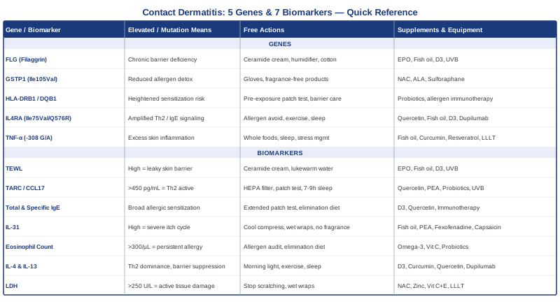
7 Biomarkers to Track for Contact Dermatitis
Contact dermatitis is not just about what touches your skin — it is about how your skin and immune system respond to that contact, and that response can be measured. The seven markers below are the most clinically meaningful for understanding disease activity, severity, and the underlying immune profile driving your reactions. Some are available through a standard blood draw at any primary care office; others require more specialized panels. All of them can meaningfully inform your next steps.
1. Transepidermal Water Loss (TEWL): Your Skin Barrier in Numbers
Why it matters
Transepidermal water loss measures how much water evaporates through your skin's outer layers. A high TEWL reading means the skin barrier is not holding moisture effectively and is allowing irritants and allergens to penetrate with much less resistance than they should. This single metric is the most important upstream factor in both irritant and allergic contact dermatitis: a compromised barrier does not just worsen existing reactions, it actively enables new sensitizations to substances that would not normally cause a problem in healthy skin.
Research shows that elevated TEWL precedes clinical flares, correlates with long-term disease severity, and persists between episodes in people with FLG gene mutations — explaining why certain people seem to have perpetually sensitive skin regardless of what they come into contact with. Measuring TEWL turns a vague sense of "sensitive skin" into a quantifiable baseline you can track over time.
How to measure it
TEWL is measured non-invasively using a Tewameter or evaporimeter — a device that detects moisture evaporation from the skin surface without any blood draw or skin disruption. Most dermatology clinics and academic skin research centers can perform this measurement, often at no additional cost during a consultation. Consumer-grade devices are available for home use in the $300–600 range; clinical instruments are more accurate, particularly for serial monitoring. Measurements are typically taken on the forearm or the site of concern in a temperature-controlled room after 20–30 minutes of acclimatization.
If the score is bad, the plan without supplements
Barrier restoration through physical means is the priority. Apply a ceramide-rich emollient (containing ceramides 1, 3, and 6-II) immediately after bathing while skin is still slightly damp — this is when absorption is highest. Use only lukewarm water; hot showers dissolve the skin's natural lipid layer. Replace any soap on affected areas with a fragrance-free, pH-balanced cleanser used sparingly. A room humidifier targeting 40–60% relative humidity reduces the evaporative gradient that pulls moisture out of already-compromised skin. Wear cotton directly against the skin, avoid extended water exposure (which paradoxically worsens barrier function by leaching natural moisturizing factors), and use cotton-lined gloves for any wet or chemical work.
If the score is bad, the plan with supplements or equipment
Evening primrose oil (providing 1–2 g of gamma-linolenic acid per day): GLA is a precursor to skin-protective prostaglandins and has been shown in multiple controlled trials to improve skin barrier function over 8–12 weeks. Take daily for a 12-week cycle, then reassess. Side effects are minimal; occasional mild GI discomfort resolves with food. Fish oil (EPA+DHA, 2–3 g/day): omega-3 fatty acids reduce arachidonic acid metabolites that degrade barrier lipid organization and also dampen the underlying inflammation. Take with meals to reduce GI side effects; no cycling required. Vitamin D3 with K2 (2000–5000 IU D3, 100 mcg K2 daily): vitamin D directly regulates gene expression for keratinocyte differentiation and skin barrier assembly. Test serum 25-OH-D before starting; target 40–60 ng/mL and recheck at 3 months. Low risk at these doses when paired with K2. For more advanced intervention, narrow-band UVB phototherapy (308 nm) can upregulate epidermal barrier proteins including filaggrin through multiple pathways — home devices exist and some are covered by insurance for appropriate diagnoses, but require dermatologist guidance and a defined treatment schedule.
2. TARC/CCL17: The Most Specific Marker of Allergic Skin Inflammation
Why it matters
TARC — Thymus and Activation-Regulated Chemokine, also known as CCL17 — is a protein released by inflamed skin that recruits Th2 immune cells into the tissue. It is among the most sensitive and specific blood markers for the severity of allergic skin inflammation, and its serum levels correlate more closely with active disease than most standard inflammatory markers. In clinical research, TARC has been used to track treatment response in atopic and allergic contact dermatitis. Critically, elevated TARC often reflects what is happening in the immune system before skin changes become visually severe.
A high TARC reading tells you your immune system is actively in Th2-skewed mode — the immune profile that drives allergic sensitization and amplifies reactions to contact allergens. This is one of the most actionable markers in this group because it points directly to the type of immune imbalance driving your skin to react.
How to measure it
TARC (CCL17) is measured from a standard serum blood sample. It is not included in routine panels, so you will need to request it specifically through a functional medicine practitioner, allergist, or immunologist. Reference ranges vary by laboratory; values above 450–700 pg/mL are typically considered elevated in adults. Cost ranges from approximately $80–180 depending on your location and insurance. In Japan and parts of Europe, TARC is used routinely as a clinical severity marker for atopic dermatitis — it is clinically validated, just not yet standard practice in most primary care settings in the United States.
If the score is bad, the plan without supplements
Elevated TARC signals an active Th2 immune environment, which means comprehensively reducing immune triggers is the first step. A thorough patch test — ideally the full European baseline series of 80+ allergens rather than the limited TRUE test — should identify all current sensitizers, followed by strict and systematic elimination. Beyond that, reducing the total allergen burden helps: HEPA air filtration at home, allergen-proof bedding covers if house dust mites are relevant, and removal of fragrance from all personal care and cleaning products. Sleep and stress management matter significantly here because cortisol spikes transiently shift immune tone toward Th2 dominance and can directly raise TARC levels. Consistent, restorative sleep (7–9 hours) is one of the most underrated anti-inflammatory interventions for this specific immune profile.
If the score is bad, the plan with supplements or equipment
Quercetin (500 mg twice daily with meals): a flavonoid with documented inhibitory effects on Th2 cytokine production, including IL-4 and IL-13, at the molecular level. Run in 8–12 week cycles with 4-week breaks. Rarely causes side effects at these doses; occasional headache reported. Palmitoylethanolamide (PEA) (600 mg twice daily): an endogenous fatty acid amide that downregulates mast cell activation — a key early step in Th2 skewing. Growing clinical evidence for its role in atopic and inflammatory skin conditions. Take continuously at this dose; no cycling required. Lactobacillus rhamnosus GG or Lactobacillus reuteri: specific probiotic strains with documented Th1/Th2 rebalancing effects in skin allergy clinical trials — take for a minimum of 12 consecutive weeks before assessing impact. Narrow-band UVB phototherapy: suppresses Th2 lymphocyte activity in skin tissue and has well-documented efficacy for both atopic and allergic contact dermatitis. Requires a dermatologist prescription; home devices are available but should be used with professional supervision to manage dose escalation and avoid burns.
3. Total IgE and Specific IgE Panel: Mapping Your Allergic Sensitization
Why it matters
Total serum IgE reflects overall allergic immune activity. People with elevated total IgE — indicating an atopic immune background — are significantly more likely to develop contact sensitization when exposed to allergens, and when they do, reactions tend to be more intense and harder to resolve. Specific IgE testing (ImmunoCAP or RAST panels) can identify exactly which substances — metals, proteins, fragrances, latex, food proteins — your immune system has produced specific antibodies against.
This matters because contact dermatitis is not always purely type IV (T-cell mediated delayed hypersensitivity). A subset of patients has overlapping IgE-mediated sensitization that makes reactions faster and more severe. Knowing both your total IgE and your specific sensitization profile helps you distinguish between irritant mechanisms, classical delayed-type allergy, and mixed presentations — each of which responds differently to intervention. It also identifies cross-reactive substances that patch testing alone might miss.
How to measure it
Total IgE is part of most standard allergy blood panels and costs approximately $25–80. Specific IgE panels typically cost $10–30 per allergen or $80–250 for a comprehensive panel covering common environmental and food allergens. These tests are available through primary care physicians, allergists, and many functional medicine practitioners. For contact dermatitis specifically, IgE testing is most useful when combined with clinical history and formal patch testing — together they provide a more complete map than either alone.
If the score is bad, the plan without supplements
High total IgE signals broadly sensitized immune activity, making comprehensive allergen identification and avoidance the priority. Extended patch testing (going beyond the standard 10-antigen panel) should be the next clinical step. An elimination diet trial lasting 2–4 weeks can identify food-based contributors — a well-documented phenomenon is the cross-reactivity between dietary nickel and nickel contact allergy, where high-nickel foods (oats, legumes, dark chocolate, shellfish) worsen skin reactions in nickel-sensitized individuals. Reducing the overall allergen burden from all sources — dietary, environmental, and chemical — gradually lowers the IgE-generating immune load over time.
If the score is bad, the plan with supplements or equipment
Vitamin D3 (2000–5000 IU/day): low vitamin D status correlates directly with elevated IgE and greater atopic sensitization in multiple population studies. Supplementation has been shown to downregulate IgE production through regulatory T-cell pathways. Test baseline levels first; target 40–60 ng/mL. Quercetin (500–1000 mg/day): inhibits IgE-mediated mast cell degranulation and reduces histamine release downstream of sensitization. Allergen-specific immunotherapy (sublingual or subcutaneous): where specific IgE testing identifies major sensitizers, allergen immunotherapy administered by an allergist can retrain immune tolerance over 3–5 years. This is the most definitively evidence-based long-term approach to reducing IgE-mediated sensitization, and it is the only intervention that addresses the root immune mechanism rather than suppressing its downstream effects.
4. IL-31: The Molecule Behind the Itch
Why it matters
IL-31 is a cytokine produced predominantly by Th2 T cells, and it is the primary molecular driver of itch in inflammatory skin conditions. Elevated serum IL-31 correlates closely with pruritus intensity in both allergic contact and atopic dermatitis — making it a direct window into how severely the itch-inflammation cycle is being activated. This matters clinically because scratching is not just a symptom that causes discomfort. It actively perpetuates barrier damage, increases the risk of secondary bacterial infection, and sustains the inflammation that triggered the itch in the first place. The itch-scratch cycle is a mechanical amplifier of the disease, and IL-31 is the molecular switch that starts it.
The clinical validation of anti-IL-31 therapy (nemolizumab, approved for atopic dermatitis in several countries) confirms that this cytokine is not peripheral to the disease — it is central to it.
How to measure it
IL-31 is measured via specialized serum immunoassay. It is not available through standard lab panels and is most commonly measured in research centers and academic medical facilities. Some functional medicine labs offer multiplex cytokine panels that include IL-31; cost typically ranges from $100–300 depending on the panel breadth. If direct measurement is not accessible, validated itch severity scales such as the NRS (Numerical Rating Scale) or POEM questionnaire correlate reasonably well with IL-31 levels and can serve as a practical proxy for tracking improvement over time.
If the score is bad, the plan without supplements
Physical itch management that does not involve scratching is essential. Cool compresses applied for 15–20 minutes provide meaningful, immediate relief by reducing the firing rate of itch-mediating C nerve fibers in the skin. Keeping skin cool generally reduces itch intensity — avoid overheating from exercise, hot showers, and heavy bedding during flares. Wet wrap therapy (a damp cotton layer applied over moisturizer, covered by a dry layer) reduces itch reliably overnight while also supporting barrier repair — a two-for-one intervention worth adopting during active flares. Removing all fragrance from personal care products is critical because fragrances are among the most potent IL-31 triggers in sensitized individuals. Habit reversal training — a structured behavioral approach to breaking the itch-scratch habit through awareness and competing responses — has genuine clinical evidence and is worth accessing through a psychologist or self-directed workbook.
If the score is bad, the plan with supplements or equipment
Fish oil (EPA+DHA 3–4 g/day with fatty meals): omega-3 fatty acids suppress IL-31 gene expression downstream of Th2 activation through multiple anti-inflammatory mechanisms. No cycling required; take long-term. PEA (palmitoylethanolamide) (600 mg twice daily): modulates peripheral nerve sensitization and reduces itch signal amplification through mast cell inhibition. Several clinical trials in pruritic skin conditions show meaningful itch reduction over 4–8 weeks. Oral fexofenadine (180 mg at night): second-generation antihistamine that reduces histamine-amplified itch without significant sedation at this dose. Not directly targeting IL-31 but reduces the converging signal that amplifies it. Topical capsaicin cream (0.025–0.075%): initially burning but causes sustained desensitization of itch nerve fibers (TRPV1 receptor downregulation) with consistent use over 2–4 weeks. Start with the lowest available concentration on a small test area. TENS (transcutaneous electrical nerve stimulation) units: consumer devices available for $40–100 that block itch signals via gate-control nerve pathway modulation — useful as an adjunct between flares or while waiting for other interventions to take effect.
5. Absolute Eosinophil Count: Reading Your Allergic Immune State
Why it matters
Eosinophils are white blood cells recruited by Th2 cytokines — particularly IL-5 — during allergic inflammation. Even a mildly elevated count (300–500 cells/µL) in someone with recurrent skin reactions suggests the immune system is persistently running in allergic mode. In the context of contact dermatitis, persistent eosinophilia indicates either ongoing allergen exposure, unresolved sensitization, or a broader atopic background that is amplifying skin reactions and preventing full resolution between flares.
Eosinophils are not passive bystanders — they release inflammatory mediators including major basic protein, eosinophil peroxidase, and cationic protein that directly damage skin tissue. A chronically elevated eosinophil count is therefore both a marker and an active contributor to the tissue injury that keeps the skin compromised.
How to measure it
Eosinophil count is included in any standard complete blood count (CBC) with differential — one of the most common and affordable blood tests in medicine, costing $15–50 at standard labs. The absolute eosinophil count (AEC) is the number to watch; values above 300–500 cells/µL in the context of recurrent skin reactions are clinically significant, even if they fall within some laboratories' "normal" range. Results are typically available within 24–48 hours and can be ordered by any primary care physician.
If the score is bad, the plan without supplements
Persistently elevated eosinophils demand systematic identification and elimination of all allergen exposures. This means professional patch testing with the extended European baseline series rather than the limited 10-antigen TRUE test, an environmental allergen audit covering workplace and home exposures, and a structured trial elimination diet if food-driven contributions are suspected. In the right geographic context, ruling out intestinal parasite infection is worthwhile — parasites are a major and frequently overlooked driver of eosinophilia. Stress reduction also matters: dysregulated HPA axis activity increases IL-5 production, which directly drives eosinophil proliferation and survival.
If the score is bad, the plan with supplements or equipment
Omega-3 fatty acids (EPA+DHA 2–3 g/day): compete with arachidonic acid for the same metabolic pathway, reducing production of the leukotriene mediators (LTC4, LTD4) that activate and sustain eosinophil activity. Take with meals; no cycling required. Vitamin C (1–2 g/day in divided doses): has mild anti-eosinophilic effects and supports tissue repair in damaged areas. Reduce dose if GI discomfort occurs. Multi-strain probiotics with Lactobacillus and Bifidobacterium species: rebalance gut microbiome composition in ways that reduce systemic IL-5 production and Th2 skewing. Use for a minimum of 12 consecutive weeks. Note: if eosinophilia is significantly elevated above 1000 cells/µL, this may indicate a condition beyond typical contact dermatitis — hypereosinophilic syndrome, eosinophilic esophagitis, or drug reaction — and warrants immunology referral rather than self-management.
6. Serum IL-4 and IL-13: The Core Drivers of Allergic Sensitization
Why it matters
IL-4 and IL-13 are the defining cytokines of the Th2 immune response — the immune branch that drives allergic sensitization, IgE class switching, and the gene expression changes in skin cells that weaken the barrier. Elevated serum levels of these cytokines signal that your immune system is actively in allergic mode right now. Critically, IL-4 and IL-13 act directly on keratinocytes (skin cells) to suppress the production of filaggrin and other structural barrier proteins, creating a feedback loop where immune activation worsens the very barrier defects that allowed the sensitization to begin. This is why simply treating the surface inflammation without addressing the Th2 driver tends to produce temporary results.
The clinical significance of this pair is also practical: these variants predict who is most likely to respond to dupilumab (which blocks the shared IL-4Rα receptor), giving their measurement a direct treatment-selection application.
How to measure it
IL-4 and IL-13 are measured through specialized cytokine panels rather than standard blood work. Functional medicine labs, academic centers, and some immunology practices offer these tests, with a combined panel typically costing $100–350. Given the cost, this marker is most useful when standard treatments have failed or when you need to make a case to a dermatologist or immunologist for targeted therapy. Elevated TARC and total IgE together serve as a reasonable proxy for Th2 dominance — if both are elevated, the assumption that IL-4 and IL-13 are elevated as well is generally safe.
If the score is bad, the plan without supplements
Every measure that reduces Th2 immune activation applies: comprehensive allergen avoidance to eliminate the sensitization triggers that sustain Th2 polarization, skin barrier repair to reduce the danger signals from keratinocytes that kick off the Th2 cascade, and consistent stress and sleep management. Morning light exposure (15–30 minutes of natural daylight without sunscreen, within an hour of waking) supports circadian regulation of immune function in ways that favor Th1 and regulatory T cell tone. Regular moderate-intensity exercise (not overtraining, which can paradoxically increase Th2 skewing) has documented Th1-supporting effects in human studies.
If the score is bad, the plan with supplements or equipment
Vitamin D3 (3000–5000 IU/day): promotes regulatory T cells (Tregs) that suppress both Th1 and Th2 excess, reducing the immune skewing that drives IL-4 and IL-13 overproduction. Curcumin with piperine (500 mg twice daily): inhibits Th2 differentiation via NF-kB pathway suppression, with multiple human studies supporting its anti-inflammatory role. Cycle 12 weeks on, 4 weeks off; may interact with anticoagulants at high doses. Quercetin (500 mg twice daily): directly inhibits IL-4 and IL-13 gene expression in T cells. Narrow-band UVB phototherapy: shifts the skin immune environment away from Th2 dominance through vitamin D synthesis in skin tissue and direct T-cell modulation — one of the most evidence-supported non-pharmacological interventions for Th2-driven skin disease. If IL-4/IL-13 are significantly elevated and disease is moderate to severe, discuss dupilumab (an anti-IL-4Rα biologic) with a dermatologist — this is the most mechanistically direct available intervention targeting this specific immune driver.
7. Lactate Dehydrogenase (LDH): The Tissue Damage Readout
Why it matters
Lactate dehydrogenase is an enzyme released by cells when they are damaged or dying. In inflammatory skin conditions, elevated serum LDH reflects the scale of active keratinocyte destruction and epidermal breakdown. Studies in atopic dermatitis — the closest well-studied analog to chronic allergic contact dermatitis — have consistently found that serum LDH correlates significantly with clinical disease severity scores, making it an accessible proxy for how much tissue damage is actively occurring. It also tracks treatment response: as the skin heals, LDH reliably returns toward normal.
LDH is inexpensive, universally available, and chronically underused in skin conditions. It will not tell you the type of inflammation occurring, but it reliably answers the question of how much cellular injury is happening right now — useful for setting treatment urgency and monitoring progress.
How to measure it
LDH is included in standard comprehensive metabolic panels (CMPs), making it one of the most accessible markers on this list — cost is typically $20–50. Normal ranges vary by laboratory but generally fall between 100–250 U/L. Importantly, LDH can be elevated for reasons unrelated to skin (muscle injury, hemolysis, liver disease), so results should always be interpreted alongside the full clinical picture. Requesting a CMP gives you LDH alongside kidney, liver, and electrolyte markers — a practical panel that provides broad context in a single blood draw.
If the score is bad, the plan without supplements
Elevated LDH from skin disease reflects active epidermal destruction. The most direct intervention is halting the mechanical damage from scratching (which destroys keratinocytes directly), combined with consistent barrier restoration through emollients. Wet wrap therapy applied nightly reduces both itch-driven scratching and the immune-mediated keratinocyte death driving LDH elevation. Keeping affected areas covered with clean cotton protects against secondary Staphylococcus aureus colonization, which amplifies epidermal damage significantly through protease activity — even in the absence of clinical infection.
If the score is bad, the plan with supplements or equipment
N-acetylcysteine (NAC) (600–1200 mg/day in divided doses): a glutathione precursor that reduces oxidative stress in damaged keratinocytes and supports cellular repair mechanisms. Well-tolerated; check for interactions with medications involving nitrates or certain chemotherapy agents. Vitamin C (1–2 g/day) combined with vitamin E (200–400 IU/day): paired antioxidants that reduce oxidative keratinocyte death; the combination is more effective than either alone. Avoid high-dose vitamin E (above 800 IU) long-term without monitoring. Zinc (30 mg/day for 8–12 weeks): essential for keratinocyte repair and proliferation, and commonly depleted in people with chronic inflammatory skin conditions. Take with food to prevent nausea; pair with 2 mg copper to avoid copper depletion with longer-term use. Photobiomodulation (LLLT) with red and near-infrared light (630–850 nm range): promotes keratinocyte survival and accelerates epidermal repair. Consumer devices are available for $150–500; apply to affected areas for 10–15 minutes daily. Avoid direct eye exposure.
What Genetics Research Reveals About Contact Dermatitis Susceptibility
Understanding your genetic profile does not predict your future with certainty, but it can explain a great deal about why your skin reacts the way it does — and why certain interventions work reliably for some people but have minimal effect in others. The five genes below have the strongest and most replicated evidence in contact dermatitis research. Where human evidence is robust, that is stated. Where evidence is more preliminary or comes primarily from smaller studies, that is clearly noted.
Gene 1: FLG (Filaggrin) — The Skin Barrier Foundation
Filaggrin is a structural protein essential for assembling the skin's cornified envelope — its outermost protective barrier. Loss-of-function mutations in the FLG gene, most commonly R501X and 2282del4 in European populations, are the single most replicated genetic risk factor for atopic and contact dermatitis. People carrying one or two FLG mutations have chronically compromised skin: elevated TEWL, reduced natural moisturizing factor, and significantly easier penetration of irritants and allergens through the skin surface. This genetic variant explains lifelong skin fragility that does not resolve between flares and persists even in people who meticulously avoid known triggers.
Population data shows FLG mutations in approximately 10% of Europeans and up to 30% of people with atopic eczema. Having a mutation substantially increases risk of developing contact sensitization, particularly to metals (nickel, cobalt) and fragrances — allergens that require deep skin penetration to trigger immune sensitization.
If the gene is bad, the plan without supplements
The FLG mutation cannot be corrected, but its functional consequences can be substantially offset through consistent barrier support. Apply ceramide-rich emollients morning and night every day — not only during flares. Use a mild, pH-balanced cleanser; avoid all soap on affected areas. Pat skin dry rather than rubbing. Use a humidifier at home year-round, but especially in winter when indoor air is driest. Wear gloves for wet work and chemical handling. Cotton-only fabrics against the skin reduce mechanical irritation. The goal is compensating externally for what the FLG gene is failing to produce internally, and this requires daily consistency rather than reactive treatment.
If the score is bad, the plan with supplements or equipment
Evening primrose oil (1–2 g GLA daily): supports ceramide-adjacent lipid synthesis in the epidermis. Fish oil (2–3 g EPA+DHA daily): reduces inflammatory barrier disruption via multiple pathways. Vitamin D3 (2000–5000 IU daily): directly regulates transcription of several skin barrier genes, partially compensating for FLG loss-of-function by upregulating alternative structural proteins including involucrin and loricrin. A 308 nm narrow-band UVB phototherapy device (2–3 sessions per week for 8–12 weeks minimum) can upregulate epidermal barrier protein expression over time — requires dermatologist guidance for safe dose escalation.
Gene 2: GSTP1 (Ile105Val) — The Chemical Detoxification Variant
GSTP1 encodes glutathione S-transferase Pi 1, an enzyme that neutralizes electrophilic chemicals by conjugating them with glutathione. The Ile105Val polymorphism reduces enzyme activity by 50–80%. Since many common contact allergens — p-phenylenediamine in hair dyes, formaldehyde releasers in preservatives, epoxy resins, acrylates in nail products — are electrophilic haptens, reduced GSTP1 activity means these chemicals are less efficiently detoxified before they bind to skin proteins and trigger an immune response. Carriers of this variant are not necessarily more likely to become sensitized initially, but once they do, reactions tend to be more severe and persistent. Evidence is strongest from occupational dermatitis cohorts in hairdressers and healthcare workers.
If the gene is bad, the plan without supplements
Chemical exposure reduction becomes the central practical priority. Consistent glove use for any chemical handling, fragrance-free and preservative-free personal care products, and avoidance of p-phenylenediamine (and cross-reactive azo dyes in clothing and some sunscreens) are the most impactful changes. In occupational settings, engineering controls — ventilation, enclosed systems, pre-moistened wipes instead of sprays — reduce absorption more than personal protective equipment alone.
If the score is bad, the plan with supplements or equipment
Supporting the glutathione detoxification pathway can partially compensate for reduced GSTP1 activity. N-acetylcysteine (600–1200 mg/day): supplies cysteine, the rate-limiting substrate for glutathione synthesis. Alpha-lipoic acid (300–600 mg/day): regenerates oxidized glutathione back to its active form. Sulforaphane from broccoli sprout extract (30–60 mg sulforaphane equivalents daily): powerfully induces NRF2, upregulating alternative phase II detoxification enzymes (GSTM1, NQO1) that partially compensate for reduced GSTP1 activity. Cycle 8–12 weeks on, 4 weeks off. High doses can cause GI discomfort; start lower and titrate up. This is one of the more compelling supplement strategies in the genetics space for contact dermatitis given its direct mechanistic relevance.
Gene 3: HLA-DRB1 and HLA-DQB1 — The Sensitization Gateway
HLA (Human Leukocyte Antigen) genes encode the proteins responsible for presenting foreign substances to T cells — and certain HLA variants strongly determine which specific contact allergens a person will develop allergy to. HLA-DRB1*01 and related HLA-DQ variants have been associated with sensitization to gold, palladium, and nickel. HLA-DR3 and DR5 show associations with sensitivity to urushiol (poison ivy and oak). This cluster explains why two people with identical workplace chemical exposures end up with entirely different sensitization profiles over time.
The evidence is moderate to strong for specific allergen-HLA pairings, though this is primarily useful for understanding individual susceptibility patterns rather than guiding direct intervention — HLA variants cannot be modified.
If the gene is bad, the plan without supplements
Primary prevention is significantly more effective than managing established sensitization. If relevant HLA variants are identified, prophylactic patch testing before starting high-chemical-exposure work (hairdressing, metalworking, dental or medical work) makes practical sense. Once sensitized, there is no reversal — the T-cell memory is permanent. Consistent barrier protection before sensitization is the highest-value application of this genetic information.
If the score is bad, the plan with supplements or equipment
No supplement directly targets HLA function. The compensatory strategy is immune modulation through regulatory T cell support: multi-strain probiotics with Lactobacillus strains (12-week courses repeated 2–3 times per year) promote Treg activity that can reduce the intensity of T-cell responses to HLA-presented antigens. Where available for specific contact allergens, allergen-specific immunotherapy is the closest clinical tool for modifying the sensitized T-cell response over time — discuss with an allergist specializing in contact allergy.
Gene 4: IL4RA (IL-4 Receptor Alpha) — Amplifying the Allergic Signal
The IL4RA gene encodes the IL-4 receptor alpha chain, shared by both the IL-4 receptor and the IL-13 receptor. Common variants — particularly Ile75Val and Q576R — increase receptor sensitivity to IL-4 and IL-13 signaling, amplifying Th2 responses and raising baseline IgE production in response to allergen exposure. Carriers of these variants respond more robustly to the same allergen exposure than non-carriers and tend to have higher total IgE even without obvious ongoing allergen contact. This gene also has a direct treatment-selection application: IL4RA variants predict better responsiveness to dupilumab, giving genetic testing practical clinical value here.
If the gene is bad, the plan without supplements
Reducing the inputs that activate the IL-4Rα pathway is the priority: comprehensive allergen avoidance, barrier repair (to reduce keratinocyte danger signals that initiate Th2 polarization), stress management, sleep quality, and morning natural light exposure to support circadian immune regulation. Moderate-intensity aerobic exercise (4–5 days per week, 30–45 minutes) has documented Th1-supporting and Treg-promoting effects in humans.
If the score is bad, the plan with supplements or equipment
Quercetin (500 mg twice daily): directly inhibits IL-4 receptor downstream signaling. Fish oil (3 g EPA+DHA daily): reduces IL-4 and IL-13 signaling at the cellular level through competitive arachidonic acid displacement and direct receptor modulation. Vitamin D3 (3000–5000 IU daily): promotes regulatory T cells that attenuate excessive Th2 signaling through the IL-4Rα pathway. If IL4RA variants are confirmed and disease is moderate to severe, dupilumab is the most mechanistically targeted available option — it blocks the exact receptor these variants hyperactivate. Discuss eligibility and insurance pathways with a board-certified dermatologist.
Gene 5: TNF-α Promoter (-308 G/A) — The Inflammatory Amplifier
The TNF-alpha promoter polymorphism at position -308 (G to A substitution) increases transcription of TNF-alpha, a master pro-inflammatory cytokine involved in virtually every stage of skin inflammation. The A allele is associated with higher TNF-alpha production, greater inflammatory responses to skin insults, and increased severity of irritant contact dermatitis in particular. In occupational cohort studies, this variant predicts who develops chronic irritant dermatitis from repeated chemical or physical skin insults — where one person's skin recovers fully between exposures, the carrier's skin accumulates progressive damage.
The evidence for this specific variant in contact dermatitis is moderate — primarily from genetic association studies and occupational cohorts. It is a plausible and biologically coherent finding, but it should be interpreted with less certainty than FLG or IL4RA.
If the gene is bad, the plan without supplements
TNF-alpha-driven inflammation responds most directly to irritant and allergen elimination and barrier repair, along with dietary patterns that avoid adding to the inflammatory load — reducing ultra-processed foods, refined sugars, and alcohol all lower baseline TNF-alpha signaling. Sleep quality and stress management reduce the cortisol-driven amplification of TNF-alpha production. These foundational changes are more impactful for this gene than for most others listed here because TNF-alpha responds so sensitively to lifestyle inputs.
If the score is bad, the plan with supplements or equipment
Fish oil (3–4 g EPA+DHA daily): EPA directly suppresses TNF-alpha production through competitive inhibition of cyclooxygenase and lipoxygenase — one of the best-supported mechanisms in the omega-3 research. Curcumin with piperine (500 mg twice daily): well-documented TNF-alpha inhibitor across multiple human trials; cycle 12 weeks on, 4 weeks off; avoid at high doses with anticoagulant medications. Resveratrol (150–500 mg daily): SIRT1 activator with documented anti-TNF effects in human studies; cycle 8 weeks on, 4 weeks off; caution with anticoagulants. Photobiomodulation (LLLT, 630–850 nm) applied to affected areas has documented local anti-inflammatory effects including TNF-alpha suppression in tissue — 10–15 minutes daily on active lesions.
The Gut-Skin Research That Could Change How You Think About Contact Dermatitis
One of the most significant shifts in dermatology research over the past decade is the growing recognition that inflammatory skin conditions are not isolated local problems — they are expressions of systemic immune dysregulation, and the gut microbiome is deeply involved in that dysregulation. Dr. Whitney Bowe, a board-certified dermatologist and clinical researcher, has been one of the most prominent voices translating this research for both clinicians and patients. Her book The Beauty of Dirty Skin (Little, Brown Spark, 2018) synthesizes the science behind the gut-brain-skin axis in a way that is both clinically grounded and practically actionable.
What follows are the ten most impactful insights from this research framework — each of which challenges the conventional model of contact dermatitis as purely a local skin problem to be managed topically.
1. Your Gut Microbiome Actively Shapes Your Skin Immune Tone
The gut and skin are in constant immunological communication via the gut-associated lymphoid tissue (GALT), circulating immune cells, and inflammatory mediators. Gut dysbiosis — an imbalance in microbial species favoring inflammatory strains over protective ones — shifts systemic immune tone toward Th2 dominance, the same immune state that drives allergic skin sensitization. Improving gut microbiome diversity is not a peripheral wellness intervention; it is a legitimate immune rebalancing strategy with direct relevance to contact dermatitis.
2. Dysbiosis Often Precedes Skin Flares
In people with atopic conditions, gut microbiome analysis frequently shows lower diversity and reduced populations of Bifidobacterium and Lactobacillus species weeks before a clinical skin flare becomes visible. The gut is signaling the immune shift before the skin shows it. This suggests a window for intervention — addressing gut imbalance when it is detected, rather than waiting for the flare to appear, may reduce its severity or prevent it entirely.
3. Not All Probiotics Are Equal — Strain Specificity Matters
The probiotic literature for skin conditions is full of contradictory results, and the reason is strain specificity. Lactobacillus rhamnosus GG, Lactobacillus plantarum, and Lactobacillus reuteri have the strongest evidence for immune modulation relevant to allergic skin conditions. Generic "probiotic blend" products with unspecified strains at low doses rarely replicate these effects. Dose matters too: effective human trials typically use 5–20 billion CFU of specific strains, not the 1–2 billion often found in grocery store products.
4. Intestinal Permeability Drives Systemic Sensitization
When the gut lining becomes more permeable than normal — a state sometimes called "leaky gut" — partially digested food proteins and bacterial components cross into the bloodstream and activate immune responses. This contributes to systemic sensitization that lowers the threshold for skin reactions to contact allergens. Zonulin (a gut permeability marker) is elevated in many people with atopic conditions, and interventions that restore gut tight junctions — reducing gluten load in sensitive individuals, optimizing zinc and L-glutamine intake, eliminating alcohol — may reduce this systemic sensitization pressure.
5. Stress Drives Both Gut Dysbiosis and Skin Inflammation Simultaneously
The gut-brain-skin axis is bidirectional. Psychological stress elevates cortisol, which directly increases gut permeability within hours, shifts the microbiome toward inflammatory species, and simultaneously activates mast cells in skin tissue. This is why contact dermatitis flares are so commonly triggered by periods of high stress — even when chemical exposure has not changed. Stress is not a soft variable; it is a documented biological driver of the same pathways that mediate contact reactions.
6. Dietary Histamine Amplifies Skin Reactions in Sensitized Individuals
Histamine is not only produced by immune cells — it is present in significant quantities in fermented foods (aged cheeses, wine, cured meats, sauerkraut, vinegar), leftover proteins, and certain fish. People with reduced diamine oxidase (DAO) enzyme activity — which metabolizes dietary histamine — accumulate histamine systemically, which lowers the itch threshold and amplifies inflammatory skin reactions. A 4-week low-histamine diet trial, combined with DAO enzyme supplements taken with meals, can identify whether this is a significant contributor to your reaction pattern.
7. Short-Chain Fatty Acids Are the Gut's Anti-Inflammatory Signal to the Skin
When gut bacteria ferment dietary fiber, they produce short-chain fatty acids (SCFAs) — particularly butyrate, propionate, and acetate. SCFAs regulate regulatory T cell development in the gut and have direct systemic anti-inflammatory effects that extend to skin tissue. Low-fiber diets starve the bacteria that produce SCFAs, and the resulting deficiency shows up both in gut inflammation and in reduced immune regulation at the skin level. Increasing diverse plant fiber intake (aiming for 30+ different plant foods per week) is the most reliable way to improve SCFA production.
8. Sleep Disruption Compounds Gut and Skin Barrier Defects
Sleep deprivation increases intestinal permeability, shifts gut microbiome composition toward less favorable species within days, and simultaneously impairs skin barrier repair — which occurs primarily during deep sleep when growth hormone secretion peaks. People with chronic contact dermatitis who sleep poorly are essentially fighting a second front against their own recovery. Improving sleep architecture (consistent sleep/wake timing, dark and cool bedroom environment, limiting blue light exposure after dark) is not optional supportive care — it is a primary intervention.
9. Fermented Foods and Prebiotic Fiber Work Better Together Than Either Alone
Clinical research comparing fermented food intake versus probiotic supplements has generally found that dietary fermented foods — when combined with adequate prebiotic fiber — produce more sustained microbiome diversity changes than supplementation alone. A practical approach combines daily fermented foods (kefir, yogurt with live cultures, kimchi, miso) with diverse prebiotic fiber sources (garlic, onion, leek, asparagus, oats, apples). The fiber feeds the beneficial organisms that fermented foods introduce — without it, they colonize transiently and then disappear.
10. The Skin Has Its Own Microbiome, and It Matters
The gut is not the only microbial ecosystem relevant to contact dermatitis. The skin microbiome — dominated by Staphylococcus epidermidis in healthy individuals — is significantly altered in people with inflammatory skin conditions, with pathogenic Staphylococcus aureus overgrowth a consistent finding in atopic and allergic contact dermatitis. S. aureus produces proteases and superantigens that directly damage the skin barrier and amplify immune reactions. Topical microbiome interventions — including dilute bleach baths (0.005% sodium hypochlorite, 2–3 times weekly), skin barrier creams that include commensal bacterial components, and avoiding antibacterial soaps that disrupt the skin microbiome — directly address this layer of the problem.
Complementary Approaches With Meaningful Clinical Evidence
The following four approaches were selected from a broader list based on having genuine human clinical evidence for inflammatory skin conditions — not just theoretical plausibility. Each is presented with a specific protocol and the honest caveat that for contact dermatitis specifically, some evidence is extrapolated from atopic dermatitis research, which shares significant immunological overlap.
Mindfulness Meditation and MBSR
Stress is one of the most reliable triggers and amplifiers of contact dermatitis flares, operating through HPA axis activation, mast cell sensitization, and direct Th2 immune skewing. Mindfulness-Based Stress Reduction (MBSR) is an 8-week structured program — typically two hours per week plus daily home practice — that has been studied specifically in inflammatory skin conditions. Jon Kabat-Zinn's research at the University of Massachusetts, including a landmark randomized controlled trial showing that MBSR accelerated skin clearing in psoriasis patients undergoing phototherapy, established the plausibility of this mechanism for skin inflammation broadly. A specific technique for the itch-scratch cycle is the STOP method: Stop what you are doing, Take a breath, Observe the itch sensation without responding, and Proceed with a competing response (pressing firmly with the back of a spoon on the itch site rather than scratching). Practiced consistently, this trains the nervous system to interrupt the habitual itch-scratch sequence. For contact dermatitis, MBSR is not a primary treatment but a meaningful adjunct that addresses a driver — chronic stress — that no topical cream can reach. Apps like Insight Timer or clinical MBSR programs through hospital wellness centers are the most accessible entry points.
Microbiome-Directed Therapies
The gut-skin axis research summarized above points to microbiome optimization as a legitimate therapeutic target for contact dermatitis with an allergic or atopic background. Multiple randomized trials have examined probiotic supplementation in atopic dermatitis — conditions with near-identical immune pathology to allergic contact dermatitis — and while results vary by strain, Lactobacillus rhamnosus GG and Lactobacillus plantarum have shown statistically significant reductions in disease severity scores (SCORAD) in adults and children. A 2021 systematic review in the Journal of the European Academy of Dermatology and Venereology confirmed benefit for specific strains in atopic skin conditions, supporting their use. The practical protocol: take a high-quality probiotic providing 10–20 billion CFU of Lactobacillus rhamnosus GG or Lactobacillus plantarum daily for 12 consecutive weeks, accompanied by a diverse high-fiber diet providing 25–35 g fiber per day from varied plant sources. Simultaneously, reduce dietary ultra-processed foods that negatively alter microbiome composition. Add dilute bleach baths (one teaspoon of unscented 6% bleach per gallon of water, 10 minutes, 2–3 times per week) to address skin microbiome dysbiosis in parallel. Evidence is stronger for prevention and mild-to-moderate disease than for severe acute flares; this is a strategy for the baseline between episodes rather than acute crisis management.
Photobiomodulation (Low-Level Laser Therapy)
Low-level laser therapy (LLLT) and red/near-infrared light therapy (600–1000 nm wavelength range) work by stimulating mitochondrial cytochrome c oxidase in cells, increasing ATP production, reducing oxidative stress, and modulating inflammatory cytokine expression — including downregulation of TNF-alpha and IL-6 in skin tissue. Multiple controlled trials have examined LLLT for inflammatory skin conditions including eczema and contact dermatitis, with a 2017 meta-analysis identifying significant reductions in skin inflammation and itch in atopic conditions. For contact dermatitis specifically, the most relevant applications are anti-inflammatory treatment of active lesions and accelerating epidermal repair between flares. The practical protocol: use a red (630–660 nm) and near-infrared (850 nm) combination device positioned 2–4 inches from affected skin, for 10–15 minutes per session, once daily during flares. Many consumer-grade devices (panel lights or handheld units, $150–500) deliver adequate irradiance; medical-grade devices are more powerful but require clinical access. Evidence is moderate and condition-specific studies are less robust than for atopic dermatitis — this is a reasonable addition to a comprehensive protocol with minimal risk, not a standalone treatment.
The Autoimmune Protocol (AIP)
Allergic contact dermatitis is an immune-mediated condition — the T-cell sensitization and effector response that drives it shares fundamental mechanisms with autoimmune tissue reactions, even if it is technically classified as a hypersensitivity rather than classical autoimmunity. Dr. Sarah Ballantyne's The Paleo Approach (2013) introduced the Autoimmune Protocol (AIP), a structured elimination and reintroduction dietary framework designed to reduce intestinal permeability, lower systemic immune activation, and remove common dietary triggers of immune dysregulation. The AIP eliminates grains, legumes, nightshades, dairy, eggs, nuts, seeds, alcohol, and refined foods during an elimination phase (30–90 days), then systematically reintroduces foods to identify personal triggers. A 2019 open-label pilot study published in Inflammatory Bowel Diseases documented significant reductions in inflammatory biomarkers and clinical symptoms in Crohn's and ulcerative colitis patients following AIP — while not specific to contact dermatitis, the mechanism (reducing gut permeability and systemic immune activation) is directly relevant. For contact dermatitis with an allergic or atopic background, the AIP is most appropriate as a time-limited investigative tool to identify dietary contributors, rather than a permanent dietary pattern. Work with a registered dietitian familiar with elimination protocols to ensure nutritional adequacy during the elimination phase — particularly for calcium, B vitamins, and phytonutrients typically provided by excluded food groups.
Conclusion
Contact dermatitis is a condition with a clear surface story — the rash, the itch, the trigger — and a much more complex underlying one involving skin barrier integrity, immune architecture, genetic susceptibility, gut microbiome balance, and systemic inflammatory state. The biomarkers and genes covered in this article are entry points into that deeper story. None of them alone will resolve a chronic case, but together they paint a picture that is far more actionable than "avoid your allergens."
The most practical next step is to start with what is accessible: request a CBC with differential (for eosinophil count), a comprehensive metabolic panel (for LDH), and a total IgE — all inexpensive, widely available, and interpretable by any primary care physician. From there, a referral to a dermatologist for extended patch testing and TEWL measurement, and a discussion of TARC or cytokine panels if standard treatments have not resolved the condition, builds a progressively more detailed picture.
The genetic angle is most useful for people with lifelong skin fragility, occupational chemical exposure, or reactions that seem disproportionate to the exposure — worth exploring through consumer genetic testing or clinical genetic panels available through dermatology and immunology centers.
Whatever your next step, the goal is the same: moving from reactive symptom management toward a biological understanding of why your skin reacts the way it does — and what, specifically, can change it.