This article contains AI generated content.
· UpdatedCoronary Ligament Sprain: 6 Genes And 6 Biomarkers To Track
Introduction
If you are dealing with a coronary ligament sprain, you are probably familiar with the frustrating pattern: pain along the knee joint line, sometimes present with every step, sometimes triggered only by squatting or rotating. You have likely been told to rest, ice, and wait it out. Some people recover in six weeks. Others are still managing symptoms months later, wondering what they are missing.
The problem is that standard guidance treats coronary ligament sprains as if recovery were a single, predictable timeline shared by everyone. The biological reality is more individual than that. How quickly your ligament heals — and how fully — depends on factors that most recovery plans never examine: the inflammatory environment in your blood, the activity of matrix-degrading enzymes, your hormonal profile, and the genetic architecture of your connective tissue. Two people can sustain an identical injury and have dramatically different outcomes. That difference is not willpower. It is biology.
This article takes a more targeted approach. The first strategy focuses on six blood biomarkers you can track through standard or specialized lab tests. These markers reflect your inflammatory load, tissue repair capacity, matrix remodeling activity, and hormonal environment in real time — offering a window into what is actually happening in the tissue between physio sessions. The second strategy covers six genetic variants that influence how your ligaments are built, regulated, and repaired at the molecular level. Knowing your genotype does not predict the future, but it explains a great deal about your baseline vulnerability and points toward interventions that work with your biology rather than against it.
Neither approach offers a guaranteed resolution. But better information leads to better decisions. For a condition as dependent on individual biology as a coronary ligament sprain, that information may be exactly what changes the recovery trajectory.
6 Biomarkers to Track During Coronary Ligament Sprain Recovery
Tracking how your body is actually responding to injury — not just how it feels day to day — removes significant guesswork from the recovery process. The following six biomarkers reflect the biological processes that determine whether a coronary ligament sprain heals cleanly or moves toward chronicity. Some are part of standard annual blood panels; others require a more targeted request. All of them are actionable.
1. High-Sensitivity C-Reactive Protein (hs-CRP)
High-sensitivity CRP is the most accessible indicator of systemic inflammation and among the first markers worth checking after a coronary ligament sprain. CRP is produced by the liver in response to inflammatory cytokine signals throughout the body. A short-term spike in the first seven to fourteen days post-injury is expected and necessary — it initiates the acute inflammatory phase that triggers the healing cascade. The concern arises when hs-CRP remains elevated weeks or months into recovery. Chronic low-grade inflammation (consistently above 2.0 mg/L) sustains an environment dominated by matrix-degrading enzymes rather than organized collagen synthesis. In that environment, the healing ligament is more likely to form disorganized scar tissue than properly aligned collagen fibers, which directly compromises long-term knee function and re-injury risk. Peter Attia places hs-CRP among his highest-priority blood markers for both musculoskeletal and cardiovascular health.
The practical recovery target is below 1.0 mg/L. Values between 1.0 and 3.0 mg/L indicate a metabolic state that warrants active intervention, and values above 3.0 mg/L are a clear signal to address root causes before expecting quality tissue repair.
Research overview on CRP and musculoskeletal healing
How to measure it
Standard blood draw at any lab or primary care facility. Request high-sensitivity CRP specifically — not standard CRP — for accurate detection at low ranges. Cost: $15–40. Available through LabCorp, Quest Diagnostics, and most direct-to-consumer testing services. Recommended schedule: at the time of injury, then every six to eight weeks during active recovery. Monthly tracking is reasonable if values remain persistently elevated.
If the score is bad, the plan without supplements
Begin with diet. A Mediterranean-pattern eating approach — oily fish, olive oil, abundant colorful vegetables, minimal ultra-processed food — consistently reduces hs-CRP in randomized controlled trials with a magnitude comparable to low-dose statin therapy. Sleep quality runs a close second: fewer than seven hours per night elevates CRP measurably within days. Cold-water immersion at 12–15°C for ten to fifteen minutes post-activity has modest anti-inflammatory effects in athletic populations and is safe during recovery when applied to the non-injured limb or in full-body cool baths. Once loading is appropriate for the knee, Zone 2 aerobic exercise (sustained, conversational-pace cycling or swimming) is among the most evidence-supported ways to reduce systemic inflammation without stressing the injured ligament.
If the score is bad, the plan with supplements or equipment
Omega-3 fatty acids (EPA + DHA, 2–4g combined daily): One of the best-studied supplements for hs-CRP reduction, with consistent effect across multiple meta-analyses. Take with a fatty meal; enteric-coated forms reduce GI side effects. Continuous use is appropriate; consult a physician if taking anticoagulants.
Curcumin (liposomal or piperine-enhanced form, 500–1000mg twice daily): Multiple meta-analyses confirm meaningful CRP reduction at this dose. Cycle eight weeks on, two to four weeks off to avoid GI tolerance. Avoid concurrent use with anticoagulant medications.
Magnesium glycinate (300–400mg at night): Low magnesium is independently associated with elevated CRP. No cycling is necessary at physiologic doses; start low and titrate up over one to two weeks.
Pneumatic compression devices (NormaTec, RecoveryAir, or similar): These improve lymphatic drainage around the knee, reducing local accumulation of inflammatory mediators. Twenty to thirty minutes per session, three to five times per week during the recovery phase.
2. COMP (Cartilage Oligomeric Matrix Protein)
COMP is a structural glycoprotein released directly from connective tissue — cartilage, tendons, and ligaments — when those tissues are mechanically stressed or actively degrading. It is detectable in blood serum and provides a real-time signal about tissue stress levels that subjective pain simply cannot offer. For a coronary ligament sprain, tracking serum COMP over time reveals whether the tissue is adapting properly to progressive loading or being degraded faster than it can recover. This is especially valuable in the sub-acute phase (weeks three through twelve), when the knee may feel significantly better subjectively but tissue remodeling remains active and vulnerable to setbacks. COMP is better known in osteoarthritis research, but its relevance to ligament recovery is well-established in the musculoskeletal science literature.
Healthy serum COMP values are generally below 12 U/L by ELISA, though laboratory-specific reference ranges apply. Persistently elevated values beyond eight to ten weeks post-injury suggest that the tissue remodeling environment needs intervention.
Research on COMP as a connective tissue biomarker
How to measure it
Specialized serum test using ELISA methodology. Not available at all standard commercial labs — request through a sports medicine physician, rheumatologist, or functional medicine provider. Cost: $100–250. Ideal testing schedule: at injury onset, then at weeks eight and sixteen of recovery to track the remodeling trajectory.
If the score is bad, the plan without supplements
Loading management is the most powerful lever. COMP spikes in response to excessive or poorly progressed mechanical load on the joint. The transition from non-impact activities (pool walking, stationary cycling with low resistance) to weight-bearing activities should be gradual, with two-week adaptation windows between each significant load increase. Adequate daily protein (1.6–2.2g per kilogram of bodyweight) provides the amino acid substrate required for collagen synthesis and matrix rebuilding — not an optional detail during this phase.
If the score is bad, the plan with supplements or equipment
Hydrolyzed collagen peptides, type I and type II (10–15g per day): Take thirty to sixty minutes before any loading exercise or physiotherapy session. When paired with vitamin C, collagen peptide supplementation measurably increases collagen synthesis markers in tendon and ligament tissue. Shaw et al. (2017), published in the American Journal of Clinical Nutrition, demonstrated this specific pre-loading protocol for supporting connective tissue collagen synthesis. Continuous use throughout rehabilitation is appropriate.
Vitamin C (250–500mg alongside collagen peptides): An obligate co-factor for collagen hydroxylation — without adequate vitamin C, collagen cross-linking is impaired and new matrix formation is structurally weaker. Generally safe at this dose range; doses above 1g per day offer diminishing returns for this purpose.
Neuromuscular electrical stimulation (NMES) applied over the quadriceps and surrounding knee musculature: improves local perfusion, reduces atrophy, and influences matrix remodeling signaling. Fifteen to twenty minutes per session, four to five times per week during recovery.
3. MMP-3 (Matrix Metalloproteinase-3)
MMP-3, also called stromelysin-1, is an enzyme that degrades the structural components of connective tissue — including the collagen and proteoglycans that form the coronary ligament. Under normal physiology, MMP-3 is tightly regulated: it removes old or damaged matrix to allow new tissue to form. Under chronic inflammation, persistent injury, or specific genetic variants, MMP-3 becomes dysregulated and degrades functional tissue faster than it can be rebuilt. Measuring serum MMP-3 gives a direct read on whether the matrix degradation environment in your knee is controlled or running ahead of repair. This is a marker that precision medicine practitioners — including those influenced by Thomas Dayspring's approach to layered biomarker analysis — would include in a comprehensive tissue-health panel for any persistent musculoskeletal condition.
Healthy serum MMP-3 levels are typically below 10 ng/mL in adults, though reference ranges vary between laboratories. Values above this threshold during recovery signal active matrix breakdown that warrants direct intervention.
Research on MMP-3 in ligament and connective tissue remodeling
How to measure it
Serum MMP-3 (stromelysin-1) can be ordered through specialty labs, sports medicine physicians, or functional medicine providers. Request it by name — "serum MMP-3" or "stromelysin-1." It may also appear in some advanced joint health panels. Cost: $150–300. Test at baseline and every three months during active recovery.
If the score is bad, the plan without supplements
MMP-3 is primarily driven by inflammatory cytokines — particularly IL-1β and TNF-α — that surge in response to mechanical overload, poor sleep, and chronic stress. In the first seventy-two hours post-injury or post-exacerbation, strategic cold application (covered ice pack, fifteen to twenty minutes per hour) limits the initial cytokine burst without fully suppressing the healing response. Prioritize sleep above all else during this window — cortisol elevation from disrupted sleep directly amplifies MMP gene expression. Avoid chronic NSAID use: while useful acutely for pain, prolonged NSAID use disrupts the regulated inflammatory phases that normally govern MMP-3 containment over time, potentially worsening long-term matrix quality.
If the score is bad, the plan with supplements or equipment
Boswellia serrata (AKBA fraction, 100–200mg twice daily): The AKBA boswellic acid fraction specifically targets MMP-3 and also modulates IL-1β and TNF-α in joint tissue. Take with food. Cycle eight to twelve weeks on, two to four weeks off. Mild GI discomfort is the most common side effect.
Green tea extract (EGCG, 400–800mg standardized daily): EGCG inhibits multiple MMP subtypes including MMP-3. Avoid on an empty stomach. Cycle eight weeks on, two weeks off. Note: may modestly reduce iron absorption — space from iron-rich meals by two hours.
Glucosamine sulfate (1500mg per day): Human clinical trial data from osteoarthritis research consistently support glucosamine sulfate for reducing MMP activity in joint tissue. Continuous use is appropriate; effects develop over four to six weeks.
4. IGF-1 (Insulin-like Growth Factor-1)
IGF-1 is the primary downstream mediator of growth hormone signaling and one of the most important drivers of anabolic tissue repair in the body — including collagen synthesis and ligament matrix rebuilding. If IGF-1 is chronically low, your biological capacity to repair the coronary ligament is meaningfully compromised, regardless of how well the rehabilitation protocol is designed. Studies confirm that local IGF-1 concentrations within injured connective tissue directly correlate with the quality and rate of healing. Peter Attia consistently identifies IGF-1 as one of the most actionable metabolic markers for anyone tracking musculoskeletal function and longevity. It is not merely a muscle-building signal — it is a maintenance and repair signal on which every connective tissue in the body depends.
In active adults, the optimal range for musculoskeletal recovery is broadly 150–250 ng/mL, adjusted for age. Values below 100 ng/mL in an otherwise healthy, physically active adult should prompt systematic intervention before expecting high-quality ligament repair.
Research on IGF-1 and connective tissue repair
How to measure it
Standard blood test, available at most commercial labs and through primary care. Cost: $50–100. Best drawn fasted in the morning. Measure at baseline, then every three months during active rehabilitation to track the response to interventions.
If the score is bad, the plan without supplements
Sleep optimization is the single most impactful lever for low IGF-1. Growth hormone — and downstream IGF-1 — is primarily secreted during slow-wave sleep. Target 7.5–9 hours with a consistent sleep-wake schedule, room temperature at 65–68°F, and complete darkness. Even modest improvements in sleep architecture produce meaningful GH-IGF-1 axis improvements within weeks.
Resistance training is the second most powerful non-pharmaceutical stimulus. During knee injury recovery, maintain the GH-IGF-1 axis by training the upper body and non-knee-loading lower body work (cable rows, presses, hip hinges that avoid rotational knee stress) consistently. The anabolic signal from whole-body resistance work is systemic.
Protein quantity and quality matter: 1.8–2.2g per kilogram of bodyweight daily, with an emphasis on leucine-rich protein sources (animal protein, whey, egg) that activate IGF-1 signaling through the mTOR pathway. Avoid fasting windows exceeding eighteen hours if IGF-1 is low — prolonged fasting suppresses IGF-1 by seventy to eighty percent.
If the score is bad, the plan with supplements or equipment
Zinc (as glycinate or picolinate, 15–30mg daily): Zinc is essential for functional GH receptors and downstream IGF-1 production. Take with food to avoid nausea. If using above 20mg long-term, pair with copper (1–2mg) to prevent depletion. Continuous use at the lower dose is appropriate.
Ashwagandha (KSM-66 extract, 300–600mg daily): Multiple well-designed randomized trials confirm modest but meaningful IGF-1 and testosterone increases in adults, with particularly notable effects when low IGF-1 is partially driven by chronic stress and elevated cortisol. Cycle eight weeks on, two weeks off. May cause mild initial drowsiness.
Bovine colostrum (10–20g daily): Contains natural IGF-1, IGF-binding proteins, and a spectrum of anabolic growth factors relevant to connective tissue repair. Evidence for human connective tissue application is still emerging but the mechanism is credible. Generally well tolerated; use caution with dairy hypersensitivity.
Red light and near-infrared therapy panels (660/850nm): Applied to the knee area and surrounding musculature, photobiomodulation at these wavelengths has been shown to increase local tissue IGF-1 expression in multiple studies. Ten to fifteen minutes per session, four to five times per week. Devices range from $150 to $800 depending on power output.
5. 25-OH Vitamin D
Vitamin D is not merely a bone nutrient — it functions as a connective tissue hormone with receptors present in ligament fibroblasts, tendon cells, and skeletal muscle. Deficiency is remarkably prevalent (estimated at forty to seventy percent of the general population in many regions, particularly in winter months) and has direct, documented consequences for ligament biology: impaired collagen synthesis, reduced proprioceptive sensitivity in the knee, diminished muscle-ligament coordination, and measurably slower recovery from soft tissue injuries. Multiple studies have confirmed that athletes and active individuals with low vitamin D sustain musculoskeletal injuries at higher rates and take longer to recover from them. For coronary ligament sprain specifically, deficiency undermines both the tissue quality required for sound healing and the neuromuscular control necessary for stable rehabilitation.
The conventional medical threshold of 20 ng/mL is widely recognized in sports medicine and functional medicine as insufficient for musculoskeletal health. Peter Attia, Andy Galpin, and others in this space consistently target 40–60 ng/mL as the meaningful therapeutic window for active individuals.
Research on vitamin D and musculoskeletal injury risk and recovery
How to measure it
Standard blood test for 25-OH vitamin D (calcidiol). Cost: $30–60. Widely available through any lab or physician. Test twice yearly — ideally at the end of winter (likely your lowest point) and at the end of summer (likely your highest). This seasonal pairing captures your full operating range.
If the score is bad, the plan without supplements
Midday sun exposure (10am–2pm) to bare arms and legs for fifteen to thirty minutes, three to five times per week, is the most natural correction. Efficacy depends on skin tone, latitude, and season — far northern latitudes in winter make meaningful sun-based correction essentially impossible. Dietary sources (fatty fish, egg yolks, fortified foods) contribute but are rarely sufficient alone to correct a significant deficiency. Maintaining a healthy body composition also matters: vitamin D is fat-soluble and can be sequestered in adipose tissue, effectively reducing circulating levels even when total body stores are adequate.
If the score is bad, the plan with supplements or equipment
Vitamin D3 (cholecalciferol): If 25-OH levels are below 30 ng/mL, a loading period of 4000–6000 IU per day for eight to twelve weeks is appropriate, followed by a maintenance dose of 2000–4000 IU per day. Always pair with vitamin K2 in MK-7 form (100–200mcg daily) to ensure calcium is directed to bone and connective tissue rather than soft tissue. Retest after ten weeks. Toxicity is rare but possible above 10,000 IU per day sustained long-term — do not exceed without lab confirmation.
Magnesium glycinate or malate (300–400mg daily): Magnesium is a necessary co-factor for vitamin D activation to its active hormonal form (1,25-OH2D). Deficiency in magnesium directly limits vitamin D's downstream effectiveness even when supplemental doses are adequate. Continuous use at this dose is appropriate.
Medical-grade UV-B lamps: A practical option during winter months or for individuals unable to access adequate outdoor sun exposure. Use per device protocol, typically five to fifteen minutes with proper skin positioning, three times per week. Eye protection is mandatory.
6. Sex Hormones: Estradiol and Testosterone
Sex hormones have a direct and systematically underappreciated role in ligament biology. Estrogen receptors are present in ligament fibroblasts throughout the body, and estrogen directly modulates ligament laxity. This is the biological explanation for why female athletes sustain ACL and other knee ligament injuries at significantly higher rates than male athletes — and why risk peaks around ovulation, when estradiol surges to its monthly maximum. For coronary ligament sprain recovery in women, high-estrogen phases of the menstrual cycle increase joint laxity and meaningfully elevate re-injury risk during rehabilitation. In men — and increasingly recognized in women as well — testosterone supports collagen protein synthesis, whole-body anabolic repair capacity, and recovery from musculoskeletal stress. Thomas Dayspring recommends a comprehensive hormonal panel as part of any precision metabolic workup, and for ligament injury recovery it is among the most actionable pieces of the puzzle.
For women: Track estradiol across the cycle. Values above 200 pg/mL around days twelve to fourteen correspond to the highest-laxity window and the period of greatest re-injury risk. For men: Total testosterone below 400 ng/dL combined with low free testosterone signals compromised anabolic repair capacity. Both sexes benefit from the full panel.
Research on estrogen, ligament laxity, and knee injury risk
How to measure it
Standard blood panel including total testosterone, free testosterone, and estradiol. Cost: $50–120. Best drawn fasted in the morning for men. For women, mid-luteal phase testing (days nineteen to twenty-two of a twenty-eight-day cycle) provides a useful baseline; additional testing around day twelve captures the estradiol peak. Available through any primary care physician or direct-to-consumer lab.
If the score is bad, the plan without supplements
For women managing relative estrogen excess: increase cruciferous vegetables (broccoli, Brussels sprouts, cauliflower) for indole-3-carbinol, which supports healthy estrogen metabolism through beneficial hydroxylation pathways. Increase dietary fiber to support fecal estrogen excretion. Reduce xenoestrogen exposure from plastic food containers, heated plastics, and cosmetics containing parabens. Resistance training supports estrogen metabolism — do not avoid it out of fear of loading during recovery; modify the exercises, not the stimulus type.
For men with low testosterone: sleep optimization (testosterone secretion is tightly coupled to deep sleep stage duration); consistent compound resistance training; moderate alcohol intake; and cortisol management through structured stress reduction. Chronic cardio overtraining at high volumes suppresses testosterone through HPA axis dysregulation — assess whether this applies.
For both sexes: excess adipose tissue drives aromatase activity, converting androgens to estrogens. Body composition improvement — particularly visceral fat reduction — is among the most impactful hormonal interventions and does not require medication.
If the score is bad, the plan with supplements or equipment
Zinc (15–30mg daily): Acts as a natural aromatase inhibitor and supports testosterone synthesis. Well-tolerated; pair with copper (1–2mg) at higher doses for long-term use.
Ashwagandha (KSM-66, 600mg daily): Well-controlled trials confirm testosterone increases in men and cortisol reduction in both sexes. Cycle eight weeks on, two weeks off.
DIM (diindolylmethane, 200–400mg daily): For women with relative estrogen dominance. Supports production of the favorable 2-hydroxy estrogen metabolites over the more stimulatory 16-hydroxy forms. Begin at 100mg and titrate up. Cycle eight weeks on, two to four weeks off. Possible minor changes in urine color; generally well tolerated.
Sauna (80–100°C, 15–20 minutes, three to four sessions per week): Associated with significant GH and testosterone optimization. Build up session duration from eight to ten minutes initially. Avoid during the acute injury phase if knee swelling is present.
Near-infrared light therapy (660/850nm panels) applied to the knee and surrounding musculature: supports local tissue anabolism and, in some research with men, gonadal VEGF and testosterone stimulation. Ten to fifteen minutes per session, four to five times per week.
The Genetics Behind Coronary Ligament Vulnerability: 6 Variants Worth Knowing
Understanding your biomarkers tells you where your recovery stands right now. Understanding your genetics tells you why your body responds the way it does — and why some individuals are biologically predisposed to ligament injury and slower recovery before any sprain ever occurs. The following six genetic variants have meaningful research support for connective tissue health, specifically around ligament vulnerability, matrix architecture, inflammatory regulation, and vascular healing capacity.
COL1A1 (rs1800012): The Collagen Output Variant
Type I collagen is the primary structural protein in ligaments, tendons, and bone. COL1A1 encodes the alpha-1 chain of this collagen, and the rs1800012 polymorphism — located in the Sp1 transcription factor binding site within the first intron — directly affects how efficiently ligament fibroblasts transcribe and produce type I collagen. The T allele, present in GT or TT genotypes, is consistently associated with lower collagen output and significantly elevated rates of ACL rupture and soft tissue ligament injury in athlete cohorts. GT and TT carriers have ligaments that are structurally more vulnerable per unit of applied load — a relevant fact for anyone managing a coronary ligament sprain or trying to prevent recurrence.
Research on COL1A1 and ligament injury risk
If the gene is bad, the plan without supplements
Progressive over-progressive loading is not optional for GT and TT carriers — the ligament adaptation curve is structurally slower. Implement longer warm-up protocols that include controlled progressive loading of the knee joint before any demanding activity (leg swings, controlled bodyweight squats, isometric holds). Allow forty-eight to seventy-two hour recovery windows between knee-challenging sessions. A high-protein diet rich in glycine sources — bone broth, gelatin, animal connective tissue — directly supports type I collagen production regardless of genetic variation.
If the score is bad, the plan with supplements or equipment
Hydrolyzed collagen peptides (15g daily) with 50mg vitamin C, taken thirty to sixty minutes before loading exercise. Evidence for this protocol increasing collagen synthesis markers specifically in connective tissue is among the strongest in the sports nutrition literature. Silicon as orthosilicic acid (10mg daily) supports collagen cross-linking at the hydroxylysine level — particularly relevant for COL1A1 risk carriers whose collagen output is already reduced. Cycle at twelve weeks on, four weeks off. Blood flow restriction (BFR) training tools: a cuff at the proximal thigh (50–80% arterial occlusion) allows collagen synthesis stimulation at loads the vulnerable ligament can tolerate. Use with low resistance (20–30% 1RM), four sets with a 30-15-15-15 rep scheme, three times per week.
COL5A1 (rs12722): The Fibril Architecture Regulator
While COL1A1 governs collagen quantity, COL5A1 governs the diameter of individual collagen fibrils. Fibril diameter is a primary determinant of ligament tensile strength — smaller, irregularly spaced fibrils produce mechanically weaker tissue even when total collagen content is normal. The TT genotype at rs12722 is associated with aberrant fibril architecture and is one of the most consistently replicated genetic risk factors for ACL injury, Achilles tendinopathy, and broad soft tissue injury susceptibility in human athlete populations. Research by Posthumus, Collins, Mokone and colleagues has examined this variant across multiple injury types and populations.
Research on COL5A1 and connective tissue injury susceptibility
If the gene is bad, the plan without supplements
Eccentric loading protocols are the most evidence-supported non-pharmacological intervention for improving collagen fibril organization in tendon and ligament tissue. For the knee, this means slow eccentric squats (three-second lowering phase), isometric quadriceps holds at thirty degrees of flexion, and Nordic-style hamstring work (if tolerated). TT carriers should allow ten to fourteen weeks of consistent eccentric-emphasis loading before expecting meaningful biomechanical improvement — fibril remodeling is a slow, cumulative process.
If the score is bad, the plan with supplements or equipment
The same collagen peptide protocol as for COL1A1 applies. Add L-proline (500mg daily) — proline is the primary structural amino acid in collagen and is rate-limiting for fibril assembly in individuals with reduced collagen matrix efficiency. L-lysine (500mg daily) supports the hydroxylysine cross-links that provide mechanical integrity between adjacent fibrils. Both are safe for continuous use at these doses; avoid lysine megadosing (above 3g daily) without physician supervision.
MMP3 (rs679620): The Matrix Degradation Amplifier
The MMP3 gene encodes matrix metalloproteinase-3 — the same enzyme tracked in the biomarker section. The rs679620 variant influences the transcriptional rate of MMP-3 production. The AA genotype is associated with inherently higher baseline MMP-3 enzyme activity, even in the absence of obvious injury or elevated inflammatory markers. AA carriers degrade connective tissue matrix more aggressively than AG or GG carriers under the same loading conditions and inflammatory environment. Studies have linked this genotype with chronic knee instability and poorer functional outcomes following soft tissue injury — making MMP-3 a particularly important target for AA individuals recovering from coronary ligament sprain.
Research on MMP3 genetic variants and joint injury outcomes
If the gene is bad, the plan without supplements
Minimize persistent rotational stress on the healing knee — this is the primary mechanical driver of the cytokine cascade (IL-1β, TNF-α) that activates MMP-3 transcription. Controlled movement within pain tolerance is consistently preferable to prolonged immobilization, which paradoxically can increase disuse-related matrix degradation. Anti-inflammatory dietary patterns directly reduce the cytokine milieu that drives MMP-3 gene expression in AA carriers — this is not a peripheral concern but a direct mechanism.
If the score is bad, the plan with supplements or equipment
Boswellia serrata (AKBA fraction, 100–200mg twice daily): Direct MMP-3 inhibition through the AKBA boswellic acid fraction. Green tea extract (EGCG, 400mg daily): Broad MMP downregulation. Glucosamine sulfate (1500mg daily): Modest but clinically meaningful MMP modulation in joint tissue confirmed in human trials. For AA carriers during the sub-acute phase (weeks four to sixteen), stacking all three during the peak remodeling window is reasonable, followed by maintenance doses thereafter.
IL6 (rs1800795): The Inflammation Amplifier
IL-6 is an interleukin with context-dependent roles — acutely pro-inflammatory and necessary for initiating healing, but chronically associated with fibrosis and impaired organized matrix regeneration when persistently elevated. The rs1800795 variant (also written as -174G>C) governs the transcriptional rate of IL-6. The GG genotype produces measurably higher IL-6 output at baseline and in response to mechanical and physiological stressors. For GG carriers recovering from coronary ligament sprain, the inflammatory signal is louder and more persistent than in CC carriers — meaning the consequences of poor sleep, overloading, or nutritional gaps are disproportionately amplified, and the window for orderly tissue repair is narrower.
Research on IL-6 gene variants and injury inflammatory response
If the gene is bad, the plan without supplements
Cold-water immersion (12–15°C, ten to fifteen minutes) immediately post-activity is particularly valuable for GG carriers in blunting the exercise-induced IL-6 spike triggered by knee loading. Sleep extension and quality are non-negotiable — even a single night of disrupted sleep amplifies IL-6 production measurably in high-IL-6 genotype carriers. Structured rest periods between knee-loading sessions (minimum forty-eight hours) allow the inflammatory cycle to resolve before the next stimulus arrives.
If the score is bad, the plan with supplements or equipment
Omega-3 fatty acids (3–4g EPA+DHA daily): among the most evidence-supported supplements for IL-6 downregulation. Tart cherry extract (480mg daily): randomized trial data in athletes specifically supports its role in reducing exercise-induced IL-6. Melatonin (0.3–3mg at night): IL-6 signaling is inversely related to melatonin adequacy; start at the lowest effective dose and cycle four weeks on, one week off to prevent receptor desensitization.
GDF5 (rs143384): The Joint Architecture Gene
GDF5 (growth differentiation factor 5) is critical for embryonic joint and tendon development and continues influencing connective tissue maintenance throughout adulthood. The rs143384 A allele is associated with reduced GDF5 expression in joint tissues and is one of the strongest genetic signals for osteoarthritis susceptibility identified in large-scale genome-wide association studies (GWAS). Beyond osteoarthritis, GDF5 A allele carriers demonstrate lower baseline connective tissue quality in joints and tendons, meaning the coronary ligament sprain may have occurred on already suboptimal tissue — and recovery is occurring in a similarly compromised biological environment.
Research on GDF5 and joint connective tissue genetics
If the gene is bad, the plan without supplements
Proprioception and neuromuscular training is the most critical non-supplemental intervention for GDF5 A allele carriers. Reduced connective tissue quality from lower GDF5 expression increases intrinsic joint instability — this gap is specifically bridged through superior neuromuscular control. Balance board training, single-leg stability drills, and reactive perturbation training (where the surface or load is unpredictably varied) build the neuromuscular coordination that compensates for the structural vulnerability. Begin earlier in recovery than standard protocols suggest, using low-demand perturbation work that does not stress the healing ligament directly.
If the score is bad, the plan with supplements or equipment
Glucosamine sulfate (1500mg daily) and chondroitin sulfate (1200mg daily): combined, these have the most established clinical trial support for joint connective tissue preservation in GDF5 risk carriers. Silicon as orthosilicic acid (10mg daily) supports the matrix quality that GDF5 expression normally underpins. BFR training tools: generate mechanical adaptation signals at loads the vulnerable ligament can tolerate — four sets at 20–30% 1RM with cuff pressure at 50–80% arterial occlusion, three times per week. This is a practical tool for building joint resilience without overloading structurally compromised tissue.
VEGF (rs2010963): The Vascular Healing Gene
Ligaments are notoriously poorly vascularized tissues — this is a primary reason for their slow healing relative to muscle. VEGF (vascular endothelial growth factor) drives angiogenesis, the process by which new blood vessels form to deliver oxygen, nutrients, and repair cells to injured tissue. The rs2010963 variant influences baseline VEGF expression; the CC genotype is associated with reduced VEGF production and, consequently, impaired revascularization at injury sites. For CC carriers with a coronary ligament sprain, the tissue receives less of the vascular supply it needs to regenerate — making strategies that upregulate VEGF specifically valuable.
Research on VEGF genetic variants and tissue healing capacity
If the gene is bad, the plan without supplements
Blood flow restriction (BFR) training is the most evidence-backed tool for locally upregulating VEGF expression in limb tissue. A cuff at the proximal thigh (50–80% arterial occlusion pressure) combined with low-resistance knee exercises creates a controlled local hypoxia that is a potent VEGF stimulus. Use the 30-15-15-15 rep scheme at 20–30% 1RM, three times per week. Intermittent altitude exposure (hiking, altitude camps, or simulated hypoxia tents at home) upregulates VEGF systemically with consistent evidence across multiple performance and recovery applications.
If the score is bad, the plan with supplements or equipment
Beetroot juice or standardized nitrate supplementation (400–500mg dietary nitrate daily): upregulates VEGF-related vasodilatory pathways and improves local knee tissue blood flow. Daily use during active recovery is appropriate. Niacin (vitamin B3, flush form, 100–500mg): stimulates VEGF expression through multiple signaling pathways. Begin at 50mg to assess and manage the flush response; cycle four weeks on, two weeks off. Near-infrared light therapy (660/850nm panels) applied to the knee: multiple peer-reviewed studies confirm that photobiomodulation at these wavelengths upregulates local VEGF expression in injured and healing tissue. Ten to fifteen minutes per session, four to five times per week.
What Kelly Starrett's Built to Move Reveals About Ligament Rehabilitation
Built to Move by Kelly and Juliet Starrett (2023) is one of the few practical health books grounded in a large body of movement science, physical therapy research, and lifestyle physiology — and it directly challenges the mainstream view that ligament and soft tissue recovery is primarily a waiting game. Starrett argues, with evidence, that how you move every day, how you breathe, how you sleep, and how you load and recover your tissues determines healing quality far more than any single treatment. The book is organized around daily practices that influence whole-body connective tissue health. The following ten insights are among the most impactful for anyone recovering from a coronary ligament sprain.
1. Ligaments Heal Through Movement, Not Passive Rest
One of the book's central arguments is that controlled, progressive movement is the primary driver of ligament healing — not rest. The premise that injured ligaments need immobilization is outdated and often counterproductive. Movement drives the orientation of new collagen fibers along the mechanical lines of stress, a process called Wolff's Law applied to soft tissue. Without progressive mechanical input, the new matrix that forms is poorly aligned and mechanically inferior. Starrett recommends starting non-impact movement — pool walking, gentle cycling, controlled range-of-motion work — as soon as pain allows, not as soon as the injury "feels better."
2. Tissue Hydration Is an Active Recovery Tool
Starrett is emphatic about hydration — not as background wellness advice, but as a direct tissue quality factor. Connective tissue is largely water-dependent for its viscoelastic properties. A dehydrated ligament is stiffer, less resilient, and more vulnerable to repeated microtrauma during rehabilitation. The recommendation is consistent adequate fluid intake throughout the day (not just before exercise), supplemented by electrolytes if sweating significantly, and combined with movement — because joint fluid circulation depends on mechanical loading, not static rest.
3. Hip Mobility Deficits Drive Compensatory Knee Overload
The coronary ligament does not exist in isolation. Restricted hip flexion, internal rotation, or extension — common in people who sit for extended periods — forces compensatory movement at the knee. The knee absorbs the rotational and translational forces that should be distributed through the hip, directly increasing stress on the meniscotibial (coronary) ligament. The book includes specific hip mobilization protocols (elevated pigeon, 90-90 hip stretching, couch stretch) that address this upstream problem. Treating the hip alongside the knee is framed not as supplementary but as structurally essential.
4. Ankle Mobility Directly Affects the Coronary Ligament's Loading Pattern
Restricted ankle dorsiflexion changes the entire lower-extremity kinematic chain during squatting and walking. Without adequate dorsiflexion (a minimum of thirty-five to forty degrees), the knee is forced to internally rotate and the tibial plateau shifts anteriorly — both of which increase strain on the meniscotibial ligament complex. Starrett dedicates significant attention to ankle mobility restoration through specific soft tissue release work (lacrosse ball to calf, banded ankle mobilizations) as a prerequisite to safe knee rehabilitation, not an afterthought.
5. Breathing Mechanics Affect Intra-Abdominal Pressure and Knee Stability
Dysfunctional breathing patterns — particularly upper-chest breathing or chronic breath-holding during movement — alter intra-abdominal pressure and reduce the stability contribution of the deep trunk musculature. This changes load transfer through the lower extremity during walking, stair climbing, and squatting, increasing rotational demands on the knee joint. The book prescribes diaphragmatic breathing restoration as an early-phase rehabilitation priority, paired with bracing strategies that stabilize the trunk before loading the knee.
6. Glute Function Is Non-Negotiable for Coronary Ligament Recovery
Gluteal weakness — particularly gluteus medius — is a nearly universal finding in individuals with knee ligament injuries. Weak glutes allow femoral internal rotation and knee valgus during gait and functional loading, dramatically increasing shear stress on the coronary and collateral ligament complex. Starrett emphasizes glute activation and progressive loading as the central lower-limb rehabilitation task for knee injuries, beginning with simple hip bridges and band-resisted clamshells and progressing to single-leg hip hinges and loaded split squats as tissue tolerance allows.
7. Fascial Lines Connect the Knee to the Whole System
Built to Move adopts a systems perspective on connective tissue that is well-supported in manual therapy and movement research: the knee is part of fascial continuity that runs from foot to hip to trunk. Restrictions anywhere along this chain — a tight calf, a restricted hip flexor, a compressed lumbar — alter tension distribution throughout the system and place disproportionate load on the coronary ligament. Systematic soft tissue work along the entire posterior and lateral chain is framed as a regular maintenance practice, not a luxury.
8. Temperature Cycling Is an Active Recovery Modality
Alternating heat and cold — particularly sauna followed by cold water — is presented with meaningful physiological justification in the book. Heat dilates blood vessels, increases nutrient delivery to healing tissue, and drives heat shock protein expression that protects cells during repair. Cold rapidly reduces swelling and inflammatory mediators. The cycling effect (repeated vasodilation and vasoconstriction) is described as a form of passive exercise for the vascular system of connective tissue. Avoid during acute injury phases when significant swelling is present; useful as a regular practice during sub-acute and maintenance phases.
9. Protein Timing Changes Connective Tissue Repair Outcomes
The book directly challenges the casual approach to protein intake in recovery. Starrett references the evidence that connective tissue synthesis is stimulated most effectively when collagen precursors — specifically collagen peptides with vitamin C — are consumed thirty to sixty minutes before mechanical loading. This window primes the fibroblast synthetic machinery for the loading-induced collagen signal. Total protein quantity across the day (1.8–2.2g/kg) matters, but the pre-load timing window for collagen-specific nutrition is an underutilized lever that most rehabilitation protocols ignore entirely.
10. Proprioception Training Must Begin Much Earlier Than Most People Expect
One of Starrett's strongest challenges to conventional rehabilitation thinking is the timing of proprioception training. Most programs introduce balance and neuromuscular control work late in rehabilitation, after structural healing is assumed to be complete. The book argues — and research supports — that proprioceptive retraining should begin in the very early phases of rehabilitation, using non-painful, low-demand tasks (single-leg standing on a stable surface, weight shifts, visual feedback training). Early proprioceptive work reduces the risk of re-injury by rebuilding the neural signaling pathways that the injury disrupts, before those pathways atrophy from disuse.
Complementary Approaches With Real Evidence
The lifestyle, nutritional, and genetic strategies above form the core of an evidence-informed recovery plan. Alongside them, several well-studied complementary modalities have meaningful clinical support for reducing pain, improving tissue healing, and accelerating functional recovery from soft tissue injuries like coronary ligament sprain. The following five modalities are selected for their condition-relevant human evidence — not general wellness appeal.
Low-Level Laser Therapy (Photobiomodulation)
Low-level laser therapy (LLLT), also known as photobiomodulation, uses specific wavelengths of red and near-infrared light (typically 630–1000nm) to stimulate cellular energy production, reduce local inflammation, and promote tissue repair at the mitochondrial level. For coronary ligament sprain, LLLT is particularly relevant because it directly targets the biological processes that limit ligament healing: it reduces pro-inflammatory cytokine expression locally, upregulates growth factors including VEGF and IGF-1 in treated tissue, and increases ATP production in fibroblasts responsible for collagen synthesis. The evidence base for LLLT in soft tissue injury is substantial and has moved well beyond early pilot studies.
A systematic review and meta-analysis by Bjordal and colleagues, published in physiotherapy research literature, identified positive effects of LLLT for soft tissue and joint injury pain and recovery across multiple randomized controlled trials. Wavelengths of 904nm and 830nm have the strongest accumulation of evidence specifically for connective tissue applications. The optimal parameters are a tissue dose of 4–8 J/cm² per session, applied directly over the joint line where the coronary ligament is located, for eight to fifteen minutes per session.
For practical application, at-home near-infrared panels and handheld LLLT devices in the 660nm and 850nm range (FDA-cleared devices are available from $150–600) are accessible and well-tolerated. Apply directly to the medial or lateral joint line (depending on injury location), five times per week during the sub-acute and rehabilitation phases. Begin with shorter sessions (eight minutes) and increase to fifteen minutes as tolerated. No significant adverse effects are documented at therapeutic doses; avoid direct ocular exposure. Evidence for acute ligament healing is stronger than for chronic pain, making early initiation preferable.
Massage Therapy
Massage therapy encompasses a range of manual techniques — effleurage, petrissage, myofascial release, and trigger point work — applied to soft tissue around and supporting the injured knee. For coronary ligament sprain, massage is relevant primarily through two mechanisms: reducing the accumulation of inflammatory fluid and metabolic waste products in the peri-articular tissue (which slows healing and amplifies pain), and addressing the secondary muscle tension patterns that develop in response to pain-protective guarding. The quadriceps, IT band, popliteus, and calf are particularly prone to dysfunctional tension following a knee ligament injury, and this tension alters knee mechanics and increases coronary ligament strain during movement.
A randomized controlled trial published in the Journal of Orthopaedic & Sports Physical Therapy and reviewed across multiple Cochrane analyses supports manual therapy combined with exercise for knee soft tissue recovery, demonstrating improvements in pain, range of motion, and functional outcomes versus exercise alone. Research on massage therapy and knee soft tissue recovery Specifically, cross-fiber friction massage applied directly over the injured ligament site (once acute inflammation has subsided, typically from week two or three onward) is a technique used clinically to reduce scar tissue formation and promote aligned collagen remodeling.
In practical application, two sessions per week with a licensed sports or orthopedic massage therapist during the sub-acute phase (weeks two through twelve) is a reasonable protocol. Cross-fiber friction over the joint line should be performed by a trained therapist, not self-applied, to avoid irritating the still-healing tissue. Self-applied foam rolling of the quadriceps, IT band, and calf is an appropriate daily adjunct that manages secondary tension without touching the injury site directly. Evidence is generally positive for this condition but study quality varies — use massage as a complement to active rehabilitation, not a replacement for it.
Biofeedback
Biofeedback uses real-time physiological data — typically surface EMG muscle activation signals or force plate data — displayed to the patient through visual or auditory feedback, allowing conscious control of neuromuscular patterns that are normally subconscious. For coronary ligament sprain, biofeedback is specifically relevant because ligament injury disrupts the afferent mechanoreceptor signals that normally coordinate knee neuromuscular control. The result is altered muscle firing timing, reduced joint position sense, and increased re-injury risk — problems that volitional exercise alone may not fully correct. Biofeedback makes these invisible coordination deficits visible and directly trainable.
EMG biofeedback targeting vastus medialis oblique (VMO) activation and quadriceps-to-hamstring co-contraction ratios has been studied in multiple clinical trials of knee rehabilitation following soft tissue injury. Research on EMG biofeedback in knee rehabilitation A meta-analysis of EMG biofeedback for knee conditions found significant improvements in muscle activation patterns and functional outcomes compared to conventional exercise alone. The technique is particularly effective for retraining the VMO, which is frequently inhibited after knee injury and whose activation is critical for medial joint line stability — directly relevant to the coronary ligament.
In practice, EMG biofeedback sessions (four to six weeks of two sessions per week) are most productive during the mid-recovery phase, once pain is manageable and basic muscle activation can be trained. A sports physiotherapist with EMG biofeedback equipment can run these sessions. The cost per session is typically $80–150, and the total course is therefore a meaningful investment. Home biofeedback devices (such as MyoPlus or similar surface EMG units) bring costs down for those who complete an initial clinic-based orientation. Combine biofeedback training with functional movement tasks (step-ups, sit-to-stand, controlled squats) for maximum transfer to real-world movement patterns.
Breathing-Based Therapies
Breathing-based therapies — including paced breathing, box breathing, and resonance frequency breathing (breathing at approximately five to six breaths per minute) — influence the autonomic nervous system in ways that are directly relevant to pain processing and tissue healing. Chronic pain from a ligament injury shifts the nervous system toward sympathetic dominance: elevated cortisol, increased MMP activity, impaired local blood flow, and heightened central pain sensitization. Structured breathing practices activate the parasympathetic branch, reducing cortisol, improving heart rate variability (HRV), and creating a physiological environment more conducive to tissue repair.
Resonance frequency breathing — exhaling slowly for approximately five seconds and inhaling for five seconds, maintaining about six breath cycles per minute — has been specifically studied for its effects on HRV, pain perception, and inflammatory markers. Research on resonance frequency breathing and pain and inflammation A randomized trial in a chronic musculoskeletal pain population found significant reductions in pain intensity and self-reported inflammatory symptom burden following eight weeks of daily paced breathing practice. While direct ligament-specific data is limited, the evidence for breathing-based modulation of pain and the autonomic-inflammatory axis is robust and growing.
For practical application, ten to twenty minutes of resonance frequency breathing (five-second inhale, five-second exhale through the nose) performed daily — ideally first thing in the morning before rising, or as a recovery practice after rehabilitation sessions — is sufficient to shift HRV measurably within two to four weeks. Free apps (Prana Breath, Breathwrk, Insight Timer guided sessions) make the pacing straightforward. The evidence for breathwork is strongest when practiced consistently over weeks rather than sporadically. No adverse effects are documented at these parameters. Consider this a low-cost, low-risk daily practice that addresses the nervous system component of recovery that most rehabilitation plans entirely overlook.
Yoga
Yoga — specifically therapeutic, rehabilitation-adapted yoga rather than advanced or hot yoga — is relevant to coronary ligament sprain recovery for three reasons: it progressively restores range of motion throughout the lower extremity (hips, ankles, and knee) while maintaining load control; it integrates proprioceptive challenge through balance-demanding postures; and it combines these physical benefits with parasympathetic nervous system activation through breath cuing. For a condition where hip mobility restriction, ankle limitation, and disturbed neuromuscular coordination are all contributing factors, appropriately adapted yoga addresses multiple recovery targets simultaneously.
A randomized controlled trial in participants with knee osteoarthritis — the closest available condition to coronary ligament recovery in terms of joint-line pain and functional limitation — demonstrated that eight weeks of adapted yoga produced significant improvements in pain, stiffness, and physical function compared to a control group. Research on yoga for knee pain and rehabilitation Specific postures with meaningful evidence for lower limb soft tissue recovery include supine hip openers, supported warrior poses (with careful knee alignment), reclined pigeon, and seated forward folds — all performed within pain-free range.
Practically, yoga should be introduced after the acute phase has resolved (typically from week three or four onward) and must be adapted for the injury. Avoid deep flexion beyond ninety degrees and any posture that requires sustained rotational load on the medial or lateral knee joint line. Working with a yoga instructor who has training in injury modification is strongly preferable to general class participation, at least for the first four to six weeks. Two to three sessions per week of thirty to forty-five minutes is a realistic, evidence-aligned starting point. Advance postures gradually and abandon any posture that reproduces pain at the coronary ligament site — pain during a yoga posture is always a signal to modify, not push through.
Conclusion
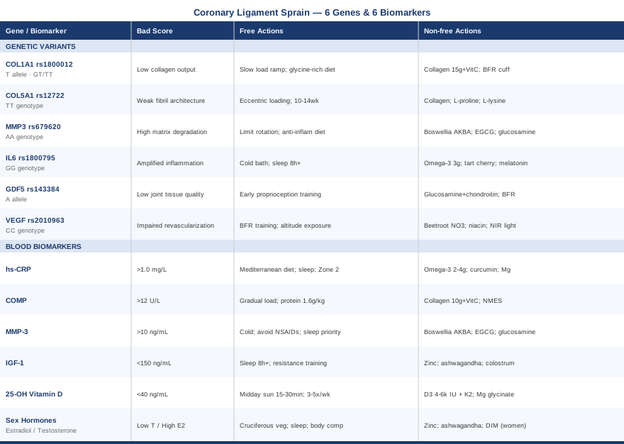
A coronary ligament sprain can be a frustrating and drawn-out injury precisely because it sits at the intersection of biomechanics, individual biology, and lifestyle factors that standard care rarely examines in combination. The biomarkers covered here — hs-CRP, COMP, MMP-3, IGF-1, vitamin D, and sex hormones — give you real-time visibility into the tissue environment that determines whether your recovery is progressing well or stalling for identifiable reasons. The genetic variants — COL1A1, COL5A1, MMP3, IL6, GDF5, and VEGF — explain a significant portion of why some people are more vulnerable to these injuries and slower to recover, and they point toward targeted interventions that work with your biology rather than ignoring it.
The next smart step is to choose two or three of the biomarkers most relevant to your situation — hs-CRP and vitamin D are the most accessible starting points for most people — and get a baseline measurement. From there, match your findings to the intervention tier that fits: lifestyle changes first, targeted supplements second, and specialized tools where the evidence supports them. If you suspect genetic factors are playing a role, consumer genomic services (such as 23andMe raw data interpreted through tools like Genetic Lifehacks or SelfDecode) can identify several of the variants discussed here at a modest cost. And whenever you are making decisions about supplementation, loading protocols, or hormonal interventions, working with a sports medicine physician or qualified physiotherapist who can contextualize your individual data remains the most important step of all.