This article contains AI generated content.
Coxa Valga: 4 Genes and 7 Biomarkers to Track
Coxa valga is not something most people go looking for. Usually a doctor mentions it after an X-ray taken for something else — a limp that would not resolve, a hip that felt loose, a routine scan in a child with cerebral palsy or a connective tissue condition. The femoral neck-shaft angle is wider than it should be, and suddenly you are holding a term you have never heard before, searching for what it means and, more importantly, what you can actually do about it.
Generic advice tends to stop at "see an orthopedist" or "it depends on the underlying cause," which is technically true and practically useless. Coxa valga is rarely a stand-alone diagnosis — it is usually a downstream sign of something else: a skeletal growth pattern, a mineralization problem, a neuromuscular condition, or a connective tissue variant. Treating it as an isolated X-ray finding, without asking what shaped the bone in the first place, misses the point.
This article takes the deeper route. Instead of stopping at "your femoral neck angle is high," it looks at the biological signals — genes and biomarkers — that are known to influence how the hip grows and remodels over time. Some of these signals are well established and clinically actionable today. Others are earlier-stage findings worth knowing about, but not worth over-interpreting. Being precise about which is which matters more than being reassuring.
None of this replaces an orthopedic evaluation, and nothing here reverses a structural bone shape that has already fully formed in an adult skeleton. But better information about the biology underneath a coxa valga finding — what may be driving it, what can be measured, and what is realistically modifiable — puts you in a stronger position to ask the right questions and make better decisions with the people actually treating you or your child.
Summary
Coxa valga sits at the intersection of bone mineralization, growth-plate biology, and mechanical loading — which is why a single X-ray angle can have very different underlying causes in different people. In one child it traces back to a rare collagen or growth-plate receptor gene; in another it is the downstream effect of low vitamin D and phosphate during a growth spurt; in an adult with cerebral palsy, muscle imbalance and loading patterns may matter more than any single gene. This article walks through the seven blood-based biomarkers most worth tracking — starting with vitamin D and calcium-phosphate-PTH physiology and moving into bone turnover markers — and explains what each one may reveal, how it is measured, and what a realistic improvement plan looks like with and without supplements. It then covers four genes with real human evidence linking them to hip and bone shape, from the well-established COL1A1/COL1A2 and FGFR3 skeletal conditions to the more preliminary GDF5 and VDR associations. A closer look at Peter Attia's framework for proactive skeletal health follows, along with a review of which complementary approaches — tai chi, yoga, massage therapy — actually have supporting evidence for hip-related joint health. By the end, the goal is a working scorecard: which numbers to ask your doctor for, what a concerning result looks like, and what to reasonably try before assuming nothing can be done.
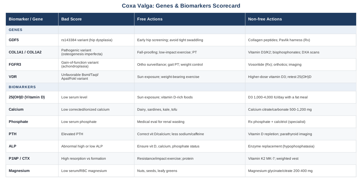
The 7 Biomarkers That Matter Most for Coxa Valga
Coxa valga develops during growth, when the femoral neck-shaft angle gradually narrows from a naturally wide angle in infancy (often over 150 degrees) down to an adult range of roughly 120 to 135 degrees. That narrowing depends on two things working together: proper mineralization of the growth plate and metaphysis, and normal mechanical loading through the hip as a child stands, walks, and bears weight. When either of those processes is disrupted — by a mineral deficiency, a hormonal imbalance, or reduced muscle loading as seen in neuromuscular conditions — the angle can stay wide instead of correcting itself, or can widen further, as documented in reviews of neuromuscular hip dysplasia in conditions like cerebral palsy (The Etiology of Neuromuscular Hip Dysplasia and Implications for Management).
That is why the biomarker panel below leans heavily on calcium-phosphate-vitamin D physiology and bone turnover, rather than on unrelated metabolic markers. These are the numbers that most directly reflect whether the biological environment around a growing or remodeling hip is supportive or working against it.
Serum 25-Hydroxyvitamin D
Vitamin D status, measured as 25-hydroxyvitamin D or 25(OH)D, governs how efficiently the gut absorbs calcium and phosphate and how well the growth plate cartilage mineralizes as it converts to bone. Chronic, unrecognized vitamin D deficiency is the classic driver of rickets, and rachitic changes at the proximal femur are a well-documented cause of abnormal femoral neck-shaft angles in children, according to the NIH Office of Dietary Supplements vitamin D fact sheet. Even outside of frank rickets, low-normal levels sustained over years can contribute to suboptimal mineralization and higher bone turnover.
How to measure it
A standard blood draw measuring serum 25(OH)D, widely available through primary care or direct-to-consumer lab services, typically costs between 35 and 120 US dollars depending on whether insurance covers it.
If the score is low: the plan without supplements
Sensible, regular sun exposure (roughly 10 to 30 minutes of midday skin exposure several times a week, adjusted for skin tone, latitude, and season) combined with vitamin D-rich foods — fatty fish, egg yolks, fortified dairy — can meaningfully raise levels in people who are only mildly deficient and otherwise healthy.
If the score is low: the plan with supplements or equipment
Vitamin D3 supplementation, typically 1,000 to 4,000 IU daily depending on baseline level and body weight, taken with a fat-containing meal for absorption, is standard practice. This is a continuous daily dose rather than a cycled one. Retest 25(OH)D after about 12 weeks to confirm the dose is working. Side effects are rare at these doses but hypercalcemia becomes a risk with sustained intake above 4,000 IU/day without monitoring, so periodic calcium checks matter if supplementing long-term.
Serum Calcium (Albumin-Corrected) and Ionized Calcium
Calcium is the literal building block of bone mineral. Chronically low calcium availability — whether from poor intake, malabsorption, or vitamin D deficiency — triggers a compensatory rise in parathyroid hormone that reshapes bone remodeling throughout the skeleton, including at the growth plate.
How to measure it
Part of a basic or comprehensive metabolic panel, costing roughly 10 to 50 US dollars; albumin-corrected calcium is more accurate than total calcium alone, and ionized calcium (a separate, slightly costlier test) is more precise still when results are borderline.
If the score is low: the plan without supplements
Dairy, canned fish eaten with bones (like sardines), leafy greens such as kale, and calcium-set tofu are practical dietary sources that close small gaps without a pill.
If the score is low: the plan with supplements or equipment
Calcium citrate or carbonate, 500 to 1,200 mg per day in split doses, is reasonable when dietary intake is genuinely insufficient — this should be confirmed by diet tracking rather than assumed. Common side effects include constipation and, at high sustained doses, an increased kidney stone risk, so this is not a "more is better" supplement; it should be dosed to close an actual measured gap and reassessed with a repeat lab, not taken indefinitely without follow-up.
Serum Phosphate
Phosphate wasting disorders, most notably X-linked hypophosphatemia caused by PHEX gene variants, cause rachitic bone changes that frequently include abnormal femoral neck-shaft angles. Even outside of a diagnosed genetic condition, unexplained low phosphate deserves attention because it directly impairs mineralization at the growth plate.
How to measure it
Included in most comprehensive metabolic panels, roughly 10 to 30 US dollars.
If the score is low: the plan without supplements
Dietary phosphate restriction is essentially never the issue in the developed world — phosphate is abundant in the food supply — so there is little to modify here through diet alone. What matters is ruling out a renal phosphate-wasting process with a clinician, since this is fundamentally a medical evaluation question, not a lifestyle one.
If the score is low: the plan with supplements or equipment
Confirmed hypophosphatemic conditions are managed with prescription phosphate supplementation and active vitamin D (calcitriol), or in some cases FGF23-targeted therapy, under a specialist's supervision — this is not a do-it-yourself supplement scenario. Side effects of phosphate/calcitriol therapy include gastrointestinal upset and hypercalciuria, both of which require lab monitoring, which is exactly why this pathway belongs to an endocrinologist rather than a supplement aisle.
Parathyroid Hormone (PTH)
PTH rises in direct response to low calcium or low vitamin D, a pattern called secondary hyperparathyroidism, and drives increased bone remodeling that can affect a growing skeleton's shape over time. It is a useful marker precisely because it reveals whether the body is already compensating for a mineral deficiency you have not yet measured directly.
How to measure it
Intact PTH blood test, roughly 50 to 150 US dollars, usually ordered alongside calcium and vitamin D.
If the score is elevated: the plan without supplements
Correcting vitamin D and calcium intake through sun and diet, as described above, along with moderating excess sodium and caffeine intake (both of which increase urinary calcium loss), is the first, most logical step.
If the score is elevated: the plan with supplements or equipment
Vitamin D repletion, following the dosing above, typically normalizes secondary hyperparathyroidism within weeks to a few months, confirmed with a repeat PTH test. If PTH is elevated alongside high (not low) calcium, this points toward primary hyperparathyroidism, which requires imaging of the parathyroid glands and is not something any supplement addresses — it is a referral, not a dosing question.
Bone-Specific Alkaline Phosphatase (or Total ALP)
ALP reflects osteoblast activity — the cells actively building bone. In growing children, markedly elevated ALP alongside other rachitic markers supports a mineralization problem, while unusually low ALP can point toward hypophosphatasia, a genetic condition (ALPL gene) that also affects hip and long-bone shape.
How to measure it
Standard on most metabolic panels, 20 to 80 US dollars; bone-specific ALP is a separate, more targeted test when total ALP is ambiguous.
If the score is abnormal: the plan without supplements
Ensuring adequate vitamin D, calcium, and phosphate status (per the sections above) addresses most of the modifiable drivers of abnormal ALP in an otherwise healthy person.
If the score is abnormal: the plan with supplements or equipment
Nutrient repletion is the practical first step. If hypophosphatasia is suspected — persistently low ALP with dental or skeletal symptoms — enzyme replacement therapy is the specialist-level treatment; this again sits outside supplement territory and belongs in a genetics or metabolic bone disease referral.
Bone Turnover Markers: P1NP and CTX
P1NP reflects bone formation and CTX reflects bone resorption; together they give a dynamic picture of remodeling balance that static tests like ALP or a bone density scan cannot capture on their own. The International Osteoporosis Foundation and International Federation of Clinical Chemistry have designated these as the reference markers for tracking bone metabolism over time (Clinical Use of Bone Turnover Markers to Monitor Pharmacologic Fracture Prevention Therapy).
How to measure it
Specialty lab panel, roughly 80 to 200 US dollars per marker; both should be drawn fasting in the morning because of significant diurnal variation.
If the score shows high resorption relative to formation: the plan without supplements
Regular resistance and impact-based exercise (within whatever range is safe given the underlying hip condition) stimulates osteoblast activity and shifts the formation-to-resorption ratio favorably, alongside adequate dietary protein to support bone matrix synthesis.
If the score shows high resorption relative to formation: the plan with supplements or equipment
Vitamin K2 (as MK-7), 90 to 180 micrograms daily, has meta-analytic support for improving bone mineral density outcomes when paired with adequate vitamin D and calcium status (Efficacy of vitamin K2 in the prevention and treatment of postmenopausal osteoporosis). Dosing is continuous rather than cycled. A weighted vest or resistance-training equipment can add safe osteogenic loading. Retest turnover markers every three to six months. The one meaningful caution: vitamin K2 can interact with warfarin and other vitamin K antagonists, so anyone on blood thinners needs to loop in their prescribing physician before starting it.
Magnesium
Magnesium is a cofactor for the enzymes that activate vitamin D and is structurally part of the bone mineral crystal itself. Low magnesium status is common and can blunt the effectiveness of vitamin D supplementation even when dosing looks adequate on paper.
How to measure it
Serum magnesium is inexpensive (20 to 40 US dollars) but a poor reflection of total body stores; red blood cell (RBC) magnesium, at 60 to 100 US dollars, is a better indicator of tissue-level status.
If the score is low: the plan without supplements
Nuts, seeds, leafy greens, and whole grains are all magnesium-dense and can close a mild gap without supplementation.
If the score is low: the plan with supplements or equipment
Magnesium glycinate or citrate, 200 to 400 mg daily in split doses, is well tolerated and continuously dosed rather than cycled. The main side effect is diarrhea at higher doses, particularly with magnesium oxide, which is best avoided in favor of the glycinate or citrate forms. Anyone with significant kidney impairment should only supplement under medical guidance, since magnesium clearance depends on renal function.
Biomarkers describe the biochemical environment the hip is growing or remodeling in; genes describe some of the underlying reasons that environment, or the skeleton's response to it, might be different from someone else's. The next section looks at four genes with real human evidence connecting them to hip shape and bone quality.
The Genetic Side: 4 Genes Linked to Hip Shape
Genetic evidence for coxa valga specifically is thinner than for the biomarkers above, and much of what is known comes from studying the conditions that most commonly produce coxa valga as a feature — skeletal dysplasias, connective tissue disorders, and developmental hip conditions — rather than from studies of coxa valga in isolation. That distinction matters: a "bad" gene here does not mean a person is destined for hip deformity, and in most cases nothing about a gene itself can be directly "fixed." What can be managed is the downstream biology and mechanical environment the gene variant influences.
GDF5 (Growth Differentiation Factor 5)
GDF5 encodes a signaling protein central to joint, cartilage, and synovial tissue formation. A promoter variant, rs143384, has one of the most replicated associations in orthopedic genetics with developmental dysplasia of the hip, carrying an odds ratio around 1.44 in large case-control and genome-wide association studies (Genome-wide association study of developmental dysplasia of the hip identifies an association with GDF5). Separately, epigenetic work has found the GDF5 promoter is abnormally hypermethylated in cartilage from children with hip dysplasia compared to healthy controls (DNA hypermethylation of GDF5 in developmental dysplasia of the hip), suggesting both genetic and epigenetic layers affect joint development. This is solid human evidence, but it is associative and modest in effect size — a risk variant nudges probability, it does not determine outcome.
If the gene variant is unfavorable: the plan without supplements
For infants with a family history or an early exam finding, timely hip screening (clinical exam and, if indicated, ultrasound) allows early bracing before the hip joint fully matures. Beyond infancy, avoiding tight, hip-adducted swaddling — a known modifiable risk factor for hip dysplasia — and maintaining healthy body weight to limit joint load are the realistic non-supplement levers.
If the gene variant is unfavorable: the plan with supplements or equipment
Collagen peptides (10 to 15 g daily) are sometimes used as a general connective-tissue support strategy, though direct evidence for altering GDF5-related joint development is indirect at best — this is a low-risk, low-certainty addition, not a treatment. The actual medical equipment used for diagnosed hip dysplasia in infancy, such as a Pavlik harness, is physician-prescribed and monitored, not a consumer decision. If using collagen peptides, a reasonable pattern is daily for three months, then reassess whether to continue; side effects are minimal, occasionally mild GI upset.
COL1A1 and COL1A2 (Type I Collagen Genes)
These genes encode the two chains of type I collagen, the main structural protein in bone matrix. Pathogenic variants cause osteogenesis imperfecta, where under-mineralized, structurally weaker bone can remodel abnormally, including coxa vara or valga patterns alongside fracture risk (COL1A1- and COL1A2-Related Osteogenesis Imperfecta, GeneReviews). This is strong, causal, well-established human evidence, not an early or preliminary association.
If a pathogenic variant is present: the plan without supplements
Home fall-proofing, low-impact loading activities like swimming or cycling that build bone stimulus without high fracture risk, and structured physical therapy for posture and gait all reduce the practical consequences of fragile bone without requiring any product.
If a pathogenic variant is present: the plan with supplements or equipment
Vitamin D3 and K2, plus calcium if dietary intake is low, optimize whatever mineralization capacity the existing collagen framework allows — they do not correct the underlying collagen defect. Bisphosphonate therapy (a prescription medication, often given as periodic IV infusions such as pamidronate) is the actual standard of care for moderate-to-severe osteogenesis imperfecta and must be managed by a specialist; cycling follows a specific infusion protocol set by that clinician. Side effects include flu-like symptoms after the first infusion and, rarely with long-term use, jaw osteonecrosis — all reasons this pathway is specialist-directed rather than self-managed. Regular DXA bone density scans and protective bracing round out the equipment side.
FGFR3 (Fibroblast Growth Factor Receptor 3)
FGFR3 normally acts as a brake on growth plate cartilage proliferation. Gain-of-function variants cause achondroplasia and hypochondroplasia, skeletal dysplasias in which altered hip geometry, including coxa valga, is a recognized clinical feature (Achondroplasia, GeneReviews). Like COL1A1/COL1A2, this is strong, causal, well-documented human evidence.
If a pathogenic variant is present: the plan without supplements
Regular orthopedic surveillance with imaging, physical therapy focused on gait and joint protection, and weight management (since disproportionate limb length changes biomechanical loading through the hip) are the realistic, ongoing management tools.
If a pathogenic variant is present: the plan with supplements or equipment
No supplement changes FGFR3 signaling. Vosoritide, a prescription CNP-analog medication approved for achondroplasia-related growth effects, is a physician-directed option evaluated on a case-by-case basis, with injection-site reactions and gastrointestinal symptoms as known side effects, dosed and monitored entirely by the treating specialist. Orthotic supports and scheduled imaging are the relevant "equipment" side of management here.
VDR (Vitamin D Receptor Gene)
VDR polymorphisms — commonly labeled BsmI, TaqI, ApaI, and FokI — modestly affect how efficiently cells respond to circulating vitamin D, and have shown associations with bone mineral density and bone turnover markers across multiple studies (Vitamin D receptor gene polymorphisms, bone mineral density and fractures in postmenopausal women with osteoporosis). The evidence here is genuinely mixed across populations and effect sizes are small — this sits closer to the early/associative end of the spectrum than COL1A1 or FGFR3.
If the gene variant is unfavorable: the plan without supplements
Regular sun exposure and weight-bearing exercise remain the foundational, low-cost levers regardless of VDR genotype.
If the gene variant is unfavorable: the plan with supplements or equipment
Because VDR variants may mean someone needs a relatively higher vitamin D intake to reach the same serum 25(OH)D level as someone without the variant, the practical approach is to simply measure 25(OH)D directly rather than guess from genotype alone. If supplementing, 1,000 to 2,000 IU of D3 daily with retesting every three months is a reasonable starting pattern, with the same hypercalcemia caution at higher doses described earlier.
Genes and biomarkers explain a lot of the biology, but they are not the only lens worth applying to skeletal health. One recent book has reframed how a growing number of physicians think about tracking bone and joint health proactively rather than reactively — and it is worth a closer look before turning to complementary approaches.
What Outlive Gets Right About Bone and Joint Health
Peter Attia's book Outlive: The Science and Art of Longevity is not written about coxa valga, or even primarily about orthopedics, but its central argument — that skeletal and muscular health deserve the same proactive tracking as cardiovascular risk — is directly relevant to anyone managing a structural hip finding long before it becomes a crisis. Below are ten of the book's most applicable ideas for someone thinking seriously about hip and bone health.
The "Marginal Decade" Changes the Math on When to Act
Attia argues that most people only think seriously about mobility and bone strength in their final decade of life, when options are already limited. Applied here, the logic is the same: addressing mineralization and joint mechanics early, whether in a growing child or a younger adult with a hip finding, is a fundamentally different problem than trying to reverse compensations decades later.
Bone Density Is a Rearview Mirror, Not a Dashboard Light
A key point in the book is that by the time bone density testing shows a problem, the underlying decline has often been happening for years. This is the argument for the biomarker panel above — catching low vitamin D, elevated PTH, or abnormal turnover markers early is catching the problem while it is still cheap and simple to correct.
Stability Training Deserves Equal Billing With Cardio and Strength
Attia treats stability — balance, proprioception, and joint control — as a fourth pillar of fitness alongside strength, cardio, and endurance, rather than an afterthought. For anyone with an altered hip angle, stability work directly addresses the compensatory movement patterns that a wider femoral neck-shaft angle can create.
The "Centenarian Decathlon" Forces Backward Planning
The book's exercise of listing physical tasks you want to still be able to do in old age, then working backward to the strength and mobility needed today, is a useful reframe for hip health specifically: what does this hip need to be capable of at 70, and what does that require building now?
Muscle Mass Around a Joint Can Compensate for Structural Imperfections
Attia repeatedly makes the point that muscular support around a joint reduces the functional impact of an imperfect structure underneath it. This lines up directly with clinical management of coxa valga in neuromuscular conditions, where hip abductor strength and muscle balance materially affect outcomes independent of the angle itself.
Zone 2 and VO2 Max Training Indirectly Protect Joints
While framed around cardiovascular longevity, Attia's argument that better aerobic capacity reduces fall risk and improves load tolerance applies just as much to joint protection — a person with better aerobic base fatigues less during the day, which reduces compensatory, joint-stressing movement patterns.
Skepticism of One-Size-Fits-All Guidelines
A recurring theme is that population-level guidelines (a single vitamin D target, a single BMD cutoff) are starting points, not personalized answers. This supports the approach in this article: track your own numbers and trends rather than assuming a "normal" lab value fully describes your situation.
Why Consistency Requires an Emotional "Why," Not Just a Plan
Attia is candid that knowing what to do is rarely the barrier — sticking with unglamorous daily habits (vitamin D, resistance training, physical therapy exercises) is. For a chronic structural issue like coxa valga, this is arguably the most underrated point in the whole framework.
Proactive Medicine Over Reactive Medicine
The book's broader critique of a medical system built to treat disease rather than prevent it maps directly onto orthopedic care: waiting for hip pain or a limp to trigger imaging misses the earlier window where biomarker correction is simplest and most effective.
Build a Personal Scorecard
The practical takeaway Attia pushes readers toward — track a small set of meaningful numbers over time rather than a one-off panel — is exactly the model this article's biomarker section is built around: 25(OH)D, calcium, phosphate, PTH, ALP, P1NP/CTX, and magnesium, rechecked on a schedule rather than measured once and forgotten.
Biomarkers, genetics, and Attia's proactive framework all sit within conventional and integrative medicine. A smaller set of complementary approaches also has genuine, if more limited, human evidence worth knowing about for hip-related joint health.
Complementary Approaches Worth Considering
Evidence for complementary therapies specific to coxa valga itself does not exist — this is too narrow and structurally specific a finding for dedicated trials. What does exist is meaningful evidence for hip and knee osteoarthritis and general musculoskeletal joint health, which is relevant to anyone managing altered hip mechanics, whether from coxa valga directly or from the compensatory patterns it can create.
Tai Chi
Tai chi combines slow, controlled weight-shifting movements with balance and muscular control, making it directly relevant to anyone whose hip mechanics are already atypical, since it builds the stabilizing strength and proprioception around the joint without high-impact loading.
A randomized controlled trial found that tai chi classes produced large and sustained improvements in physical function among older adults with chronic hip or knee osteoarthritis (Physical activity for osteoarthritis management: a randomized controlled clinical trial evaluating hydrotherapy or Tai Chi classes), and tai chi is now recommended in several osteoarthritis management guidelines based on broader systematic review evidence (The Effectiveness of Tai Chi for Knee Osteoarthritis: An Overview of Systematic Reviews).
A realistic starting point is two sessions per week for at least eight weeks before expecting to notice functional changes, ideally with an instructor experienced in adapting movements for people with existing hip involvement. It is low-risk, but anyone with significant hip instability should get orthopedic clearance before starting a new weight-shifting practice.
Yoga
Yoga's combination of controlled hip range-of-motion work, strength, and body awareness makes it relevant for maintaining functional hip mobility around a structural finding like coxa valga, though most direct evidence covers knee rather than hip osteoarthritis specifically.
A randomized controlled trial found that integrated yoga therapy produced significantly better quality-of-life improvements than standard physiotherapy exercises in knee osteoarthritis (Effect of an integrated approach of yoga therapy on quality of life in osteoarthritis of the knee joint), and a later trial found yoga to be noninferior to a dedicated strengthening program for pain and quality of life.
For someone with an atypical hip angle, this points toward a therapist-guided or adapted class rather than an unsupervised general class, with particular caution around deep hip-flexion poses until a clinician has confirmed they are appropriate for that specific hip.
Massage Therapy
Chronic muscular tension around a mechanically compromised hip — from the abductors, iliopsoas, or piriformis compensating for altered joint geometry — is common, and massage therapy is one of the better-evidenced ways to address that secondary muscular component, even though it does nothing to the underlying bone angle itself.
A broad evidence map found massage therapy produces short-term benefits for common musculoskeletal pain conditions compared to no treatment (Massage for Pain: An Evidence Map), with effects concentrated in the days to weeks after treatment rather than lasting indefinitely.
Used realistically, this means scheduling massage around periods of increased activity or flare-ups in muscular tightness rather than as a stand-alone fix, and treating it as one part of a broader plan that includes the strength and stability work described above.
Putting It All Together
Coxa valga is a description of hip shape, not a single disease with one cause or one fix. The most useful thing a biomarker and genetic lens can offer is precision: knowing whether a vitamin D and phosphate problem is quietly shaping bone growth, whether a well-documented gene like COL1A1 or FGFR3 is driving a recognized skeletal condition, or whether the picture is more about muscular loading and compensation than any lab value at all. That precision is what turns "see an orthopedist" into a specific, useful conversation.
The most productive next step is rarely dramatic. It is asking for a basic panel — vitamin D, calcium, phosphate, PTH, and ALP — the next time you are already at a lab, tracking it over time rather than once, and bringing the results to whoever is managing the hip in question, whether that is a pediatric orthopedist, a geneticist, or a primary care physician. Better numbers do not rewrite an X-ray on their own, but they do make every decision built on top of them a more informed one.