This article contains AI generated content.
· UpdatedCoxa Vara: 6 Genes and 7 Biomarkers to Track
Introduction
If you have received a coxa vara diagnosis, the first conversation with a clinician likely centered on the structural problem: the abnormal angle of the femoral neck, the limp, the leg length difference, possibly surgical options. That is the appropriate medical starting point. What gets covered less often is that the shape of that bone reflects, in many cases, years of metabolic activity — how the body managed phosphate, how effectively bone was mineralized, and what genetic parameters were in play before any symptoms appeared.
That metabolic layer matters practically. The same hip deformity can arise from nutritional vitamin D deficiency, from a PHEX mutation that causes chronic phosphate wasting, from a broken alkaline phosphatase enzyme, or from a collagen defect that makes bone fracture and remodel under normal loads. These are not variants of the same problem. They respond differently — sometimes oppositely — to the same interventions. Knowing which mechanism is operating changes what to test, what to supplement, what to avoid, and what trajectory is likely.
Generic bone health advice — calcium, sunlight, weight-bearing exercise — covers the basics for the average case but does not account for an inherited FGF-23 excess or a VDR polymorphism that makes standard vitamin D dosing insufficient. Those distinctions are only visible through targeted testing and, increasingly, genetic panels that are more accessible than they were a decade ago.
This article covers two complementary frameworks. The first is practical biomarker tracking: seven markers that give a real-time view of the metabolic environment in which bone is forming — or failing to form. The second is genetic: six genes with direct links to coxa vara and related mineralization disorders, with specific plans for each. Beyond that, there is a summary of the most actionable ideas from Peter Attia's Outlive on bone health, and a section on three complementary modalities with meaningful human clinical evidence.
Summary
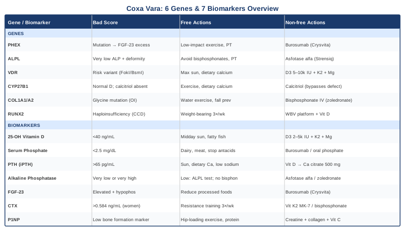
What this article covers: Seven biomarkers — 25-OH vitamin D, serum phosphate, PTH, alkaline phosphatase, FGF-23, CTX, and P1NP — that collectively reveal the metabolic state of bone in coxa vara, with specific plans for abnormal readings. Six genes — PHEX, ALPL, VDR, CYP27B1, COL1A1/COL1A2, and RUNX2 — that explain why coxa vara develops in some people despite seemingly adequate nutrition, including plans for carriers. Beyond that: ten actionable ideas from Peter Attia's Outlive that challenge the standard medical approach to bone health, and three complementary modalities (photobiomodulation, yoga, tai chi) with condition-specific evidence and practical application protocols.
7 Biomarkers That Reveal What Is Actually Happening in Coxa Vara
Coxa vara does not develop in a metabolic vacuum. Whether it appears in a child with nutritional rickets or in an adult whose proximal femur has progressively deformed under ongoing bone disease, the underlying biochemical state can be measured. These seven biomarkers cover vitamin D status, phosphate metabolism, parathyroid activity, enzyme function, FGF-23–driven phosphate wasting, and bone remodeling dynamics. Tracking them as a panel rather than in isolation gives a working picture of what is driving the condition and what can be done about it.
1. 25-OH Vitamin D (Calcidiol)
Why it matters: Vitamin D deficiency is the most common preventable cause of rickets globally and the leading metabolic driver behind nutritional coxa vara in children. 25-OH vitamin D — the circulating storage form — is the standard clinical measurement. Low levels impair intestinal calcium absorption, trigger secondary hyperparathyroidism, and reduce the rate of calcification of the bone collagen matrix. Decades of data confirm that adequate vitamin D is a non-negotiable foundation for normal skeletal development, especially during the rapid growth phases when coxa vara risk is highest.
How to measure it
A standard blood test, fasting or non-fasting. Cost: $30–80 at most labs; direct-to-consumer services such as Ulta Lab Tests or Walk-In Lab frequently offer it for $15–30. The optimal range for bone health — as referenced by clinicians including Peter Attia and endocrinologist Michael Holick — is 40–60 ng/mL (100–150 nmol/L), substantially above the lab deficiency cutoff of 20 ng/mL, which marks the minimum to prevent frank deficiency rather than support optimization. Retest every 3–4 months during supplementation adjustment, annually once stable. The NIH Office of Dietary Supplements vitamin D fact sheet provides a solid evidence summary.
If the score is low, the plan without supplements
Midday sun exposure is the most efficient non-supplemental strategy: 15–30 minutes on arms and legs for fair skin tones, 30–60 minutes for darker skin tones, without sunscreen during peak UVB hours (roughly 10 AM to 2 PM). Fatty fish (salmon, sardines, mackerel), whole eggs, and vitamin D-fortified foods contribute meaningfully to status but rarely bring levels above 30 ng/mL as a sole strategy. Daily weight-bearing activity does not directly raise 25-OH-D but supports the downstream utilization of circulating vitamin D through mechanosensitive signaling in osteoblasts.
If the score is low, the plan with supplements or equipment
Vitamin D3 (cholecalciferol): 2,000–5,000 IU daily taken with a fatty meal to maximize absorption. People with VDR polymorphisms, obesity (vitamin D is sequestered in adipose tissue), or darker skin tones may need 5,000–10,000 IU/day to reach the 40–60 ng/mL target. Always co-administer vitamin K2 MK-7 (100–200 mcg/day) — as vitamin D raises serum calcium availability, K2 activates matrix Gla protein and osteocalcin to direct that calcium into bone rather than arterial walls. Magnesium glycinate or malate (300–400 mg/day) is a required cofactor for vitamin D hydroxylation; deficiency significantly blunts the response to supplementation. No cycling needed — daily use. Monitor for hypercalcemia symptoms (excessive thirst, frequent urination, fatigue, nausea) at doses above 5,000 IU, and retest serum 25-OH-D at 3–4 month intervals until stable.
2. Serum Phosphate (Inorganic Phosphorus)
Why it matters: Hydroxyapatite — the mineral phase of bone — requires both calcium and phosphate. Without adequate circulating phosphate, bone mineralization is impaired regardless of vitamin D and calcium status: the organic collagen matrix forms but remains unmineralized as osteoid. Chronic hypophosphatemia is the defining metabolic feature of X-linked hypophosphatemia (XLH), the most common heritable cause of rickets and coxa vara. It is also seen in renal tubular disorders, malabsorption, and antacid overuse. Serum phosphate is not routinely flagged in general health panels but is an essential test whenever skeletal deformity is present. The NIH Phosphorus fact sheet is a useful clinical reference.
How to measure it
Included in the basic and comprehensive metabolic panel (BMP/CMP). Cost: $10–30. Normal range: 2.5–4.5 mg/dL. Values consistently below 3.0 mg/dL in a patient with bone deformity warrant investigation, particularly FGF-23 measurement. Test in a fasting morning state — postprandial insulin drives phosphate into cells and can temporarily lower serum levels by 0.5–1.0 mg/dL.
If the score is low, the plan without supplements
Increase dietary phosphorus through whole food sources: dairy products, meat, poultry, fish, legumes, nuts, and seeds. Eliminate phosphate-binding antacids (aluminum-containing antacids are a frequently overlooked cause of secondary hypophosphatemia and should be discontinued where possible). Identify the root driver — dietary adjustments will not overcome FGF-23–driven renal phosphate wasting.
If the score is low, the plan with supplements or equipment
For confirmed XLH, burosumab (Crysvita) addresses the FGF-23 mechanism directly and is now the preferred approach over conventional oral phosphate supplementation. Where burosumab is unavailable: oral phosphate (20–40 mg/kg/day in 4–5 divided doses across the day, using neutral phosphate preparations) combined with calcitriol (0.25–0.5 mcg twice daily) partially corrects hypophosphatemia. Side effects of oral phosphate: GI intolerance (diarrhea, nausea), and paradoxical long-term FGF-23 elevation that can create a worsening cycle of phosphate loss. This combination requires monitoring of serum calcium, phosphate, PTH, and urinary calcium at 3-month intervals. No cycling — daily consistency is essential for maintaining trough serum phosphate.
3. Intact Parathyroid Hormone (iPTH)
Why it matters: PTH is the body's calcium stabilizer. When calcium or vitamin D is low, PTH rises to extract calcium from bone, increase renal reabsorption, and stimulate vitamin D activation. Chronically elevated PTH — secondary hyperparathyroidism — drives sustained osteoclast activity, progressively degrading bone quality over time. In the context of coxa vara, persistent PTH elevation means bone is being actively broken down in a structure that is already mechanically compromised. PTH is also a direct readout of whether upstream vitamin D and calcium inputs are adequate, making it one of the most useful feedback indicators in metabolic bone disease management.
How to measure it
Intact PTH blood test. Cost: $50–100. Normal range: 10–65 pg/mL. Interpret always in conjunction with serum calcium and 25-OH-D. Elevated PTH with low-normal calcium and low vitamin D = secondary hyperparathyroidism from deficiency. Elevated PTH with elevated calcium = primary hyperparathyroidism, a different condition requiring separate workup. Morning fasting preferred for reproducibility.
If the score is high, the plan without supplements
Address the upstream deficiency: increase daily sun exposure and dietary calcium from food (dairy products, calcium-set tofu, leafy greens, fortified plant milks). Reduce urinary calcium losses by lowering sodium intake (high dietary sodium drives calcium excretion in urine and has a direct effect on PTH independently of intake). Moderate animal protein excess for the same reason. Daily weight-bearing physical activity supports PTH normalization by providing the mechanical stimulation that reduces bone's dependency on hormonal calcium mobilization.
If the score is high, the plan with supplements or equipment
Correct vitamin D first — PTH typically normalizes within 3–6 months when 25-OH-D reaches the 40–60 ng/mL range. If PTH remains elevated despite adequate vitamin D, add calcium citrate (500 mg twice daily with meals; citrate has superior absorption to carbonate in older adults and anyone on acid-suppressing medications). For persistent severe secondary hyperparathyroidism: calcitriol (0.25–0.5 mcg/day) under physician supervision, with quarterly monitoring of serum and urinary calcium to avoid hypercalcemia. Daily dosing, no cycling required.
4. Alkaline Phosphatase — Total and Bone-Specific
Why it matters: ALP is the most frequently measured metabolic bone marker — and the one most likely to be misread in the context of coxa vara. In most bone diseases with active remodeling (rickets, Paget's disease, healing fractures, secondary hyperparathyroidism), ALP is elevated as osteoblasts work harder. But in hypophosphatasia — a rare inherited enzyme deficiency that directly causes rickets-like skeletal deformity including coxa vara — ALP is paradoxically and markedly low. Missing a very low ALP in a patient with coxa vara means missing a specifically treatable enzymatic diagnosis. Both extremes of ALP carry distinct clinical meaning. Bone-specific ALP (BSAP) eliminates liver contribution and gives a cleaner signal of skeletal remodeling activity.
How to measure it
Total ALP is included in the standard CMP ($10–30). Bone-specific ALP (BSAP/BAP) costs $80–150 and is more specific when liver disease is also a concern. Normal total ALP in adults: 44–147 IU/L. In children, physiological ALP values during bone growth can reach 400–500 IU/L, which makes identifying an abnormally low ALP in a child with bone deformity especially important. Any adult total ALP consistently below 30–35 IU/L alongside bone deformity is a flag for hypophosphatasia until proven otherwise.
If the score is high, the plan without supplements
Identify the driving condition: rule out Paget's disease (bone scan, urinary NTX/CTX), active liver disease (check GGT and transaminases to differentiate bone vs. liver origin), and bone metastases if indicated by clinical context. For elevated ALP from active nutritional rickets, correction of vitamin D and phosphate deficiency typically normalizes total ALP over 6–12 months. Reduce alcohol intake independently (alcohol elevates liver ALP). Age-appropriate weight-bearing activity at sustainable intensity supports healthy bone remodeling without excessive osteoclast stimulation.
If the score is low, the plan without supplements
A persistently low ALP in the presence of bone deformity is the primary indication for ALPL genetic testing. Avoid high-dose vitamin D supplementation without specialist guidance — it will not address an enzyme defect and may worsen hypercalcemia risk. Critically, avoid bisphosphonates in suspected hypophosphatasia, as they inhibit the already-deficient ALP and can dramatically worsen bone quality. Physical therapy to maintain gait mechanics and hip range of motion is the primary non-pharmacological intervention while diagnosis is confirmed.
If the score is abnormal, the plan with supplements or equipment
For confirmed hypophosphatasia: asfotase alfa (Strensiq), enzyme replacement therapy given subcutaneously at 1–2 mg/kg three times per week. Randomized trials have demonstrated significant improvement in radiographic bone mineralization and skeletal deformity scores, including in pediatric patients with rickets-related hip deformity. Pyridoxine (vitamin B6) supplementation (5–100 mg/day titrated to seizure response) is essential for hypophosphatasia patients experiencing pyridoxine-responsive seizures, as accumulated PLP blocks GABA synthesis. For elevated ALP from Paget's disease: zoledronic acid (5 mg IV, single infusion) is the standard therapy, with long remissions typically achieved.
5. FGF-23 (Fibroblast Growth Factor 23)
Why it matters: FGF-23 is a phosphaturic hormone secreted by osteocytes. Its primary functions are to instruct the kidney to excrete phosphate in urine and to suppress renal activation of vitamin D. In X-linked hypophosphatemia, the PHEX gene product normally degrades FGF-23, but loss-of-function mutations in PHEX allow FGF-23 to accumulate unchecked. The result is chronic renal phosphate wasting, impaired active vitamin D production, and the progressive rickets and coxa vara that characterize XLH. FGF-23 is also elevated in autosomal recessive and dominant hypophosphatemic rickets, tumor-induced osteomalacia, and certain kidney diseases. Measuring FGF-23 is the critical mechanistic step that distinguishes FGF-23–excess disorders from other causes of hypophosphatemia and guides the most targeted intervention.
How to measure it
Specialized blood test available through major reference laboratories (Quest, LabCorp). The C-terminal FGF-23 assay is most widely available; the intact FGF-23 assay measures only the biologically active molecule and is more specific. Cost: $100–250, typically requires physician order. An elevated FGF-23 in the context of hypophosphatemia and skeletal deformity is functionally diagnostic of an FGF-23–excess disorder and should prompt genetic testing to identify the specific mutation. Test in fasting morning state for consistency across serial measurements.
If the score is high, the plan without supplements
Limit dietary intake of highly bioavailable inorganic phosphate: reduce processed foods and soft drinks using phosphoric acid as a preservative (the phosphate in these products is absorbed at rates of 80–100%, compared to 40–60% from organic food sources). Maintain adequate caloric intake — caloric restriction and metabolic stress independently amplify FGF-23 secretion. These dietary adjustments are supportive but cannot meaningfully reverse a genetic FGF-23 excess; medical intervention is required.
If the score is high, the plan with supplements or equipment
Burosumab (Crysvita) is the targeted treatment: a subcutaneous monoclonal antibody that binds and neutralizes excess FGF-23. Dosing is every 2 weeks (children, weight-based) or every 4 weeks (adults, 1 mg/kg), titrated to serum phosphate response. A randomized controlled trial published in The Lancet (Imel et al., 2019) documented superior serum phosphate normalization, improved rickets severity scores on radiographic scoring systems, and enhanced linear growth in children treated with burosumab compared to oral phosphate plus calcitriol. Side effects include injection site reactions, extremity pain, and dental abscess risk (an XLH-related complication independent of oral hygiene). Access programs exist for cost in many countries; specialist endocrinology or nephrology oversight is required. The alternative regimen of oral phosphate combined with calcitriol remains available but does not address elevated FGF-23 and carries a higher burden of GI side effects and secondary hyperparathyroidism.
6. CTX (C-Telopeptide of Type I Collagen)
Why it matters: CTX-I (beta-CrossLaps) is a bone resorption marker — released when osteoclasts digest the cross-links of old type I collagen matrix during bone breakdown. Elevated CTX indicates that bone is being dismantled faster than it can be rebuilt. This matters in coxa vara associated with high-turnover bone states: secondary hyperparathyroidism, active Paget's disease, prolonged immobilization, or the remodeling aftermath of fractures that progressively distort the femoral neck. Tracking CTX before and after metabolic intervention provides confirmation that treatment is actually reducing bone destruction — a signal that does not show up on imaging until months or years later.
How to measure it
Fasting blood draw, before 10 AM — CTX has a pronounced diurnal pattern, peaking in early morning and reaching its lowest point in the afternoon. Consistent timing is critical for reproducible results. Cost: $80–150. Reference range for premenopausal women: below 0.584 ng/mL; varies with age in men (roughly below 0.3 ng/mL in younger men). Always interpret CTX alongside P1NP to understand the full remodeling picture: high CTX with high P1NP indicates balanced high turnover; high CTX with low P1NP is the red flag for net bone loss.
If the score is high, the plan without supplements
Progressive resistance training and impact loading consistently reduce CTX over 12–16 weeks through OPG/RANKL signaling — mechanical load suppresses osteoclast recruitment while simultaneously stimulating osteoblasts. Minimum effective stimulus: three sessions per week of progressive resistance exercise targeting large muscle groups with compound movements. Eliminate the independent CTX drivers: smoking (roughly doubles bone resorption rates), excess alcohol, chronic sleep deprivation (growth hormone deficiency secondary to poor sleep impairs bone formation and worsens the CTX/P1NP ratio), and prolonged caloric restriction.
If the score is high, the plan with supplements or equipment
Vitamin K2 MK-7 (180–360 mcg/day) activates osteocalcin, which modulates osteoclast activity alongside its role in bone mineralization; several clinical trials show modest CTX reduction with MK-7 supplementation. Hydrolyzed collagen peptides (10 g/day) provide bone matrix substrate and have shown signal in some trials for reducing bone resorption markers. For severe, clinically-driven high-turnover states requiring pharmacological anti-resorptive therapy: bisphosphonates (alendronate 70 mg once weekly, or zoledronic acid 5 mg IV annually) or denosumab (60 mg subcutaneous every 6 months) are the most potent agents. No cycling needed for MK-7 or collagen; current guidelines recommend bisphosphonate drug holidays after 5 years of continuous use to reduce atypical fracture risk. Side effects: oral bisphosphonates require strict dosing protocol to avoid GI ulceration; rare risks of osteonecrosis of the jaw (particularly with dental procedures) and atypical femur fractures with very long-term use.
7. P1NP (Procollagen Type I N-Terminal Propeptide)
Why it matters: P1NP is the gold-standard marker of bone formation — cleaved from procollagen and released into the bloodstream when osteoblasts actively lay down new type I collagen matrix. Where CTX measures destruction, P1NP measures construction. Peter Attia uses the CTX/P1NP pair as his preferred bone metabolic assessment precisely because the ratio and directional change over time provides more actionable information than a static bone density scan. In coxa vara management, a rising P1NP during treatment confirms that metabolic correction, exercise, and supplementation are translating into actual new bone formation — not just slowing breakdown.
How to measure it
Blood test; fasting morning preferred for consistency. Cost: $100–200. Desired trend during treatment: CTX declining, P1NP increasing — or both in a balanced elevated range in young people actively building bone. Low P1NP with normal or elevated CTX is the worst combination: bone is being broken down faster than it is being formed, yielding net architectural loss. Retest every 3–6 months during active intervention; annual monitoring once stable.
If the score is low, the plan without supplements
Progressive resistance training with compound, hip-loading movements is the most evidence-backed strategy for raising P1NP: mechanical loading activates osteoblasts through Wnt/LRP5, integrin-mediated, and IGF-1 signaling pathways. Hip-specific exercises — hip thrusts, single-leg press, step-ups, weighted walking lunges — are particularly relevant for stimulating bone formation in the proximal femur region affected by coxa vara. Minimum effective stimulus: three sessions per week, progressively increasing load. Total protein intake of 1.6–2.0 g/kg/day provides the amino acid substrate for collagen synthesis; bone collagen is especially glycine- and proline-dependent.
If the score is low, the plan with supplements or equipment
Creatine monohydrate (3–5 g/day continuously, no cycling needed) has supporting evidence for increasing bone formation markers in combination with resistance training, with modest effects seen in trials of older adults. Hydrolyzed collagen peptides (10 g) with vitamin C (50–100 mg) taken 30–60 minutes before a workout supports periosteum remodeling and bone collagen synthesis through mechanically-stimulated collagen-producing fibroblasts. Whole-body vibration platforms (25–40 Hz, 10–20 minutes, 3×/week) have shown modest P1NP increases in trials of older adults and children with mobility limitations — a useful adjunct when impact exercise is limited by hip pain or significant gait disturbance. Sex hormone optimization under physician oversight (testosterone in hypogonadal men; estrogen-progesterone therapy in postmenopausal women) is among the most powerful drivers of P1NP — clinically low sex hormones are a frequently unaddressed cause of persistently low bone formation markers regardless of exercise and supplementation.
The Genetics Behind Coxa Vara: 6 Genes Worth Understanding
Most of the biomarkers above reflect metabolic states that are, at least in part, under genetic control. In some cases, knowing which gene is involved changes the treatment approach entirely — what works for nutritional rickets does the opposite in hypophosphatasia, and standard vitamin D supplementation fails completely in CYP27B1 deficiency. The six genes below cover the most clinically relevant genetic causes of coxa vara and related mineralization disorders.
PHEX — The Central Gene in X-Linked Hypophosphatemia
What it does: PHEX encodes phosphate-regulating endopeptidase homolog X-linked, an enzyme expressed in osteoblasts and osteocytes that participates in the degradation of FGF-23. Loss-of-function mutations prevent FGF-23 clearance, allowing it to accumulate and chronically instruct the kidney to waste phosphate in urine while suppressing active vitamin D production. The result is persistent hypophosphatemia, defective bone mineralization, and rickets — including coxa vara — appearing in early childhood. XLH is X-linked dominant, affecting both males and females, though males typically present with more severe bone deformity. Prevalence is approximately 1 in 20,000. Testing: gene panel for heritable rickets or PHEX-specific sequencing.
If the gene is bad, the plan without supplements
Maintain a healthy body weight to reduce mechanical load on the deformed hip joint. Low-impact aerobic exercise — swimming, cycling, elliptical — preserves cardiovascular fitness and maintains hip mobility without excessive stress on an abnormal femoral neck. Physical therapy focused on hip abductor and external rotator strengthening compensates for the gait abnormality and reduces secondary lumbar and knee loading. Monitor for XLH-specific dental complications (dental abscesses from poorly mineralized dentin form independently of hygiene and require proactive dental surveillance). Genetic counseling for family members of childbearing age.
If the gene is bad, the plan with supplements or equipment
Burosumab (Crysvita) is now the standard of care for XLH: subcutaneous injection every 2 weeks in children (weight-based dosing), every 4 weeks in adults (1 mg/kg). Patients previously on oral phosphate plus calcitriol should transition only under specialist guidance to avoid rebound hyperphosphatemia. For patients where burosumab is unavailable, oral neutral phosphate (20–40 mg/kg/day divided into 4–5 daily doses) plus calcitriol (0.25–0.75 mcg twice daily) partially corrects hypophosphatemia. Orthopedic surgery — corrective osteotomy, intramedullary rodding — is indicated for severe coxa vara deformity once metabolic control is established, as deformity correction attempted without metabolic stabilization has a high recurrence rate.
ALPL — The Hypophosphatasia Gene
What it does: ALPL encodes tissue-nonspecific alkaline phosphatase (TNSALP). Loss-of-function mutations — both autosomal dominant and recessive forms exist — impair the degradation of the natural ALP substrate inorganic pyrophosphate (PPi). Accumulated PPi is a potent inhibitor of hydroxyapatite crystal growth, effectively blocking bone mineralization from within. The hallmark is a serum ALP that is paradoxically, often dramatically, low — the opposite of what is expected in a bone disease presenting as rickets. Other characteristic features include premature tooth loss before age 5, recurrent stress fractures, and in infants, respiratory compromise from poor rib mineralization. Coxa vara is a recognized skeletal complication.
If the gene is bad, the plan without supplements
Avoid bisphosphonates entirely — this is critical. Bisphosphonates work by inhibiting bone resorption in part through accumulation of PPi-related compounds, effectively worsening the primary defect in hypophosphatasia. Avoid high-dose vitamin D supplementation without specialist guidance. Physical therapy focused on low-impact strengthening and gait mechanics. Seek specialist care from a metabolic bone disease unit with experience in rare skeletal dysplasias. Genetic counseling for family members; test first-degree relatives with a serum ALP.
If the gene is bad, the plan with supplements or equipment
Asfotase alfa (Strensiq) is enzyme replacement therapy approved for hypophosphatasia: subcutaneous injection at 1 mg/kg three times per week, or 2 mg/kg three times per week for severe perinatal-onset disease. Clinical trials reported in The Lancet Diabetes and Endocrinology documented significant improvement in radiographic mineralization and functional outcomes in pediatric patients. Side effects include injection site reactions and the theoretical risk of ectopic calcifications at sustained supratherapeutic doses. Pyridoxine (vitamin B6) supplementation at physician-directed doses is essential for patients with pyridoxine-responsive seizures caused by PLP accumulation blocking cerebral GABA synthesis. Cost of asfotase alfa is very high; access programs through the manufacturer exist in many countries.
VDR — The Vitamin D Receptor Gene
What it does: VDR encodes the intracellular receptor that mediates the genomic effects of active vitamin D (1,25-dihydroxyvitamin D) on bone, calcium homeostasis, immune regulation, and hundreds of gene targets. Common VDR polymorphisms — FokI, BsmI, TaqI, ApaI — affect receptor transcription start site, mRNA stability, or receptor-DNA binding affinity. The FokI f allele encodes a longer receptor isoform with reduced transcriptional efficiency; the BsmI B allele has been associated with reduced bone mineral density in multiple population studies. VDR variants do not cause coxa vara directly but modulate the degree of vitamin D needed to achieve adequate bone mineralization, which is relevant for anyone managing bone health longitudinally.
If the gene is bad, the plan without supplements
Maximize non-supplemental vitamin D: consistent daily midday sun exposure (20–40 minutes uncovered arms and legs), combined with daily dietary sources (fatty fish, egg yolks, liver). Maximize dietary calcium from whole food sources at the high end of adequate intake (1,200 mg/day). Consistent resistance training and impact exercise throughout life is particularly important when receptor efficiency is reduced — the mechanical signal for osteoblast activation operates through pathways that partially complement the VDR pathway, providing a non-receptor-dependent route to bone formation.
If the gene is bad, the plan with supplements or equipment
Higher-dose vitamin D3 is the logical response to reduced receptor efficiency: 5,000–10,000 IU/day may be required to achieve the same serum 25-OH-D response and downstream bone effect as 2,000 IU achieves in a person with normal VDR function. Magnesium (400 mg/day) and vitamin K2 MK-7 (200 mcg/day) remain essential co-factors. VDR polymorphism testing is available through direct-to-consumer genetic panels (23andMe, AncestryDNA with third-party analysis) or clinical genetics labs. Frequency: daily supplementation, no cycling. Recheck 25-OH-D every 3–4 months to confirm the 40–60 ng/mL target is reached, since a person with reduced receptor function who has not yet achieved that level may require further dose escalation.
CYP27B1 — The Vitamin D Activation Gene
What it does: CYP27B1 encodes 1-alpha-hydroxylase, the renal enzyme that converts 25-OH vitamin D (the storage form measured in standard blood tests) into 1,25-dihydroxyvitamin D (calcitriol), the biologically active hormone. Biallelic loss-of-function mutations cause vitamin D–dependent rickets type 1A (VDDR1A): 25-OH-D levels are normal or elevated, but active vitamin D is absent. Children present with hypocalcemia, secondary hyperparathyroidism, rickets, and coxa vara. The critical diagnostic clue: serum 25-OH-D is normal but bone disease is severe. Conventional vitamin D supplementation — even at massive doses — does not help because the conversion step is broken.
If the gene is bad, the plan without supplements
Standard dietary and sun-based strategies do not address a conversion enzyme defect — 25-OH-D accumulates normally with these approaches, but none of it reaches the active form. Without pharmacological intervention, the key priorities are protecting bone through impact-loading exercise calibrated to pain and fracture tolerance, and ensuring adequate dietary calcium to partially suppress secondary hyperparathyroidism. Fall prevention and hip protection measures are important given the associated gait disturbance.
If the gene is bad, the plan with supplements or equipment
Calcitriol (1,25-dihydroxycholecalciferol) is the direct treatment — it bypasses the defective hydroxylation step entirely. Typical doses: 0.25–1.0 mcg/day orally, with response calibrated to serum calcium, urinary calcium, and PTH normalization. Response is typically dramatic: calcium and phosphate normalize, PTH falls, and radiographic rickets improves over months. Side effects: hypercalcemia and hypercalciuria with excessive dosing, requiring quarterly monitoring of serum calcium, urine calcium/creatinine ratio, and renal ultrasound (to screen for nephrocalcinosis). Calcium supplementation alongside calcitriol is sometimes needed when dietary intake is insufficient. Prescription only; requires endocrinology supervision. No cycling — continuous daily treatment.
COL1A1 and COL1A2 — The Collagen Genes and Osteogenesis Imperfecta
What they do: COL1A1 and COL1A2 encode the alpha-1 and alpha-2 chains of type I collagen, which forms the organic scaffold of bone. Dominant mutations — particularly glycine substitutions in the triple-helical domain — cause osteogenesis imperfecta (OI), a spectrum of bone fragility disorders. Coxa vara is a recognized skeletal complication, particularly in OI types III and IV, where repeated microfractures of the proximal femur cumulatively distort the neck-shaft angle over time. Even mild OI (type I) can present with coxa vara in the context of high physical activity. Severity of the collagen mutation — its position in the triple helix and which amino acid replaces glycine — predicts the severity of the bone phenotype.
If the gene is bad, the plan without supplements
Avoid high-impact activities with significant fracture risk (contact sports, jumping, high-intensity plyometrics). Water-based exercise (swimming, aqua jogging) provides mechanical loading benefits with substantially lower fracture risk and is the safest cardiovascular option. Occupational therapy to modify the home environment for fall risk reduction. Fitted orthotic devices and walking aids as needed. Physical therapy focused on proprioception and muscle strengthening to reduce falls — muscle strength is an independent protective factor for fracture risk in OI.
If the gene is bad, the plan with supplements or equipment
Bisphosphonates (pamidronate IV or zoledronic acid IV) are the pharmacological standard for moderate-to-severe OI: they reduce fracture frequency and measurably improve bone mineral density by suppressing the excessive bone resorption. In children with OI and coxa vara, bisphosphonate therapy combined with corrective osteotomy and telescoping intramedullary rodding is the standard combined approach; metabolic stabilization before surgery and continued treatment after reduces recurrence. Vitamin D and calcium at optimized levels remain important adjuncts regardless of collagen mutation. Frequency for zoledronic acid: 0.025–0.05 mg/kg IV once yearly. Side effects: acute phase reaction with first IV infusion (fever, myalgia, arthralgias), rare osteonecrosis of the jaw, theoretical atypical fracture risk with multi-decade use. Supplemental agents under investigation in OI include anti-sclerostin antibodies and parathyroid hormone analogs; these are not yet standard of care.
RUNX2 — The Osteoblast Transcription Factor
What it does: RUNX2 (Runt-related transcription factor 2) is the master regulator of osteoblast differentiation — it drives mesenchymal stem cells toward the bone-forming osteoblast lineage rather than adipocyte or chondrocyte fates. Haploinsufficiency causes cleidocranial dysplasia, a skeletal dysplasia that includes delayed fontanelle closure, supernumerary teeth, and skeletal deformities including coxa vara. Beyond this rare condition, common RUNX2 variants in the general population affect bone mineral density and fracture risk, and emerging epigenetics research suggests RUNX2 expression is regulated by histone acetylation and DNA methylation patterns that respond to diet, exercise, and environmental exposures.
If the gene is bad, the plan without supplements
Mechanical stimulation is the most direct way to upregulate RUNX2 expression in osteoblast precursors: regular weight-bearing loading activates RUNX2 transcription through integrin/focal adhesion kinase signaling and the Wnt/beta-catenin pathway. Even a partially reduced RUNX2 activity level can be compensated by consistent mechanical input. Hip-specific resistance exercises (hip thrusts, single-leg press, squat variations) are particularly relevant for driving osteoblast activity in the proximal femur. Three sessions per week minimum; progressive overload is the key variable.
If the gene is bad, the plan with supplements or equipment
Whole-body vibration platforms (25–50 Hz, 10–20 minutes, 3×/week) have been shown in cell studies and some clinical trials to upregulate RUNX2 through mechanosensitive pathways independent of conventional exercise — a useful option when hip pain or gait disturbance limits conventional loading. Vitamin D at optimal levels (40–60 ng/mL) directly amplifies RUNX2 transcription through VDR response elements in the RUNX2 promoter region. Early research on quercetin and resveratrol suggests these polyphenols modulate RUNX2 expression through epigenetic mechanisms (HDAC inhibition, Wnt pathway activation), but current evidence is primarily from cell and animal studies — it is interesting enough to note but should not be treated as established human evidence. Where RUNX2 reduction is due to cleidocranial dysplasia, orthopedic management of any specific deformity is the primary intervention alongside the general bone health measures above.
Ten Things Peter Attia's "Outlive" Gets Right About Bone Health
The biomarker and genetics framework above gains practical traction when placed within a broader framework for how to think about bone across the lifespan. Peter Attia's 2023 book Outlive: The Science and Art of Longevity dedicates significant attention to musculoskeletal health, bone density biomarkers, and the structural inadequacy of how conventional medicine currently manages bone. The following ten ideas from the book are the most directly applicable to someone managing coxa vara or monitoring bone quality long-term.
1. Bone Is a Living Tissue That Responds to Everything You Do
Bone is not a passive scaffold. It is one of the most metabolically active tissues in the body, continuously remodeling in response to mechanical load, hormonal signals, nutritional inputs, sleep, and metabolic health. Attia frames bone health as an integrated outcome — not an isolated organ system — which means that choices in exercise, nutrition, sleep quality, and metabolic health all feed directly into whether bone is accumulating or eroding. For coxa vara specifically, this means the metabolic environment surrounding the deformed bone is as important as the mechanical load on it.
2. The Conventional Diagnostic Threshold Is Set at the Wrong Point
Standard medicine intervenes when a DXA scan shows a T-score below −2.5 (osteoporosis diagnosis) or −1.0 to −2.5 (osteopenia). By that point, decades of bone loss have already occurred. Attia's framework argues for tracking bone biomarkers actively in the 30s and 40s — when intervention is most effective — rather than waiting for a threshold event. In coxa vara, this translates to tracking CTX and P1NP from the moment the diagnosis is made, regardless of whether bone density imaging shows a problem yet.
3. The CTX/P1NP Pair Is More Useful Than a Snapshot DXA
A DXA scan captures a static moment in time. CTX and P1NP reveal the direction of bone metabolism — whether bone is currently being gained or lost, and whether an intervention is working. Attia specifically recommends this paired assessment as the most actionable bone monitoring tool available. For anyone managing coxa vara and adjusting supplementation, exercise, or hormonal status, these two markers provide quarterly feedback that imaging cannot.
4. Optimal Vitamin D Is Not Anywhere Near the Lab's "Normal" Range
Attia explicitly criticizes the clinical use of 20 ng/mL as the target for vitamin D sufficiency — noting it was derived from fracture prevention thresholds, not bone optimization data. His target of 40–60 ng/mL reflects a higher evidentiary standard for what supports ongoing bone formation. This distinction is directly applicable to coxa vara management: a patient consistently told their vitamin D is "normal" at 22 ng/mL may still be operating in a range insufficient for optimal mineralization.
5. Protein Is as Important as Calcium for Bone
The dominant cultural narrative around bone health focuses on calcium. Attia's treatment of the evidence points equally at protein — specifically, adequate total protein intake (he recommends at minimum 1.6 g/kg/day, trending toward 2.0 g/kg) as a substrate for bone collagen synthesis and a driver of IGF-1, which is a primary anabolic stimulus for osteoblast activity. Low protein intake — which is common in older adults and dieters — is an underappreciated driver of low P1NP and inadequate bone formation independent of calcium and vitamin D status.
6. Progressive Resistance Training Cannot Be Replaced by Any Supplement
This point is stated plainly in the book and is backed by a substantial evidence base. No supplement or medication produces the mechanosensitive osteoblast activation signal that comes from progressive mechanical loading — the Wnt/LRP5 pathway, sclerostin suppression, and the IGF-1/mTOR anabolic cascade are all primarily load-triggered. Attia recommends at least three sessions per week of progressive resistance training targeting all major movement patterns. For coxa vara, this should be guided by a physical therapist familiar with hip biomechanics to load the proximal femur appropriately without excessive stress on an abnormal neck-shaft angle.
7. Sex Hormones Are Critical, Undermonitored Drivers of Bone Remodeling
Estrogen suppresses osteoclast activity; testosterone drives osteoblast anabolic signaling. The decline of both in midlife (menopause in women, andropause in men) is one of the largest predictable drivers of bone loss in the general population. Attia argues this is undermonitored and undertreated, particularly in men with subclinical hypogonadism. For coxa vara management, checking testosterone (total, free) or estradiol as appropriate alongside the bone biomarkers is warranted — hormonal insufficiency is often the missing link when CTX is elevated and P1NP remains low despite adequate vitamin D and exercise.
8. Grip Strength Is a Practical Proxy for Musculoskeletal Health
Attia cites grip strength as one of the most predictive physical metrics for longevity — and specifically for hip fracture risk in aging studies. Low grip strength in midlife predicts subsequent fractures even after adjusting for bone density. The practical implication: grip training is not vanity work. Farmer's carries, dead hangs, and loaded carrying exercises build the systemic anabolic environment that benefits hip bone specifically, while also serving as a simple monitoring metric across quarterly assessments.
9. Sleep Directly Affects Bone Formation — Not Just Recovery
Growth hormone — the primary physiological stimulus for P1NP and bone formation — is secreted predominantly during slow-wave deep sleep. Chronically shortened or fragmented sleep (less than 7 hours, poor sleep architecture) reduces growth hormone pulsatility, which suppresses bone anabolic activity. This is not widely discussed in bone health management. For coxa vara patients whose P1NP remains low despite otherwise adequate intervention, sleep quality assessment (sleep staging via consumer wearables as a starting point, PSG if needed) is a worthwhile addition to the standard workup.
10. Bone Health Is Part of Metabolic Health — They Are Not Separate
Attia draws explicit links between insulin resistance, visceral adiposity, and bone quality — elevated insulin resistance independently reduces bone formation markers through disrupted IGF-1 signaling and adipokine interference with osteoblast function. The common tendency to manage bone conditions in isolation from metabolic health means patients who are also insulin-resistant may be fighting their bone therapy upstream. Tracking fasting insulin or HOMA-IR alongside bone markers is not standard practice but aligns with Attia's "medicine 3.0" framework of proactive integrated metabolic monitoring.
Complementary Approaches Worth Knowing
The strategies above cover the primary metabolic and genetic levers. Several complementary modalities have meaningful human clinical evidence specifically for bone conditions affecting the hip and proximal femur, and are worth considering alongside — not instead of — the medical and nutritional framework.
Low-Level Laser Therapy / Photobiomodulation
Low-level laser therapy (LLLT) and broader photobiomodulation (PBM) use specific wavelengths of red and near-infrared light (typically 630–1064 nm) to stimulate mitochondrial activity in target tissues, including bone cells. In bone biology, PBM has been shown in human and animal studies to promote osteoblast differentiation, increase bone mineral density at treated sites, and accelerate fracture healing. The relevance for coxa vara lies primarily in supporting bone remodeling during the recovery period following corrective osteotomy, and in partially compensating for reduced mechanical loading when hip pain limits exercise.
A 2016 study in Lasers in Medical Science (Pinheiro et al.) demonstrated enhanced bone formation markers and radiographic bone density improvement in patients receiving PBM alongside conventional fracture rehabilitation, compared to controls. A 2018 systematic review of photobiomodulation for musculoskeletal bone conditions found consistent osteogenic effects across multiple study designs, with near-infrared wavelengths (808–904 nm) showing the most consistent bone-specific results. Evidence is still emerging in the context of specific metabolic bone diseases, so PBM is best considered a supportive adjunct rather than a primary therapy.
For coxa vara management, a practical protocol uses a near-infrared device (808–830 nm, class 3B or class 4) applied to the lateral hip and proximal thigh, 10–15 minutes per session, 3×/week. Home devices in the 50–300 mW range are available; clinical devices are more powerful and typically offered through physiotherapy or sports medicine clinics. Side effects at therapeutic doses are minimal; avoid direct eye exposure and use over active malignancy. Cost of home devices: $200–800; clinical sessions $50–150 each. No evidence currently supports stopping or cycling in healthy individuals.
Yoga — Hip-Targeted Practice for Range of Motion and Muscle Balance
Yoga is relevant to coxa vara primarily through two mechanisms: maintaining hip range of motion against the progressive stiffening associated with abnormal joint mechanics, and strengthening the hip stabilizing muscles (abductors, external rotators, hip flexors) that are chronically under- or over-loaded in the compensatory gait pattern of coxa vara. The altered biomechanics of coxa vara — reduced neck-shaft angle, often with resulting internal rotation of the femur and compensatory lumbar and knee adaptations — create predictable muscle imbalance patterns that yoga practice directly addresses.
A 2015 randomized controlled trial published in Journal of Rheumatology (Moonaz et al.) demonstrated meaningful improvements in hip mobility, pain, and physical function in participants with inflammatory arthropathy following an 8-week yoga program, with effects persisting at 9 months. While this evidence base applies to hip arthritis rather than coxa vara specifically, the shared biomechanical context (reduced hip motion, altered gait, muscle compensation patterns) makes the functional outcomes directly transferable. Modified hip-opening poses — such as Baddha Konasana, Supta Kapotasana, and Warrior II with adjusted stance width — are best adapted by a yoga instructor familiar with hip pathology.
For realistic application in coxa vara, begin with a yin or restorative yoga format (2–3×/week, 20–40 minutes) to build hip mobility passively with minimal loading risk. Progress to a modified vinyasa or hatha practice as hip mobility and stability allow. Avoid poses requiring extreme internal or external rotation at the limit of range until assessed by a physical therapist familiar with the condition. An instructed class rather than self-guided video practice is strongly preferred for this population given the need for individualized modification.
Tai Chi — Balance, Fall Prevention, and Proximal Hip Conditioning
The gait disturbance of coxa vara — shortened limb, altered trunk shift, compensatory lumbar changes — increases fall risk, particularly as individuals age. Tai chi has one of the strongest evidence bases among any complementary modality for fall risk reduction in older adults, and its slow-movement, weight-shifting practice provides low-impact mechanical loading through hip and leg muscles that directly conditions the proximal femur environment relevant to coxa vara.
A 2012 meta-analysis published in Journal of the American Geriatrics Society (Liu and Frank) analyzing 20 randomized controlled trials found that regular tai chi practice reduced fall incidence by approximately 43% in older adults — a magnitude of effect larger than most pharmaceutical interventions for fall prevention. A 2018 trial published in Osteoporosis International found that 24 weeks of Yang-style tai chi significantly improved hip bone mineral density in postmenopausal women compared to stretching controls, with corresponding improvements in dynamic balance. Though direct coxa vara trials do not exist, the biomechanical loading pattern of tai chi — repeated unilateral weight-bearing on a planted hip while the contralateral limb moves through space — provides a progressive hip stability stimulus without impact.
For practical application, Yang-style tai chi beginners' programs (24-form or 8-form) are widely available through community classes, physiotherapy departments, and online platforms. Three sessions per week of 30–45 minutes each is the standard studied dose. All individuals with significant limb length discrepancy from coxa vara should begin with an instructor rather than video practice, and a shoe lift or orthotic correction addressing the discrepancy before beginning practice will improve both safety and the quality of the hip loading stimulus.
Conclusion
Coxa vara is a structural condition with metabolic roots that are measurable, and in many cases, modifiable. The seven biomarkers described here — particularly the vitamin D/phosphate/PTH triad and the CTX/P1NP formation pair — provide a dynamic picture of bone metabolism that imaging alone cannot offer. The six genetic variables add a layer of explanation for why standard approaches fail in specific individuals, and each comes with practical, protocol-level guidance rather than vague recommendations to "support bone health."
The next smart step is not to implement everything at once. Begin with the most accessible panel: 25-OH vitamin D, serum phosphate, PTH, and total ALP. These four tests together cost less than $100 through direct-to-consumer labs and will immediately reveal whether a metabolic driver is present. From there, add CTX and P1NP to establish your bone remodeling baseline. If hypophosphatemia or an unexpectedly low ALP is found, pursue FGF-23 testing and specialist referral before adjusting supplementation. A metabolic bone disease specialist, endocrinologist, or rheumatologist experienced in bone disorders is the right partner for interpreting these results in the context of coxa vara specifically.
Musculoskeletal: Bone Conditions Joint Conditions