This article contains AI generated content.
· UpdatedCoxsackievirus Arthritis: 5 Genes And 6 Biomarkers To Track
Introduction
Joint pain that arrives in the weeks following a viral illness is one of the most disorienting experiences a person can have. You recover from what seemed like a run-of-the-mill infection — fatigue, mild fever, maybe a sore throat — and then your knees swell, your wrists ache, and your mornings feel like you aged ten years overnight. When the culprit turns out to be a Coxsackievirus, most people find themselves staring at a diagnosis that their physician may have little experience explaining, let alone treating systematically.
Generic inflammation advice — rest, NSAIDs, ice — addresses the symptom layer but not the underlying biology. Why does one person clear a Coxsackievirus B infection without incident while another develops persistent joint involvement that lingers for months? The answer almost certainly lies in individual genetic architecture and the specific immune and metabolic patterns those genes produce. Without that lens, you are essentially guessing.
This article takes a more targeted approach. It examines both the measurable biomarkers that reveal how your body is currently responding to viral-triggered joint inflammation, and the genetic variants that may determine why you are more susceptible in the first place. Neither layer is a silver bullet, but together they give you a much clearer map to work from.
Better information does not guarantee a cure, but it reliably leads to better decisions. The primary section below covers six trackable biomarkers — each one a window into a different part of the post-viral immune cascade — with concrete guidance on measurement, interpretation, and intervention. A second section covers five key genes and what their variants may mean for your risk and recovery plan. From there, you will find a curated look at a landmark resource that challenges standard medical thinking on viral immunity, followed by complementary approaches with real clinical evidence behind them.
Summary
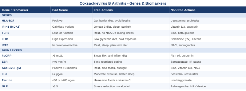
This article examines Coxsackievirus-related arthritis from two angles that most standard medical consultations never reach: trackable blood biomarkers that reveal what is actually happening in your immune and inflammatory pathways right now, and the genetic variants that explain why some people develop persistent joint inflammation after infection while others do not.
The six biomarkers covered — hsCRP, ESR, IL-6, anti-CVB antibodies, ferritin, and the neutrophil-to-lymphocyte ratio — are not just academic curiosities. Each one has a specific story to tell about where the immune dysregulation is happening, how active it currently is, and what type of intervention is most likely to help. For each, you will find practical guidance on how to measure it, what a concerning result looks like, and what to do about it — with and without supplements.
The genetic section covers five genes that shape susceptibility and severity: HLA-B27, IFIH1, TLR3, IL1B, and IRF3. Understanding your genetic risk is less about fatalism and more about identifying which biological levers are most worth pulling.
Beyond biology, the article also covers a landmark resource challenging conventional thinking on viral immunity, and five complementary approaches — including the Autoimmune Protocol and low-level laser therapy — that have meaningful clinical evidence for this type of condition.
---
6 Biomarkers to Track When Coxsackievirus Has Triggered Arthritis
Tracking the right biomarkers is not about collecting numbers for their own sake. In post-viral arthritis triggered by Coxsackievirus B, the inflammatory process is complex and multi-layered: innate immune activation, molecular mimicry where immune cells misidentify joint tissue, cytokine amplification loops, and metabolic stress all contribute. Different biomarkers illuminate different parts of that process. Knowing which ones are elevated — and by how much — lets you and your clinician build a picture of how active the disease is, whether it is resolving or persisting, and which interventions are most appropriate at a given moment.
Biomarker 1: High-Sensitivity C-Reactive Protein (hsCRP)
Why it matters
CRP is produced by the liver in response to IL-6 and other pro-inflammatory cytokines. In the context of Coxsackievirus arthritis, it serves as a real-time readout of systemic inflammation. High-sensitivity CRP (hsCRP) is the more precise version of the standard CRP test and can detect low-grade inflammation that standard CRP misses. This distinction matters in post-viral arthritis, where inflammation may be chronic but sub-acute — significant enough to damage joints over time, yet too subtle for a basic blood panel to catch.
Peter Attia consistently identifies hsCRP as one of the most useful markers for tracking inflammation-linked chronic disease risk, precisely because it responds both to acute and to smoldering inflammatory states. In reactive and post-viral arthritis, serial hsCRP measurements over weeks and months are more informative than any single reading.
How to measure it
A standard blood draw. Most commercial labs offer hsCRP as a standalone test or as part of a cardiovascular panel. Cost: typically $10–$40 USD depending on the lab and whether it is ordered through a physician. Optimal range: below 0.5 mg/L. Values between 1 and 3 mg/L represent moderate concern. Above 3 mg/L suggests active inflammation that warrants investigation.
If the score is bad — the plan without supplements
Before adding any supplements, address the fundamentals that have the most effect on hsCRP. Sleep is the most underrated lever here: reducing sleep to six hours or fewer for even a few nights raises CRP meaningfully in controlled studies. Aim for 7.5–9 hours in a consistent window. Eliminate processed seed oils (soybean, sunflower, corn) from the diet, as their high omega-6 content drives the arachidonic acid pathway and downstream CRP elevation. A Mediterranean-pattern or anti-inflammatory diet with abundant polyphenols, fatty fish, and olive oil has been shown in multiple randomized trials to reduce hsCRP by 20–40% over 12 weeks. Cold water immersion (contrast showering or cold plunge, 2–4 minutes at 10–15°C, 3–5x per week) may also reduce basal inflammation, though evidence in post-viral arthritis specifically is preliminary.
If the score is bad — the plan with supplements or equipment
Fish oil (EPA+DHA combined, 2–4g per day, taken with the largest meal) has the strongest evidence for hsCRP reduction among over-the-counter interventions, with multiple meta-analyses confirming a meaningful effect. Cycle: 12 weeks on, assess, continue if CRP improving. Side effects: fishy aftertaste, mild blood thinning at doses above 3g per day — relevant if you take anticoagulants. Curcumin with piperine (500mg curcumin, 5mg piperine, twice daily with food) has shown CRP-lowering effects in arthritis trials; cycle 8 weeks on, 4 weeks off due to limited long-term safety data. Low-level laser therapy (LLLT) devices targeting inflamed joints may also reduce local and systemic CRP, discussed further in the complementary section.
Biomarker 2: Erythrocyte Sedimentation Rate (ESR)
Why it matters
ESR measures how quickly red blood cells settle in a tube of blood — a proxy for fibrinogen, immunoglobulins, and other acute-phase proteins elevated during inflammation. It is a slower-moving signal than hsCRP (it lags behind clinical changes by days) but provides complementary information. In Coxsackievirus arthritis, ESR tends to stay elevated for longer than hsCRP, making it useful for tracking whether a chronic inflammatory process is truly resolving or merely entering a quieter phase. When ESR remains elevated long after hsCRP has normalized, that discordance can indicate persistent immune activity in the joint or systemic compartment.
How to measure it
Included in most comprehensive inflammatory panels. Cost: $10–$25 as a standalone test. Normal range: generally below 20 mm/hr in men and below 30 mm/hr in women, though age affects reference ranges. In active post-viral arthritis, values of 40–80 mm/hr are not uncommon.
If the score is bad — the plan without supplements
ESR is elevated by anemia, high fibrinogen, elevated immunoglobulins, and active infection or autoimmune activity. Address the root cause first: confirm that the Coxsackievirus infection has cleared (check anti-CVB IgM below). If it has, assess for secondary autoimmune triggers — gut permeability, dental infections, and chronic sleep disruption are all known ESR drivers. Time-restricted eating (compressing meals to an 8–10 hour window without calorie restriction) has shown some evidence for reducing inflammatory protein levels.
If the score is bad — the plan with supplements or equipment
Serrapeptase (10–60mg enterically coated, on an empty stomach, 3x per week) is a proteolytic enzyme sometimes used to reduce fibrinogen and ESR. Evidence is modest and mostly from older European trials; use cautiously, cycling 6 weeks on and 2 weeks off. Nattokinase (2000 FU per day) has a similar but better-studied fibrinolytic profile. Both interact with anticoagulants — check with a physician. Infrared sauna sessions (20–30 minutes at 55–65°C, 3–4x per week) have shown reductions in ESR in fibromyalgia and chronic inflammatory conditions, though CVB-specific data is lacking.
Biomarker 3: Anti-Coxsackievirus B Antibodies (IgM and IgG)
Why it matters
This is the most condition-specific biomarker on this list. IgM antibodies against CVB indicate recent or active infection — they typically appear within a week of exposure and decline over 1–3 months. IgG antibodies indicate past exposure and immune memory. In post-viral arthritis, a persistent IgM signal beyond three months is concerning and may indicate ongoing viral activity, incomplete viral clearance, or molecular mimicry — where the immune system's response to viral proteins cross-reacts with joint tissue.
Understanding where you sit on this antibody timeline changes the management approach entirely. Someone with active IgM elevation may need antiviral support; someone with only IgG and a clean IgM suggests the infection has cleared and the arthritis is a post-infectious inflammatory or autoimmune residue.
How to measure it
Requires a specific CVB serology panel — not always ordered automatically. Many specialty labs (LabCorp, Quest in the US; equivalent reference labs in Europe) offer CVB1–6 IgM and IgG panels. Cost: $80–$200 depending on the panel breadth. Request specifically "Coxsackievirus B serotype panel" with separate IgM and IgG results.
If the score is bad — the plan without supplements
If IgM remains elevated, the priority is supporting viral clearance. Zinc-rich foods (oysters, pumpkin seeds, red meat) and vitamin D optimization through sunlight exposure (15–30 minutes midday, skin exposed) are foundational. Reducing immunosuppressants where possible (including high-dose anti-inflammatory medications that blunt innate immunity) may allow the immune system to complete viral clearance. Rest — genuine, extended rest, not just reduced activity — is a non-negotiable pillar; the literature on enterovirus-associated myalgic conditions consistently shows that overexertion during active IgM elevation correlates with chronification.
If the score is bad — the plan with supplements or equipment
Zinc acetate or picolinate (25–40mg per day with food) has solid antiviral mechanistic evidence for enteroviruses including Coxsackievirus; supplementation should be cycled (8 weeks on, 2 weeks off) to avoid copper depletion — add 1–2mg copper if cycling long-term. Vitamin D3 supplementation (2000–5000 IU per day with vitamin K2 MK-7) may enhance innate antiviral immunity; target a serum 25(OH)D level of 40–60 ng/mL. N-acetylcysteine (600mg twice daily with food) supports glutathione levels needed for immune cell function; side effects are generally mild (GI upset at higher doses). Elderberry standardized extract (600–900mg per day during active infection phase) has shown modest antiviral effects in rhinovirus and influenza trials; CVB-specific data is limited.
Biomarker 4: Interleukin-6 (IL-6)
Why it matters
IL-6 is the upstream driver of most acute-phase reactants including CRP and ferritin. In Coxsackievirus arthritis, it is elevated both by the direct viral stimulus and by the subsequent autoimmune response in the synovium. IL-6 is also the cytokine responsible for the fever, fatigue, and systemic malaise that accompany both the infection and the arthritis flares. Its measurement helps distinguish a purely infectious inflammatory state from a more complex dysregulated autoimmune process — and it is the primary target of several biologic medications used in severe cases of inflammatory arthritis.
Tracking IL-6 over time allows you to see whether the inflammatory cascade is dampening or whether a chronic overproduction pattern is developing. Research published via PubMed has consistently linked elevated serum IL-6 to worse joint outcomes in reactive and post-infectious arthropathies.
How to measure it
Serum IL-6 via ELISA — available at most reference labs though sometimes requires a specific physician order or functional medicine panel. Cost: $50–$150. Normal range: typically below 7 pg/mL. In active viral arthritis, values can reach 20–100 pg/mL.
If the score is bad — the plan without supplements
IL-6 is acutely sensitive to obesity (particularly visceral fat), sedentary behavior, and poor sleep. Even 30 minutes of moderate aerobic exercise 5 days per week (walks, cycling — nothing that strains inflamed joints) has been shown in meta-analyses to reduce IL-6 by 10–35% over 8–12 weeks. Caloric restriction sufficient to reduce visceral adiposity has an even stronger effect. Address sleep apnea if relevant — intermittent hypoxia is a potent IL-6 inducer.
If the score is bad — the plan with supplements or equipment
Boswellia serrata extract (200–400mg of AKBA-standardized extract, twice daily with fat-containing meals) has shown IL-6 reductions in clinical trials of inflammatory joint conditions. Cycle: 12 weeks on, then reassess; well tolerated with minimal side effects. Resveratrol (500mg per day) has demonstrated IL-6 suppression in human studies, with best absorption from trans-resveratrol taken with fat; cycle 8 weeks on, 4 off. Continuous glucose monitors (CGMs) used short-term can identify glucose spikes that drive IL-6 production — a practical tool for identifying specific foods worth eliminating.
Biomarker 5: Serum Ferritin
Why it matters
Ferritin is both an iron storage protein and an acute-phase reactant — it rises sharply during inflammation and infection. In Coxsackievirus arthritis, elevated ferritin often signals active immune system engagement and can persist for weeks to months after the initial infection. At the same time, very low ferritin (below 30–40 ng/mL) independently impairs immune function and worsens fatigue — a common compounding problem in post-viral states where poor appetite reduces dietary iron intake. This dual role makes ferritin an unusually informative biomarker: it tells you simultaneously whether inflammation is active and whether iron-dependent immune processes are adequately supported.
Thomas Dayspring has emphasized the importance of interpreting ferritin in context — a ferritin of 80 ng/mL in a healthy athlete and a ferritin of 80 ng/mL in someone with active viral arthritis tell very different stories when evaluated alongside other inflammatory markers.
How to measure it
Standard blood panel, usually included in comprehensive metabolic or iron studies. Cost: $10–$30. Optimal range (excluding active infection): 50–150 ng/mL. Above 200 ng/mL in the absence of recent illness warrants investigation for hemochromatosis or ongoing systemic inflammation.
If the score is bad — the plan without supplements
If ferritin is high due to inflammation (rather than iron overload), the priority is reducing the inflammatory driver rather than modifying iron intake. If ferritin is low, dietary adjustments — heme iron sources like lean red meat, liver, and dark poultry, consumed with vitamin C-containing foods to enhance absorption — are the first tool. Avoid coffee or tea within 1 hour of iron-rich meals as polyphenols inhibit non-heme iron absorption.
If the score is bad — the plan with supplements or equipment
For low ferritin: iron bisglycinate (18–27mg elemental iron per day, on an empty stomach or with vitamin C) is better tolerated than ferrous sulfate with fewer GI side effects. Reassess every 6–8 weeks. For high ferritin driven by inflammation: lactoferrin (100–300mg per day) has shown modest ferritin-lowering effects in small trials and supports mucosal immunity simultaneously; generally safe for extended use.
Biomarker 6: Neutrophil-to-Lymphocyte Ratio (NLR)
Why it matters
The NLR is derived from a simple complete blood count (CBC) with differential — no extra blood draw needed. It is calculated by dividing the absolute neutrophil count by the absolute lymphocyte count. A high NLR reflects a state of immune stress: neutrophils rise during acute infection and tissue injury while lymphocytes fall under cortisol elevation and systemic inflammation. In post-viral arthritis, the NLR provides a quick proxy for how hard the innate immune system is working and whether a counter-regulatory (lymphocyte-depleting) stress response is still active.
Studies across multiple inflammatory conditions have shown that a persistently elevated NLR (above 3.0–3.5) correlates with worse clinical outcomes and delayed recovery. For Coxsackievirus arthritis, tracking the NLR over serial visits can tell you whether the immune system is regaining balance or whether the stress response is being chronically perpetuated.
How to measure it
Derived from any standard CBC with differential — no additional cost. Calculate by dividing neutrophils by lymphocytes from the same blood draw. Optimal range: 1.0–2.5. Values above 3.5 are clinically meaningful; above 5.0 in the absence of acute bacterial infection suggests significant immune dysregulation.
If the score is bad — the plan without supplements
NLR is particularly sensitive to psychological stress and poor sleep through the cortisol axis. Cortisol drives neutrophilia and lymphopenia simultaneously. Evidence-based stress reduction — any consistent practice that demonstrably lowers cortisol output (yoga, breathing protocols, cognitive behavioral therapy, social connection) — is the most direct lever. Eliminate or significantly reduce alcohol, which raises neutrophil counts. Moderate-intensity exercise (not high intensity, which acutely spikes neutrophils) over a 10–12 week program reliably improves NLR.
If the score is bad — the plan with supplements or equipment
Ashwagandha KSM-66 extract (300mg twice daily with meals) has shown meaningful cortisol reductions and NLR normalization in multiple randomized controlled trials in stressed populations. Cycle: 8 weeks on, 4 weeks off; avoid in thyroid conditions. Phosphatidylserine (100mg three times per day) has evidence for blunting exercise-induced cortisol and may help reduce background cortisol-driven NLR elevation. HRV biofeedback devices (Polar, Garmin, or dedicated HRV monitors) can serve as a real-time guide for recovery status — training when HRV is depressed drives the NLR higher; rest drives it lower.
---
The Genetic Layer: 5 Key Genes That Shape Susceptibility and Recovery
Understanding your genetic profile does not create fatalism — it creates specificity. In Coxsackievirus arthritis, certain gene variants change the probability of developing joint involvement after infection and influence how prolonged or severe that involvement becomes. Ali Torkamani's work on polygenic risk and Gary Brecka's popularization of functional genomics have both contributed to broader public awareness that these variants are not destiny — they are signals that guide lifestyle and nutritional compensations.
Gene 1: HLA-B27
What it is
HLA-B27 is the most studied genetic risk factor for reactive arthritis, the category into which Coxsackievirus-triggered arthritis often falls. It is a human leukocyte antigen that shapes how the immune system presents viral peptides to T cells. Approximately 5–8% of the general population carries HLA-B27, but among patients with reactive arthritis following enteric or urogenital infections, prevalence rises to 40–80% in published case series. Its role in CVB arthritis specifically is less well characterized than in Chlamydia- or Salmonella-triggered reactive arthritis, but the mechanistic overlap is substantial.
If the gene is bad — the plan without supplements
HLA-B27 positivity does not cause arthritis on its own — it requires an infectious trigger. The key intervention is therefore reducing exposure to and impact of those triggers. Gut barrier integrity matters enormously: HLA-B27 positive individuals appear more susceptible to gut-mediated molecular mimicry. A diet that minimizes lectins, excess gluten exposure, and industrial seed oils supports mucosal barrier function. High-intensity endurance training in HLA-B27 carriers may increase intestinal permeability transiently — structure exercise to allow adequate recovery.
If the score is bad — the plan with supplements or equipment
L-glutamine (5g per day between meals) is the primary mucosal support supplement with meaningful gut barrier evidence; 8 weeks on, assess. Probiotic strains Lactobacillus rhamnosus GG and Bifidobacterium longum have shown gut barrier strengthening effects in multiple trials. Infrared sauna protocols (3–4x per week) may reduce the systemic inflammatory load in HLA-B27 carriers without the joint-stressing effects of aggressive exercise.
Gene 2: IFIH1 (MDA5)
What it is
IFIH1 encodes Melanoma Differentiation-Associated protein 5 (MDA5), a cytoplasmic RNA helicase that acts as a sensor for viral double-stranded RNA — exactly the type produced during Coxsackievirus replication. When MDA5 detects viral RNA, it triggers a type I interferon response, the first wave of antiviral defense. Variants in IFIH1 are associated with both susceptibility to enteroviral infection and with autoimmune risk: some variants lead to reduced sensing (allowing the virus more time to replicate), while gain-of-function variants produce excessive interferon responses that can drive autoimmune arthritis even after viral clearance.
If the gene is bad — the plan without supplements
For reduced-function variants: optimizing innate immune responsiveness through consistent sleep, adequate vitamin D status, and avoiding immunosuppressant exposures (chronic alcohol, excess cortisol) is the primary tool. For overactive variants: anti-inflammatory dietary patterns that reduce interferon-driven inflammation — emphasizing omega-3 fats, polyphenols, and reducing refined carbohydrates — are particularly relevant.
If the gene is bad — the plan with supplements or equipment
Vitamin D3 (target serum 25(OH)D 50–70 ng/mL) directly modulates MDA5 pathway activity and is the most evidence-backed supplement for interferon pathway calibration. Quercetin (500mg per day with fat-containing food) has shown MDA5-modulatory effects in cell studies; human evidence is early. Side effects are minimal. Cycle 8 weeks on, 4 off.
Gene 3: TLR3 (Toll-Like Receptor 3)
What it is
TLR3 encodes a transmembrane receptor on immune cells that specifically recognizes double-stranded RNA, the molecular signature of Coxsackievirus and other enteroviruses. TLR3 activation triggers NF-κB and interferon regulatory pathways — the rapid inflammatory escalation that follows viral detection. Loss-of-function variants in TLR3 reduce the initial immune response to enteroviruses, which can allow deeper viral replication, more extensive tissue spread, and greater subsequent joint involvement. This has been particularly studied in the context of CVB-associated cardiomyopathy, but the joint consequences follow a similar logic.
If the gene is bad — the plan without supplements
For individuals with reduced TLR3 activity, the initial immune response to Coxsackievirus may be delayed and blunted. This makes early rest and early sleep optimization critical during any febrile illness — do not push through viral symptoms. Avoid non-steroidal anti-inflammatory drugs (NSAIDs) during the acute phase if possible; by reducing fever prematurely, they may blunt TLR3-dependent early immune clearance and allow viral persistence.
If the gene is bad — the plan with supplements or equipment
Zinc again is centrally relevant here — TLR3 signaling is zinc-dependent, and deficiency blunts the pathway. Beta-glucans (250–500mg per day from Saccharomyces cerevisiae or oat-derived sources) have shown TLR3 upregulation in immunological studies; practical application in enteric viral infection is plausible but needs more specific trial evidence. Well tolerated for extended use.
Gene 4: IL1B (Interleukin-1 Beta)
What it is
The IL1B gene encodes one of the most potent pro-inflammatory cytokines in the body. In Coxsackievirus arthritis, IL-1β is activated via the NLRP3 inflammasome — a danger-sensing complex that detects viral RNA and metabolic stress signals simultaneously. High-expression variants in IL1B produce an exaggerated inflammasome response, leading to greater joint swelling, more severe synovitis, and higher risk of chronic inflammatory changes. This gene has been directly studied in reactive arthritis cohorts and consistently associates with disease severity.
If the gene is bad — the plan without supplements
Reducing NLRP3 inflammasome triggers is the central strategy: excess dietary glucose and fructose are potent NLRP3 activators, as is saturated fat in combination with refined carbohydrates. A low-glycemic, Mediterranean-style dietary pattern with time-restricted eating has shown NLRP3 suppression in human studies. Cold exposure (cold showers or cold water immersion 4–5x per week) also blunts IL-1β-dependent inflammatory signaling through adrenergic and anti-inflammatory pathways.
If the gene is bad — the plan with supplements or equipment
Colchicine at low doses (0.5mg per day) directly inhibits NLRP3 inflammasome assembly and has been studied in both pericarditis and inflammatory arthritis — discuss with a physician as it requires a prescription. OTC alternative: luteolin (100–200mg per day, from supplement or parsley/artichoke-rich diet) shows IL-1β inhibition via NLRP3 suppression in preclinical and early clinical studies. Cycle 8 weeks.
Gene 5: IRF3 (Interferon Regulatory Factor 3)
What it is
IRF3 is a master transcription factor for type I interferon production — the antiviral cytokine cascade triggered by MDA5 and TLR3. After viral detection, IRF3 is phosphorylated and translocates to the nucleus to drive interferon-beta production. Variants that impair IRF3 phosphorylation reduce type I interferon output, leaving the host more susceptible to deep viral spread. Conversely, constitutively active variants drive chronic interferon production that can maintain joint inflammation long after the virus is gone. This dual risk profile makes IRF3 genotyping particularly informative for understanding both vulnerability and the specific nature of chronic post-CVB arthritis.
If the gene is bad — the plan without supplements
For reduced-function variants: the management mirrors TLR3 above — aggressive rest during acute illness, sleep optimization, and vitamin D repletion. For overactive variants contributing to chronic interferon-driven joint inflammation: a low-starch, plant-rich diet limiting lectins and nightshades has shown clinical benefit in autoimmune arthritis cohorts — the mechanistic pathway involves reduced TLR stimulation from gut-derived bacterial products.
If the gene is bad — the plan with supplements or equipment
NAC (N-acetylcysteine, 600mg twice daily) reduces oxidative signals that abnormally activate IRF3 in the absence of true viral threat. Andrographis extract (200–400mg per day, standardized to 10% andrographolides) modulates interferon regulatory pathways and has shown clinical anti-inflammatory effects in viral respiratory conditions; CVB-specific evidence is early. Cycle 6 weeks on, 2 off; can cause GI discomfort at higher doses.
---
What a Landmark Resource Suggests About Viral Immunity and Joint Recovery
Few resources have synthesized the intersection of viral immunology, chronic inflammation, and lifestyle intervention as comprehensively as the Rhonda Patrick Podcast — specifically her multi-part series on viral persistence, type I interferons, and the role of micronutrients in resolving post-infectious syndromes. Drawing on dozens of peer-reviewed studies, these episodes challenge the dominant clinical narrative that post-viral arthritis is simply "inflammation that will resolve with time and NSAIDs."
Below are the ten most impactful concepts from this body of work, each of which carries direct relevance for Coxsackievirus arthritis.
1. Viral Persistence Is Underestimated
Enteroviruses including CVB can persist in muscle, cardiac, and joint tissues for months after the acute phase resolves. PCR studies of muscle biopsies and synovial fluid from chronic post-viral patients have detected enteroviral RNA long after serology turns negative. This means the immune system may not be "overreacting" — it may be responding to ongoing low-level viral antigen presentation.
2. Vitamin D Deficiency Fundamentally Impairs Viral Clearance
Vitamin D receptor expression on immune cells is required for full TLR and MDA5 pathway activation. At 25(OH)D levels below 30 ng/mL — which describes much of the northern-hemisphere population in winter months — innate antiviral immunity is functionally impaired. Correcting deficiency to 50–70 ng/mL is not supplementation for marginal benefit; it is restoring a baseline requirement.
3. Magnesium Is Required for Over 300 Enzymatic Reactions in Immune Defense
Magnesium deficiency (very common, as serum magnesium is a poor marker of cellular stores) impairs NLRP3 inflammasome regulation, IL-6 production control, and mitochondrial energy metabolism in immune cells. Testing red blood cell magnesium is more informative than serum magnesium.
4. Exercise Timing Matters More Than Exercise Type
Moderate aerobic exercise reduces IL-6 and hsCRP when performed during the recovery phase, but high-intensity exercise during active viral replication — even subclinical replication — can disseminate virus to more tissues including joints. The safest protocol post-CVB: walk-only activity until IgM antibodies clear, then gradually reintroduce resistance and aerobic training.
5. Omega-3 Index Is a Critical Modifier of Joint Inflammation
The ratio of EPA+DHA in red blood cells (the omega-3 index) determines how quickly leukotriene-mediated joint inflammation resolves. A low omega-3 index (below 4%) dramatically prolongs the resolution phase of inflammatory arthritis. Target: above 8%, achievable with 2–4g EPA+DHA per day over 12–16 weeks.
6. Gut Microbiome Composition Shapes Systemic Inflammatory Tone
CVB enters the body via the gut. The composition of the intestinal microbiome at the time of infection influences both initial viral replication speed and the subsequent immune response. Bifidobacterium and Lactobacillus-rich microbiomes appear to limit viral replication; antibiotic exposure prior to infection may worsen outcomes.
7. Molecular Mimicry Is Probably More Common Than Diagnosed
CVB protein sequences share homology with several human joint proteins. Following infection, T cells and antibodies trained against viral antigens may cross-react with synovial tissue. This mechanism explains why arthritis persists after the virus is cleared — and why immunosuppressant monotherapy without addressing the antigen-presentation loop often produces incomplete results.
8. Cortisol Is a Double-Edged Sword
Acute cortisol release during infection appropriately suppresses the most destructive inflammatory responses. But chronic HPA axis activation in post-viral fatigue states paradoxically promotes IL-6 production, reduces lymphocyte counts, and impairs tissue repair. Managing the stress response is not "soft medicine" — it is mechanistically central to recovery.
9. Time-Restricted Eating Activates AMPK and Reduces NLRP3
Eating compressed into an 8–10 hour window activates AMPK (the cellular energy sensor), which directly suppresses the NLRP3 inflammasome responsible for IL-1β production in inflamed joints. This is achievable through meal timing alone without caloric restriction.
10. Tracking Is Not Optional — It Is the Intervention
The repeated finding across these episodes is that patients who track their biomarkers, monitor their sleep and HRV, and adjust behaviors based on real data improve substantially faster and more consistently than those who follow static protocols. The act of measurement itself changes behavior.
---
Complementary Approaches With Real Evidence for Post-Viral Arthritis
The Autoimmune Protocol (Sarah Ballantyne)
The Autoimmune Protocol (AIP), developed and systematized by Dr. Sarah Ballantyne in The Paleo Approach, is a structured dietary and lifestyle elimination protocol designed specifically for conditions where the immune system attacks or chronically inflames the body's own tissues. Coxsackievirus arthritis sits in this territory — molecular mimicry and persistent immune activation in joint tissue make it functionally similar to autoimmune joint conditions. The AIP eliminates dietary antigens most likely to drive intestinal permeability and immune reactivity (grains, legumes, dairy, eggs, nightshades, nuts, seeds, alcohol) while emphasizing organ meats, fatty fish, fermented vegetables, and micronutrient-dense whole foods.
A 2017 open-label clinical trial published via PubMed (PMID 28858071) examined the AIP in inflammatory bowel disease and showed significant reductions in clinical disease activity and inflammatory markers within 6 weeks, providing evidence that the protocol produces measurable systemic anti-inflammatory effects beyond symptom management. While direct CVB arthritis trials do not yet exist, the mechanistic overlap with gut-mediated immune dysregulation is strong.
To apply the AIP in Coxsackievirus arthritis: commit to a strict 30–60 day elimination phase followed by systematic reintroduction. The reintroduction phase is as important as the elimination — it identifies individual trigger foods rather than prescribing permanent avoidance. Work with a practitioner familiar with the protocol to ensure nutritional adequacy, particularly for calcium, iodine, and selenium during the dairy and seed elimination phases.
Low-Level Laser Therapy / Photobiomodulation
Low-level laser therapy (LLLT), also called photobiomodulation, uses red and near-infrared light (typically 630–850nm) at non-thermal intensities to modulate cellular metabolism, reduce inflammation, and promote tissue repair. The primary mechanism is photon absorption by cytochrome c oxidase in mitochondria, leading to increased ATP production, reduced reactive oxygen species, and downstream modulation of NF-κB — the central transcription factor governing IL-6 and TNF-alpha production. For inflamed synovial tissue in viral arthritis, this offers a non-pharmacological pathway to reducing local inflammatory cytokine production.
A meta-analysis published via PubMed (PMID 29394171) examining LLLT in rheumatoid arthritis found statistically significant reductions in pain and morning stiffness compared to sham controls. The effect sizes were moderate rather than large, but the risk profile is exceptional — virtually no adverse effects at therapeutic doses.
For Coxsackievirus arthritis application: target affected joints (knees, ankles, wrists are most commonly involved) with a device emitting 630–850nm at 5–50mW/cm². A practical consumer-grade protocol involves 10–20 minute sessions directly over each affected joint, 4–5 times per week for 6–8 weeks. Devices from established manufacturers (Joovv, Mito Red Light, or clinical-grade devices) are preferred over uncharacterized LED panels with poorly calibrated irradiance.
Mindfulness-Based Stress Reduction (MBSR)
MBSR is an 8-week standardized program developed by Jon Kabat-Zinn that combines body scan meditation, sitting meditation, mindful movement, and group discussion. Its relevance to post-viral arthritis goes beyond pain psychology: chronic psychological stress independently elevates IL-6, hsCRP, and cortisol — all of which worsen joint inflammation and impair viral clearance. Reducing the stress response through consistent mindfulness practice is not merely palliative; it mechanistically reduces the inflammatory burden the body must resolve.
A systematic review and meta-analysis of MBSR in inflammatory and autoimmune conditions, referenced via PubMed (PMID 24395196), found meaningful reductions in psychological distress and modest but consistent reductions in pro-inflammatory markers including IL-6 and cortisol. The effect on immune regulation — specifically on the balance between Th1 and Th2 cell populations — appears particularly relevant in post-viral immune dysregulation states.
For practical application in CVB arthritis: access MBSR through certified instructors (in-person or via platforms such as Palouse Mindfulness, which offers a free online adaptation of the original curriculum). Commit to the full 8 weeks before assessing outcomes. Daily practice of 20–45 minutes is required for clinical-level effects — shorter casual practice produces weaker results.
Tai Chi
Tai chi is a traditional Chinese mind-body practice combining slow, flowing movement sequences, postural alignment, and diaphragmatic breathing. For individuals with post-viral arthritis, it offers a uniquely valuable profile: it improves joint range of motion and proprioception without the mechanical loading stress of conventional exercise, while simultaneously activating parasympathetic nervous system pathways that reduce systemic inflammatory tone. Unlike vigorous aerobic exercise — which should be avoided during the active inflammatory phase of CVB arthritis — tai chi can be practiced safely even with acutely inflamed joints when modified appropriately.
A systematic review published in Seminars in Arthritis and Rheumatism and indexed via PubMed (PMID 19656578) examined tai chi in rheumatoid arthritis and found significant improvements in pain, disease activity, and physical function compared to control groups. Effects on serum inflammatory markers including IL-6 were also observed in several included trials.
For CVB arthritis: begin with a seated or standing beginner-level tai chi program (15–20 minutes, 5 days per week) during the acute phase, progressing to full sequences as joint inflammation subsides. The 24-form Yang style is the most studied and accessible starting point. Consistency over 8–12 weeks is required before joint-specific benefits become measurable.
Microbiome-Directed Therapies
The gut-joint axis is directly relevant to Coxsackievirus arthritis in a way that is rarely discussed in standard clinical care. CVB is an enteric virus — it enters and initially replicates in the gastrointestinal tract. The gut microbiome composition at the time of infection modulates viral replication speed, intestinal permeability, and the systemic immune response that determines whether joint involvement occurs. Post-infection, gut dysbiosis persists in many patients and maintains systemic inflammatory activation through lipopolysaccharide translocation and short-chain fatty acid deficiency — both of which directly affect joint inflammation.
Research published via PubMed (PMID 30046556) examined the role of specific microbiome signatures in reactive arthritis and found consistent dysbiosis patterns including reduced Faecalibacterium prausnitzii (the primary producer of butyrate, a key anti-inflammatory short-chain fatty acid) and elevated Prevotella copri in active disease. Targeted microbiome restoration — through specific probiotic strains, prebiotic fiber intake, and fermented food consumption — represents a genuinely mechanistic intervention rather than a vague "gut health" recommendation.
Application: introduce fermented foods gradually (kefir, kimchi, sauerkraut — 1–2 servings per day, increasing over 4 weeks to allow tolerance). Supplement with a multi-strain probiotic containing L. rhamnosus GG, B. longum, and L. plantarum at 20–50 billion CFU per day for a 12-week period. Prioritize prebiotic fiber (15–30g per day from diverse whole plant sources) to feed the restored microbiome — probiotic supplementation without prebiotic support produces limited lasting benefit.
---
Conclusion
Coxsackievirus arthritis sits at the intersection of infectious medicine, immunology, and individual biology — and that is precisely why generic protocols so often fall short. The biomarkers outlined above give you a real-time picture of where inflammation is concentrated and whether the immune system is resolving or perpetuating the problem. The genetic layer explains the why: why some people respond to CVB infection with joint involvement while others clear the virus without incident, and which biological systems most need support in any individual case.
The clearest next step is to get a targeted panel — at minimum hsCRP, ESR, ferritin, a CBC with differential, and CVB serology — and bring those results to a physician or functional medicine practitioner who can interpret them in the context of your clinical picture. From there, layering in the lifestyle and nutritional interventions described above — starting with the ones that address your most elevated markers — gives you a structured, evidence-grounded approach that is far more likely to produce meaningful improvement than waiting for inflammation to spontaneously resolve. You have more leverage over this condition than the standard protocol suggests.