This article contains AI generated content.
· UpdatedCushing's Syndrome Genes and Biomarkers: 6 Genes and 7 Biomarkers To Track
Introduction
Living with Cushing's syndrome — or trying to finally identify what's causing unexplained symptoms — places you in a frustrating and often disorienting position. The condition is serious, its effects are wide-ranging, and yet the medical conversation tends to stay focused on finding the cortisol source and removing it. That's the right first priority. But it doesn't explain why two people with nearly identical cortisol levels experience such different outcomes, or why metabolic dysfunction, bone loss, and cognitive fog can persist long after treatment succeeds.
Generic health advice doesn't offer much here. "Reduce stress" and "exercise more" are not useful when your body is running on cortisol dysregulation driven by a tumor, a genetic variant in your glucocorticoid receptor, or overactive local cortisol regeneration in adipose tissue. Cushing's syndrome is a biochemical problem, and it needs biochemically specific answers.
This article takes a more granular approach. It covers seven biomarkers that provide specific, trackable information about how cortisol excess is affecting different systems in your body — and what targeted actions may improve each one. It also examines six genes that influence cortisol production, receptor sensitivity, and tissue-level metabolism, which may help explain why your personal experience with this condition looks the way it does. Beyond that, a deep look at one of the most influential bodies of cortisol research ever compiled adds important mechanistic context, and several evidence-based complementary modalities round out a picture that is genuinely more complete than the standard overview.
None of this replaces working with an endocrinologist. But it may help you understand your own biology more precisely, identify where interventions are most needed, and have more productive conversations with the specialists who are treating you.
Summary
Seven biomarkers — urinary free cortisol, late-night salivary cortisol, plasma ACTH, DHEA-S, HOMA-IR, bone mineral density, and high-sensitivity CRP — reveal exactly where cortisol is doing damage and how quickly recovery is progressing. Six genes — PRKAR1A, MEN1, NR3C1, POMC, GNAS, and HSD11B1 — explain susceptibility, receptor hypersensitivity, and why some people metabolize cortisol differently even after treatment. The HSD11B1 enzyme, which regenerates active cortisol in fat tissue, may be the most underappreciated reason metabolic dysfunction persists after cortisol normalizes. The NR3C1 glucocorticoid receptor gene explains why some people experience severe metabolic and psychological effects at cortisol levels that don't bother others at all. The Sapolsky section covers ten mechanistic insights from the most rigorous cortisol research ever synthesized for a general audience — including how perceived control modifies cortisol damage, how exercise directly reverses hippocampal atrophy, and why slow-wave sleep matters more than total sleep duration in this condition. Four evidence-based complementary approaches — MBSR, biofeedback, breathing-based therapies, and yoga — each have specific protocols backed by clinical trials, not vague recommendations.
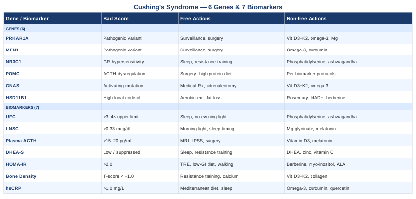
7 Biomarkers to Track When You Have Cushing's Syndrome
Biomarkers in Cushing's syndrome are not just diagnostic checkboxes. They're tools for understanding severity, monitoring treatment response, detecting complications early, and measuring whether lifestyle and supplement interventions are producing real change. The seven below cover the most clinically meaningful territory — from confirming cortisol excess itself to tracking the downstream metabolic consequences that often outlast the primary disease.
Biomarker 1: 24-Hour Urinary Free Cortisol (UFC)
Why it matters and what it reveals
The 24-hour urinary free cortisol test measures the amount of unbound, biologically active cortisol excreted in urine over a full day. Unlike a single blood draw that captures only a moment in time, UFC reflects average cortisol output across the entire diurnal cycle, making it one of the most robust first-line tests for Cushing's syndrome. A consistently elevated UFC — particularly when more than three to four times above the upper limit of normal — is strongly suggestive of true hypercortisolism.
In monitoring, a falling UFC in response to treatment confirms the intervention is working. Persistently elevated UFC after pituitary surgery signals incomplete remission or recurrence. UFC also helps distinguish genuine Cushing's from pseudo-Cushing's states such as depression, alcoholism, and severe obesity, which can produce mild elevations but rarely exceed three to four times the normal upper limit.
How to measure it
UFC requires a timed 24-hour urine collection. Your lab provides a container; you collect all urine for exactly 24 hours and return it for analysis. Normal reference ranges vary by laboratory but typically fall between 10 and 50 micrograms per 24 hours (28–138 nmol/24h). Cost: $50–$150 through standard labs, often covered by insurance with a documented clinical indication. Accuracy depends on complete collection — a missed void will underestimate results. Creatinine is frequently measured simultaneously to verify collection adequacy, and kidney disease can artificially suppress UFC values.
If the UFC is elevated: the plan without supplements
In active Cushing's syndrome, an elevated UFC is a signal that medical intervention is required — surgery, medication, or both. Lifestyle measures alone cannot normalize UFC caused by a true cortisol-secreting source. However, for milder elevations, pseudo-Cushing's states, or post-treatment residual dysregulation, targeted behavioral changes matter.
Sleep architecture has direct effects on cortisol production: aim for 7–9 hours with consistent timing seven days a week. Irregular sleep schedules chronically dysregulate the HPA axis. Reducing evening artificial light — particularly blue-spectrum light from screens — supports the normal cortisol decline that should occur in the hours before midnight. Moderate-intensity aerobic exercise (not excessive high-intensity training, which transiently spikes cortisol) improves HPA axis regulation over time when practiced consistently. Eliminating exogenous glucocorticoids — including prescribed topical steroids, some supplements, and certain nasal sprays — is essential before interpreting UFC.
If the UFC is elevated: the plan with supplements or equipment
Phosphatidylserine is the most rigorously studied nutraceutical for blunting the cortisol stress response. It attenuates ACTH and cortisol responses to exercise-induced and psychological stress without impairing performance. Dosing: 300–400 mg/day in divided doses, or 400–800 mg taken 30–60 minutes before an anticipated stressor. Cycling: continuous use is acceptable; some practitioners recommend 5 days on, 2 days off. Side effects: mild GI upset at higher doses; otherwise well-tolerated.
Ashwagandha (KSM-66 extract) has multiple double-blind RCTs showing cortisol reductions of 14–28% in chronically stressed adults. Dosing: 300–600 mg once or twice daily. Cycling: 8–12 weeks on, 2–4 weeks off. Side effects: potential thyroid effects (monitor TSH if you have thyroid disease); rare hepatotoxicity with very high doses or poor-quality products; mild GI symptoms initially.
Important caveat: if you are in active diagnosis, these supplements may partially blunt the clinical picture and complicate interpretation of your UFC. Discuss timing with your endocrinologist before using them during a diagnostic workup.
Continuous glucose monitors (CGMs) can indirectly track cortisol surges through their effect on blood glucose — particularly the afternoon and early-morning rises driven by cortisol's gluconeogenic activity. Wearable HRV monitors track autonomic recovery, which correlates with HPA axis regulation.
Biomarker 2: Late-Night Salivary Cortisol (LNSC)
Why it matters and what it reveals
Under normal physiology, cortisol follows a strict diurnal rhythm: it peaks approximately 30–45 minutes after waking, then declines throughout the day to reach its nadir around midnight. Cushing's syndrome disrupts this rhythm. Even when daytime cortisol levels overlap with normal ranges, midnight values remain inappropriately elevated — a loss of the circadian pattern that is one of the earliest and most sensitive markers of hypercortisolism.
Late-night salivary cortisol is one of the three first-line recommended tests in the Endocrine Society Clinical Practice Guideline for diagnosing Cushing's syndrome, alongside UFC and the overnight dexamethasone suppression test. Its sensitivity for detecting mild or intermittent Cushing's is particularly high, making it valuable not only at initial diagnosis but during long-term monitoring for recurrence.
How to measure it
LNSC is collected at home at 11 PM to midnight using a small absorbent swab placed in the mouth. The swab is sealed and sent to a certified lab. Two measurements on separate (ideally non-consecutive) nights are standard. Normal midnight salivary cortisol is generally below 0.33 mcg/dL (9 nmol/L), though reference ranges vary by lab. Cost: $30–$80 per collection; $60–$160 for the recommended two. Comprehensive hormone panels such as the DUTCH test include LNSC measurements alongside urinary cortisol metabolites.
Collection rules matter: nothing in the mouth 30 minutes before the swab, no alcohol or cigarettes in the evening, and ideally no unusual stress events on collection nights.
If LNSC is elevated: the plan without supplements
Morning bright light exposure — 10–30 minutes of natural sunlight or a 10,000-lux lamp within 30 minutes of waking — anchors the cortisol awakening response and strengthens the overall diurnal pattern, making the evening nadir deeper and more consistent. Eliminating artificial light after 8 PM allows melatonin to rise, which supports the cortisol decline.
Late-night eating disrupts circadian rhythms through nutrient-sensing signals that partially reset peripheral clocks; ending food intake at least 3 hours before sleep is a practical intervention. A consistent sleep-wake schedule — same bedtime and wake time seven days a week — is among the most powerful stabilizers of the HPA axis circadian pattern. Cognitive behavioral therapy for insomnia (CBT-I) has demonstrated improvements in cortisol dynamics in individuals with circadian disruption and is worth pursuing if sleep quality is poor.
If LNSC is elevated: the plan with supplements or equipment
Magnesium glycinate (200–400 mg taken 30–60 minutes before bed) supports GABA signaling and may contribute to evening cortisol reduction. Magnesium is depleted by chronic cortisol excess; deficiency worsens HPA axis reactivity in a self-reinforcing cycle. Side effects: loose stools at higher doses; otherwise well-tolerated. Continuous use is safe; test serum magnesium periodically.
Low-dose melatonin (0.3–1 mg, taken 60–90 minutes before target bedtime) anchors the circadian clock. Lower doses are more physiologically appropriate than the commonly marketed 5–10 mg options; higher doses produce pharmacological effects rather than mimicking normal physiology. Side effects: daytime grogginess if taken too late.
Phosphatidylserine (100–200 mg taken in the late afternoon) may specifically help blunt the abnormal evening cortisol elevation characteristic of circadian dysfunction in Cushing's. Cycling as above.
Wearable devices tracking HRV and sleep staging (Oura Ring, Whoop, Garmin) allow objective confirmation that sleep architecture is improving — which correlates with progressive normalization of the cortisol nadir.
Biomarker 3: Plasma ACTH
Why it matters and what it reveals
Once cortisol excess is confirmed, the most critical question is where is it coming from? Plasma ACTH answers this precisely. ACTH is produced by the pituitary gland and drives cortisol production in the adrenal cortex. Measuring it at 8 AM — its normal physiological peak — reveals the mechanism of disease.
High or inappropriately normal ACTH (typically above 15–20 pg/mL in confirmed hypercortisolism) indicates ACTH-dependent Cushing's: either a pituitary adenoma (Cushing's disease, the most common endogenous cause) or ectopic ACTH secretion from a tumor outside the pituitary, such as a lung carcinoid or thymic tumor. Suppressed ACTH (below 5 pg/mL) indicates ACTH-independent disease: the adrenal gland is producing cortisol autonomously, typically from a benign adenoma, bilateral hyperplasia, or, less commonly, adrenal carcinoma. This distinction completely redirects the diagnostic and treatment pathway.
How to measure it
Plasma ACTH requires a carefully handled draw — the sample must be kept on ice and processed within 15–20 minutes because ACTH degrades rapidly at room temperature. It is drawn at 8 AM. Cost: $50–$120, generally covered when clinically indicated. Results in 1–3 days.
If ACTH is inappropriately elevated: the plan without supplements
High ACTH in the context of confirmed Cushing's indicates upstream disease — pituitary or ectopic. The primary intervention is medical: MRI of the pituitary, and often inferior petrosal sinus sampling (IPSS) to confirm pituitary versus ectopic source. There is no lifestyle or supplement intervention that will meaningfully reduce ACTH from a secreting adenoma. Monitoring post-surgical ACTH is critical — undetectable ACTH in the immediate post-operative period confirms successful adenoma removal, and temporary adrenal insufficiency (requiring hydrocortisone replacement) should be anticipated.
If ACTH is inappropriately elevated: the plan with supplements or equipment
The evidence for supplements directly modulating pituitary ACTH secretion in Cushing's disease is limited. Ensuring vitamin D sufficiency (serum 25-OH-D between 40–60 ng/mL) is reasonable given pituitary tissue expressing vitamin D receptors and evidence that deficiency may affect pituitary function broadly. Dosing: vitamin D3 1,000–4,000 IU/day depending on baseline (re-test every 3–6 months), paired with K2 MK-7 at 100–200 mcg/day. Side effects: hypercalcemia at very high doses; monitor calcium alongside 25-OH-D.
Low-dose melatonin (0.3–1 mg at bedtime) has receptors in pituitary tissue and some early evidence for modulating ACTH secretion rhythms; the risk-to-benefit ratio is favorable for safe adjunctive use, though clinical evidence in Cushing's disease specifically remains preliminary.
Biomarker 4: DHEA-S (Dehydroepiandrosterone Sulfate)
Why it matters and what it reveals
DHEA-S is an adrenal androgen that serves as an important diagnostic marker and a window into adrenal reserve. In healthy individuals, both cortisol (from the zona fasciculata) and DHEA-S (primarily from the zona reticularis) are produced under ACTH stimulation. Their relationship in Cushing's syndrome carries diagnostic weight.
In benign cortisol-secreting adrenal adenomas, DHEA-S is characteristically suppressed: the adenoma produces cortisol autonomously, suppressing pituitary ACTH, which in turn withdraws stimulation from the zona reticularis. A low or undetectable DHEA-S alongside elevated cortisol is a strong fingerprint of a benign adrenal adenoma — and its degree of suppression correlates roughly with the duration and severity of autonomous cortisol production.
In adrenal carcinoma, DHEA-S may instead be elevated, because carcinomas frequently co-secrete androgens. A significantly elevated DHEA-S alongside cortisol excess is a red flag warranting urgent evaluation for malignancy. Beyond diagnostics, DHEA-S tracks adrenal reserve and plays roles in mood, immunity, bone health, and metabolic regulation — all compromised in Cushing's syndrome.
How to measure it
DHEA-S is measured from a fasting morning blood draw and is a stable analyte. Cost: $30–$80; frequently included in hormone panels. Normal ranges are strongly age- and sex-dependent — DHEA-S peaks in the mid-20s and declines approximately 2% per year thereafter, so always interpret relative to age-matched references.
If DHEA-S is low: the plan without supplements
In Cushing's syndrome, DHEA-S will often recover on its own after successful cortisol-normalizing treatment as ACTH stimulation of the zona reticularis resumes. Supporting this recovery requires adequate slow-wave sleep (DHEA-S is partially regulated by sleep depth and quality), resistance training (strength exercise acutely stimulates DHEA-S and may support zona reticularis recovery), and reduction of overall cortisol load through the behavioral strategies covered above.
If DHEA-S is low: the plan with supplements or equipment
DHEA supplementation is available over the counter but requires medical supervision and regular testing, particularly in the context of Cushing's syndrome where hormonal dynamics are already complex. When DHEA-S deficiency is confirmed and the treating endocrinologist approves, typical dosing is 25–50 mg/day for women and 50–100 mg/day for men, taken in the morning. Cycling: 3-month cycles with DHEA-S re-testing before each continuation. Side effects: acne, oily skin, hair thinning, potential worsening of hormone-sensitive conditions. Do not use without lab-confirmed deficiency and physician review.
Zinc (15–30 mg/day with food) and vitamin C (500–1,000 mg twice daily) support adrenocortical function broadly and carry favorable safety profiles as lower-risk adjuncts to support adrenal recovery without direct hormone replacement.
Biomarker 5: Fasting Glucose and HOMA-IR
Why it matters and what it reveals
Cortisol is a glucocorticoid — its effects on blood sugar are profound and central to the pathology of Cushing's syndrome. Cortisol elevates blood glucose by stimulating hepatic gluconeogenesis, suppressing insulin signaling in peripheral tissues, and impairing glucose uptake in muscle. Chronic cortisol excess produces a state of insulin resistance that closely resembles type 2 diabetes, with elevated fasting glucose, compensatory hyperinsulinemia, and eventual frank hyperglycemia in a significant proportion of patients.
HOMA-IR (Homeostatic Model Assessment of Insulin Resistance), calculated as (fasting glucose in mg/dL × fasting insulin in µIU/mL) / 405, is considerably more sensitive than fasting glucose alone for detecting insulin resistance. A HOMA-IR above 2.0 suggests meaningful insulin resistance; above 2.9 is more clearly abnormal. In active Cushing's syndrome, HOMA-IR is often significantly elevated even in patients with fasting glucose that appears only marginally elevated. Tracking HOMA-IR over time provides a sensitive signal of metabolic recovery — it often improves before fasting glucose alone would show a meaningful change.
Peter Attia has emphasized the critical importance of measuring both fasting glucose and fasting insulin together — not glucose alone — to assess metabolic health meaningfully. This principle is especially important in cortisol-driven insulin resistance, where fasting glucose may appear relatively normal while insulin is significantly elevated.
How to measure it
Both fasting glucose and fasting insulin require an 8–12 hour overnight fast before the draw. Fasting glucose is included in most standard metabolic panels, but fasting insulin must be specifically requested — it is frequently omitted from routine labs. Cost: fasting glucose alone is under $20; adding fasting insulin brings the total to $30–$70. A CGM complements fasting values by revealing glucose variability across the day — particularly useful in Cushing's since cortisol's glucose effects peak in the afternoon and may be missed by morning-only measurements.
If HOMA-IR is elevated: the plan without supplements
Time-restricted eating (TRE) within a consistent 8–10 hour window reduces daily insulin signaling without requiring caloric restriction and has strong evidence for improving insulin sensitivity. A low-glycemic, high-fiber diet — reducing refined carbohydrates and added sugars while emphasizing non-starchy vegetables, legumes, and whole grains — directly counters cortisol's glucose-raising effects.
Resistance training is arguably the most powerful non-pharmacological intervention for insulin resistance. Muscle contraction drives GLUT4 translocation to cell surfaces, enabling glucose uptake independently of insulin signaling — a direct bypass of the cortisol-impaired pathway. Aim for 3–4 sessions per week of compound movements. Post-meal walking (10 minutes after any meal) significantly blunts postprandial glucose excursions and requires no equipment.
If HOMA-IR is elevated: the plan with supplements or equipment
Berberine has demonstrated efficacy comparable to metformin for reducing insulin resistance in multiple clinical trials, acting primarily via AMPK activation. Dosing: 500 mg with meals, 2–3 times per day. Cycling: 8–12 weeks on, 4 weeks off — sustained continuous use may alter gut microbiome composition; cycling allows partial recovery. Side effects: GI symptoms (nausea, diarrhea, constipation) especially early; potential additive hypoglycemia risk if combined with metformin; interactions with CYP450-metabolized drugs.
Myo-inositol (2–4 g/day in powder form) improves insulin receptor sensitivity and has clinical trial evidence specifically in insulin-resistant conditions. Continuous use is generally safe; GI discomfort at higher doses is the main limitation.
Alpha-lipoic acid (300–600 mg/day) acts as a mitochondrial cofactor with modest but consistent evidence for improving insulin sensitivity. Caution: can lower blood glucose significantly in diabetics — monitor closely if used alongside diabetes medications. Long-term high-dose ALA may compete with biotin; supplementing biotin (300–500 mcg/day) alongside is advisable.
A CGM such as Dexcom Stelo or Libre provides real-time glucose data revealing which meals, stressors, and activities most significantly affect glucose — particularly valuable in Cushing's syndrome where the afternoon cortisol-glucose surge may be missed by fasting measurements. Cost: approximately $70–$100 per month without prescription.
Biomarker 6: Bone Mineral Density (DEXA Scan)
Why it matters and what it reveals
Chronic cortisol excess is among the most potent drivers of secondary osteoporosis in medicine. Glucocorticoids suppress osteoblast activity (bone formation), increase osteoclast activity (bone resorption), and impair intestinal calcium absorption while increasing urinary calcium losses. The combined result is an accelerated bone loss that can be rapid and severe — patients with Cushing's syndrome may lose significant trabecular density within months of disease onset, and vertebral compression fractures can occur with minimal trauma.
Bone mineral density (BMD) measured by DEXA scan quantifies bone density at the lumbar spine and hip, the sites most commonly affected in glucocorticoid-driven osteoporosis, and expresses it as T-scores and Z-scores. Tracking BMD at diagnosis and annually (or every 1–2 years after cortisol normalization) documents both the impact of disease and recovery trajectory. Critically, trabecular bone recovery after successful treatment is slow and may never reach pre-disease levels — making early detection and intervention genuinely important rather than precautionary.
How to measure it
DEXA scanning is widely available at imaging centers and endocrinology practices. The scan takes 15–30 minutes and delivers minimal radiation. Cost: $100–$300 out of pocket; generally covered by insurance when Cushing's syndrome or glucocorticoid exposure is documented. T-score interpretation: above -1.0 is normal; -1.0 to -2.5 is osteopenia; below -2.5 is osteoporosis.
If BMD is low: the plan without supplements
Resistance training provides the strongest osteogenic stimulus of any exercise type. Bone responds to mechanical loading — heavy compound movements (squats, deadlifts, presses) generate the highest bone formation signals. Weight-bearing activities (walking, jogging, hiking) complement this. High-impact activities (jumping, stair-climbing) add further stimulus where joint integrity allows.
Dietary calcium from food — distributed across meals throughout the day rather than in single large amounts — is better absorbed than supplements and has fewer associated adverse effects. Dairy, canned fish with bones, tofu, and fortified foods are practical sources. Aim for 1,000–1,200 mg/day from food.
Daily walking (7,000–10,000 steps) provides consistent low-intensity mechanical loading that complements structured exercise and supports bone maintenance between training sessions.
If BMD is low: the plan with supplements or equipment
Vitamin D3 + K2 (MK-7 form) is the foundational combination for bone health. Vitamin D3 supports intestinal calcium absorption; K2-MK7 activates osteocalcin and matrix GLA protein, directing calcium into bone and away from arterial tissue. Dosing: D3 2,000–5,000 IU/day (titrated to achieve serum 25-OH-D between 40–60 ng/mL), K2-MK7 100–200 mcg/day. Take together with a fat-containing meal for optimal absorption. Test 25-OH-D and serum calcium every 3–6 months. Side effects: hypercalcemia at very high sustained D3 doses; K2 may interact with warfarin (monitor INR carefully).
Calcium citrate (if dietary intake is genuinely below 1,000 mg/day from food) at 500 mg twice daily with meals. Citrate absorbs better than carbonate at lower stomach acid levels and causes less constipation. Avoid single doses above 500 mg (absorption efficiency drops sharply). Side effects: constipation; potential cardiovascular concerns with long-term high-dose calcium supplements — prioritize dietary sources.
Collagen peptides (10–15 g/day) have emerging evidence for supporting bone matrix alongside mineral supplements; they also support connective tissue integrity that is separately compromised by chronic glucocorticoid exposure. Generally well-tolerated. In severe osteoporosis, medical treatments including bisphosphonates, denosumab, or teriparatide may be necessary — discuss with your endocrinologist. Whole-body vibration platforms (20–30 Hz) have modest evidence as adjunctive support for bone density maintenance.
Biomarker 7: High-Sensitivity CRP and Interleukin-6
Why it matters and what it reveals
The relationship between cortisol and inflammation defies simple categorization. In acute settings, cortisol is profoundly anti-inflammatory — it is used pharmacologically for exactly this purpose. But in chronic Cushing's syndrome, this suppressive relationship paradoxically coexists with chronic low-grade systemic inflammation, measurable through elevated high-sensitivity CRP (hsCRP) and interleukin-6 (IL-6).
The mechanism: chronic cortisol excess drives visceral fat accumulation, and visceral adipose tissue is itself a major source of pro-inflammatory cytokines. IL-6 and TNF-alpha secreted from visceral fat perpetuate systemic inflammation even as some immune branches remain suppressed. Elevated hsCRP in Cushing's syndrome correlates with cardiovascular risk, metabolic syndrome severity, and overall disease burden. Even after cortisol normalization, inflammatory markers may remain elevated for months — reflecting the sustained pro-inflammatory activity of the visceral adipose depot that accumulated during active disease.
How to measure it
hsCRP is measured from a standard blood draw; fasting is not strictly required, but avoid acute illness or injury in the preceding two weeks as these cause sharp reactive CRP elevations. Cost: $20–$50; widely available. Optimal hsCRP is below 1.0 mg/L; 1.0–3.0 mg/L indicates moderate cardiovascular risk; above 3.0 mg/L in the absence of acute illness signals elevated systemic inflammatory load. IL-6 is less commonly ordered but more directly reflects cytokine activity; available through specialty labs ($50–$100).
If hsCRP is elevated: the plan without supplements
The Mediterranean-style diet — emphasizing extra-virgin olive oil, fatty fish, vegetables, legumes, and nuts, while minimizing ultra-processed foods and refined grains — has the strongest dietary evidence for reducing hsCRP. Multiple large clinical trials confirm CRP reductions of 20–30% with sustained adherence.
Sleep duration powerfully modulates inflammatory cytokines: consistently sleeping below 6 hours or above 9 hours is associated with significantly elevated CRP and IL-6. Seven to eight hours of high-quality, consistent sleep is the most evidence-backed single behavioral intervention for inflammation management.
Moderate-intensity aerobic exercise (150 minutes per week or more) has consistent meta-analytic support for reducing hsCRP. Excessive high-intensity training without adequate recovery can transiently elevate CRP — balance and recovery time matter as much as the training itself.
If hsCRP is elevated: the plan with supplements or equipment
Omega-3 fatty acids (EPA + DHA, 2–4 g/day with meals) are among the most robustly evidenced supplements for reducing hsCRP and IL-6. Multiple RCTs and meta-analyses confirm meaningful anti-inflammatory effects at these doses. Cycling: continuous use is safe; reassess hsCRP at 90-day intervals. Side effects: fishy aftertaste (enteric-coated formulations minimize this), mild blood-thinning at high doses (relevant if anticoagulated).
Curcumin with piperine (500–1,000 mg of curcumin standardized to 95% curcuminoids, paired with 5–10 mg piperine for bioavailability) has RCT evidence for reducing CRP, IL-6, and TNF-alpha. Take with a fat-containing meal, once or twice daily. Cycling: 12 weeks on, 4 weeks off. Side effects: GI upset; mild blood-thinning properties; potential interactions with chemotherapy agents.
Quercetin (500–1,000 mg/day) has mechanistic and early clinical evidence for IL-6 reduction and is being actively studied in metabolic syndrome — a phenotype that closely overlaps with Cushing's syndrome pathophysiology. Pair with vitamin C for enhanced absorption. Cycling: 6–8 weeks on, 2–4 weeks off.
---
These seven biomarkers give you a measurable, trackable picture of where cortisol excess is causing harm and how recovery is progressing. Understanding the genetic layer beneath them adds another dimension — one that helps explain why your specific experience looks the way it does.
What Your Genes Reveal About Cushing's Syndrome Risk and Response
Genetics in Cushing's syndrome operates on two distinct levels: genes that cause the disease by producing hormone-secreting tumors, and genes that shape how severely cortisol affects you through receptor sensitivity, enzymatic activity, and metabolic processing. Both levels are worth understanding — the first for risk stratification and surveillance, the second for personalizing intervention strategies.
Researchers like Ali Torkamani at the Scripps Research Translational Institute have emphasized that genetic profiling in endocrine tumors is moving rapidly toward clinical actionability, with individual genetic context increasingly explaining heterogeneity in disease expression and treatment response. The approach popularized by Gary Brecka — using genetic variant data to build personalized lifestyle and supplement protocols — has limits in the absence of rigorous trial data, but the underlying principle of using known genetic variants to target interventions more precisely has genuine scientific grounding in several well-studied genes.
Gene 1: PRKAR1A — The Carney Complex Driver
What this gene does
PRKAR1A encodes the regulatory subunit 1A of protein kinase A (PKA), a central enzyme in the signaling cascade that drives adrenal cortisol production. Normally, PRKAR1A holds PKA in check. Pathogenic loss-of-function mutations in PRKAR1A — inherited in an autosomal dominant pattern — release this brake, causing constitutively elevated PKA activity in adrenocortical cells. The result is primary pigmented nodular adrenocortical disease (PPNAD), a bilateral adrenal condition producing ACTH-independent Cushing's syndrome, typically presenting in young adults.
PPNAD is the adrenal manifestation of Carney Complex, which also includes cardiac myxomas, spotty skin pigmentation, and various other tumors. The cortisol production in PPNAD is characteristically autonomous and shows a paradoxical increase during dexamethasone suppression testing — one of the diagnostic fingerprints. Genetic testing for PRKAR1A pathogenic variants is available through comprehensive endocrine genetics panels.
If PRKAR1A has a pathogenic variant: the plan without supplements
This is structural disease — the primary intervention is medical and surgical. Surveillance per Carney Complex protocols is the foundation: annual echocardiography (cardiac myxomas can be life-threatening), periodic adrenal imaging, thyroid ultrasound, and testicular examination (in males). Bilateral adrenalectomy is frequently necessary for PPNAD-associated Cushing's syndrome. Genetic counseling for first-degree relatives is strongly recommended, as the mutation is autosomal dominant. Lifestyle support (anti-inflammatory diet, bone protection, metabolic management) helps manage downstream cortisol effects but cannot address the structural adrenal disease.
If PRKAR1A has a pathogenic variant: the plan with supplements or equipment
Given the medical severity, supplementation should focus on managing the consequences of cortisol excess rather than the genetic defect itself. Prioritize vitamin D3 + K2 for bone protection (as above), omega-3 fatty acids for cardiovascular and inflammatory support, and magnesium glycinate for HPA axis and sleep support. All supplements should be reviewed with your endocrinologist, as some may interact with monitoring protocols or ongoing treatment.
Gene 2: MEN1 — Hereditary Pituitary ACTH Excess
What this gene does
The MEN1 gene encodes menin, a tumor suppressor involved in chromatin remodeling, transcription regulation, and cell cycle control. Loss-of-function mutations cause Multiple Endocrine Neoplasia Type 1 (MEN1), predisposing carriers to tumors of the parathyroid, pancreas, and pituitary. Pituitary tumors occur in approximately 40% of MEN1 carriers; ACTH-secreting adenomas causing Cushing's disease account for 2–5% of MEN1 pituitary tumors — representing a hereditary cause of ACTH-dependent hypercortisolism.
MEN1-associated Cushing's disease tends to present at younger ages than sporadic Cushing's disease and may have a more aggressive clinical course.
If MEN1 has a pathogenic variant: the plan without supplements
Annual surveillance per MEN1 guidelines includes serum calcium and PTH (parathyroid tumors are the most common manifestation), fasting gut hormone panel (pancreatic neuroendocrine tumors), and periodic pituitary MRI. When an ACTH-secreting pituitary adenoma is identified, transsphenoidal surgery is the standard treatment, with post-operative management similar to sporadic Cushing's disease. Genetic counseling for relatives.
If MEN1 has a pathogenic variant: the plan with supplements or equipment
Calcium and vitamin D management in MEN1 is complex because primary hyperparathyroidism frequently co-occurs and can produce hypercalcemia. Work directly with your endocrinologist before supplementing either. Beyond those constraints, anti-inflammatory lifestyle support (omega-3 fatty acids, curcumin) and consistent stress management are reasonable adjuncts that support overall HPA axis function without interfering with surveillance protocols.
Gene 3: NR3C1 — Glucocorticoid Receptor Sensitivity
What this gene does
NR3C1 encodes the glucocorticoid receptor (GR) — the cellular receptor through which cortisol exerts virtually all of its physiological effects. This is arguably the most clinically relevant gene for understanding why different people experience profoundly different symptom severity at similar cortisol levels.
Several well-studied NR3C1 polymorphisms alter receptor sensitivity in meaningful ways. The BclI variant is associated with increased glucocorticoid sensitivity — carriers may experience greater metabolic effects (insulin resistance, fat redistribution, bone loss) at a given cortisol level. The ER22/23EK variant is associated with relative glucocorticoid resistance — carriers may require higher cortisol levels to suppress ACTH normally, potentially complicating interpretation of suppression tests. The N363S variant increases glucocorticoid sensitivity specifically for metabolic effects.
These variants don't cause Cushing's syndrome, but they may explain why your symptom burden or treatment response diverges from what lab values alone predict.
If NR3C1 variants suggest increased receptor sensitivity: the plan without supplements
Increased glucocorticoid receptor sensitivity means cortisol's downstream effects on metabolism, bone, immune function, and mood are amplified — raising the priority of every intervention aimed at reducing cortisol's downstream reach. This makes sleep quality, resistance training for insulin sensitivity and bone preservation, anti-inflammatory diet, and stress reduction all more important than they would be for someone with normal receptor sensitivity.
Reducing cortisol-stimulating inputs matters more than average: excessive high-intensity training, chronic psychosocial stress, poor sleep, and high caffeine intake all drive cortisol production, and with enhanced receptor sensitivity, these inputs carry outsized downstream consequences.
If NR3C1 variants suggest increased receptor sensitivity: the plan with supplements or equipment
Phosphatidylserine blunts the ACTH and cortisol response at the HPA axis level — reducing the cortisol signal reaching the hypersensitive receptor, which is precisely where the leverage is. Dosing: 300–400 mg/day; 30–60 minutes before anticipated stressors. Cycling: 5 days on, 2 days off or continuous. Side effects: minimal.
Ashwagandha (KSM-66) reduces adrenal cortisol output, reducing the substrate reaching the sensitized receptor. Dosing: 300–600 mg/day; 8–12 weeks on, 2–4 weeks off. Monitor thyroid function (TSH) if used long-term.
Rhodiola rosea acts on the cortisol release mechanism and has clinical evidence for reducing cortisol and stress-related fatigue. Dosing: 200–400 mg of standardized extract (3% rosavins, 1% salidroside), taken in the morning. Cycling: 6 weeks on, 1–2 weeks off. Side effects: stimulating — avoid evening use; mild agitation or headache in sensitive individuals; contraindicated in bipolar disorder.
Gene 4: POMC — The Cortisol Precursor Chain
What this gene does
POMC (pro-opiomelanocortin) is expressed in the pituitary and serves as the molecular precursor for ACTH and several melanocortin peptides including alpha-MSH. When processed correctly in the pituitary, POMC yields ACTH, which travels to the adrenal gland to drive cortisol synthesis.
In Cushing's syndrome, POMC matters in two relevant contexts. First, ectopic ACTH production — from lung carcinoids, thymic tumors, pancreatic neuroendocrine tumors, and other sources — involves ectopic expression of POMC with subsequent processing to ACTH. Second, rare germline POMC mutations can cause early-onset obesity and disrupted cortisol axis regulation, a distinct phenotype. More commonly, POMC methylation patterns (an epigenetic modification) have been associated with altered HPA axis setpoints in population studies, suggesting epigenetic regulation of this gene contributes to cortisol variation beyond rare mutations.
If POMC signaling is dysregulated: the plan without supplements
Ectopic ACTH from POMC-expressing tumors requires identification and surgical or ablative removal of the source — typically involving chest CT, octreotide scans, or PET imaging. This is entirely medical. For the rarer germline POMC mutation phenotype, appetite regulation and weight management are the central clinical challenges; a specialist in obesity medicine alongside an endocrinologist provides the right framework.
If POMC signaling is dysregulated: the plan with supplements or equipment
Supporting the broader cortisol axis during ectopic ACTH-driven Cushing's syndrome includes all the downstream biomarker management strategies already described. A protein-rich, high-fiber dietary pattern helps manage the appetite dysregulation associated with POMC pathway disruption. No supplement directly addresses ectopic ACTH production effectively; the downstream support strategies above are the appropriate focus.
Gene 5: GNAS — Constitutional Adrenal Activation
What this gene does
The GNAS gene encodes the alpha subunit of a stimulatory G protein (Gsα), a central signal transducer in the ACTH-cortisol signaling cascade. Activating somatic mutations in GNAS cause McCune-Albright syndrome (MAS), in which the mutant Gsα protein is constitutively active — it continuously signals the adrenal gland to produce cortisol without requiring any ACTH stimulation. The result is ACTH-independent bilateral adrenal hyperplasia and cortisol excess that presents in infancy or childhood.
Because the mutation is somatic and mosaic (not all cells carry it), phenotypic expression varies widely. The adrenal cortex cells that do carry the mutation produce cortisol autonomously and suppress pituitary ACTH, producing the biochemical profile of ACTH-independent Cushing's syndrome.
If GNAS has an activating mutation: the plan without supplements
Management is challenging and specialist-directed. Medical steroidogenesis inhibitors (ketoconazole, metyrapone, osilodrostat) are often used as a bridge while definitive management is planned. Bilateral adrenalectomy is frequently required for cortisol control; the multi-system nature of MAS requires long-term multidisciplinary follow-up covering bone disease, thyroid, and gonads alongside adrenal disease.
If GNAS has an activating mutation: the plan with supplements or equipment
Strictly adjunctive. Vitamin D3 + K2 for bone protection, omega-3 fatty acids for cardiovascular and inflammatory support, and the glucose management strategies above are all appropriate as complements to medical management. Discuss all supplements with your endocrinologist given potential effects on hormone monitoring.
Gene 6: HSD11B1 — The Local Cortisol Amplifier
What this gene does
HSD11B1 (11-beta-hydroxysteroid dehydrogenase type 1) may be the most metabolically actionable gene in the cortisol story for people recovering from Cushing's syndrome. This enzyme, expressed primarily in the liver and adipose tissue, converts inactive cortisone back to active cortisol at the tissue level. It acts as a local cortisol amplifier — even when the adrenal glands produce normal amounts of cortisol, HSD11B1 activity in visceral fat determines how much active cortisol your cells are actually exposed to.
A landmark Science paper by Masuzaki et al. (2001) demonstrated that overexpression of HSD11B1 specifically in adipose tissue of mice produced a full metabolic syndrome phenotype — central obesity, insulin resistance, hypertension, and dyslipidemia — without any increase in circulating cortisol. This elegantly demonstrated that local tissue cortisol regeneration is sufficient to drive metabolic disease independently of systemic cortisol levels.
In Cushing's syndrome recovery, persistently elevated HSD11B1 activity in the visceral fat depot accumulated during active disease may explain why some patients continue experiencing insulin resistance, central adiposity, and metabolic dysfunction long after their systemic cortisol normalizes. Genetic variants affecting HSD11B1 expression or promoter activity are common in the general population and may modulate metabolic disease risk across the spectrum.
If HSD11B1 activity is high: the plan without supplements
Visceral fat reduction is the most powerful intervention — adipose tissue is the primary site of HSD11B1 activity, and reducing its volume directly reduces the organ's overall cortisol-regenerating capacity. A caloric deficit achieved through any sustainable dietary approach, combined with consistent resistance and aerobic exercise, is the foundation.
Moderate-intensity aerobic exercise (150–300 minutes per week) has direct evidence for reducing HSD11B1 gene expression in adipose tissue — the exercise signal directly downregulates this enzyme. This is one of the clearest mechanistic reasons why aerobic exercise is particularly important in post-Cushing's metabolic recovery.
If HSD11B1 activity is high: the plan with supplements or equipment
Rosemary extract containing carnosic acid has in-vitro evidence for HSD11B1 inhibition. Standardized rosemary extract at 1,000–2,000 mg/day is broadly safe, though clinical human trials demonstrating HSD11B1 inhibition in vivo remain limited — this is early-stage evidence with a favorable safety profile.
NAD+ precursors (NMN 250–500 mg/day, or NR 300–600 mg/day) support mitochondrial efficiency in adipose and liver tissue and have emerging evidence for improving metabolic function — which may indirectly reduce HSD11B1-driven cortisol amplification through improved cellular energy metabolism. Cycling: 5 days on, 2 days off or continuous. Generally well-tolerated; the primary limitation is cost. Side effects: generally mild — some GI sensitivity early; flush-free compared to niacin forms.
Berberine (500 mg with meals, 2–3 times daily) has also shown indirect HSD11B1-modulating effects through its AMPK-activating and metabolic-improving properties — another reason it is one of the more broadly useful supplements in Cushing's syndrome's metabolic aftermath.
---
What Robert Sapolsky's Cortisol Research Can Change for You
Few scientists have contributed more to understanding what chronic cortisol does to the human body than Robert Sapolsky, professor of biology, neurology, and neurological sciences at Stanford and author of Why Zebras Don't Get Ulcers. While his research centers on chronic psychological stress rather than Cushing's syndrome specifically, the mechanisms are identical — because cortisol doesn't distinguish between the signal from a pituitary adenoma and the signal from an overactivated hypothalamus. The cellular damage downstream is the same.
1. Cortisol Was Designed for Acute Emergencies, Not Sustained Activation
Cortisol's adaptive role is to mobilize energy rapidly under genuine threat. Every effect that makes it useful acutely — suppressing digestion, immune function, reproduction, and growth to redirect resources — becomes destructive when sustained for months or years. This isn't a failure of design; it's a mismatch between evolutionary programming and modern conditions, including endocrine disease.
2. Cardiovascular Risk Is Mechanistically Real, Not Just Statistical
Cortisol excess accelerates atherosclerosis through multiple converging pathways: chronic hyperglycemia damages endothelial cells, elevated aldosterone-like effects raise blood pressure, and direct pro-inflammatory effects on arterial walls promote plaque formation. Sapolsky's work clarifies that cardiovascular mortality in Cushing's syndrome isn't a coincidence — it is mechanistically driven.
3. The Hippocampus Literally Shrinks Under Cortisol Load
The hippocampus is packed with glucocorticoid receptors and is profoundly vulnerable to cortisol excess. Sapolsky's primate research and subsequent human MRI studies confirm measurable hippocampal volume loss in states of chronic hypercortisolism, including Cushing's syndrome. This provides a neurological explanation for the memory impairment, brain fog, and spatial disorientation many patients describe.
4. Muscle Wasting Is a Direct Cortisol Effect, Not Secondary Inactivity
Cortisol signals muscle protein breakdown for gluconeogenesis. The proximal muscle wasting of Cushing's syndrome — particularly shoulders, hips, and thighs — is a direct biochemical consequence, independent of exercise habits. This explains why maintaining high protein intake and resistance training during Cushing's syndrome is simultaneously more important and more physiologically difficult than normal.
5. Immune Dysregulation Is Bidirectional, Not Simple Suppression
Sapolsky's synthesis helped clarify that chronic glucocorticoid exposure doesn't uniformly suppress immunity — it dysregulates it. Some branches are suppressed while others are paradoxically over-activated, producing the combination of infection susceptibility and chronic inflammatory conditions that many Cushing's patients experience simultaneously, which confounds clinical management.
6. Slow-Wave Sleep Is the Specific Target
Cortisol specifically suppresses slow-wave (deep, restorative) sleep — the stage when growth hormone is secreted and tissue repair occurs. This explains why many Cushing's patients report unrefreshing sleep regardless of total sleep duration. Sleep quality interventions targeting sleep architecture (consistent bedtime, darkness, reduced evening light, magnesium) are more relevant than simply increasing time in bed.
7. Perceived Control Amplifies or Reduces Cortisol Harm
Among Sapolsky's most counterintuitive findings: the subjective experience of having no control over a stressor amplifies the cortisol response far beyond the stressor's objective severity. For patients who spent years without diagnosis — experiencing body changes with no explanation — this mechanism may have compounded cortisol's harm through an additional, psychologically driven HPA axis activation. Diagnosis itself begins to reverse this.
8. Social Connection Has a Direct Neuroendocrine Effect
Sapolsky's primate work, confirmed in human studies, shows that social support measurably lowers glucocorticoid responses to equivalent stressors. Social isolation amplifies them. This is not metaphorical — it is a real neuroendocrine effect. Maintaining social connection during the isolating experience of Cushing's syndrome is biologically protective, not just psychologically important.
9. Exercise Directly Reverses Cortisol's Neurological Damage
Regular moderate-intensity aerobic exercise reduces glucocorticoid receptor density in stress-sensitive brain regions and promotes hippocampal neurogenesis — directly partially reversing the volume loss caused by chronic cortisol excess. Sapolsky identifies this as the single most robustly evidence-backed intervention for reducing cortisol's neurological damage, more than any pharmaceutical or supplement.
10. Tonic vs. Phasic Cortisol: The Distinction That Changes Everything
Sapolsky distinguishes between phasic cortisol responses (acute spikes in response to real stressors, which are healthy and necessary) and tonic cortisol elevation (the baseline that remains chronically high). Building resilience — through sleep, consistent exercise, social connection, and perceived agency — reduces the tonic baseline, not the phasic response. For post-treatment Cushing's patients rebuilding HPA axis regulation, this distinction is practically meaningful: the goal is not a flat cortisol curve, but a low, healthy baseline with preserved reactive capacity.
---
Evidence-Based Complementary Approaches
For people managing the systemic consequences of cortisol excess — whether during active Cushing's syndrome, in recovery, or while managing post-treatment metabolic residuals — several well-studied complementary modalities can meaningfully support the strategies above.
Mindfulness-Based Stress Reduction (MBSR)
MBSR is an 8-week structured program combining mindfulness meditation, body scan practices, and gentle movement developed by Jon Kabat-Zinn. Its relevance to Cushing's syndrome lies in the fact that chronic psychosocial stress activates the HPA axis and compounds cortisol burden regardless of the primary endocrine cause. Even when the main cortisol driver is a tumor, the autonomic and psychological overlay meaningfully affects downstream outcomes.
In a study published in Psychoneuroendocrinology, Carlson and colleagues demonstrated that MBSR produced measurable reductions in cortisol levels and improvements in immune parameters in cancer patients, suggesting direct HPA axis modulation beyond subjective stress reduction. Similar cortisol-reducing effects have been documented in stressed healthy adults and in conditions characterized by cortisol dysregulation. MBSR consistently shows stronger evidence than informal meditation practices.
Commit to the full 8-week structured curriculum rather than informal daily meditation — the dose-response data is strongest for the structured format. Many hospital-based endocrinology programs offer affiliated MBSR courses. Frequency: 20–45 minutes of daily practice, 6–7 days per week for the 8-week program, then 4–5 days per week for maintenance. Occasional increases in anxiety during initial practice have been reported in individuals with unprocessed trauma; proceed with guidance if this is a concern.
Biofeedback and HRV Training
Biofeedback uses real-time physiological monitoring — most commonly heart rate variability (HRV), skin conductance, or muscle tension — to train the autonomic nervous system toward greater parasympathetic dominance, which directly reduces HPA axis activation and cortisol output. The mechanism is direct: improved vagal tone reduces sympathetic drive, which reduces hypothalamic CRH release, which reduces pituitary ACTH, which reduces adrenal cortisol output.
HRV biofeedback specifically has been studied in stress-related and anxiety conditions, with multiple RCTs demonstrating significant improvements in autonomic regulation and self-reported stress. A systematic review by Wheat and Larkin confirmed that HRV biofeedback interventions produced reliable improvements in autonomic balance across controlled trials. For cortisol management, the physiological pathway is straightforward and well-supported.
Practical application: clinical biofeedback with a certified practitioner (20–30 sessions initially) provides the most structured protocol. Home HRV biofeedback devices — the HeartMath Inner Balance sensor or a Polar H10 with compatible app — allow daily practice at significantly lower cost. Protocol: 20 minutes daily of slow-paced breathing at approximately 5.5–6 breaths per minute (resonance frequency breathing), observing real-time HRV feedback. Daily practice is ideal; improvements in autonomic regulation begin to appear after 4–6 weeks of consistent use. Side effects: essentially none; time commitment is the primary barrier.
Breathing-Based Therapies
Paced, slow breathing at 4–7 breaths per minute directly activates the vagus nerve through a mechanical pathway — stimulating baroreceptors in the carotid and aortic arch, which signal the brainstem to reduce sympathetic tone. This is distinct from meditation in that it works through a physiological rather than primarily cognitive mechanism, making it accessible even for individuals who struggle with thought-focused mindfulness practices.
Multiple RCTs have demonstrated that slow-paced breathing interventions (15–20 minutes/day at 4–6 breaths per minute) significantly reduce salivary cortisol, lower perceived stress, improve HRV, and reduce blood pressure in stressed adults. Research from the Huberman Lab at Stanford (Balban et al., 2023) using a randomized design confirmed that brief daily breathwork — particularly double-inhale followed by extended exhale patterns — produced rapid and measurable reductions in cortisol and anxiety, with effects appearing faster than those from mindfulness meditation alone.
Begin with box breathing (4 counts in, 4 hold, 4 out, 4 hold) for 10 minutes in the morning and again before sleep. Progress to extended-exhale patterns (4 counts in, 8 counts out) once comfortable — the longer exhale provides stronger vagal activation through greater baroreceptor stimulation. Daily practice produces compounding autonomic benefits over 2–4 weeks. No equipment is required, though HRV monitoring adds objective feedback. Side effects: brief lightheadedness if breathing is too slow or deep initially — sit comfortably and reduce depth until your body acclimates.
Yoga
Yoga combines physical postures, controlled breathwork, and mindfulness into a multi-mechanism practice that simultaneously addresses several downstream consequences of cortisol excess: muscle weakness, reduced bone density, insulin resistance, inflammation, and autonomic dysregulation. This makes it one of the more broadly useful complementary modalities in the Cushing's syndrome context.
A 2017 meta-analysis in Complementary Therapies in Medicine reviewing 14 RCTs found significant reductions in salivary cortisol across yoga interventions compared to controls. Hatha yoga — emphasizing posture, controlled breathing, and sustained relaxation — has the most robust cortisol-reduction evidence among yoga styles. In Cushing's syndrome specifically, the muscle weakness and elevated fracture risk associated with chronic glucocorticoid exposure make gentler styles preferable to vigorous vinyasa flows, which can transiently spike cortisol.
Protocol: 2–3 sessions per week, 45–60 minutes per session, of Hatha, Yin, or restorative yoga. Avoid hot yoga (thermal stress) and high-intensity vinyasa in the context of active Cushing's-related complications. Communicate any musculoskeletal limitations — particularly proximal muscle weakness, joint fragility, or vertebral osteoporosis — to your instructor before beginning. Online platforms provide accessible entry points, but an initial session with a certified instructor experienced with medical conditions helps identify appropriate modifications. Side effects: joint strain with improper form is the primary risk; an experienced instructor largely eliminates this.
Conclusion
Cushing's syndrome is complex, but it is not an unknowable condition. The biomarkers covered here — from late-night salivary cortisol to HOMA-IR to bone mineral density — give you concrete, measurable targets to track alongside any medical treatment. The genetic variants, particularly NR3C1 and HSD11B1, add a layer of explanation that can help make sense of why your experience looks the way it does, even when lab values seem comparable to others with the same diagnosis.
The most practical next step is not trying to address everything at once. If you are in active diagnosis, start with UFC and late-night salivary cortisol. If you are in post-treatment recovery, prioritize HOMA-IR, bone mineral density, and hsCRP — the metabolic and inflammatory aftermath is where ongoing intervention has the most leverage. Build your monitoring framework, discuss findings with your endocrinologist, and use the lifestyle and supplement information here as a complement to, not a substitute for, specialist care.
Better information leads to better questions, and better questions lead to better decisions. That is the most reliable path forward from here.
Mental Health Endocrine & Metabolic
Musculoskeletal: Bone Conditions
Endocrine & Metabolic: Diabetes & Blood Sugar Adrenal Conditions Metabolic Syndrome
Women's Health: Hormonal Conditions