This article contains AI generated content.
· UpdatedCutaneous Lupus Erythematosus - 6 Genes And 7 Biomarkers To Track
Introduction
Living with cutaneous lupus erythematosus means navigating a condition that behaves differently in every person — flares that arrive without obvious warning, treatments that work for one person but barely touch another, and advice that often stops at "avoid the sun and take your medication." That advice is not wrong, but it is incomplete. The skin is only where the storm shows up. What drives it runs far deeper.
The frustration many people feel is not that doctors are indifferent. It is that the standard framework often lacks the granularity to explain why some people develop scarring discoid lesions while others experience widespread photosensitive rashes, or why a flare can be triggered in someone who has been careful for months. The missing piece is usually biological individuality — the specific immune pathways that are overactive, the genetic variants that amplify certain responses, and the measurable signals that warn of trouble before it becomes visible on skin.
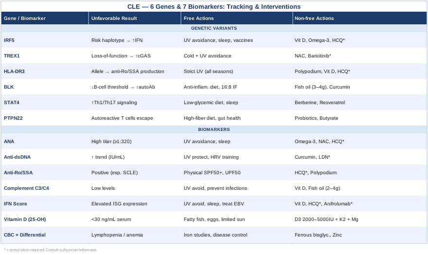
This article takes a more precise approach. Rather than repeating general lifestyle guidance, it focuses on the specific biomarkers that reveal what is actually happening in your immune system and the genetic variants most associated with CLE subtypes. Both of these angles are actionable: biomarkers can be tested repeatedly to track progress and response to interventions, while genetic data can inform long-term strategy even before symptoms escalate.
The goal is not to replace medical care — it is to make you a far more informed participant in it. When you understand what your anti-Ro/SSA levels mean, what your interferon score signals, or what an IRF5 variant implies for your immune regulation, conversations with your specialist become much more productive. Better information leads to better decisions, and better decisions compound over time into meaningful changes in how this condition behaves.
Summary
This article covers two interconnected lenses for understanding and managing cutaneous lupus erythematosus beyond standard care. The first and most actionable is a panel of 7 key biomarkers — including the type I interferon score, anti-Ro/SSA antibodies, complement levels, and vitamin D — each explained with what it reveals, how to measure it affordably, and what you can do to improve it with or without supplements. The second lens covers 6 genetic variants — IRF5, TREX1, HLA-DRB1*03:01, BLK, STAT4, and PTPN22 — with practical guidance on what each variant means for your immune behavior and how to compensate. Beyond these two core sections, the article also covers insights from Dr. Terry Wahls' research-backed autoimmune protocol and four complementary modalities — including the Autoimmune Protocol, MBSR, microbiome therapy, and photobiomodulation — that have meaningful clinical evidence for conditions like CLE. If you have wondered whether there is a smarter, more personalized path through this condition, this is where to start.
7 Biomarkers That Can Reveal What Is Driving Your Cutaneous Lupus
Most laboratory panels ordered for lupus focus on diagnosis and safety monitoring. That is necessary, but it is not the same as understanding the mechanism behind your individual presentation. The following seven biomarkers go further — they reveal immune system activity levels, nutritional deficiencies, and inflammatory drivers that respond to targeted interventions. Tracking them over time gives you a dynamic picture rather than a static snapshot.
Biomarker 1: Antinuclear Antibodies (ANA)
The ANA test is the entry point for almost every lupus evaluation, and for good reason: it is positive in over 95% of people with systemic lupus and in the majority of those with cutaneous subtypes. But most people are told only whether it is positive or negative. The titer and the pattern matter far more than a binary result.
A titer of 1:80 in a healthy person carries different significance than a titer of 1:640 in someone with active skin lesions. The pattern — whether speckled, homogeneous, nucleolar, or centromere — points toward different autoantibody profiles and disease subtypes. A nucleolar pattern, for example, is more common in systemic disease, while a speckled pattern often corresponds to anti-Ro or anti-Smith antibodies relevant to specific CLE phenotypes. Following changes in titer over time, particularly before and after interventions, can be a useful proxy for overall immune system modulation even when no other tests change.
How to Measure It
ANA is measured by indirect immunofluorescence (IIF) on HEp-2 cells, which remains the gold standard, or by ELISA-based multiplex assays (less sensitive but more widely available). Cost ranges from $30 to $100 through standard labs. Most rheumatologists order it routinely, but you can also request it through direct-to-consumer lab services in many regions. Ask specifically for titer and pattern, not just a positive/negative result.
If the Score Is Elevated: The Plan Without Supplements
A high ANA titer reflects ongoing immune activation. The first non-pharmacological priority is reducing the triggers that sustain that activation. UV avoidance is non-negotiable — even UVA through glass causes photosensitive immune activation in CLE. Broad-spectrum SPF 50+ sunscreen applied daily (not just on sunny days), UV-protective clothing with UPF 50 rating, and window film for cars and homes make a meaningful difference. Sleep quality is the second lever: poor sleep elevates inflammatory cytokines and drives ANA upward in lupus-prone individuals. Consolidating sleep to consistent hours, eliminating blue light two hours before bed, and treating any underlying sleep disorder should be early priorities.
If the Score Is Elevated: The Plan With Supplements or Equipment
Omega-3 fatty acids (EPA and DHA, 2–4 g per day from fish oil or algae-based sources) have demonstrated modest but consistent immunomodulatory effects in lupus, including reductions in inflammatory cytokine production. Take with a fat-containing meal; cycle with a break every 12 weeks to reassess. N-acetylcysteine (NAC) at 600–1200 mg per day has been studied in SLE — a small randomized trial found it reduced disease activity scores. NAC replenishes glutathione, which is often depleted in autoimmune disease. Avoid in people with asthma (may trigger bronchospasm). Note that hydroxychloroquine, the most effective pharmacological option for reducing ANA titers and disease activity in CLE, must be prescribed by a physician — it is not a supplement but should be considered a central tool in any management plan.
Biomarker 2: Anti-dsDNA Antibodies
If ANA is broad surveillance, anti-double-stranded DNA antibodies are the precision signal. Anti-dsDNA is far more specific to lupus (specificity >95%) and, critically, its levels rise and fall in closer correlation with disease activity than ANA does. Longitudinal tracking of anti-dsDNA is one of the most reliable ways to anticipate a flare before skin lesions appear or worsen.
In CLE specifically, anti-dsDNA positivity is more common in the systemic forms (SLE with cutaneous involvement) than in purely cutaneous disease, but elevated levels in any CLE patient signal increased risk of systemic progression, particularly renal involvement. A rising anti-dsDNA titer over two to three consecutive measurements — even within the technically normal range — warrants attention.
How to Measure It
Anti-dsDNA is measured by ELISA or by the Crithidia luciliae indirect immunofluorescence test (considered more specific). Standard pricing is $40 to $80 through most clinical labs. Request quantitative results (IU/mL) rather than a simple positive/negative, as trend tracking requires numbers.
If the Score Is Elevated: The Plan Without Supplements
The same UV protection strategy applies here, as UV exposure directly triggers the release of nuclear material (including DNA) from skin cells, which fuels anti-dsDNA production. Beyond UV control, stress management becomes mechanistically relevant: elevated cortisol impairs immune tolerance mechanisms and promotes the type of neutrophil extracellular trap (NET) formation that releases chromatin and DNA into circulation, directly stoking anti-dsDNA antibody production. Structured breath-work (4-7-8 breathing, 10 minutes twice daily) and heart rate variability (HRV) biofeedback have measurable effects on HPA axis dysregulation within four to eight weeks.
If the Score Is Elevated: The Plan With Supplements or Equipment
Curcumin (from turmeric, ideally in a bioavailable form such as phosphatidylcholine-bound or piperine-enhanced) at 500–1000 mg per day has demonstrated anti-dsDNA lowering effects in preliminary lupus trials by modulating NF-κB signaling. It should be used with caution in people on blood thinners and cycled (12 weeks on, 4 weeks off). Chlorogenic acid-rich foods (green coffee extract, artichoke) and low-dose naltrexone (LDN) (1.5–4.5 mg prescribed, taken at bedtime) are emerging tools — LDN modulates toll-like receptor signaling and has preliminary evidence in lupus and autoimmune conditions. LDN requires a prescription but is increasingly available through integrative physicians.
Biomarker 3: Anti-Ro/SSA and Anti-La/SSB Antibodies
For anyone with cutaneous lupus, anti-Ro/SSA may be the single most clinically relevant autoantibody to know. It is present in over 70% of subacute cutaneous lupus erythematosus (SCLE) cases — the most photosensitive CLE subtype — and it predicts both the nature of skin involvement and the photosensitivity mechanism. Anti-Ro/SSA positivity means that the Ro antigen is being trafficked to the cell surface under UV stress, creating a target for antibody attack precisely in sun-exposed skin.
Anti-La/SSB often accompanies anti-Ro and carries additional significance: it is associated with neonatal lupus in pregnancies, which is critical information for anyone of reproductive age. Both antibodies are highly relevant to Sjögren's overlap syndrome, which occurs in a meaningful subset of CLE patients and can go undiagnosed for years.
How to Measure It
Standard ELISA panels test anti-Ro/SSA and anti-La/SSB together; cost is typically $30 to $60 through a rheumatology panel. Results are reported in units with lab-specific cutoffs. Quantitative titers are more useful than qualitative positive/negative results for monitoring. This test is part of most autoimmune antibody panels and can be requested directly.
If the Score Is Elevated: The Plan Without Supplements
Anti-Ro/SSA positivity demands strict, consistent UV protection — not just on beach days. Even ambient UVA exposure through windows is enough to trigger the Ro antigen surface translocation that drives SCLE lesions. Tinted car windows, UV-blocking window film for home and office, SPF 50+ broad-spectrum sunscreen applied daily year-round, and UV-protective hats and clothing rated UPF 50 are all relevant. Physical sunscreens (zinc oxide, titanium dioxide) are preferred over chemical UV filters, which have shown preliminary evidence of hormonal disruption and may not offer the same inert protection.
If the Score Is Elevated: The Plan With Supplements or Equipment
Hydroxychloroquine (prescribed, 200–400 mg daily depending on weight) is standard of care and directly reduces anti-Ro/SSA-driven skin inflammation by interfering with toll-like receptor signaling in plasmacytoid dendritic cells. It is not optional for most SCLE cases. Supplementary support includes Polypodium leucotomos extract (240–480 mg daily, taken before anticipated sun exposure), a fern-derived antioxidant with published human trials showing reduced UV-induced skin damage, reduced MED (minimal erythema dose), and modulation of UV-triggered immune activation. It should not replace sunscreen but meaningfully enhances photoprotection. Side effects are minimal; monitor for GI discomfort.
Biomarker 4: Complement Proteins C3 and C4
Complement levels sit at the intersection of diagnosis, disease monitoring, and genetic risk in lupus. In active disease, immune complexes — clusters of autoantibodies bound to their targets — activate and consume complement proteins. Low C3 and C4 therefore signal active immune complex deposition. In CLE with systemic involvement, falling complement often precedes clinical flares and can serve as an early warning system when tracked serially.
C4 carries additional complexity: C4A null alleles are a genetic variant strongly associated with lupus susceptibility in certain populations. These individuals may have consistently low C4 not because of active disease consuming it, but because they simply produce less. Distinguishing between a constitutional C4 deficiency and active complement consumption requires tracking alongside disease activity markers, but either situation informs the clinical picture.
How to Measure It
C3 and C4 are standard chemistry tests available at any clinical laboratory; cost is $20 to $50 per test. They are often included in rheumatology panels. CH50 (total hemolytic complement) provides an overall complement function measure and costs $30 to $80. For genetic C4 copy number assessment (if baseline C4 is persistently low without active disease), specialized testing is available through academic medical centers.
If the Score Is Low: The Plan Without Supplements
Low complement during active disease requires addressing the upstream trigger — the immune complex burden. This means optimizing disease control through existing pharmacological therapies (hydroxychloroquine, topical steroids, or systemic immunosuppression as appropriate) while reducing immune activation triggers. UV avoidance, stress reduction, and sleep optimization all reduce the pace of immune complex generation. Infections are major triggers for complement consumption through a separate pathway, so prevention — vaccination, hand hygiene, and prompt treatment of infections — matters more than many people realize.
If the Score Is Low: The Plan With Supplements or Equipment
There is no direct supplement that restores low complement in active disease — the goal is reducing what depletes it. Vitamin D (see below) modulates complement regulatory proteins and may indirectly support complement balance. Fish oil at 2–4 g EPA+DHA daily reduces immune complex formation over time by modulating membrane phospholipid composition and reducing antibody-driven inflammation. For persistently low C4 due to genetic null alleles, the focus shifts to reducing the infectious and inflammatory triggers that activate complement in a person who has limited reserve.
Biomarker 5: Type I Interferon Score
The type I interferon (IFN) pathway is arguably the central biological mechanism in lupus, both systemic and cutaneous. Plasmacytoid dendritic cells activated by nuclear material — especially after UV-induced cell death releases DNA and RNA — produce massive amounts of interferon-alpha. This cytokine then activates B cells, primes autoreactive T cells, and drives the inflammatory cascade that produces skin lesions. Research published in Proceedings of the National Academy of Sciences by Baechler and colleagues was among the first to document a pervasive interferon gene expression signature in lupus that tracked directly with disease severity.
The IFN score is measured by assessing the expression of interferon-stimulated genes (ISGs) in peripheral blood. A high IFN score correlates with anti-dsDNA positivity, complement consumption, and disease flares. More importantly, it has become a key stratification tool: patients with high IFN scores respond better to anti-interferon biologics like anifrolumab, which was approved for active SLE in 2021 and has shown benefit in cutaneous disease.
How to Measure It
IFN score testing is increasingly available but requires a specialized laboratory. Some academic rheumatology centers offer it as part of a comprehensive lupus panel. Commercial options are emerging; cost ranges from $100 to $350 depending on the platform. The test measures relative expression of four to seven ISGs (such as IFI27, IFI44L, IFIT1, RSAD2) and reports a normalized score. Standard clinical labs do not yet offer this routinely, but the landscape is changing rapidly.
If the Score Is Elevated: The Plan Without Supplements
Reducing interferon production begins with removing its triggers. UV radiation is the dominant environmental activator of the IFN pathway in CLE: photons damage keratinocyte DNA, the resulting debris is sensed by cGAS-STING and TLR7/9 pathways in dendritic cells, and interferon-alpha production escalates. Every layer of UV protection — topical, physical, behavioral — directly suppresses IFN pathway activation. Sleep deprivation is a secondary but significant driver: even one night of sleep restriction measurably increases ISG expression. Treating any latent viral infections (Epstein-Barr reactivation is documented in lupus flares) also reduces the immunological background noise that keeps the IFN system primed.
If the Score Is Elevated: The Plan With Supplements or Equipment
Hydroxychloroquine (prescribed) is the most evidence-backed pharmacological suppressor of the TLR-IFN axis in CLE; it physically blocks TLR7 and TLR9 from sensing nucleic acid ligands inside endosomes. Vitamin D at therapeutic levels (targeting serum 25-OH-D of 50–70 ng/mL) directly suppresses plasmacytoid dendritic cell interferon-alpha output — the cell type most responsible for the elevated IFN signature in lupus. This makes vitamin D optimization particularly high-yield in CLE. Resveratrol (500 mg daily, taken with food) has shown preliminary evidence of STING pathway modulation in cell studies; human evidence is limited but side effects are minimal. For those with very high IFN scores not responding to existing therapies, anifrolumab (anti-IFNAR1 monoclonal antibody, prescribed) represents a biologic option with approval for SLE and emerging evidence in refractory cutaneous disease.
Biomarker 6: Vitamin D (25-OH)
Vitamin D is not merely a bone health nutrient in the context of autoimmune disease. The vitamin D receptor (VDR) is expressed on virtually every immune cell, and its activation directly suppresses Th17 cell development, promotes regulatory T cells (Tregs), and reduces B cell autoantibody production. This makes it structurally relevant to all three of the major immune dysfunctions in CLE.
The problem is that CLE creates a cruel self-reinforcing cycle: photosensitivity forces UV avoidance, UV avoidance prevents cutaneous vitamin D synthesis, and vitamin D deficiency worsens the immune dysregulation that drives photosensitivity. Multiple studies, including a cross-sectional analysis published in Lupus by Amital and colleagues, have documented that vitamin D deficiency is significantly more prevalent in lupus patients than in matched controls, and that lower levels correlate with higher disease activity scores. This is not a marginal effect.
How to Measure It
The 25-hydroxyvitamin D test (25-OH-D) is a standard blood test available at any clinical laboratory; cost is $20 to $60. Insurance often covers it when ordered by a physician. Optimal range for immune modulation in the context of autoimmune disease is generally considered 50–80 ng/mL by functional medicine practitioners (notably Peter Attia and Rhonda Patrick), compared to the conventional threshold of 30 ng/mL. Testing every six months — spring and autumn — is sufficient for most people.
If the Score Is Low: The Plan Without Supplements
Because UV avoidance is obligatory in CLE, dietary sources become more important than in the general population. Fatty fish (salmon, mackerel, sardines), egg yolks, and fortified foods provide some vitamin D, but dietary amounts alone are rarely sufficient to maintain optimal serum levels in someone who avoids sun. The most useful non-supplemental strategy is aggressive dietary inclusion of vitamin D-rich whole foods and, where medically appropriate, carefully timed and limited, well-protected sun exposure to non-affected body sites under physician guidance.
If the Score Is Low: The Plan With Supplements or Equipment
Vitamin D3 supplementation of 2,000–5,000 IU daily (based on baseline levels and monitored by testing every 3–6 months) is appropriate for most CLE patients with documented deficiency. Always pair with vitamin K2 (MK-7 form, 100–200 mcg daily) to ensure calcium is directed appropriately and not deposited in arteries. Magnesium glycinate (200–400 mg daily) is required as a cofactor for vitamin D hydroxylation — many people are magnesium deficient, which blunts the effectiveness of vitamin D supplementation regardless of dose. Toxicity is rare below 10,000 IU daily in most adults but always retest before escalating dose. Target 50–70 ng/mL in serum.
Biomarker 7: Complete Blood Count with Differential
The CBC is often considered a routine safety test for people on immunosuppressive medications, but in CLE it carries diagnostic and mechanistic information that goes beyond medication monitoring. Lymphopenia (total lymphocyte count below 1,000/μL) is one of the eleven American College of Rheumatology classification criteria for SLE and reflects the direct attack on lymphocytes by autoantibodies and immune complexes in active disease. It is underappreciated as a disease activity marker in its own right.
Anemia — particularly normocytic, normochromic anemia of chronic disease — occurs when pro-inflammatory cytokines suppress erythropoiesis. This form of anemia does not respond to iron supplementation and instead reflects the degree of systemic immune activation. Thrombocytopenia, when present, may indicate antiphospholipid antibody syndrome overlap, which significantly changes cardiovascular and thrombotic risk management. The CBC therefore captures three parallel windows into immune and systemic disease status simultaneously.
How to Measure It
A complete blood count with differential is one of the most affordable standard tests in medicine; cost is $10 to $30 at most laboratories. Request the differential specifically to get lymphocyte, neutrophil, and monocyte counts separately, not just the total white cell count. Serial testing every three to six months provides trend data that is more valuable than any single measurement.
If the Score Is Abnormal: The Plan Without Supplements
Lymphopenia that tracks with disease activity will improve with disease control — this makes managing the upstream immune drivers (UV protection, stress, medication compliance) the primary intervention. If thrombocytopenia is found, antiphospholipid antibody syndrome should be evaluated before attributing it to lupus alone. Anemia of chronic disease is best addressed by treating the underlying inflammatory state rather than supplementing iron blindly; iron studies (ferritin, serum iron, TIBC) should always be checked alongside the CBC to distinguish anemia types.
If the Score Is Abnormal: The Plan With Supplements or Equipment
For iron-deficiency anemia confirmed by low ferritin (below 30 ng/mL), iron supplementation is appropriate: ferrous bisglycinate (25 mg elemental iron) is better tolerated than ferrous sulfate with fewer GI side effects. Take every other day (not daily) — a 2017 study in Blood showed every-other-day dosing increases net absorption by reducing hepcidin suppression. For lymphopenia driven by autoimmune attack, no direct supplement restores counts; the goal is reducing the autoimmune burden. Zinc (15–25 mg daily) supports lymphocyte maturation and immune signaling; thymosin alpha-1 (a prescription peptide in some countries) has emerging evidence for immune reconstitution in autoimmune lymphopenia.
6 Genetic Variants That Shape Your Risk and How to Work With Them
Genetics do not write your fate in autoimmune disease — they set the parameters of your immune system's behavior. The variants associated with CLE are almost all gain-of-function mutations in inflammatory pathways or loss-of-function in regulatory ones. Understanding which variants you carry tells you which immune circuits are likely running hot and therefore where targeted lifestyle and pharmacological interventions will have the most leverage. Consumer genetic tests (23andMe, AncestryDNA) provide raw data that can be analyzed through third-party tools such as Genetic Genie or Promethease; clinical genetic testing through rheumatology departments offers more curated interpretation.
IRF5: The Interferon Amplifier
What This Gene Does
IRF5 (Interferon Regulatory Factor 5) encodes a transcription factor that directly controls the production of interferon-alpha and pro-inflammatory cytokines (IL-6, IL-12, TNF-α) in plasmacytoid dendritic cells and macrophages. Risk variants — particularly the haplotype containing rs2004640 and rs10488631 — increase IRF5 expression and stability, resulting in chronically elevated interferon production even in the absence of overt infection or injury. Research by Graham and colleagues in Nature Genetics identified IRF5 as one of the strongest genetic risk factors for SLE and CLE across multiple ethnic populations. In CLE, carrying an IRF5 risk haplotype correlates with higher basal IFN scores and more aggressive skin involvement.
If the Gene Is Unfavorable: The Plan Without Supplements
The most impactful non-pharmacological strategy for someone with IRF5 risk variants is minimizing the inputs that activate the IRF5 pathway: UV radiation, viral triggers (particularly Epstein-Barr virus reactivation and cytomegalovirus), and chronic psychological stress (which elevates corticotropin-releasing factor, a known pDC activator). Maintaining consistent, quality sleep is also disproportionately impactful in IRF5 carriers — sleep deprivation amplifies the very interferon output that this gene variant already produces in excess. Infection prevention through vaccination and hand hygiene deserves more attention than it typically receives in this context.
If the Gene Is Unfavorable: The Plan With Supplements or Equipment
Vitamin D (targeting 60–70 ng/mL) directly suppresses IRF5 transcriptional activity through VDR binding — this is one of the most mechanistically sound supplement interventions for IRF5 carriers. Omega-3 fatty acids at 3–4 g EPA+DHA daily reduce the lipid mediators (PGE2, LTB4) that amplify IRF5 pathway signaling; take with food, 12 weeks on/4 weeks off. Hydroxychloroquine (prescribed) blocks the endosomal TLRs that activate IRF5 and is strongly indicated in any CLE patient with confirmed IRF5 risk variants and active disease.
TREX1: When DNA Becomes Its Own Enemy
What This Gene Does
TREX1 encodes a 3'-5' DNA exonuclease — an enzyme that degrades cytoplasmic DNA to prevent it from activating innate immune sensors. Loss-of-function mutations in TREX1 allow single-stranded DNA to accumulate in the cytoplasm, where it is sensed by the cGAS-STING pathway and triggers a massive interferon response. In the skin, this mechanism is highly relevant: UV radiation causes keratinocyte death and releases DNA; in someone with functional TREX1, this DNA is cleared; in someone with a TREX1 mutation, it persists and chronically activates innate immunity. Heterozygous mutations in TREX1 are associated with chilblain lupus erythematosus — a rare CLE subtype featuring acral ulcerations triggered by cold exposure — as well as familial CLE more broadly, as established in multiple European cohort studies.
If the Gene Is Unfavorable: The Plan Without Supplements
For chilblain CLE specifically, cold avoidance is as critical as UV avoidance: cold triggers vasoconstriction and local hypoxia that synergizes with the TREX1-driven interferon response at acral sites. Warm clothing, glove use, heated environments, and avoidance of wet-cold exposure in autumn and winter are baseline necessities. For all TREX1 carriers, the priority is reducing the sources of cytoplasmic DNA accumulation — which means optimizing UV protection and considering antiviral strategies if herpetic reactivation is documented.
If the Gene Is Unfavorable: The Plan With Supplements or Equipment
NAC (N-acetylcysteine) at 600–900 mg twice daily supports glutathione production, which reduces oxidative DNA damage — the very form of DNA modification that TREX1 variants fail to clear efficiently. It is one of the most rational supplements for TREX1 carriers. Baricitinib (a JAK1/2 inhibitor, prescribed) has shown particular efficacy in TREX1-related interferonopathies by blocking the downstream JAK-STAT signaling that STING activates; it is off-label for CLE but increasingly considered in treatment-resistant cases. Reverse transcriptase inhibitors (antiretroviral drugs) have been studied experimentally in Aicardi-Goutières syndrome (a severe TREX1-related condition) for their ability to block retroelement reverse transcription, but clinical evidence in CLE specifically remains preliminary.
HLA-DRB1*03:01 (HLA-DR3): The Molecular Matchmaker for Autoantibodies
What This Gene Does
HLA-DRB1*03:01, commonly called HLA-DR3, encodes a cell surface molecule that presents protein fragments to CD4+ T cells. Its specific binding groove shape is unusually well-suited to presenting Ro/SSA peptides, making it the strongest genetic predictor of anti-Ro/SSA antibody production. Individuals carrying this allele are far more likely to develop SCLE — the photosensitive, annular-polycyclic rash that is the most prevalent CLE subtype — and to carry the clinical features associated with anti-Ro positivity. DR3 is also associated with primary Sjögren's syndrome, and CLE patients carrying this allele have higher rates of Sjögren's overlap. Understanding whether you carry DR3 helps predict which CLE subtype you are most susceptible to and informs the degree of UV protection needed.
If the Gene Is Unfavorable: The Plan Without Supplements
The logic here flows directly from what DR3 does: it drives anti-Ro/SSA production, and anti-Ro/SSA drives photosensitive skin lesions upon UV exposure. The intervention is the same as managing confirmed anti-Ro positivity — strict, layered UV protection applied consistently across all seasons. For DR3 carriers who have not yet developed overt anti-Ro positivity, consider it a reason to be proactive rather than reactive: beginning UV protection habits before detectable antibody elevation may prevent the autoimmune cascade from accelerating.
If the Gene Is Unfavorable: The Plan With Supplements or Equipment
Polypodium leucotomos (240–480 mg daily, split dose) provides systemic photoprotection through multiple mechanisms including antioxidant scavenging of UV-induced free radicals and modulation of UV-triggered immune activation — directly relevant for DR3 carriers whose immune system overreacts to UV-released nuclear material. Vitamin D supplementation at therapeutic levels modulates antigen-presenting cell behavior and may reduce the efficiency with which DR3 presents autoantigens to Th cells; while this is mechanistically plausible rather than proven in clinical trials, the cost-to-benefit ratio strongly favors supplementation given the multiple other benefits. Hydroxychloroquine (prescribed) disrupts the endosomal antigen processing pathway through which DR3 most efficiently presents Ro antigens.
BLK: When B Cells Lose Discipline
What This Gene Does
BLK encodes B lymphoid tyrosine kinase, a signaling enzyme critical for setting the activation threshold of B cells. Risk variants (most notably the rs13277113-rs2248932 haplotype) reduce BLK expression, lowering the activation threshold so that B cells respond more readily to self-antigen stimulation. The result is expanded autoreactive B cell populations and, consequently, higher autoantibody titers — particularly ANA, anti-Ro/SSA, and anti-dsDNA. BLK was identified as a lupus risk gene in a landmark genome-wide association study and subsequently confirmed in independent CLE cohorts. In practical terms, BLK risk variants translate to a higher autoantibody burden even in the absence of clinical symptoms, making early monitoring of antibody panels particularly important.
If the Gene Is Unfavorable: The Plan Without Supplements
Autoreactive B cells in BLK carriers are, in essence, easier to activate. The non-pharmacological strategy is therefore reducing the antigens and inflammatory signals that activate them. This means optimizing gut barrier integrity — gut dysbiosis increases systemic antigen load from microbial products, which drives bystander B cell activation — through an anti-inflammatory, low-processed-food diet. Intermittent fasting (16:8 or time-restricted eating) has documented effects on reducing B cell receptor signaling tone and lowering inflammatory cytokines that co-stimulate B cell activation; preliminary data in autoimmune models is supportive.
If the Gene Is Unfavorable: The Plan With Supplements or Equipment
Fish oil at 3–4 g EPA+DHA daily directly modulates B cell function: EPA incorporates into B cell membrane phospholipids and alters BCR signaling efficiency, reducing the response to low-level antigen stimulation — the precise mechanism that BLK variants amplify. Curcumin (phospholipid-complexed, 500 mg twice daily) inhibits NF-κB in B cells, reducing their production of inflammatory cytokines independently of the BCR signal. Both are well-tolerated with minimal side effects; monitor for increased bleeding time if on anticoagulants.
STAT4: The Inflammatory Amplifier in T Cell Signaling
What This Gene Does
STAT4 (Signal Transducer and Activator of Transcription 4) is the intracellular messenger for IL-12 and IL-23, cytokines that drive Th1 and Th17 T cell differentiation. The rs7574865 risk variant increases STAT4 expression in response to IL-12 stimulation, producing a more aggressive Th1 and Th17 response than in non-carriers. In SLE and CLE, STAT4 risk variants are associated with higher anti-dsDNA titers, more severe disease, and increased risk of lupus nephritis. From a skin perspective, STAT4 amplification means more interferon-gamma production from Th1 cells and more IL-17 from Th17 cells — both of which contribute to the inflammatory infiltrate found in CLE lesions. A study by Remmers and colleagues published in the New England Journal of Medicine (2007) established STAT4 as a shared genetic risk factor for both SLE and rheumatoid arthritis.
If the Gene Is Unfavorable: The Plan Without Supplements
The Th1/Th17 axis that STAT4 amplifies is highly responsive to diet. A low-glycemic, anti-inflammatory diet — emphasizing diverse vegetables, fatty fish, olive oil, and legumes, while minimizing refined carbohydrates, seed oils, and ultra-processed foods — has documented effects on reducing IL-17 and IL-12 production. This is not an optional lifestyle suggestion for STAT4 carriers; it is a mechanistically grounded intervention in the specific pathway the variant upregulates. Disrupted circadian rhythms also amplify Th17 responses; consistent sleep and light timing supports regulatory T cell function that counterbalances STAT4-driven inflammation.
If the Gene Is Unfavorable: The Plan With Supplements or Equipment
Berberine (500 mg twice daily, taken with meals) has published evidence of STAT3/STAT4 pathway modulation and has been studied in autoimmune conditions as an IL-17 suppressor. Cycle 8 weeks on/4 weeks off; monitor for GI tolerance. Resveratrol (500 mg daily, fat-soluble form taken with food) inhibits STAT4 phosphorylation in vitro and has anti-Th17 properties in human studies. Quercetin (500–1000 mg daily, divided doses) broadly suppresses STAT-mediated transcription and reduces the IL-12 sensitivity that STAT4 risk variants create. All three can be taken together with no known interactions; avoid berberine during pregnancy.
PTPN22: The Self-Tolerance Gatekeeper
What This Gene Does
PTPN22 encodes LYP (Lymphoid Phosphatase), a phosphatase that dampens T cell receptor signaling after antigen recognition, acting as a brake on T cell activation. The R620W variant (rs2476601) produces a gain-of-function mutation that paradoxically increases LYP phosphatase activity — but in the wrong direction: instead of properly eliminating autoreactive T cells in the thymus, it impairs central tolerance. The result is that autoreactive T cells survive thymic selection and enter the periphery, where they are more likely to attack self-tissues. PTPN22 R620W is associated with multiple autoimmune diseases — type 1 diabetes, rheumatoid arthritis, vitiligo, and SLE — making it one of the broadest autoimmune susceptibility variants known.
If the Gene Is Unfavorable: The Plan Without Supplements
The most leverage in a PTPN22 R620W carrier lies in gut health. The thymic tolerance defect is established developmentally, but peripheral tolerance — the second line of defense against autoreactive T cells — is heavily influenced by the gut microbiome. Short-chain fatty acids produced by gut bacteria (particularly butyrate from fermentation of dietary fiber) directly promote regulatory T cell development in the colon and circulation. This means a high-fiber, diverse-plant diet is not generic advice for PTPN22 carriers — it is targeted therapy for a known immune regulatory deficit. Minimizing antibiotic use and processed food consumption protects the microbial diversity that produces these regulatory metabolites.
If the Gene Is Unfavorable: The Plan With Supplements or Equipment
Probiotic supplementation with multi-strain formulas including Lactobacillus rhamnosus, Bifidobacterium longum, and Lactobacillus acidophilus supports the microbial populations most associated with Treg induction. Take on an empty stomach or with a small, low-fat meal; 12-week cycles with microbiome diversity testing to guide strain selection. Sodium butyrate or tributyrin (500–1000 mg twice daily) provides direct butyrate delivery to colonic epithelium when dietary fiber is insufficient. Quercetin (500 mg twice daily) modulates regulatory T cell/Th17 balance and has shown efficacy in multiple preclinical autoimmune models; it is among the safest and most evidence-supported supplements for broad autoimmune susceptibility.
What Dr. Terry Wahls' Research Reveals About Autoimmune Disease — 10 Findings Worth Understanding
Dr. Terry Wahls is a clinical professor of medicine at the University of Iowa who reversed her secondary progressive multiple sclerosis using a structured dietary and lifestyle protocol, then spent the following decade conducting clinical trials to validate the approach. Her work — published in peer-reviewed journals and detailed in The Wahls Protocol — challenges the assumption that autoimmune disease progression is inevitable. While her trials focused primarily on MS, the mechanistic framework applies directly to CLE and other autoimmune conditions because it targets the shared biological drivers: mitochondrial dysfunction, microbiome disruption, nutrient depletion, and neurological dysregulation.
1. Mitochondria Are Immunological Engines
Every immune cell — T cell, B cell, dendritic cell — depends on mitochondrial energy production to function. In autoimmune disease, chronic inflammation impairs mitochondrial efficiency, creating a feedback loop where dysfunctional immune cells produce more inflammation, which further impairs mitochondria. Wahls identified specific nutrients essential for mitochondrial function: B vitamins (particularly B1, B2, B3, B6, B9, B12), coenzyme Q10, carnitine, and alpha-lipoic acid. Deficiency in any of these nutrients slows mitochondrial electron transport chain function and shifts immune cells toward inflammatory (Th17, M1 macrophage) rather than regulatory phenotypes.
2. Nine Cups of Vegetables Per Day Is Not Arbitrary
The Wahls Protocol prescribes nine cups of vegetables and fruits daily, categorized into three groups: three cups of leafy greens (kale, collards, beet greens — for B vitamins and minerals), three cups of sulfur-rich vegetables (onion, garlic, cabbage, mushrooms — for glutathione precursors), and three cups of deeply pigmented produce (beets, berries, carrots — for antioxidants and polyphenols). Each group targets a different axis of immune regulation. For CLE specifically, the leafy green category provides the folate and vitamin B9 that support methylation reactions critical for DNA repair — directly relevant to the UV-induced DNA damage mechanism in CLE pathogenesis.
3. Gluten and Casein Removal Has Immunological Logic
Wahls eliminates gluten and dairy casein from her protocol, not on ideological grounds but because both proteins have documented effects on intestinal permeability. Increased gut permeability allows bacterial lipopolysaccharide (LPS) and other microbial products to enter systemic circulation, where they activate toll-like receptors — the same innate immune receptors that drive the interferon response central to CLE. In genetically susceptible individuals (including those with IRF5 and TREX1 variants), this chronic TLR activation may sustain the inflammatory baseline that makes CLE harder to control.
4. Omega-3 to Omega-6 Ratio Matters More Than Total Fat
The modern Western diet provides omega-6 to omega-3 ratios of 15–20:1; the Wahls Protocol targets 4:1 or better. This matters because omega-6 fatty acids are precursors to pro-inflammatory eicosanoids (PGE2, LTB4, thromboxane A2), while omega-3s compete for the same enzymes and produce anti-inflammatory resolvins and protectins. Shifting this ratio — through elimination of seed oils and addition of fatty fish, walnuts, and flaxseed — takes approximately 6–12 weeks to measurably alter membrane phospholipid composition and reduce inflammatory eicosanoid output.
5. Iodine and Thyroid Function Are Often Overlooked
Thyroid dysfunction occurs in lupus at rates significantly higher than in the general population, and conversely, thyroid autoimmunity (Hashimoto's, Graves') frequently co-occurs with CLE. Wahls emphasizes that iodine deficiency impairs thyroid function and that thyroid hormone regulates mitochondrial biogenesis — another link between thyroid health and immune cell energy metabolism. Seaweed, seafood, and dairy are primary dietary iodine sources; testing thyroid function (TSH, free T3, free T4, anti-TPO antibodies) is warranted in any CLE patient without a recent thyroid workup.
6. Electrostimulation Has Emerging Relevance for Immune Regulation
Wahls herself uses functional electrical stimulation (FES) cycling, and her broader framework acknowledges the vagal nerve as a major anti-inflammatory pathway. Vagal tone suppresses TNF-α, IL-6, and IL-12 production through the cholinergic anti-inflammatory pathway. Transcutaneous vagal nerve stimulation (tVNS) devices — now available without a prescription in some countries — provide a practical way to engage this pathway. HRV biofeedback (using a chest strap monitor and corresponding app) is a lower-barrier entry point to vagal training.
7. Sulfur-Rich Foods Directly Support Glutathione
Glutathione is the master antioxidant of every cell, and its depletion in autoimmune disease is well-documented. Cruciferous vegetables and alliums (garlic, onion, leeks) provide cysteine and glutamate — the rate-limiting precursors for glutathione synthesis — without the toxicity risks of direct glutathione supplementation. For CLE patients, this matters because UV-induced oxidative stress depletes glutathione in keratinocytes faster than it can be replenished; dietary support of glutathione synthesis provides ongoing replenishment that topical antioxidants cannot match.
8. Food Elimination Can Serve as a Diagnostic Tool
Wahls recommends a structured elimination protocol as an experimental diagnostic: removing the most common inflammatory food categories (gluten, dairy, eggs, nightshades, legumes) for 90 days, then reintroducing one at a time every two weeks while tracking symptoms and relevant biomarkers. In CLE, this approach can reveal food-specific triggers of disease activity that are otherwise invisible in standard care. It requires commitment but produces personalized data that no laboratory test can provide.
9. Stress-Induced Immune Dysregulation Is Mechanistically Specific
Wahls' framework emphasizes that psychological stress activates the HPA axis, which elevates glucocorticoids; chronic glucocorticoid exposure ultimately shifts immunity from Th1 to Th2 dominance, paradoxically — at the same time, corticotropin-releasing hormone locally activates mast cells and skin-resident immune cells. This means stress does not simply worsen lupus through a vague "immune suppression" mechanism; it engages specific pathways (mast cell activation, skin-local neuroinflammation, HPA-driven immune skewing) that directly overlap with CLE pathogenesis.
10. The Protocol Is Not All-or-Nothing
One of the most practically important insights from Wahls' research is that partial implementation still produces measurable benefits. Participants in her randomized feasibility trials who could not achieve the full dietary changes still showed improvements in fatigue and quality of life measures proportional to their degree of adherence. For CLE patients who find comprehensive dietary change overwhelming, prioritizing the leafy greens and omega-3 components first — as the two categories with the most direct immune relevance — provides a rational starting point.
Complementary Approaches With Clinical Support
The interventions below are not replacements for medical treatment. They are modalities with published human evidence for autoimmune conditions including cutaneous lupus, selected from a larger pool on the basis of clinical relevance and quality of evidence. Each is practical to implement alongside standard care.
The Autoimmune Protocol (AIP)
The Autoimmune Protocol, developed by Dr. Sarah Ballantyne (a biomedical scientist and former autoimmune patient), is a structured dietary and lifestyle intervention specifically designed for autoimmune conditions. It extends the Paleo framework by removing nightshades, eggs, nuts, seeds, and all grains — foods identified in immunology literature as potential drivers of intestinal permeability and molecular mimicry — while emphasizing nutrient-dense meats, organ meats, vegetables, and fermented foods. The rationale for CLE is direct: by reducing intestinal permeability and the resulting systemic antigen load, AIP targets one of the most modifiable drivers of autoimmune activity upstream of the specific CLE immune pathways.
A pilot randomized controlled trial published in Inflammatory Bowel Diseases by Konijeti and colleagues (2017) found significant reductions in clinical disease activity in IBD patients following the AIP, with measurable reductions in inflammatory markers at six weeks. While no CLE-specific AIP trial exists yet, the IBD data establishes clinical proof-of-concept for meaningful anti-inflammatory effects in autoimmune disease. Ballantyne's book The Paleo Approach summarizes over 1,200 peer-reviewed references supporting the mechanistic rationale.
To implement AIP for CLE: begin with a strict 60–90 day elimination phase, then reintroduce eliminated foods one at a time every five to seven days while tracking skin lesions and antibody titers. The elimination phase requires careful planning to maintain caloric and nutritional adequacy. Ballantyne's approach also mandates sleep optimization (eight to nine hours), stress management, and moderate physical activity — elements that independently have documented effects on CLE disease activity. This is not a temporary diet; it is a framework for identifying and managing personal inflammatory triggers through systematic experimentation.
Mindfulness-Based Stress Reduction (MBSR)
MBSR is an eight-week structured program developed by Jon Kabat-Zinn at the University of Massachusetts Medical Center. It combines body scan meditation, mindful movement, and breath awareness in a format that has now been studied in over 800 clinical trials across chronic illness populations. For CLE and lupus specifically, MBSR addresses the HPA-mediated stress-inflammation cycle: chronic psychological stress activates corticotropin-releasing hormone in skin-resident nerve fibers, directly triggering mast cell degranulation and keratinocyte pro-inflammatory cytokine production — a local inflammatory mechanism independent of systemic immune activation.
A randomized controlled trial by Greco and colleagues published in the Journal of Rheumatology assessed MBSR in lupus patients and found improvements in pain, psychological well-being, and fatigue measures over controls. A separate systematic review on MBSR in autoimmune disease found consistent improvements in quality of life and inflammatory biomarkers across multiple conditions. The stress-skin connection in CLE is not metaphorical — it is anatomically grounded in the skin-associated lymphoid tissue and its direct innervation by stress-responsive neuropeptides.
To implement: enroll in a certified eight-week MBSR course (available in-person or online through established providers). Practice formally for 30–45 minutes daily during the eight-week program and at least 20 minutes daily thereafter. Track flares and skin lesions in a symptom diary alongside the course to identify personal stress-flare relationships. Heart rate variability monitoring (using a wearable device) provides objective feedback on autonomic regulation improvements over weeks three to six, which correlates with reduced neuroinflammatory skin signals.
Microbiome-Directed Therapies
The gut-immune connection in lupus has moved from theoretical to mechanistically established over the past decade. Lupus patients show characteristic gut dysbiosis patterns — reduced Lactobacillaceae and Bifidobacterium abundance, overgrowth of Ruminococcus gnavus (a species found at elevated levels during lupus flares in multiple cohort studies), and reduced microbial diversity overall. These dysbiotic patterns reduce short-chain fatty acid production, impair regulatory T cell induction, and increase gut permeability — collectively amplifying the systemic antigen exposure that sustains autoantibody production. A study by Azzouz and colleagues published in Annals of the Rheumatic Diseases (2019) specifically implicated R. gnavus overgrowth in lupus nephritis flares through an LPS-mediated TLR4 activation mechanism.
Microbiome-directed strategies include high-fiber dietary interventions to support SCFA production, targeted probiotic supplementation, and avoidance of gut-disrupting inputs (unnecessary antibiotics, NSAIDs, proton pump inhibitors). Fecal microbiota transplantation (FMT) has been explored in murine lupus models with promising results but remains investigational in human autoimmune disease outside of Clostridioides difficile infection.
For practical CLE management: incorporate 30+ distinct plant foods per week (diversity drives microbial diversity, which drives SCFA production), use a quality multi-strain probiotic during and after any antibiotic course, and consider a six-week high-dose probiotic trial (50–100 billion CFU daily) during periods of increased disease activity. Stool microbiome testing through commercial providers (Viome, Biomesight, Doctor's Data) can identify specific dysbiosis patterns and guide more targeted probiotic strain selection, though the clinical interpretation of consumer microbiome reports remains an emerging science.
Photobiomodulation (Red and Near-Infrared Light Therapy)
An important clarification upfront: ultraviolet light is a documented trigger for CLE and must be avoided. Photobiomodulation (PBM), also called low-level laser therapy, uses red (620–700 nm) and near-infrared (800–1100 nm) wavelengths — entirely distinct from UV — that penetrate tissue and stimulate mitochondrial cytochrome c oxidase, increasing ATP production and reducing oxidative stress in target cells. This distinction matters because red and NIR light do not carry the energy to cause DNA photoproduct formation (the mechanism by which UV triggers CLE), yet they do stimulate anti-inflammatory and tissue repair pathways that may benefit CLE-affected skin.
A double-blind randomized trial by Dompe and colleagues published in Cells (2020) reviewed PBM mechanisms in dermatological conditions and found documented anti-inflammatory effects including reduced IL-1β, IL-6, TNF-α, and NF-κB activation in skin — pathways directly relevant to CLE lesion biology. Evidence specifically in CLE is still limited, but PBM has established evidence in discoid wounds, chronic inflammatory skin conditions, and as a complementary modality for accelerating resolution of erythematous lesions.
For CLE patients, PBM should be approached cautiously and selectively: use a device emitting 630–670 nm red and/or 810–850 nm near-infrared wavelengths at 5–50 mW/cm² irradiance on non-active, healing, or scarring lesion sites only — never on acutely inflamed or erosive areas without dermatological guidance. Treatment protocols typically involve 10–15 minutes per session, three times per week for six to eight weeks. Consult a dermatologist before initiating PBM therapy for any CLE subtype, as individual skin response and UV sensitivity may affect the safety profile. Home panel devices (10 × 20 cm to full-body sizes) are available at $200–$800; handheld clinical devices range higher.
Conclusion
Cutaneous lupus erythematosus is not one disease — it is a family of immune-mediated skin presentations shaped by individual genetic architecture, environmental exposures, microbiome composition, and lifestyle inputs. The seven biomarkers outlined here give you a dynamic, trackable picture of what your immune system is actually doing. The six genetic variants provide context for why your disease may behave differently from someone else's, and how to target interventions more precisely. The dietary and lifestyle frameworks — from the Wahls Protocol to the AIP and MBSR — are not generic wellness advice; they are mechanistically grounded tools for reducing the specific inflammatory drivers of CLE.
The most productive next step is not trying to implement everything at once. Start with the biomarkers: bring the tracking panel to your rheumatologist or dermatologist, and begin building a longitudinal picture of your immune activity. If you have access to genetic testing, review your raw data through a third-party tool and note whether the variants discussed here apply to you. Then layer in one lifestyle intervention at a time — starting with the one that addresses your most elevated biomarker — and measure whether it moves the needle. This is the difference between managing a condition reactively and understanding it deeply enough to change its trajectory.