This article contains AI generated content.
· UpdatedCutaneous Vasculitis Genes And Biomarkers – 7 Biomarkers And 6 Genes To Track
Introduction
Living with cutaneous vasculitis means navigating something most people around you have never encountered. Palpable purpura concentrated on the lower legs. Patches of livedo reticularis that come and go. Sometimes blisters, ulcers, or skin breakdown that takes weeks to resolve. The condition is real, it is often painful, and it is rarely explained in terms that actually help you understand what is happening or what to do beyond waiting for the current flare to pass.
The challenge is that cutaneous vasculitis is not one disease. It is a clinical presentation — inflamed blood vessel walls visible in the skin — that can arise from a dozen different underlying mechanisms. What matters for IgA vasculitis may be entirely irrelevant for ANCA-mediated disease. What applies to lupus-associated vasculitis may not apply to cryoglobulinemic vasculitis. Generic advice about anti-inflammatory diets and sunscreen covers almost everyone and helps almost no one specifically. You need to know what is driving your situation before any guidance becomes useful.
This article takes a more targeted approach. The first section identifies seven biomarkers — blood and urine tests — that rheumatologists use to characterize and monitor cutaneous vasculitis, explaining what each one reveals, how to measure it affordably, and what to do when a result comes back unfavorable. The second section examines six genes whose variants have been meaningfully linked to vasculitis susceptibility or severity, with practical plans for each. Beyond those two frameworks, the article also summarizes what one evidence-grounded book on autoimmunity gets right about inflammatory skin conditions, and identifies which complementary approaches have real human clinical evidence supporting their use.
Better information does not replace medical care. But it gives you something more useful than waiting: a structured way to understand your own biology and to make evidence-informed decisions in conversation with your doctor and on your own time.
Summary
This article covers two frameworks for approaching cutaneous vasculitis at a deeper level. The biomarker section examines seven key tests — including ANCA, complement C3 and C4, cryoglobulins, CRP and ESR, IgA, ANA with anti-dsDNA, and urinalysis with eGFR — explaining what each one reveals about the underlying disease mechanism, how to measure it (with cost ranges), and what to do when a result is abnormal, both with and without supplements. For each supplement mentioned, you will find doses, cycling recommendations, and known side effects. The genetics section covers six key variants — HLA-DRB1, PTPN22, IRF5, TNFA, IL-10, and PRTN3 — with actionable plans for each. After that, the article draws on ten high-impact ideas from Amy Myers MD's The Autoimmune Solution, a book that challenges conventional thinking about how autoimmune diseases develop and what actually reverses them. It closes with four complementary approaches backed by meaningful clinical evidence for autoimmune and inflammatory skin conditions. If you have ever felt that the guidance you received for vasculitis was too broad or too vague, this article is built to give you something more specific.
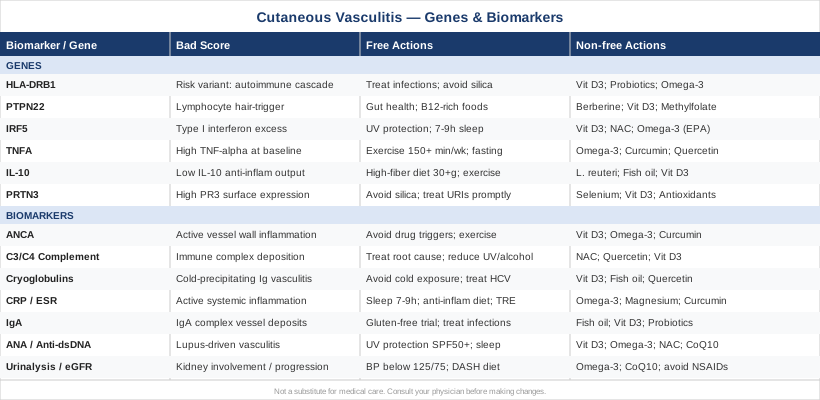
7 Biomarkers That Reveal What Is Happening in Your Vessels
Understanding which biomarkers to test — and what the results actually mean for your specific situation — is one of the most practical steps you can take when managing cutaneous vasculitis. Not all of these tests are ordered on every initial workup. Knowing which ones apply to your clinical picture helps you have a far more productive conversation with your physician, and allows you to track disease activity over time rather than relying solely on visible skin changes.
Biomarker 1: ANCA – The Antibody That Points Directly to Vessel Inflammation
Why it matters and what it reveals: Antineutrophil cytoplasmic antibodies (ANCA) are autoantibodies that target proteins inside neutrophils — specifically proteinase 3 (PR3) and myeloperoxidase (MPO). A positive c-ANCA (anti-PR3) is strongly associated with granulomatosis with polyangiitis (GPA), while p-ANCA (anti-MPO) is more commonly seen in microscopic polyangiitis and eosinophilic granulomatosis with polyangiitis. In cutaneous vasculitis, ANCA positivity suggests the skin involvement may be part of a systemic small-vessel process — not an isolated or benign rash. ANCA titers often correlate with disease activity, making serial testing useful for monitoring flares and treatment response. The updated Chapel Hill Consensus Nomenclature (Jennette et al., Arthritis & Rheumatism, 2013) formalized ANCA-associated vasculitis as a distinct mechanistic category and remains the clinical framework used today.
How to measure it: ANCA testing is a standard blood draw available at any reference laboratory. Most labs offer both an immunofluorescence screen and ELISA-based testing for specific PR3 and MPO antibodies. Cost typically ranges from $50 to $200 depending on whether you receive a bundled vasculitis panel or order tests individually. Retesting every 3 to 6 months during active disease, or after treatment changes, gives useful trend data.
If the score is bad, the plan without supplements: If ANCA is positive, the immediate priority is identifying and eliminating external triggers. Known drug-induced causes include hydralazine, propylthiouracil (PTU), minocycline, and cocaine — ruling these out is the first clinical step. Occupational silica dust exposure is the most strongly established environmental trigger for ANCA vasculitis in genetically susceptible individuals, and respiratory protection in at-risk environments is directly relevant. Treating chronic infections — particularly upper respiratory infections, dental foci, and sinusitis — matters because persistent infections can maintain elevated ANCA titers through molecular mimicry between bacterial proteins and PR3. A Mediterranean-style dietary pattern (rich in vegetables, legumes, olive oil, and fatty fish; low in refined carbohydrates and ultra-processed foods) provides a structural anti-inflammatory foundation. Regular moderate-intensity aerobic exercise — 150 minutes or more per week — consistently reduces neutrophil activation markers across inflammatory conditions.
If the score is bad, the plan with supplements or equipment: Vitamin D3 at 5,000 IU daily has broad immunomodulatory effects in autoimmune disease, including documented reductions in ANCA-relevant neutrophil activation markers. Target serum 25-OH-D between 50 and 80 ng/mL and monitor levels every 3 months. Omega-3 fatty acids (EPA + DHA combined) at 3–4 g daily reduce neutrophil priming and activation through effects on pro-inflammatory eicosanoid synthesis; note mild anticoagulant effects at this dose, relevant if anticoagulants are already prescribed; no cycling required. Curcumin at 500 mg twice daily with piperine (5 mg per dose for absorption) targets the NF-κB pathway, which is central to ANCA-mediated vascular inflammation; cycle 8–12 weeks on, followed by 4 weeks off to prevent tolerance; GI discomfort is the most common side effect. None of these replace medical evaluation or standard immunosuppressive treatment for ANCA-positive systemic vasculitis, but they are rational low-risk adjuncts.
Biomarker 2: Complement Levels (C3 and C4) – The Immune Complex Signal
Why it matters and what it reveals: Complement proteins are effectors of the innate immune system. When immune complexes deposit in vessel walls — as occurs in lupus vasculitis, cryoglobulinemic vasculitis, and certain forms of hypersensitivity vasculitis — complement is consumed in situ. Low C3 and C4 levels therefore serve as a flag for ongoing immune complex deposition in vessels. They help distinguish complement-consuming vasculitis (lupus, cryoglobulinemia, serum sickness) from ANCA-mediated or IgA-mediated forms, which typically maintain normal complement levels. Understanding which mechanism is active is clinically important because each responds to fundamentally different treatments.
How to measure it: C3 and C4 are standard serum protein tests available at any reference lab, costing approximately $30 to $80 for the pair. Normal adult ranges are approximately 90–180 mg/dL for C3 and 16–47 mg/dL for C4. A CH50 (total hemolytic complement) test assesses the entire complement cascade at once for a similar cost and is useful when a global picture of complement function is needed. These values fluctuate with disease activity; retesting every 3 months in active disease provides clinically useful trend data. It is worth tracking complement alongside anti-dsDNA antibodies, since they typically move in opposite directions during lupus flares.
If the score is bad, the plan without supplements: Low complement in active cutaneous vasculitis almost always indicates an underlying condition driving immune complex deposition. The highest-leverage step is identifying and treating that root cause — whether active SLE, hepatitis C, or a lymphoproliferative disorder. Reducing UV light exposure matters specifically when complement-consuming vasculitis occurs in the context of lupus, as UV-B upregulates anti-dsDNA antibody production and worsens immune complex formation. Reducing alcohol intake is consistently important in cryoglobulinemia-associated cases, where it accelerates underlying liver disease and worsens immune complex clearance. Improving sleep quality and reducing psychological stress reduces the neuroimmune signaling that amplifies immune complex-mediated inflammation.
If the score is bad, the plan with supplements or equipment: N-acetylcysteine (NAC) at 600 mg twice daily supports glutathione production and reduces the oxidative stress that accompanies and worsens complement-driven vessel damage; cycle 8 weeks on, 2 weeks off; nausea is possible at higher doses. Quercetin at 500 mg twice daily has complement-modulating and mast cell-stabilizing properties; no cycling needed; well-tolerated in most people. Vitamin D3 at 5,000 IU daily has documented effects on downregulating several complement-activating autoantibody pathways through its nuclear receptor activity in immune cells. Evidence for these supplements specifically in complement-consuming vasculitis is early-stage; use them as adjuncts to, not replacements for, treatment of the underlying condition.
Biomarker 3: Cryoglobulins – The Cold-Precipitating Clue
Why it matters and what it reveals: Cryoglobulins are immunoglobulins that precipitate from serum at temperatures below 37°C and redissolve upon rewarming. Three types are recognized: Type I (monoclonal immunoglobulin, associated with myeloma or Waldenström's macroglobulinemia), Type II (mixed monoclonal-polyclonal, strongly linked to chronic hepatitis C infection), and Type III (polyclonal, seen in autoimmune conditions including Sjögren's syndrome and SLE). Cryoglobulinemic vasculitis classically presents with the triad of palpable purpura, arthralgia, and weakness, with skin lesions concentrated on the lower extremities and buttocks. Identifying cryoglobulins as the mechanism changes the diagnostic workup and treatment approach entirely — there is no other biomarker that so directly points to the treatment solution (hepatitis C antivirals in most Type II cases).
How to measure it: Cryoglobulin testing requires careful specimen handling — blood must be kept at 37°C during collection and transport to prevent premature precipitation, which causes false negatives. This handling requirement means processing errors are relatively common, and results may need to be repeated if clinical suspicion remains high. Cost ranges from $50 to $200. A positive result should always be followed by immunofixation electrophoresis to type the cryoglobulin and by full hepatitis C serology with viral load testing, since the majority of mixed (Type II) cryoglobulinemia is HCV-driven.
If the score is bad, the plan without supplements: For hepatitis C-associated Type II cryoglobulinemia, treatment with modern direct-acting antivirals is the single most impactful available intervention. Current regimens achieve sustained virologic response in over 95% of treated patients, and cryoglobulinemic vasculitis resolves or substantially improves in the majority of those who clear the virus. For non-HCV cryoglobulinemia, treating the underlying B-cell disorder or autoimmune condition is the priority. Practical daily measures include avoiding cold exposure — warm clothing, heated environments in winter, avoiding cold water immersion — which reduces acute triggering of cryoprecipitation and symptom burden. Eliminating alcohol is consistently recommended when underlying liver disease or active hepatitis is present.
If the score is bad, the plan with supplements or equipment: Vitamin D3 at 5,000 IU daily is broadly immunomodulatory and relevant across cryoglobulin types; monitor 25-OH-D quarterly. Fish oil at 3 g EPA + DHA daily reduces the inflammatory signaling driving immune complex-mediated vessel damage; note mild anticoagulant effects at this dose, particularly when combined with HCV antiviral therapy. Quercetin at 500 mg twice daily has mast cell-stabilizing and anti-inflammatory properties relevant to immune complex disease; well-tolerated, no cycling required. Direct supplement trial evidence specific to cryoglobulinemic vasculitis is absent; these are rational low-risk additions to standard care, not substitutes for treating the underlying cause.
Biomarker 4: CRP and ESR – The Inflammation Barometer
Why it matters and what it reveals: C-reactive protein (CRP) and erythrocyte sedimentation rate (ESR) are general-purpose systemic inflammation markers. Neither is specific for vasculitis, but both serve as reliable indicators of ongoing inflammation and are useful for tracking disease activity over time. A markedly elevated ESR (above 80–100 mm/hr) or CRP (above 10 mg/L) alongside cutaneous vasculitis suggests significant systemic inflammation and warrants workup for systemic organ involvement. High-sensitivity CRP (hs-CRP) is more sensitive than standard CRP and responds more rapidly to changes in inflammation level, making it the preferred serial monitoring marker. Peter Attia consistently recommends hs-CRP as a core routine cardiovascular inflammation marker; in vasculitis it serves the same role as a real-time disease activity signal that updates faster than autoantibody titers.
How to measure it: Both tests are inexpensive — $10 to $30 each — available at any standard laboratory. Hs-CRP below 1.0 mg/L is low-risk; values above 3.0 mg/L are elevated, and values above 10 mg/L indicate significant active inflammation. ESR above 30–40 mm/hr in adults warrants explanation in the context of vasculitis symptoms. Retesting every 4 to 8 weeks during active disease, and every 3 months during remission, provides practically useful trend information without unnecessary costs.
If the score is bad, the plan without supplements: Sustained sleep duration of 7–9 hours per night is one of the most consistently effective CRP reducers documented — multiple epidemiological studies show chronically short sleep (below 6 hours) raises hs-CRP by 40–80% independent of other factors. An anti-inflammatory dietary pattern built around diverse vegetables, legumes, fatty fish, extra-virgin olive oil, and whole fruits — with minimal refined carbohydrates and ultra-processed foods — addresses multiple upstream inflammation drivers simultaneously. Time-restricted eating (a 10 to 12-hour eating window, typically skipping late-night eating) has shown modest hs-CRP-lowering effects in randomized trials. Regular moderate-intensity exercise — at least 150 minutes per week — consistently reduces both ESR and hs-CRP in inflammatory conditions; the benefit appears most robust in people with elevated baseline values.
If the score is bad, the plan with supplements or equipment: Omega-3 (EPA + DHA combined at 4 g/day) is the most evidence-backed supplement for CRP reduction, with robust data from both cardiovascular and autoimmune populations; mild anticoagulant effect at this dose is worth noting; no cycling required. Magnesium glycinate at 300–400 mg nightly reduces NF-κB-driven inflammation at the cellular level; no cycling required; possible loose stools at higher doses, which resolves by starting at 150 mg and titrating up. Curcumin at 500 mg twice daily with 5 mg piperine reduces hs-CRP in multiple randomized controlled trials; cycle 8–12 weeks on, 4 weeks off to prevent tolerance; GI discomfort is the main dose-limiting side effect. Vitamin D3 at 5,000 IU daily is worth adding if serum 25-OH-D is below 40 ng/mL; deficiency independently raises CRP.
Biomarker 5: IgA Levels – The Forgotten Antibody
Why it matters and what it reveals: IgA vasculitis — formerly known as Henoch-Schönlein purpura — is the most common systemic vasculitis in children and a meaningful proportion of adult cases, often presenting with greater severity and more renal involvement in adults than children. It is defined by IgA-dominant immune complex deposits in small vessel walls and classically presents with palpable purpura on dependent areas (lower extremities, buttocks), arthritis, abdominal cramping, and in adults, renal involvement that overlaps mechanistically with IgA nephropathy. Serum IgA may be elevated during active disease, though the definitive diagnostic test remains skin or kidney biopsy with immunofluorescence demonstrating IgA deposits. For anyone with recurring palpable purpura below the waist without a clear alternative diagnosis, IgA vasculitis deserves to be on the differential.
How to measure it: Serum IgA is a standard immunoglobulin quantification test costing $20 to $60 at most labs. Normal adult range is approximately 70–400 mg/dL; values above 400 mg/dL in the appropriate clinical context support the diagnosis. Definitive confirmation requires a skin biopsy with direct immunofluorescence staining ($200–$600 depending on facility) showing IgA deposits in dermal capillary walls. Serum IgA should be retested every 3 months during active disease, and urinalysis with eGFR checked simultaneously given the frequency of renal involvement.
If the score is bad, the plan without supplements: A structured gluten-free trial of 60 to 90 days is worth considering for adults with IgA vasculitis or IgA nephropathy. The association between mucosal IgA dysregulation and gluten sensitivity has clear biological plausibility — gliadin stimulates zonulin release and mucosal IgA production — and the intervention is low-risk when done with adequate nutritional planning. Treating infections promptly is particularly important in IgA vasculitis, where upper respiratory tract infections and tonsillitis reliably trigger flares; some adult patients benefit from tonsillectomy evaluation when recurrent pharyngitis is a consistent flare trigger. A structured food sensitivity elimination protocol — removing gluten and dairy — followed by systematic reintroduction can identify individual dietary drivers of IgA dysregulation.
If the score is bad, the plan with supplements or equipment: Fish oil at 3 g EPA + DHA daily has clinical trial evidence in IgA nephropathy, including the STOP-IgAN and OMEGA trials, showing modest but meaningful reductions in proteinuria; given the mechanistic overlap between IgA vasculitis and IgA nephropathy, this is a rational adjunct. Vitamin D3 at 5,000 IU daily modulates mucosal IgA production and supports the immune regulation that moderates IgA overproduction; monitor 25-OH-D quarterly. Probiotics — particularly multi-strain formulas including Lactobacillus rhamnosus GG and Bifidobacterium longum at 20–50 billion CFU daily — support gut mucosal IgA homeostasis; take daily with food, no cycling required, minimal side effects. When renal involvement is confirmed, these adjuncts should complement, not replace, nephrology or rheumatology oversight.
Biomarker 6: ANA and Anti-dsDNA Panel – Checking for Lupus-Associated Vasculitis
Why it matters and what it reveals: Antinuclear antibodies (ANA) are autoantibodies targeting nuclear antigens across a spectrum of specificities. A positive ANA at titer 1:160 or higher — combined with anti-double-stranded DNA (anti-dsDNA) antibodies — significantly raises the probability of systemic lupus erythematosus as the underlying driver of cutaneous vasculitis. Anti-dsDNA titers are particularly valuable for disease monitoring: rising titers frequently precede lupus flares by two to four weeks, while declining titers track with clinical remission. Thomas Dayspring and other precision medicine physicians consistently emphasize that ANA and anti-dsDNA should be interpreted together with complement levels, since the three move in a predictable pattern during flares — antibodies rise, complement falls — making this combination one of the most informative monitoring panels available.
How to measure it: ANA with reflex testing (triggering anti-dsDNA, anti-Sm, and anti-Ro/La on a positive screen) is a standard rheumatology panel costing $50 to $150 for the complete combination. ANA screening is performed by immunofluorescence; a positive result triggers specific antibody testing on the same draw. Anti-dsDNA should be retested every 3 months in active lupus. For highest sensitivity in disease monitoring, the Crithidia luciliae immunofluorescence assay for anti-dsDNA is preferred over ELISA-based methods, which have lower specificity for active disease.
If the score is bad, the plan without supplements: Minimizing UV light exposure is among the most evidence-supported lifestyle interventions for ANA-positive lupus vasculitis. UV-B radiation exposes nuclear antigens in apoptotic skin cells, directly upregulating anti-dsDNA production through toll-like receptor activation. Broad-spectrum SPF 50+ sunscreen, UV-protective clothing (UPF 50), and avoiding peak-sun hours (10 AM–4 PM) are well-supported by mechanistic and clinical data — this is not cosmetic advice for this group, it is disease management. Consistent sleep of 7–9 hours reduces the cortisol and sympathetic drive that amplifies Th17 dominance and worsens ANA-mediated vasculitis flares; sleep optimization functions as a structural anti-inflammatory intervention.
If the score is bad, the plan with supplements or equipment: Vitamin D3 at 5,000–10,000 IU daily has documented immunomodulatory effects specific to lupus-related autoimmunity, including reductions in anti-dsDNA titers in multiple observational studies; monitor 25-OH-D and serum calcium every 3 months at these doses and do not exceed a 25-OH-D level of 100 ng/mL. Omega-3 at 3–4 g EPA + DHA daily reduces pro-inflammatory prostaglandins relevant to lupus pathophysiology; no cycling required. NAC at 600 mg twice daily supports the glutathione system, which is consistently depleted in active lupus and exacerbates oxidative vessel damage; cycle 8 weeks on, 2 weeks off; occasional nausea at initiation. CoQ10 at 200 mg daily in ubiquinol form addresses mitochondrial dysfunction common in autoimmune conditions and supports energy metabolism in immune cells; no cycling required.
Biomarker 7: Urinalysis and eGFR – Tracking Systemic Spread
Why it matters and what it reveals: Cutaneous vasculitis can be an isolated skin condition or the most visible surface of a systemic process affecting kidneys, lungs, and peripheral nerves. The kidneys are among the most vulnerable organs in most forms of systemic vasculitis. Hematuria (blood in urine), proteinuria, and red cell casts on urinalysis microscopy indicate renal involvement — even in patients who feel entirely well and have no urinary symptoms. eGFR (estimated glomerular filtration rate) translates kidney function into a measurable number. A declining eGFR in a patient with known vasculitis is a sign of disease progression requiring urgent medical evaluation — this is not a situation to address with lifestyle changes while deferring a physician visit.
How to measure it: Urinalysis with microscopy costs $10 to $30. eGFR is calculated from serum creatinine, available in any basic metabolic panel ($15–$50). A spot urine protein-to-creatinine ratio ($20–$50) quantifies proteinuria more precisely than dipstick screening alone. Anyone with a form of vasculitis that could be systemic — ANCA-positive, complement-low, ANA-positive, or with cryoglobulins — should have urinalysis and eGFR checked at every clinical visit, or at minimum every 3 months. During active renal vasculitis, monthly monitoring is clinically justified.
If the score is bad, the plan without supplements: Active urinary sediment or declining eGFR in vasculitis is a medical urgency — medical treatment should not be delayed to try lifestyle interventions first. Once medical management is underway, supportive lifestyle measures include: blood pressure control (target below 125/75 mmHg when proteinuria is present, consistent with KDIGO nephrology guidelines), adequate hydration (approximately 2 liters of water daily), a DASH-pattern diet (low sodium at 2g/day, adequate potassium from whole foods), and strict avoidance of nephrotoxic agents including NSAIDs, iodinated contrast when avoidable, and herbal preparations containing aristolochic acid (found in some traditional herbal teas and supplements).
If the score is bad, the plan with supplements or equipment: Omega-3 at 2–4 g EPA + DHA daily has demonstrated renoprotective effects in IgA nephropathy (STOP-IgAN trial data), with modest but meaningful proteinuria reduction; relevant given the mechanistic overlap between IgA nephropathy and IgA vasculitis-related renal involvement. CoQ10 at 200–400 mg daily in ubiquinol form addresses the mitochondrial energy demands of proximal tubule cells, which are highly vulnerable to the oxidative stress in autoimmune kidney disease; no cycling needed. Avoid high-dose vitamin C (above 2 g/day), which can contribute to oxalate deposition in kidneys with compromised filtration. The priority at this stage is always medical stabilization; supplements are secondary adjuncts once the clinical situation is stable.
What Your Genes May Reveal About Vasculitis Susceptibility
The biomarkers above tell you what is happening in your body right now. Genetics offers a different but complementary perspective: it tells you why your immune system may respond to certain triggers more intensely than average, and which biological pathways are working against you even before environmental exposures appear. Genetic information does not determine outcomes, and carrying a risk variant does not mean disease is inevitable. What it does provide is clarity about which preventive and compensatory strategies are most biologically relevant for your specific profile.
Gene 1: HLA-DRB1 – The Immune Recognition Gatekeeper
Human Leukocyte Antigen DR beta 1 (HLA-DRB1) is part of the MHC class II complex, the system the immune system uses to recognize foreign and self-antigens and present them to T cells. Specific HLA-DRB1 alleles have been associated with susceptibility to ANCA-associated vasculitis and multiple other autoimmune conditions involving cutaneous vasculitis. The HLA region is the most consistently replicated genetic locus across autoimmune disease genome-wide association studies, reflecting how foundational antigen recognition is to autoimmune susceptibility. Ali Torkamani's genetic interpretation frameworks emphasize that HLA variants establish immune responsiveness thresholds — they do not cause disease, but they lower the bar for how much environmental provocation is needed to initiate an autoimmune cascade.
If the gene is bad — the plan without supplements: Reducing antigen exposure at mucosal surfaces is the strategic priority for HLA risk carriers. Treat chronic infections aggressively rather than managing symptoms — dental infections, chronic sinusitis, and recurring pharyngitis all provide persistent antigenic stimulation that HLA risk alleles amplify. Minimize occupational exposure to silica dust, petrochemicals, and other immune-activating inhalants. Maintain mucosal barrier integrity through a high-fiber diet, consistent sleep, and stress management; all three lower the intestinal antigen load that HLA risk variants tend to translate into elevated autoimmune reactivity.
If the score is bad — the plan with supplements or equipment: Vitamin D3 at 5,000 IU daily directly modulates HLA expression on antigen-presenting dendritic cells, shifting them toward tolerogenic rather than inflammatory presentations; monitor 25-OH-D quarterly. Multi-strain probiotics at 20–50 billion CFU daily (including Lactobacillus acidophilus and Bifidobacterium longum) support the mucosal immune milieu that interacts closely with HLA-mediated antigen presentation; take daily with food, no cycling required; well-tolerated. Omega-3 at 3 g EPA + DHA daily modulates downstream T-helper cell polarization away from the Th17 dominance that HLA risk alleles tend to amplify; mild anticoagulant effect at this dose.
Gene 2: PTPN22 – The Autoimmune Hair-Trigger
PTPN22 encodes protein tyrosine phosphatase non-receptor type 22, an enzyme that regulates the activation threshold for T and B lymphocytes. The rs2476601 variant (R620W) lowers the activation threshold for autoreactive lymphocytes and is among the most replicated genetic associations in autoimmune disease, linked to rheumatoid arthritis, SLE, type 1 diabetes, and vasculitis in multiple populations. Gary Brecka's genetic interpretation work has highlighted that PTPN22 variants function like a hair-trigger on the immune system: the system is not broken, but it takes far less environmental provocation to activate autoreactive T and B cells than in people without the variant. The clinical implication is that environmental triggers — infections, toxins, gut permeability — have a larger impact on disease onset in PTPN22 risk carriers than in the general population.
If the gene is bad — the plan without supplements: Treating infections promptly and completely rather than waiting them out is critical for PTPN22 risk carriers, where lingering antigenic stimulation has a disproportionate impact. Improving gut permeability through diet is equally important — intestinal barrier dysfunction provides a constant source of bacterial lipopolysaccharide that PTPN22 variants process with exaggerated immune activation. A high-fiber, low-ultra-processed-food diet is the most practical tool for reducing this steady-state antigen load. Optimizing folate and methylcobalamin (B12) intake through food (dark leafy greens, eggs, fish) matters because PTPN22 interacts with methylation pathways that regulate immune gene expression.
If the score is bad — the plan with supplements or equipment: Berberine at 500 mg twice daily with meals has demonstrated effects on lymphocyte signaling pathways and gut microbiome composition that are relevant to PTPN22-driven autoimmune activation; cycle 8 weeks on, 4 weeks off to avoid tolerance; GI discomfort (loose stools, cramping) is the most common side effect, particularly at the start, and usually resolves after the first week. Vitamin D3 at 5,000–10,000 IU daily modulates PTPN22-driven lymphocyte hyperactivation; monitor 25-OH-D and calcium every 3 months. Methylcobalamin (active B12) at 1,000 mcg daily and methylfolate at 400–800 mcg daily support the epigenetic methylation that regulates immune gene expression adjacent to PTPN22 function; no cycling needed, excellent safety profile.
Gene 3: IRF5 – The Interferon Switch
Interferon Regulatory Factor 5 (IRF5) is a transcription factor that drives the production of type I interferons and pro-inflammatory cytokines including TNF-alpha, IL-6, and IL-12. Risk variants — particularly rs2004640 and rs10954213 — are strongly associated with SLE, and more broadly with autoimmune conditions that include cutaneous vasculitis as a feature. High IRF5 activity tips the immune system toward pro-inflammatory interferon signaling, amplifying vessel wall inflammation. The evidence for IRF5 variants in isolated cutaneous vasculitis is mostly indirect, derived from SLE and related autoimmune GWAS data; direct vasculitis-specific IRF5 studies remain limited.
If the gene is bad — the plan without supplements: UV light directly activates the type I interferon pathway — UV-exposed dying skin cells release nuclear DNA fragments that act as interferon-inducing ligands through toll-like receptors. For IRF5 risk carriers, rigorous daily sun protection is not cosmetic but mechanistically central to reducing interferon drive. Sleep deprivation independently upregulates interferon signaling; 7–9 hours of consistent, restorative sleep is one of the most effective available non-pharmacological IRF5 interventions. Stress reduction through consistent autonomic practices (described in the complementary section) matters because chronic sympathetic activation upregulates interferon-mediated inflammation independent of UV or infectious triggers.
If the score is bad — the plan with supplements or equipment: Vitamin D3 at 5,000 IU daily has documented ability to downregulate IRF5-dependent interferon production in immune cell models; monitor quarterly. NAC at 600 mg twice daily reduces the oxidative stress that amplifies interferon signaling in IRF5 risk carriers; cycle 8 weeks on, 2 weeks off; occasional nausea, especially without food. Omega-3 (EPA specifically at 4 g/day) inhibits the prostaglandin and leukotriene pathways that synergize with IRF5-driven inflammation; no cycling required. Direct supplement evidence for IRF5 variants in vasculitis does not yet exist; these recommendations derive from the SLE literature where the variant data are strongest.
Gene 4: TNFA – The Inflammatory Volume Setting
The TNFA gene encodes TNF-alpha, one of the master regulators of acute and chronic inflammation. The G308A promoter polymorphism (rs1800629) is associated with higher TNF-alpha transcript levels and has been linked to more severe inflammatory responses across immune conditions including systemic vasculitis. Carrying the high-expression variant means your immune system effectively runs its inflammatory cascade at an elevated baseline, lowering the threshold at which vasculitis flares occur and increasing the intensity once they do. TNF-alpha is also one of the primary cytokines released by visceral adipose tissue, making body composition directly relevant to TNFA variant carriers.
If the gene is bad — the plan without supplements: Sustained aerobic exercise at moderate intensity for 150 or more minutes weekly is among the most consistently effective interventions for downregulating TNF-alpha expression, working through both direct immune cell effects and improved adipose tissue metabolism. Reducing visceral adiposity is particularly relevant for TNFA risk carriers, since fat tissue is a major tissue-level TNF-alpha source that operates independently of immune system activation. Time-restricted eating in a 12:12 or 16:8 pattern has shown TNF-alpha-reducing effects in multiple randomized trials, making it an accessible non-supplement approach with broad supportive evidence.
If the score is bad — the plan with supplements or equipment: Omega-3 at 4 g EPA + DHA daily consistently reduces circulating TNF-alpha across clinical studies in inflammatory conditions; mild anticoagulant effect at this dose. Curcumin at 500 mg twice daily with piperine inhibits NF-κB, the primary downstream transcriptional pathway of TNF-alpha; cycle 8–12 weeks on, 4 weeks off; GI discomfort is the most frequent side effect. Quercetin at 500 mg daily provides complementary TNF-alpha inhibition through distinct molecular pathways including JAK-STAT; no cycling required, minimal side effects. Resveratrol at 150–500 mg daily activates SIRT1, which independently dampens NF-κB activity; cycle 8 weeks on, 4 weeks off to maintain cellular sensitivity.
Gene 5: IL-10 – The Anti-Inflammatory Thermostat
IL-10 is one of the body's primary anti-inflammatory cytokines. It suppresses macrophage activation, inhibits pro-inflammatory cytokine cascades, and expands regulatory T cell populations that maintain immune tolerance. Promoter polymorphisms — particularly the -1082G/A, -819C/T, and -592C/A variants — determine how much IL-10 is produced in response to immune activation. Low-expression haplotypes are associated with more severe and persistent inflammatory responses across multiple autoimmune conditions, including vasculitis. Think of IL-10 as the volume-down control on the immune response: if yours is genetically set too low, inflammatory cascades run longer and more intensely before they resolve.
If the gene is bad — the plan without supplements: The gut microbiome is the primary source of the microbial metabolites — particularly butyrate — that stimulate IL-10 production in intestinal immune cells and downstream in systemic circulation. A high-fiber diet providing 30 or more grams daily from diverse plant sources (legumes, vegetables, intact whole grains, fruits) consistently raises butyrate production and supports IL-10 expression without any supplement needed. Regular moderate exercise also upregulates IL-10 release as part of the well-documented post-exercise anti-inflammatory response. Minimizing unnecessary antibiotic courses protects the microbial diversity that underlies this microbiome-driven IL-10 production pathway.
If the score is bad — the plan with supplements or equipment: Lactobacillus reuteri — specifically the ATCC PTA 6475 strain, which has the strongest published IL-10 induction data — at 1–2 billion CFU daily is the most biologically targeted supplement for low IL-10 producers; take daily with food, no cycling required; well-tolerated. Fish oil at 3 g EPA + DHA daily supports IL-10 production through effects on resolvin and protectin synthesis in immune cells; no cycling required. Vitamin D3 at 5,000 IU daily directly upregulates IL-10 transcription through the vitamin D response element in the IL-10 gene promoter region — one of the more direct gene-to-supplement connections in this list; monitor 25-OH-D quarterly.
Gene 6: PRTN3 – The ANCA Target Gene
Proteinase 3 (PRTN3) is the neutrophil granule serine protease that serves as the primary antigenic target for c-ANCA (anti-PR3) autoantibodies. Polymorphisms in the PRTN3 gene affect how much proteinase 3 is expressed on the neutrophil surface membrane — higher surface expression means more ANCA binding, more neutrophil activation, greater vessel wall damage, and more frequent disease relapses. The PR3HI genotype (high surface-expression variant) has been associated with GPA susceptibility and higher relapse rates in European GWAS cohorts studying ANCA-associated vasculitis. Evidence for PRTN3 variants in isolated cutaneous vasculitis (without systemic involvement) is limited and should be interpreted cautiously; most data derive from systemic GPA populations.
If the gene is bad — the plan without supplements: Avoiding silica dust exposure is the most strongly established environmental modifier for ANCA-associated vasculitis in genetically susceptible individuals, including those with PRTN3 risk variants. In construction, mining, and agricultural contexts with dusty soils, appropriate respiratory protective equipment is directly relevant. Maintaining excellent respiratory tract health through prompt treatment of upper respiratory infections matters because PR3 shares structural sequence homology with certain bacterial proteins — providing a molecular mimicry route through which infections can initiate or amplify anti-PR3 antibody production even in the absence of silica exposure.
If the score is bad — the plan with supplements or equipment: Selenium at 200 mcg daily supports neutrophil antioxidant defense and has shown inhibitory effects on neutrophil granule proteases in experimental models; do not exceed 400 mcg/day as selenium is toxic at supraphysiological doses; no cycling required at 200 mcg. Vitamin D3 at 5,000 IU daily has shown ability to reduce neutrophil surface PR3 expression in animal models; direct human PRTN3 variant-specific data are limited. An antioxidant combination — vitamin C at 500 mg, vitamin E at 200 IU, and mixed carotenoids — supports the cellular redox balance around neutrophil activation; take daily with food, no cycling needed.
What "The Autoimmune Solution" Gets Right About Inflammatory Skin Conditions
Amy Myers MD published The Autoimmune Solution in 2015, drawing on her clinical experience treating autoimmune patients and on her own diagnosis with Graves' disease. The book does not address cutaneous vasculitis by name, but its systems-level model for how autoimmune diseases develop and what reverses them is mechanistically relevant — particularly for IgA vasculitis, lupus-associated vasculitis, and hypersensitivity vasculitis. Several of its core claims, which were ahead of mainstream acceptance in 2015, have since been supported by growing research in intestinal permeability, the microbiome, and epigenetic immune regulation. The following ten ideas from the book are the most impactful for anyone dealing with autoimmune-driven vessel inflammation.
1. Leaky Gut Is Upstream of Autoimmune Disease, Not a Consequence of It
Myers argues that intestinal permeability — the breakdown of tight junction proteins including zonulin, claudin, and occludin — is a necessary upstream condition for autoimmune disease, not simply a downstream effect. When the gut barrier fails, bacterial endotoxins (LPS) and partially digested food proteins enter systemic circulation, priming innate immune cells for chronic activation. Alessio Fasano's team at Harvard gave this mechanism substantial scientific grounding, particularly through the discovery of zonulin as a regulated intestinal permeability modulator. For vasculitis, the implication is that gut barrier integrity may need to be treated as directly as the vessels themselves.
2. Gluten Triggers Intestinal Permeability in All Humans, Not Just Celiac Patients
Myers cites research demonstrating that gliadin stimulates zonulin release from intestinal epithelial cells through the CXCR3 receptor in all people — not only those with celiac disease — though the magnitude of response varies with individual genetics. The practical implication is that for patients with IgA vasculitis, elevated serum IgA, or poorly controlled autoimmune vasculitis, a structured 60 to 90-day gluten elimination trial has a plausible biological rationale that extends beyond popular wellness trends into measurable mucosal immunology.
3. Chronic Infections Drive Molecular Mimicry Toward Blood Vessel Antigens
The book explains how chronic or recurring infections — Epstein-Barr virus, Candida overgrowth, SIBO — produce antigens that structurally resemble self-proteins in blood vessel walls and connective tissue. The immune system, attempting to clear the infection, generates cross-reactive antibodies that also recognize vessel antigens. This molecular mimicry mechanism is particularly well-documented in ANCA vasculitis, where PR3 shares sequence homology with certain bacterial proteins. Myers argues that identifying and systematically treating occult chronic infections is a clinically underutilized intervention in autoimmune management.
4. Environmental Toxins Accumulate in Immune Cells and Shift the Tolerance Threshold
Myers details how heavy metals (mercury, lead, cadmium), mold mycotoxins, and organochlorine pesticide residues impair liver phase II detoxification and accumulate in macrophages and other immune cells, pushing the immune system away from tolerance and toward chronic reactivity. While this perspective is not standard rheumatology, functional medicine testing — urinary heavy metal panels and mycotoxin urine tests — has made these burdens more accessible and quantifiable. For vasculitis patients who have not responded adequately to standard treatment, toxin burden is a variable worth systematically evaluating.
5. MTHFR and Methylation Directly Regulate Immune Gene Expression
The book covers MTHFR gene variants that impair methyl group production, disrupting the epigenetic regulation of immune gene promoters. Methylation marks on immune gene DNA control whether pro-inflammatory pathways remain switched on or off. Myers recommends active forms — methylcobalamin (B12) and methylfolate — specifically over synthetic cyanocobalamin and folic acid, which MTHFR risk carriers cannot efficiently convert. This recommendation connects directly to the PTPN22 and IRF5 gene sections earlier in this article, where methylation support is a consistent adjunct recommendation.
6. Autoimmunity Exists on a Spectrum With a Years-Long Pre-Disease Phase
One of the book's most clinically actionable concepts is that autoimmune disease does not begin with a formal diagnosis — it begins years earlier with elevated autoantibodies, fluctuating fatigue, and episodic unexplained symptoms. ANA positivity at low titers, recurring purpuric episodes, and elevated hs-CRP in the absence of a definitive vasculitis diagnosis are all early-spectrum autoimmune signals. Myers argues persuasively that this is the phase where lifestyle and nutritional interventions have their greatest leverage — before the autoimmune process entrenches into structural vessel damage.
7. The Autoimmune Paleo Protocol as a Dietary Reset
Myers advocates the Autoimmune Paleo (AIP) protocol — eliminating grains, legumes, nightshades, eggs, dairy, nuts, seeds, and alcohol for 30 to 60 days, followed by systematic reintroduction. The evidence base has strengthened significantly since 2015: a 2017 pilot study in Inflammatory Bowel Diseases (Konijeti et al.) and a 2019 randomized trial both demonstrated meaningful improvements in disease activity scores and inflammatory markers following AIP. For vasculitis specifically, the nightshades reintroduction phase — tomatoes, peppers, eggplant — is worth monitoring carefully, as alkaloids in these plants have documented effects on intestinal tight junction integrity.
8. Thyroid Autoimmunity Is a Frequently Overlooked Comorbidity That Adds to Inflammatory Burden
Myers documents the high co-occurrence of thyroid autoimmunity (Hashimoto's thyroiditis, Graves' disease) with other autoimmune conditions. In patients with cutaneous vasculitis, undiagnosed thyroid autoimmunity adds to the overall inflammatory burden and may be a concurrent driver of systemic immune dysregulation. She recommends testing not only TSH but free T3, free T4, anti-TPO antibodies, and anti-thyroglobulin antibodies as a complete thyroid autoimmune panel. For vasculitis patients whose disease is poorly controlled despite standard treatment, this panel is a worthwhile addition to the existing workup.
9. Chronic Stress Maintains the Th17 Dominance That Drives Vessel Wall Inflammation
Myers explains the immunological mechanism linking chronic psychological stress to autoimmune flares: sustained cortisol dysregulation and elevated sympathetic nervous system tone shift T-helper cell populations toward Th17 (pro-inflammatory, tissue-attacking) and away from Treg (regulatory, tolerance-maintaining) cells. This is the biological underpinning of the well-observed clinical pattern where vasculitis flares cluster around periods of major life stress. It makes autonomic interventions — sleep, slow breathing, mindfulness — mechanistically relevant to disease management rather than merely supportive.
10. Identifying Your Specific Driver Matters More Than Treating All Autoimmunity the Same Way
The book's central thesis is that autoimmune disease can be significantly improved when its specific upstream drivers — gut permeability, infections, toxins, or stress — are systematically identified and addressed. For cutaneous vasculitis, the practical translation is: knowing whether your condition is IgA-driven, ANCA-driven, immune complex-mediated, or drug-induced matters enormously, because each mechanism responds to different interventions. Generic immune suppression without identifying the specific driver addresses the symptom — the inflamed vessel — without addressing the reason the immune system is attacking it.
Other Approaches with Meaningful Clinical Evidence
The following modalities do not replace medical management of cutaneous vasculitis — particularly when organ involvement is present. They address inflammatory burden, immune regulation, and stress physiology through pathways that are clinically relevant and supported by human evidence across autoimmune and inflammatory skin conditions.
Mindfulness-Based Stress Reduction (MBSR)
Mindfulness-Based Stress Reduction (MBSR), the structured 8-week program developed by Jon Kabat-Zinn at the University of Massachusetts Medical School, combines body scan meditation, sitting meditation, and gentle mindful movement. Its relevance to cutaneous vasculitis is mechanistic: chronic psychological stress shifts the T-helper cell balance toward Th17 dominance through cortisol dysregulation and sustained sympathetic activation, directly maintaining the pro-inflammatory vessel wall infiltration characteristic of vasculitis. MBSR consistently shifts this balance toward parasympathetic, Treg-favoring physiology.
A 2015 randomized trial published in Brain, Behavior, and Immunity (Creswell et al.) found that MBSR reduced circulating CRP and IL-6 in adults under chronic stress. A subsequent meta-analysis in Psychoneuroendocrinology (2019) confirmed that mindfulness-based interventions reliably reduce inflammatory cytokines across autoimmune and inflammatory conditions, with the strongest effects in conditions where psychological stress is a primary amplifier of immune activity. Evidence specific to vasculitis remains indirect, derived from studies in related inflammatory conditions.
Begin with the free Palouse Mindfulness curriculum (a validated digital MBSR adaptation built on Kabat-Zinn's original program) at 20 to 30 minutes of daily practice for 8 weeks. The inflammation-reducing effects documented in randomized trials typically appear between weeks 4 and 8 of consistent practice — not at week 1. Combining MBSR with the slow breathing protocol below amplifies the autonomic benefits; both can be integrated into a single 40-minute morning practice without requiring additional time commitment. No adverse effects have been documented at standard program doses.
Breathing-Based Therapies
Slow diaphragmatic breathing — approximately 5 to 6 breath cycles per minute, or a 4-second inhale paired with a 6- to 8-second exhale — activates the vagus nerve and shifts the autonomic nervous system from sympathetic to parasympathetic dominance. This shift directly suppresses the neuroimmune pro-inflammatory cascade. The physiological mechanism is well-characterized: vagal activation reduces macrophage TNF-alpha and IL-6 release through α7-nicotinic acetylcholine receptors on immune cells — the cholinergic anti-inflammatory pathway described in detail by Tracey et al. in Frontiers in Immunology (2017). TNF-alpha and IL-6 are central mediators of vessel wall inflammation in vasculitis; any intervention that durably reduces their production through a non-pharmacological mechanism has direct disease relevance.
Heart rate variability (HRV) biofeedback devices — including the HeartMath Inner Balance sensor and the Lief Therapeutics wearable patch — make the autonomic shift produced by slow breathing visible in real time. Clinical trials using HRV biofeedback in inflammatory conditions have demonstrated reductions in inflammatory markers and improved disease-related quality of life, with the effect driven by the breathing technique itself rather than the device. A 2017 review in Applied Psychophysiology and Biofeedback found that HRV biofeedback produced significant reductions in inflammatory markers in several autoimmune and chronic inflammatory conditions.
Practice 5 to 10 minutes of slow breathing twice daily — once on waking and once before sleep. A 4-7-8 pattern (inhale 4 counts, hold 7, exhale 8) is one accessible entry point; a simple 5-second inhale, 5-second exhale pattern achieves the same autonomic target. Consistent twice-daily practice for 4 to 6 weeks is required before autonomic tone changes stabilize measurably. No equipment is necessary; HRV monitoring devices ($80–$300) are optional tools for people who find objective feedback helpful for adherence.
The Autoimmune Protocol (AIP)
The Autoimmune Protocol, developed by Sarah Ballantyne PhD and published in The Paleo Approach (2013), is a dietary elimination-and-reintroduction framework designed specifically for autoimmune conditions. It removes food categories that either increase intestinal permeability or directly stimulate immune cell activation — grains, legumes, nightshades, eggs, dairy, nuts, seeds, alcohol, and NSAIDs — while emphasizing organ meats, diverse vegetables, fermented foods, and bone broth to support mucosal healing and immune regulation.
The AIP is one of the few alternative dietary protocols with published randomized controlled trial evidence in autoimmune disease. A 2017 open-label study in Inflammatory Bowel Diseases (Konijeti et al.) and a 2019 randomized pilot trial both showed significant improvements in disease activity scores and inflammatory markers following the AIP in IBD. For cutaneous vasculitis, direct AIP trial data do not exist, but the protocol addresses intestinal permeability, IgA dysregulation, microbiome composition, and food antigen-driven immune activation — all of which are mechanistically relevant to the condition. The autoimmune protocol is consistently recommended by Ballantyne as a foundational intervention for any autoimmune condition, given its effects on the gut-immune axis that underlies autoimmune vasculitis.
The elimination phase lasts 30 to 60 days, after which individual foods are systematically reintroduced over 10 days each, allowing clear identification of personal triggers. The protocol is demanding, and planning ahead — particularly for social eating — makes adherence significantly more practical. Ballantyne provides a detailed reintroduction schedule and food list at no cost through her published materials. For vasculitis specifically, monitoring symptoms closely during the nightshades and eggs reintroduction phases is important, as both are common triggers of immune reactivity in autoimmune skin conditions.
Microbiome-Directed Therapies
The gut microbiome exerts substantial influence over systemic immune tone through production of short-chain fatty acids (particularly butyrate), regulation of mucosal IgA output, and control of the Treg-versus-Th17 balance that determines whether the immune system maintains tolerance or attacks self-tissues. A 2020 study published in Arthritis & Rheumatology found significant gut dysbiosis in ANCA-associated vasculitis patients compared to healthy controls, with notable reductions in butyrate-producing bacterial genera including Roseburia and Faecalibacterium. While no microbiome-specific intervention trial for cutaneous vasculitis has been published, a 2021 meta-analysis in Frontiers in Immunology found that probiotic supplementation reduced CRP and autoimmune activity markers across multiple autoimmune diseases, including conditions with direct mechanistic overlap with vasculitis.
The approach works best through two simultaneous inputs. First, a high-fiber dietary foundation providing at least 30 grams daily from diverse plant sources — legumes, colorful vegetables, whole fruits, and intact whole grains if tolerated — feeds the beneficial microbial populations that produce butyrate and support IL-10 production. Second, a multi-strain probiotic including Lactobacillus rhamnosus GG, Lactobacillus acidophilus, Bifidobacterium longum, and Lactobacillus reuteri at 20 to 50 billion CFU daily provides targeted microbial reinforcement. Adding at least one daily serving of fermented food (kefir, kimchi, sauerkraut, or live-culture yogurt) provides additional microbial diversity that probiotic capsules alone do not fully replicate.
Take probiotics with food, preferably alongside the highest-fiber meal of the day, to maximize survival through the upper GI tract. No cycling is required; continuous use for a minimum of 12 weeks is needed before microbiome composition changes stabilize. The combination of dietary fiber, probiotic supplementation, and fermented foods has no meaningful adverse effects at standard doses and complements every other intervention described in this article — making it a practical foundation layer rather than a standalone strategy.
Conclusion
Cutaneous vasculitis is not one thing, and managing it well requires knowing which specific mechanisms are active in your case. The seven biomarkers covered here — ANCA, complement C3/C4, cryoglobulins, CRP and ESR, IgA, ANA with anti-dsDNA, and urinalysis with eGFR — provide a meaningful map of what may be driving your condition and how active it is at any given point in time. The six genes — HLA-DRB1, PTPN22, IRF5, TNFA, IL-10, and PRTN3 — offer context for why your immune system may respond to certain triggers more intensely than average, and which compensatory strategies are most biologically relevant to your specific profile.
None of this replaces the judgment of a rheumatologist, nephrologist, or dermatologist when organ-threatening disease is present. But it does give you the language, the tests, and the actionable plans to participate more meaningfully in your own care — to ask better questions, to track the right numbers, and to build an adjunct strategy grounded in your actual biology rather than generic advice.
The most useful next step is concrete: if you have not yet had a complete vasculitis biomarker panel, that is where to begin. Review your results with a clinician familiar with the full spectrum of vasculitis mechanisms, not just the most common presentations. If genetics interests you, direct-to-consumer panels can identify several of the variants discussed here, which you can then interpret using the plans above. Better information leads to better decisions — and in a condition as heterogeneous as cutaneous vasculitis, that is the most grounded and honest form of progress available.
Cardiovascular: Blood Vessel Conditions Vascular Conditions
Skin: Inflammatory Skin Conditions
Autoimmune: Inflammatory Conditions Autoimmune Skin Conditions
Urological: Kidney Conditions